The molecular stress response in the Indo-Pacific model scleractinian coral, Acropora millepora
135
0
0
Full text
(2) Chapter 1. 1. Chapter 1.0. Background and General Introduction. Escalating concerns about coral health have prompted an increase in effort put into understanding the complex organisms responsible for the formation and maintenance of reef ecosystems.. The study of the coral molecular stress response. (MSR) has been driven by a need to fill two knowledge gaps: 1) the processes involved in the establishment and maintenance of the symbiosis between the coral host and its algal symbionts; and, 2) appropriate biomarkers for diagnosing coral health, which may then be used to identify the source of the problem by pointing at genes specific to different stressors. In this chapter, I will review current understanding of the coral MSR in the context of these two objectives, with a particular focus on coral bleaching as the most prominent and highly observable stress response in corals.. 1.1. Corals under threat Coral reefs of the GBR and around the world have increasingly lost live coral. cover following events linked to elevated temperature and high light, disease, changes in salinity, runoff from land based pollution and sediment, predation outbreaks (e.g. crown of thorn starfish), and destructive storms. Coral bleaching (i.e. the loss of visual coloration; see section 1.3 for formal definition), has been directly associated with many of these stressors including; for the most part, abnormally high and low temperatures, high UV radiation, and severe drops in salinity due to storm events. However, among these environmental stressors, the combination of increasing maximum sea surface temperatures (SST) (Lough 2000) and long summer days of intense UV exposure (Harriott 1985) has been implicated as the predominant trigger for mass bleaching episodes.. Unfortunately, with global climate change (GCC) and decreasing water.
(3) Chapter 1. 2. quality, massive coral mortality following global and regional bleaching episodes has become progressively more frequent in the past 40 years (Hoegh-Guldberg 1999). However, it is possible that we face similarly critical issues for reef survival with the degradation of ecosystem function by overfishing, the yet to be understood effects of ocean acidification due to increased atmospheric CO2 concentrations (Marubini et al. 2008), and imminent sea level rise from thermal expansion of seawater and glacial melting. Currently, the correlation between global warming and the decline in live coral cover may be better understood given that: 1) many coral species are suggested to exist within an relatively narrow temperature range and often within 2-3°C from their upper thermal limit (Jokiel and Coles 1990; Berkelmans and Willis 1999; Podesta and Glynn 2001); and, 2) global surface temperature has increased approximately 0.2°C per decade in the past 30 years (Hansen et al. 2006).. Clearly, coral bleaching poses an. increasingly forbidding threat to the persistence of reef ecosystem as a whole (Hughes et al. 2003; Hoegh-Guldberg et al. 2007). Live corals support an extraordinary biomass and diversity of life in reef habitats including benthic and pelagic fish species in an environment often relatively poor in nutrients (i.e. oligotrophic), the clear waters of the tropics. These ecosystems not only possess an intrinsic value of natural beauty analogous to the tropical rainforests, but like their terrestrial counterparts coral reefs also provide an invaluable array of human and ecosystem services. The massive accumulations of biomass have sustained various human populations for millennia with food and other resources. The physical structure of reefs protects shorelines from storms and wave action, making them suitable for human settlement and prosperity. More recently, tourism generated by coral reefs has globally developed into a multi-billion dollar industry (Hoegh-Guldberg et al. 2007). In addition, with modern molecular technologies, the reef presents a vast source of.
(4) Chapter 1. 3. potentially life-saving pharmaceutical compounds. For example, therapeutic treatments for cancer have been isolated from a reef-associated sponge (reviewed in Fenical 1997). Clearly, we must appreciate that if corals are threatened by extinction, it is the whole coral reef ecosystem (Connell 1973) as well as the ecosystem services that will also disappear.. 1.2. Background on coral biology Scleractinian corals (Phylum Cnidaria) play an essential role in shallow tropical. oceans, in particular because of their capacity to efficiently deposit an external calcium carbonate (aragonite) skeleton modifying the substratum into the vast three-dimensional structures known as tropical coral reefs. These calcifying organisms are typically colonial animals comprised of many genetically identical units (polyps) that form the ecological individual referred to as the coral colony. The extensive calcification by scleractinian corals depends on a mutualistic symbiosis with microscopic dinoflagellates (Symbiodinium spp. aka zooxanthellae) living within the coral cells.. These. photosynthetic unicellular algae inhabit coral tissues in concentrations of millions of cells per square centimeter and use sunlight to derive energy via photosynthesis. The transfer of algal photosynthates to the coral host in the form of carbohydrates meets the majority of daily carbon requirements for growth and skeletal deposition in most coral species (Muscatine and Cernichiari 1969; Trench 1979; Gates et al. 1995). This is the phenomenon known as light enhanced calcification (Tentori and Allemand 2006). In exchange for essential energy production, the host provides the algae with inorganic nitrogen, phosphorus and carbon as well as a high light exposure environment and protection from predation (Venn et al. 2008; Yellowlees et al. 2008). In addition to the zooxanthellae, corals associate with a wide range of bacteria, fungi and viruses (Shashar et al. 1994; Le Campion-Alsumard et al. 1995; van Oppen et al. 2009). Collectively,.
(5) Chapter 1. 4. this complex biosystem comprised of the coral and all its symbionts is commonly referred to as the coral holobiont.. 1.3. Coral bleaching and the molecular stress response. 1.3.1. Coral bleaching definition In general, corals thrive within a relatively narrow set of environmental. conditions (e.g. thermal, saline and light), the ranges of which can vary according to species and geographic location. Under adverse circumstances the equilibrium between the partners of the holobiont may be compromised and often lead to the phenomenon known as coral bleaching. Bleaching refers to the loss in the overall coloration of the coral colony, however this visual indication can be induced by three different processes: 1) decrease in Symbiodinium cell density (Douglas 2003); 2) reduction of Symbiodinium photosynthetic pigments (Glynn 1993); and, 3) coral host depigmentation (personal observation). Depending on the stressor and the length and intensity of exposure, bleaching can be caused by one or any combination of these three processes. However, regardless of which process ensues, a prolonged state of bleaching can lead to negative impacts on coral health and survival. Moreover, although corals can recover from bleaching, susceptibility to the stress response seems to vary within and between species, it remains unclear which genetic characteristics make a species or an individual more resilient than others. 1.3.2. General bleaching response Both field and laboratory investigations have shown that bleaching in. scleractinians and other symbiotic cnidarians can be characterized as a general stress response. Bleaching can be induced by various factors, including heat stress (HoeghGuldberg and Smith 1989; Glynn and D'Croz 1990), cold stress (Coles and Fadlallah.
(6) 5. Chapter 1. 1991; Muscatine et al. 1991), elevated irradiance (Hoegh-Guldberg and Smith 1989; Lesser and Shick 1989), prolonged absence of light (Yonge and Nichols 1931a), increased UV radiation exposure (Lesser et al. 1990; Kinzie 1993), low salinity (Goreau 1964; Fang et al. 1995), heavy sedimentation and general exposure to pollutants (Glynn 1991), herbicides (reviewed in Owen et al. 2002; Jones 2005), pesticides (reviewed in Brown 2000), heavy metals (Morgan et al. 2001), starvation (Yonge and Nichols 1931a), and bacterial infection (reviewed in Rosenberg et al. 1999). The combination of high irradiance, UV radiation and elevated SST is commonly agreed to be the most frequent culprit of coral mass bleaching events (Lesser et al. 1990; Gleason and Wellington 1993; Glynn 1993) causing dramatic alterations in coral fitness.. For. example, several studies have shown that corals undergoing bleaching generally exhibit slower metabolism (Szmant and Gassman 1990; Fitt et al. 2000; Grottoli et al. 2004), including a decline in the rate of calcification (Porter et al. 1989; Carriquiry et al. 1994; Suzuki et al. 2003; Jones and Berkelmans 2010). Subsequent effects may include decreased growth and fecundity (Coles and Brown 2003) and increased susceptibility to disease (Rosenberg et al. 2008). Nevertheless, corals can survive successive exposure to various stressors, and in some cases may even gain experience-acquired tolerance (Brown et al. 2002). Background levels of stress from less acute sources, including widespread anthropogenic pollutants, may additionally increase the sensitivity of reef corals to environmental fluctuations and further reduce their resilience to thermally induced bleaching. Thus, determining the relative contribution of a specific pressure, for example a chemical pollutant, towards the overall stress response would greatly enhance our capacity to prioritize adequate conservation efforts and mediate the greatest risk factors. Unfortunately, in a confounded stress response, physiological measurements.
(7) 6. Chapter 1. cannot identify the different causes, which ultimately trigger the bleaching reaction. However, pollutant compounds in the water may affect specific cellular, metabolic or molecular targets leading to differences in the gene expression profile of the individual. 1.3.3. Molecular stress response and understanding symbiosis Unraveling the molecular bleaching response is a complex task as we currently. lack sufficient information about how the coral/Symbiodinium symbiosis establishes in the first place. The cellular mechanisms and molecular pathways involved in the intake and retention of unicellular algae in symbiotic marine organisms are largely unresolved.. As such, extensive study and comprehensive understanding of. photosynthesis in higher plants has doubtlessly biased the subject of coral bleaching, or the breakdown of symbiosis, to an algal perspective. However, it is likely that the maintenance of photosynthetic algae within coral cells involves constant cellular communication between the two partners for such an intimate relationship to persist (Weis 2008). Therefore, it is imperative that we expand our perspective to equally incorporate the role of the coral host in our understanding of the partner dissociation. Here, I will highlight the current state of knowledge in regards to the cellular mechanisms of cnidarian bleaching, which as indicated above has been largely focused on processes occurring or initiated within the algal symbiont. The available information addressing the role of the coral host in the bleaching process will also be included, but is limited by comparison. A more comprehensive review of this subject is available in a recent article by Weis (2008) detailing the existing understanding and hypotheses regarding the cellular mechanisms and molecular perpetrators of cnidarian bleaching. Iglesias-Prieto et al. (1992) first suggested that bleaching may be primarily caused by the effect of high temperature on photosynthetic activity in the algal symbiont. Their study demonstrated that photosynthesis in cultured Symbiodinium was.
(8) Chapter 1. 7. impaired at 30ºC and stopped completely at 34ºC, which would imply dramatic consequences for the energy dependent host. The initial site of action of elevated temperature was first thought to be on the reaction centre of PSII (Iglesias-Prieto 1995), possibly at the D1 protein (Warner et al. 1996). Today the favored hypothesis more specifically links thermally associated bleaching with an excessive excitation energy flowing to the MAP cycle beyond the processing ability of both the photochemical and non-photochemical pathways (Jones et al. 1998). The excessive excitation energy in the presence of molecular oxygen favors the formation of reactive oxygen species (ROS), which have the capacity to oxidize lipid membranes, proteins and nucleic acids (Lesser 2006). This has been supported by several studies that have reported an increased ROS production in symbionts exposed to elevated temperature (Lesser and Shick 1989; Shick et al. 1995; Lesser 1996; Downs et al. 2002). Therefore, the initial loss of coloration during thermally associated bleaching is believed to be the result of the destruction of chlorophyll by ROS, ultimately caused by photo-damage to the lightharvesting antennae (Smith et al. 2005) and subsequently impairing photosynthesis. Furthermore, under high light pressure excessive photon absorption also leads to the formation of chlorophyll triplets, which can then interact with oxygen molecules and form singlet oxygen (Owens 2004). Singlet oxygen can in turn react with carotenoids, chlorophylls and lipids to produce high concentrations of cytotoxic peroxides (Halliwell 1991).. Potential damages caused by peroxides include the oxidation of. membrane components resulting in membrane dysfunction and disturbed organization, and more importantly, the peroxidation of chlorophylls leading to depigmentation (Smith et al. 2005)..
(9) Chapter 1. 8. Fig. 1.1. Photosynthetic electron transport chain of the thylakoid membrane (reprint based on Taiz and Zeiger 1998).. Processes leading to photo-inhibition and photo-damage of the PSII reaction centre are known to occur when photo-protective mechanisms cannot dissipate the excess excitation energy effectively. Two independent mechanisms, either at the acceptor or donor side of the PSII reaction centre can be responsible for its photoinactivation. In both cases, the result is inhibition of electron transfer through PSII and the degradation of the D1 protein (Figure 1.1). Acceptor-side photo-inhibition is the most likely to play a major role in coral bleaching, as it will occur under high light conditions when the plastoquinone molecules involved in the electron transport chain are fully reduced and leads to subsequent damage of the D1 protein (see Smith et al. 2005 for review). In support of this hypothesis, a decrease in D1 protein content in the coral symbiont under excess light conditions has been observed (Warner et al. 1999; Lesser 2004).. At the donor end of the photosynthetic apparatus, the PSI reaction. centre provides protection by directly reducing O2 via the Mehler reaction, and thereby preventing the full reduction of PSII quinone acceptors at high light intensities. And,.
(10) 9. Chapter 1. under normal conditions anti-oxidant enzymes should rapidly detoxify the ROS produced through the Mehler reaction.. However, temperature-increased ROS. production may result from an imbalance between an increased rate of the Mehler reaction and the rate at which antioxidant enzymes can scavenge ROS. Conversely, it has been suggested that in the case of high temperature-induced coral bleaching the primary site of perturbation could be a metabolic or transport process (Smith et al. 2005), essentially affecting carbon dioxide assimilation. For example, Rubisco activity was reduced when temperature was increased in Symbiodinium culture (Lesser 1996; Leggat et al. 2004). Another possible site of perturbation in the Calvin-Benson cycle could be the supply of CO2 itself.. Any. disturbances to the CO2 concentrating mechanism of the symbiont could result in reduced CO2 supply to Rubisco and limit photosynthesis (Weis et al. 1989). However, Leggat et al. (2004) have shown that the affinity of cultured Symbiodinium cells for inorganic carbon was not affected by elevated temperature, implying that the carbon concentrating mechanism was not altered. With similar metabolic consequences, Tchernov et al. (2004) suggested that coral susceptibility to bleaching could depend on the lipid composition of the symbiont thylakoid membranes. This hypothesis is based on the increased thermal stability of eukaryotic thylakoid membrane supplied by higher relative concentration of a specific polyunsaturated fatty acid, which makes the membrane less susceptible to ROS oxidation.. Thermal bleaching may be associated with a disconnection of. photosynthetic electron transport in which the thylakoid transmembrane proton gradient dissipates without generating ATP (Finazzi et al. 1993). Restricted carbon assimilation from the ATP deficit would lead to an increase in electron transport activities and production of ROS (Smith et al. 2005), which could subsequently result in bleaching..
(11) 10. Chapter 1. In a transport process involving both partners of the symbiosis, disruption to the transfer of photosynthate from Symbiodinium to the coral has been proposed as another source of photo-inhibition and photo-damage (Smith et al.. 2005).. Normally,. photosynthate is discharged from the algae upon stimulation by amino acids produced by the host (Gates et al. 1995). Conversely, thermal damage to the signal molecules may cause dysfunction in the host/symbiont amino acid communication.. The. consequent build-up of photosynthates could result in the feedback inhibition of photosynthetic electron transport leading to additional cellular damage. Finally, ROS are also produced in damaged host cell mitochondria as a result of elevated temperature and light (Dykens et al. 1992; Nii and Muscatine 1997). These additional ROS can inflict more damage to the host DNA (Lesser and Farrell 2004), proteins, and membranes (Richier et al. 2005) in addition to those leaking from dysfunctional Symbiodinium (Tchernov et al. 2004; Lesser 2006). 1.3.4. Cellular mechanisms of bleaching Whatever the mechanisms, excessive production of ROS has been accepted as the. trigger of the molecular cascade leading to bleaching. It is still unclear, whether the production of ROS from the multiple sources mentioned above occurs simultaneously or chronologically during natural bleaching. However, the consequences are well documented and include degradation of the symbiont and its photosynthetic pigments (Hoegh-Guldberg and Smith 1989; Brown et al. 1995; Fitt and Warner 1995; Ainsworth and Hoegh-Guldberg 2008), exocytosis of the symbiont cells (Steen and Muscatine 1987; Brown et al. 1995), symbiont-containing host cell detachment (Gates et al. 1992; Brown et al. 1995; Fitt et al. 2001), necrosis and apoptosis of both host and symbiont cells (Dunn et al. 2002, 2004, 2007; Lesser and Farrell 2004; Strychar et al. 2004; Richier et al. 2006), and digestion of the symbiont by the coral host (Brown et al..
(12) 11. Chapter 1. 1995). A mechanistic role for temperature-induced oxidative stress leading to bleaching was proposed based on the diffusion of hydrogen peroxide produced via Mehler reaction out of the algae to the host, where it would act as the signaling molecule for exocytosis of the symbiont (Smith et al. 2005). Furthermore, Perez and Weis (2006) built on this theory to including the role of nitric oxide (NO) as a cell-signaling molecule for apoptosis in Symbiodinium-containing host cells. In their model, NO produced within the Symbiodinium directly diffuses through membranes to activate the pro-apoptotic transcription factor p53, which in turn activates caspase-dependent apoptosis. This suggestion is supported by a finding of increased NO levels in tissues of the anemone Aiptasia pallida in response to elevated temperature or photosynthesis inhibition (Perez and Weis 2006), providing evidence for thermally-induced increase in p53 protein levels in corals (Lesser and Farrell 2004) as well as presence of caspases in cnidarians (Cikala et al. 1999; Dunn et al. 2006; Richier et al. 2006). 1.3.5. Quest for coral health biomarker Recently much effort has been directed towards the identification of protein or. gene biomarkers, such as heat shock proteins (HSPs) and oxidative stress response proteins, with the goal to molecularly identify stress in corals (see Table 2 in van Oppen and Gates 2006 for review). Some identified genes (e.g. the antioxidant enzymes mentioned above) suggest the potential cellular pathways involved in the rupture of symbiosis during bleaching and provide further valuable information to understand the symbiotic relationship between host and symbiont.. The sensitivity of corals to. moderate change in temperature has led coral-reef researchers to overwhelmingly target the heat-shock response of corals for potential biomarkers. HSPs are components of the heat-shock response studied in many organisms,.
(13) 12. Chapter 1. and they are believed to play a role in experience thermo-tolerance acquisition (Parsell et al. 1993). Sharp and colleagues (1994) were the first to find heat-induced HSP70 in the coral Goniopora djiboutiensis by immunoblotting. As a result of this discovery, many other groups immediately became interested in HSP expression in scleractinians. First, Black et al. (1995) induced the production of seven different HSPs in Montastraea faveolata with experimental heat treatments. Next, Acropora grandis was found to synthesize three common HSPs following heat shock, including the oxidative stress-induced enzyme, heme oxygenase (Fang et al. 1997). Subsequently, Sharp and colleagues (1997) showed that there was a positive correlation between the level of HSP70 expression and increased temperature in G. djiboutiensis. Tom et al. (1999) were the first to clone and characterize coral HSP70 gene, with the goal to use it as a tool for future studies on coral stress response. Later Downs and colleagues (2000) designed immunochemical assays of ten biomarkers with known or suspected functions, including two HSPs, to target specific cellular parameters in the host and algae singularly, and both together. These biomarkers are useful for indicating that the cells are under oxidative stress or experiencing structural protein denaturation, however, none is stressor specific.. For example, an increase in the formation of. superoxide radicals may be induced similarly by high UV radiation, irradiance, or temperature (Lesser et al. 1990) and consequently produce the synthesis of the biomarker superoxide dismutase. Furthermore, ubiquitin marks denatured proteins for rapid degradation by proteasome activity (Hershko and Ciechanover 1998), and as a result, can be involved in proteolysis induced by many different stressors. Moreover, HSPs are generally recognized to be ubiquitous, multi-functional, and induced by a variety of stressors, such as heat, cold, anoxia, heavy metals, UV radiation and changes in salinity (Sanders 1993; Choresh et al. 2004)..
(14) Chapter 1. 13. To overcome the non-specificity issue associated with previous biomarkers, Morgan et al. (2001) used differential display PCR to develop six molecular probes in Northern dot blots. Each target of mRNA was labeled with a digoxigenin (DIG) probe and changes in gene transcription associated with each toxicant exposure (permethrin and copper) were then determined by densitometry of chemiluminescence (Morgan et al. 2001). Although, these gene probes showed specificity between the two tested toxicants, further confirmation is required with a greater variety of stressors. Subsequently, differential mRNA display was also examined in Pocillopora damicornis following exposure to red soil, elevated temperature, and low salinity (Hashimoto et al. 2004). Hashimoto et al. (2004) found that a coral homolog of the human GRP78, a member of the HSP70 family, was induced by red soil and temperature exposure, but not by reduced salinity. The identification of coral genes specifically induced by a type of stressor has the potential to significantly improve our understanding of the overall coral bleaching response which in most cases results from the confounding effect of a variety of pressures. For example, coral populations located at varying distances from the coast are susceptible to combinations of anthropogenic disturbances such as boat paint chemicals leaching, human waste pathogens, and runoff pollutions including sedimentation, herbicides, pesticides, fertilizers, and heavy metals. All of these are potentially playing a role in the fitness and resilience of corals in the face of environmental events such as SST anomalies, high irradiance and reduced salinity. Determining the key molecular components of the coral response to one stressor in isolation can help understand: 1) how it affects the organism, 2) its contribution to the overall stress response; and, 3) unknown aspects of coral biology. In the search for alternative methods for assessing coral health, a variety of other.
(15) Chapter 1. 14. molecular parameters have been investigated, including lipid concentration change induced by elevated irradiance (Harriott 1993) and bleaching (Grottoli-Everett 1995), adenosine triphosphate content in response to desiccation and bleaching (Fang et al. 1991), enzymes production after pesticide treatment (Firman 1995). Although all of these biomarkers are informative of cellular status, they neither detect specific effects of individual stressors, nor quantitative levels of stress. Investigating coral gene expression could fill these gaps.. 1.4. Challenges in coral molecular ecology The application of molecular techniques to address questions related to coral. ecology is challenging, in particular the measurement of coral gene expression levels. The study of coral gene expression is relatively new to the field of coral research; hence, the techniques require optimization, extensive testing, and validation.. First, the. extraction of pure, non-degraded RNA can be difficult and variable depending on the species. Furthermore, extraction of RNA devoid of Symbiodinium contamination is still under development. Overcoming this particular obstacle is essential as Symbiodinium contamination can have a diluting effect of the host RNA and consequently creates a bias in studies comparing gene expression between samples harboring different algal densities. Second, a large percentage of coral sequences do not match any homologous sequences in the databases, which diminishes the ability to satisfactorily link those significantly regulated genes to potential biological pathways and cellular functions. To date, genomic sequences for scleractinian corals are limited. Third, typically, whole coral colony branches or fragments are ground up for mRNA isolation, causing current gene expression results to correspond to an amalgamated response of all tissues. This is particularly problematic given that Symbiodinium cells are contained in the gastrodermal host cells and usually absent in the epithelium. Coral cell culture has not.
(16) 15. Chapter 1. yet been successfully maintained for experimentation, and therefore is not available to address tissue-specific responses. Despite these difficulties, molecular techniques, such as microarray and qRTPCR, coupled with the development of complementary DNA (cDNA) libraries for a number of scleractinian corals have generated interest and enthusiasm for an increasing number of coral genomic studies.. Initially, a small cDNA array of 32 genes from. Acropora cervicornis and M. faveolata blotted on nylon membranes was used to assess the effects of elevated temperature, salinity and ultraviolet light on M. faveolata colonies in controlled laboratory conditions (Edge et al. 2005). Subsequently, the same array helped to detect responses to heavy metals, sedimentation and oxidative stress in corals adjacent to a municipal dump in Bermuda (Morgan et al. 2005). In addition, a medium-scale cDNA array containing 1310 genes from M. faveolata has identified further cellular processes affected by thermal stress and bleaching in two laboratory experiments (Desalvo et al. 2008). Most recently, cDNA microarray experiments have also investigated the thermal stress response in A. millepora larvae (Rodriguez-Lanetty et al. 2009) and M. faveolata embryos (Voolstra et al. 2009a). In the near future, several coral genome-sequencing projects will complete and considerably expand the amount of genetic information available to coral reef research creating exciting prospects for future studies.. 1.5. Implications for management on coral reefs As presented earlier in this chapter, the current state and prognosis for corals and. their. dependent. reef. ecosystems. require. immediate. and. continued. action.. Understanding the coral MSR, coral bleaching and symbiosis, and the ability to identify the effects of particular stressors will be essential to successful efforts towards management and conservation, as well as providing valuable information on an.
(17) Chapter 1. 16. organism positioned at the evolutionary base of the animals. Gene expression studied in response to environmental or anthropogenic stimuli (i.e. which genes are turned on or off and at what level are these expressed) can identify genes and suggest the cellular machinery involved in managing that stress.. Such data help to explain the. consequences of stress on coral physiology, but also provide guidance for where to direct future studies on understanding coral/Symbiodinium symbiosis. Furthermore, comparing the molecular stress response of sympatric individuals within and between coral species may help our understanding of coral adaptation and acclimatization to environmental stressors. Variation in bleaching, for example, has been observed within a colony, between co-specific individuals, and between different species of the same population. Understanding how some species or individuals survive and recover from such stress, while others do not, may help to provide valuable information to reef managers to choose the best candidates for conservation and future restoration of coral reefs. Similarly, identification of the relative contribution of different anthropogenic stressors to coral bleaching can prioritize efforts to mitigate the impacts of those stressors.. 1.6. Thesis objectives In this thesis I aim to characterize the transcriptomic stress response in reef-building. corals, emphasizing the effect of coral bleaching on the model coral species, Acropora millepora. A. millepora is a widespread, ecologically important Indo-Pacific coral, and it is currently the most represented species in sequence databases. First, I report the development of an assay of reference genes and adapt the relative quantification method for measurement of gene expression to investigate samples from a population of corals undergoing bleaching in the field. Second, I investigate for the first time on the natural bleaching stress response from onset to recovery using large-scale microarray analysis..
(18) Chapter 1. 17. And finally, I describe a novel metazoan gene family, discovered in A. millepora (Technau et al. 2005a) that may play an important role in the molecular stress response of corals. In the next two chapters, I employ technologies novel to coral molecular biology, including microarray and qRTPCR. -. In Chapter 2, I detail the development of the first-ever internal control gene assay for relative quantification of gene expression data in a coral. The assay is then used in the qRTPCR experiment to detect the regulation of selected genes of interest in order to investigate their potential role in the coral bleaching stress response.. -. In Chapter 3, a large-scale cDNA microarray hybridization was conducted to determine patterns of transcriptomic change between four time points during the onset of bleaching and four others during recovery in the same A. millepora population as was studied in Chapter 2. The results of Chapter 3 reveal genes and identify or infer the biological processes they represent within the context of natural coral bleaching.. -. In my final data chapter (chapter 4), I report the presence of the extensive USP gene family in A. millepora, Nematostella vectensis, and a third cnidarian, Hydra magnipapillata as well as other ‗lower‘ metazoans.. Phylogenetic. analysis shows that all animal sequences are monophyletic and divergent from the plant and bacterial sequences. However, some of those sequences retain an ATP binding motif also found in some bacterial USPs. In situ hybridization in post-settlement juveniles shows USP expression co-localized with the first deposition of the skeleton, suggesting that some USPs may play a role in calcification. However, no change in USP expression was detected during stress experiments of this work..
(19) Chapter 1. 18. In conclusion, this thesis presents pioneering work in coral molecular ecology and reports the first exploration of gene expression in a population of reef-building corals undergoing natural bleaching. The numerous genes identified by this thesis suggest that bleaching widely affects most of the cellular machinery in the coral host. Nevertheless, several specific biological processes are highlighted by the data and this has improved our understanding of the bleaching stress response in scleractinian corals. I propose a hypothetical model for coral bleaching in Chapter 3, which builds on the literature and the results of this thesis work..
(20) 19. Chapter 2. Chapter 2.0. Patterns of gene expression in a scleractinian. coral undergoing natural bleaching. This chapter is inserted without abstract as published in the journal Marine Biotechnology (2009) DOI 10.1007/s10126-009-9247-5: François O. Seneca, Sylvain Forêt, Eldon E. Ball, Carolyn Smith-Keune, David J. Miller, Madeleine J. H. van Oppen.. All the gene expression data was collected and analyzed by F.O. Seneca, who also wrote the chapter and manuscript after intellectual contributions by all co-authors..
(21) Chapter 2. 2.1. 20. Introduction The future of coral reefs is uncertain because many reef-building corals live. close to their upper thermal limit (Jokiel and Coles 1990; Berkelmans and Willis 1999; Podesta and Glynn 2001) and may be unable to adapt to the rapidly warming seas. Global surface temperature (over land and sea) has increased approximately 0.2°C per decade over the past 30 years (Hansen et al. 2006) and is projected to increase by another 0.4°C degrees over the next 20 years (Bates et al. 2008). These environmental changes will most likely translate into substantial loss of coral reef biomass and diversity (Connell and Slatyer 1977; Maynard et al. 2008). To date, loss of coral cover has mainly occurred through mass coral bleaching events that will likely increase in intensity and frequency with increasing sea surface temperatures (reviewed in Brown 1997; Hoegh-Guldberg 1999). Coral bleaching is the breakdown of the symbiosis between a coral host and its endosymbiotic dinoflagellates (Symbiodinium spp., aka zooxanthellae), which results in coral depigmentation via the loss of Symbiodinium cells and/or their pigments. As reefbuilding corals receive most of their nutrition from Symbiodinium photosynthesis (Muscatine and Porter 1977), the loss of symbiont cells deprives the coral of essential nutrients leading to reduced growth, decreased reproductive output and/or mortality (reviewed in Glynn 1993). The available evidence suggests that one key trigger for coral bleaching is damage to the photosynthetic apparatus (and/or downstream enzymes) of the dinoflagellate symbionts and subsequent cellular damage resulting from continued absorption of excitation (light) energy beyond the processing ability of heat affected symbionts (Iglesias-Prieto et al. 1992; Lesser 1996, 1997; Jones et al. 1998; Warner et al. 1999). The cellular damage triggering bleaching has been widely attributed to increased production of reactive oxygen species (ROS) and the inability of detoxifying.
(22) Chapter 2. 21. antioxidant enzymes to keep pace with the rate at which ROS are produced (Lesser et al. 1990; Lesser 1997; Nii and Muscatine 1997; Downs et al. 2002; Dunn et al. 2002; Franklin et al. 2004; Smith et al. 2005). Endogenous antioxidant enzymes, including superoxide dismutases and catalase, are produced as part of the first line of defense against excessive ROS production in symbiotic cnidarians (Dykens and Shick 1982; Nii and Muscatine 1997; Brown et al. 2002; Richier et al. 2003; Merle et al. 2007). This makes these enzymes potentially sensitive indicators for the onset of bleaching stress in reef-building corals. The role of heat, light and ROS in triggering the bleaching response of corals is increasingly evident but many novel stress response pathways remain to be discovered and understood. For instance, recent studies raise interesting questions about the potential for green fluorescent protein (GFP) homologs to protect corals against heat induced bleaching. Scleractinian corals have a diverse complement of GFP-like proteins and non-fluorescent chromoprotein homologs (Dove et al. 1995; Mazel 1995; Dove et al. 2001; Miyawaki 2002; Mazel et al. 2003). These are located within the tissue of the host (Mazel 1995; Salih et al. 2000) and their localization to vesicles above Symbiodinium cells in high light environments is suggestive of a photo-protective role for the symbionts (Salih et al. 2000). A GFP from the hydromedusa, Aequorea victoria, quenches superoxide radicals in vitro indicating that some of these proteins could also act in an antioxidant capacity (Bou-Abdallah et al. 2006). The putative photo-protective role of GFP-like proteins, and their ability to confer any protection against bleaching, remain however both uncertain and contentious (Mazel et al. 2003; Dove 2004; Dove et al. 2006; Smith-Keune and Dove 2008). General stress response proteins such as heat shock proteins and antioxidant enzymes have both been identified in bleaching corals in the past and quantification of.
(23) Chapter 2. 22. these proteins has been proposed with a view to using protein-based assays as sensitive biomarkers for diagnosing or identifying stress (reviewed in van Oppen and Gates 2006). However, changes in gene transcript levels usually precede those in protein levels, therefore changes in gene expression may serve as even more sensitive indicators of the onset of a bleaching stress. Using this approach to understand coral stress responses is only recently becoming possible with the availability of large databases of expressed sequence data for certain scleractinian corals (Edge et al. 2005; Technau et al. 2005a; Meyer et al. 2009) and other symbiotic cnidarians (Sunagawa et al. 2009). With this in mind, Acropora millepora represents an ideal model species, as it is easy to identify, occurs with high abundance on the GBR and throughout the Indo-West Pacific, is common on inshore reefs of the GBR, and is the most represented species in sequence databases. Inshore reefs are generally the most impacted by anthropogenic stressors and consequently are the most extensively and severely impacted by recent mass bleaching events (Berkelmans and Willis 1999; Berkelmans et al. 2004). The identification of appropriate internal control genes (ICGs), for which expression patterns remain stable despite the physiological stress and substantial loss of algal symbionts associated with bleaching, is essential if gene expression assays are to be used as sensitive tools for coral bleaching field studies. The present study is the first to develop and validate qRTPCR assays for biomonitoring of genes of interest (GOI) expression levels in a widespread, Indo-Pacific, scleractinian coral (Acropora millepora) during a natural bleaching event. I identify three suitable ICGs for this species as well as two novel candidate stress response genes for future study..
(24) 23. Chapter 2. 2.2. Materials and methods. 2.2.1. Sample collection Thirty A. millepora colonies were tagged between 1.5 - 3 meters depth on the. reef flat of Nelly Bay, Magnetic Island (19º10‘S, 146º50‘E), in the central GBR, Australia. Samples were collected at 21 time points over a two-year period. Sampling started in November 2000 and covered a bleaching event from January to March 2002. Small branch fragments, ~5 cm long, were obtained from the centre of tagged colonies at approximately mid-day on each sampling occasion. Branches were promptly snapfrozen in liquid nitrogen at the end of each dive and brought back to the laboratory to be stored in a -80C freezer until molecular analysis. Samples from a subset of the nine colonies, from two time points, the 24th of January 2001 and the 24th of January 2002, were used for an assessment of gene expression levels in this study. These represent a non-bleaching and bleaching summer, respectively. Water temperatures were obtained from the GBR Marine Park Authority (GBRMPA) water-temperature monitoring program. The GBRMPA temperature logger on the Nelly Bay reef flat was located ~50 - 100 m from the tagged colonies. Daily mean, minimum and maximum temperatures on the Nelly Bay Reef flat were calculated for the duration of the monitoring period from half-hourly logger data. 2.2.2. Bleaching condition: Symbiodinium cell counts and chlorophyll content The extent and severity of coral bleaching was monitored for each colony by. measuring algal symbiont density and the ratio of visually degenerate to healthy symbionts as described in Bourne et al. (2008). Chlorophyll a content of algal cells was also measured in a sub-sample of isolated Symbiodinium for each colony using methanol extraction. Sub-samples were centrifuged at 250 xg for 10 min and algal cell.
(25) 24. Chapter 2. pellets were resuspended in 4 ml of 100 % methanol and extracted overnight at -20 °C in the dark. Extracts were clarified by centrifugation at 500 xg (10 min) and absorbance at 635 nm and 668 nm was measured in a quartz cuvette of 1 cm path length. Chlorophyll a content was calculated using the equation, 13.8 x A. 668. – 1.3 x A. 635. (Jeffrey and Haxo 1968). For each sub-sample the values from two consecutive extracts of algal cell pellets were combined, adjusted for total sample volume and the data were normalized to estimated numbers of algal cells (both healthy and degenerate). 2.2.3. Total RNA extraction Total RNA was extracted using TRI-reagent (Ambion, Australia) in an. optimised protocol for adult coral tissue as follows. Frozen pieces of coral branch were first crushed in liquid nitrogen (LN2) in a pre-chilled stainless steel mortar and pestle. Samples were crushed with a bench press (LabTek) that applied 12 tons of pressure reducing the samples to a 0.5 millimetre chip, subsequently loosened to a fine powder using a ceramic pestle. Two ml of TRI-reagent were added to ~100 mg of powder per sample and vortexed at low speed for five minutes at room temperature. Samples were spun at 12,000 xg for 10 minutes at 4ºC to pellet skeletal material. The TRI-reagent manufacturer‘s protocol was then followed to isolate RNA from the aqueous phase with the inclusion of an additional chloroform separation step to reduce mucosal polysaccharide contamination. RNA samples were DNase-treated using the DNAfreeTM kit (Ambion). Integrity of the total RNA was determined by running RNA 6000 nano assays on the Bioanalyzer (Agilent Technologies). Integrity values over 6.0 reflect minimal degradation of the RNA and are suitable for downstream gene expression applications. For samples used in this study, integrity values ranged between 6.1 and 9.5. Nucleic acid concentrations were measured at 260 nm with the ND-1000 Spectrophotometer. (NanoDrop. Technologies).. Total. RNA. samples. with. a.
(26) Chapter 2. 25. 260nm/280nm ratio falling between 1.8 and 2 were considered pure and included the nine selected colonies. 2.2.4. ICGs and GOIs selection qRTPCR is complicated in symbiotic organisms because of the potential. contribution of each partner to the total pool of nucleic acid (Mayfield et al. 2009). Therefore, in addition to being stable under the experimental conditions, the primers used to amplify ICGs must be specific to the host. To meet these criteria, ICG expression stability was determined by pairwise comparison of expression levels between all candidate reference genes, using the same amount of total RNA in each qRTPCR reaction for bleached and unbleached samples of the same colony. Furthermore, host-specific primers were designed in open-reading frames of aposymbiotic (devoid of symbionts) larval A. millepora EST sequences (Grasso 2008) and checked for specificity in silico using the Primer-BLAST tool from NCBI. Extensive mechanical grinding was avoided to limit breakage of the symbiont cell wall, and minimise contamination from Symbiodinium RNA. Symbiont contamination was evaluated with Symbiodinium-specific primers (supplementary Table1) on three pooled cDNAs from aposymbiotic planula larvae, juvenile and adult tissues, harboring low and high Symbiodinium densities, respectively. Despite efforts to minimize contamination, non-normalized values for gene expression of host genes were likely to be somewhat overestimated due to a higher host/symbiont RNA ratio in bleached versus nonbleached samples, but this error appears to be minimal (supplementary Figure 1). Following DNase treatment, no genomic DNA contamination could be detected from either the host or the symbionts using intron-annealing primers in standard PCR reactions..
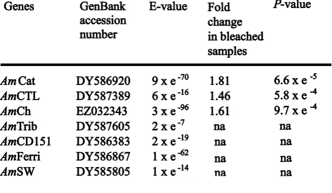
(27) Chapter 2. 26. Beta actin, glyceraldehyde 3-phosphate dehydrogenase (GAPDH) and the ribosomal protein L13a (rpL13a) are commonly used ICGs in gene expression experiments (Vandesompele et al. 2002) and were therefore considered in this study. Other potential ICGs were also evaluated including a coral homolog of adenosylhomocysteinase (Levy et al. 2007), filamin, tropomyosin, rpL9, rpS7 and two unknown sequences, Ctg1913 and Ctg3235. These genes were selected based on their stable expression at the time points used in this study in a small pilot microarray experiment (data not shown) based on a method of reference gene selection similar to that of Rodriguez-Lanetty et al. (2008). A quick test using a subset of samples in preliminary qRTPCR runs isolated the following candidate ICGs: GAPDH, rpL13a, rpL9, rpS7, Ctg1913 and Ctg3235, which were thoroughly tested in the full-scale qRTPCR experiment. GOIs were selected based on their known or suspected functions relevant to the coral bleaching response (e.g., Morgan et al. 2005; Desalvo et al. 2008; Edge et al. 2008). More specifically, we selected homologs of catalase (AmCat), ferritin (AmFerri), and selenoprotein W (AmSW) as indicators of oxidative stress (reviewed in Whanger 2000; Lesser 2006; Mladenka et al. 2006). Further, the chromoprotein (AmCh) represents a host pigment and adds information to the work performed on GFP-like genes in corals (Dove 2004; Bou-Abdallah et al. 2006). C-type lectin (AmCTL), tetraspanin CD151 (AmCD151), and tribble (AmTrib) were chosen as potential indicators of stress or an immune response (Kvennefors et al. 2008), cell-cell adhesion (Shigeta et al. 2003) and mitogen-activated protein kinase (MAPK) regulation (KissToth et al. 2004), respectively. Defense mechanisms may play an important part in the bleaching stress response, as bleached corals have been found to be more susceptible to disease (Muller et al. 2008). One hypothesis for this increased susceptibility is an.
(28) Chapter 2. 27. increased microbial community due to elevated temperature combined with a decrease in coral tissue thickness due to bleaching, leading to bacterial infection. Therefore, regulation of genes with functions in immunity, such as C-type lectins, is expected. In addition, symbiont-containing cell detachment has been observed as one of the cellular mechanisms involved in coral bleaching in the field (Brown et al. 1995). Therefore, cell-cell adhesion related genes, such as tetraspanins, represent good targets. Finally, genes regulating MAPK expression, such as tribble, are also of interest because MAPKs are present in corals and may play a role in osmoregulatory cascades during coral bleaching (reviewed in Mayfield and Gates 2007). 2.2.5. Two step qRTPCR In order to validate the stability of six candidate ICGs and the change in. expression for seven GOIs, qRTPCR assays were performed on nine individual colonies. The same amount of pure total RNA per colony was reverse-transcribed to cDNA in a Mastercycler personal (Eppendorf, Hamburg, Germany) following the SYBR GreenERTM Two-Step qRT-PCR universal kit protocol (Invitrogen, Australia). cDNA samples were diluted five-fold prior to qRTPCR runs. Primer pairs were designed (see supplementary Table 1 for primer sequences) to amplify fragments of between 90 and 110 bp, with a GC content ranging from 50 to 60 % and a melting temperature between 57ºC and 63ºC using the OligoPerfectTM designer software (Invitrogen). Primers were synthesized commercially (GeneWorks Pty Ltd, Hindmarsh, Australia). An optimum annealing temperature of 60°C was determined on the Mastercycler Gradient (Eppendorf). After UV-sterilization of all blocks and tubes, qRTPCR reactions were loaded in triplicate using the CAS-1200 loading robot (Corbett Research) to minimize pipetting error. Assays were performed following the program proposed in the SYBR GreenERTM Two-Step qRT-PCR kit (Invitrogen) on the Rotor-.
(29) Chapter 2. 28. Gene 3000 (Corbett Research). The master-mix was scaled down to 20 µL as follows: 10 µL SYBR GreenERTM qRTPCR SuperMix (Invitrogen), 1 µL of each primer (at 4 µM), 2 µL of 5x diluted cDNA, and 6 µL DEPC-treated water. Runs were followed by melting curves for detection of any non-specific amplification or primer dimerization, with heating starting at 57ºC and a rate of 0.1ºC per second up to 95ºC with continuous measurement of fluorescence. The efficiency of each primer was evaluated using the dilution method (Rebrikov and Trofimov 2006) on a pool of all cDNA samples and used in the calculation of gene expression level using the normalized relative quantification method as in Hellemans et al. (2007). Data on the expression levels of each sample were obtained in the form of quantification cycle (Cq) by manually positioning the lowest possible threshold above background fluorescence on the exponential phase of all amplification plots viewed using the logarithmic scale for the fluorescence axis. This provides the most accurate Cq measurements as this part of the plots represents the optimum qRTPCR-kinetics (Bustin 2004). 2.2.6. Statistical analysis Hellemans et al. (2007) suggested two measures to assess a reference gene for. qRTPCR: (1) the coefficient of variation (CV) of the normalized relative quantities in all conditions; and, (2) a stability value, M, that indicates the stability of the nonnormalized relative quantities of a transcript with respect to the other transcripts. In their experiment with heterogeneous samples, Hellemans et al. (2007) observed that M and CV could be values up to 1 and 50%, respectively, and advised the use of at least three genes for the normalization of qRTPCR data. The qRTPCR data were therefore transformed and normalized according to the methods proposed by Hellemans et al. (2007) using the ICGs described below, with M and CV values lower than 1 and 50%,.
(30) 29. Chapter 2. respectively. Differences in gene expression were assessed with linear mixed models analysis of variance (Demidenko 2004) using condition (healthy-looking or severely bleached) as the fixed effect, colony as the random effect and the log of the transcript relative quantity as the dependent variable. The statistical analysis was conducted using the. R. statistical. environment. (R_Development_Core_Team. 2008).. Statistical. significance was evaluated at the = 0.05 level for the F statistic.. 2.1. Results. 2.1.1. Changes in Symbiodinium densities, chlorophyll a content and water. temperature At the January 24th 2001 time point, the average symbiont density was 2.07 x 10⁶ (± 0.25 x 10⁶) cells cm-2 (Fig. 2.1A) and none of the nine colonies showed visual signs of bleaching (Smith 2005). Furthermore, on the preceding sampling date (3rd of November 2000), the same colonies looked healthy and the average symbiont density was 2.50 x 10⁶ (± 0.37 x 10⁶) cells cm-2. In comparison to the overall fluctuation in symbiont population over the two-year experimental period (Bourne et al. 2008), these values fall within the range of Symbiodinium densities found in healthy-looking individuals. An average symbiont density of 1.10 x 10⁶ (± 0.18 x 10⁶) cells cm-2 was measured in the same colonies exactly a year later, at the January 24th 2002 time point (Fig. 2.1A). This corresponds to a drastic drop, which was emphasized by severe loss of color in all nine colonies. The symbiont density and chlorophyll a concentration were both significantly reduced in 2002 (paired two-tailed t-test, P < 0.05; Fig. 2.1). The water temperature monitoring data revealed that temperature stayed below 29 ºC for three weeks prior to sampling in 2001. However, during an equivalent period of time prior to the 2002 sample, water temperature rose to 32ºC over a few days and reached a plateau for two weeks before dropping to 30 ºC during the third week. Bleaching was.
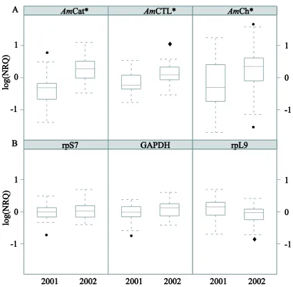
(31) Chapter 2. 30. first observed on the 7th of January 2002, approximately two weeks prior to the collection of January 24th 2002 (Bourne et al. 2008).. Fig. 2.1. Comparison of (A) the mean Symbiodinium cell densities and (B) chlorophyll a concentrations in samples used in the qRTPCR experiment (n = 9) at two time points, 24th of January 2001 (2001) and 24th of January 2002 (2002). *paired two-tailed t-test, P < 0.05. The percentages of degenerate Symbiodinium cells are indicated.. 2.1.2. ICGs, differentially expressed genes and inter-colony variability Consistent differences in transcript quantities between conditions (bleached and. non-bleached) were observed among the six candidate ICGs for almost all colonies under investigation, indicating stability in expression (supplementary Fig. 1). Moreover, GAPDH, rpS7 and rpL9 had the best CV (29 – 32%) and M (0.58 – 0.62) values (supplementary Table 2), and were therefore used for normalization of GOIexpression data (Fig. 2.2)..
(32) Chapter 2. 31. Fig. 2.2. Box plots comparing the log-normalized relative quantity (log (NRQ)) for nine Acropora millepora colonies in healthy-looking (2001) and severely bleached (2002) condition for: (A) three GOIs, AmCat, AmCTL and AmCh, and (B) three ICGs, rpS7, GAPDH and rpL9. *linear mixed models analysis of variance, P < 0.05 (• outliers, superposed outliers).. Using mixed model analysis of variance, three genes were found to be significantly differentially expressed in the qRTPCR experiment: AmCat (F = 19.8, P = 6.6 x 10⁻⁵), AmCTL (F = 13.8, P = 5.8 x 10⁻⁴) and AmCh (F = 12.5, P = 9.7 x 10⁻⁴). These three genes were up-regulated in bleached colonies by factors of 1.81, 1.46 and 1.61, respectively (Table 2.1), relative to the three best ICGs mentioned above..
(33) Chapter 2. 32. Table 2.1. Regulation of seven GOIs (based on qRTPCR) in nine field colonies of A. millepora undergoing bleaching. The same nine colonies were sampled exactly one year apart and were healthylooking at the first and severely bleached at the second sampling time point. The fold changes of gene expression are shown with the P-values for those genes that were significantly differentially expressed. The E-value corresponds to the best match of a Tblastx search in the nucleotide collection (nr/nt).. A significant difference among colonies was detected for two putative GOIs (AmTrib, F = 89.18, P = 8.0 x 10⁻⁵; AmSW, F = 6.9, P = 3.4 x 10⁻²) and one potential ICG, Ctg1913 (F = 15.6, P = 5.5 x 10⁻³). This intercolony variability is not only caused by differences in amplitude of the response to bleaching but also by dramatic changes in the direction of individual regulation (Fig. 2.3)..
(34) Chapter 2. 33. Fig. 2.3. Log (NRQ) data for three genes showing great inter-colony variability in expression among the nine colonies. *linear mixed models analysis of variance, P < 0.05 (qRTPCR reaction triplicates for one colony are depicted by the same symbol).. 2.2. Discussion Gene expression studies using qRTPCR can help identify the onset of the. physiological changes indicative of stress that leads to the phenomenon of coral bleaching. However, ICGs that exhibit stable expression under field conditions are essential if gene expression is to be used as an indicator for the onset of stress. We have identified three such genes from a widely distributed Indo-Pacific coral, which are therefore promising candidates for the normalization of gene expression data during.
(35) Chapter 2. 34. natural coral bleaching events. Furthermore, my finding of three up-regulated GOIs (catalase, chromoprotein and C-type lectin homologs) in naturally bleached colonies of A. millepora supports and enhances our understanding of the cellular processes underpinning bleaching. Large inter-colony variation in the expression patterns for three other genes, AmTrib, AmSW and an unknown gene Ctg1913, were observed during this natural bleaching event, highlighting the need for appropriate gene selection and adequate sample sizes for future gene expression studies on natural coral populations. 2.2.1. ICGs for coral bleaching ICGs were selected based on their consistency in expression despite the drastic. decrease in symbiont densities between bleached and non-bleached samples and technical discrepancies across samples using the gNorm method (Vandesompele et al. 2002). The ribosomal protein rpL9 and rpL13a genes encode components of the 60S subunit and therefore function together, while the rpS7 protein is a component of the 40S subunit. Nevertheless, the expression of rpS7 and rpL9 appeared to be more stable than rpL13a and were therefore used in the present analysis. In support of this, another 60S subunit ribosomal protein gene, rpL12, has been suggested as a potential ICG in cnidarian studies (Rodriguez-Lanetty et al. 2008). GAPDH, also a commonly used ICG, was relatively stable in its expression between bleached and unbleached A. millepora colonies and was therefore included in the ICG panel for normalization. In model organisms, GAPDH, beta actin and the 16S ribosomal subunit are commonly used ICGs in qRTPCR gene expression assays (Vandesompele et al. 2002). Although these genes are suitable ICGs for some controlled laboratory experiments they are not uniformly applicable across all species, individuals, tissues and experimental conditions. For example, beta actin has been used to normalize qRTPCR gene.
(36) Chapter 2. 35. expression data from both A. millepora (Levy et al. 2007) and other cnidarians (Moya et al. 2008; Rodriguez-Lanetty et al. 2008), however this gene did not perform well in preliminary trials of the field-bleached colonies sampled here (data not shown) and was found significantly regulated in a recent coral bleaching microarray study (Chapter 3). Additional challenges are present when identifying stably expressed ICGs for qRTPCR assays in field coral samples. For corals undergoing bleaching in the field, individuals from the same sampling site (tens of square meters) may experience strong microhabitat differences (e.g. light and temperature exposure), which may cause subsequent differences in their responses. In some species, multiple Symbiodinium types (with varying physiological performances) may co-exist on the same reef producing staggered phenotypic responses to widespread stressors (Rowan et al. 1997; Jones et al. 2008). Genetically determined variation in bleaching susceptibility of the coral host may further contribute to variance in the bleaching response among colonies. As a result, qRTPCR assays on coral field samples need to include multiple carefully selected ICGs. In the present study, I demonstrated that my samples were contaminated with Symbiodinium cDNA. Primers for the proliferating cell nuclear antigen gene in Symbiodinium (Boldt et al. 2009) failed to amplify cDNA from aposymbiotic planula larvae (Cq > 33), but amplified juvenile (Cq < 23) and adult cDNAs (Cq < 21). Naturally, one would expect this contamination to affect healthy and bleached tissues in such way that the Cq values for host ICGs would be lower in bleached samples due to the dilution effect of higher symbiont density in healthy coral samples. However, the small shift in Cq values for the best performing ICG, GAPDH, rpL9 and rpS7 presented here, was not uniform in direction across all colonies (supplementary Fig. 1). This inconsistency in direction of the shift for Cq values between bleached and healthy.
(37) 36. Chapter 2. samples suggests that the symbiont contamination is minimal and most likely confounded with other technical artifacts such as variations in cDNA efficiency and/or messenger RNA yield between samples. Here, I present an alternative method of normalization to that developed by Mayfield et al. (2009) which consists of using a spike of exogenous RNA/DNA to normalize for differential extraction of Symbiodinium nucleic acids in symbiotic systems. I suggest that one should develop an ICG-system of at least three genes exhibiting stable expression levels throughout the experimental period before assessing normalized relative quantification of scleractinian coral gene expression. Potential ICGs can be selected using microarray data as described in Rodriguez-Lanetty et al. (2008) before being further tested and used in qRTPCR experiments, whether to validate microarray data as in DeSalvo et al. (2008) or monitor gene regulation in the field as presented here. I do not claim that the ICGs presented here will be universally useful across conditions and species but, rather, that they are the best under the conditions in this study to accurately measure change in gene expression and should be considered as candidates in other studies, especially those with A. millepora. 2.2.2. Regulation of GOIs during bleaching The regulated genes discovered here are uncharacterized in corals and little is. known about their specific function in cnidarians. Their function in other organisms is summarized below. The consistent up-regulation of three genes in nine independent A. millepora colonies during a natural coral bleaching event suggests, however, that these three genes in particular, warrant further investigation for their role in the bleaching process and their potential as useful stress indicators in field sampling studies..
(38) 37. Chapter 2. 2.2.2.1 AmCatalase The most studied of my up-regulated genes is an A. millepora catalase homolog (AmCat). Catalase is an antioxidant enzyme that limits the production of highly cytotoxic hydroxyl radicals (HO) (Lesser 2006). My finding of a significant upregulation of catalase in A. millepora during natural coral bleaching supports previous studies indicating that oxidative stress likely affects host coral tissue during bleaching events (Lesser 1996, 1997; Lesser and Farrell 2004; Lesser 2006) and is consistent with its up-regulation in a time course experiment looking at heat-induced bleaching in laboratory conditions by DeSalvo et al. (2008). Catalase reduces the H2O2 produced in excess by photosynthetic organisms undergoing photo-inhibition (reviewed in Smith et al. 2005) to water and molecular oxygen (Mladenka et al. 2006). During coral bleaching, heat and light may induce photoinhibition in endosymbiotic zooxanthellae, leading to the overproduction of ROS, which may diffuse through membranes and damage host cells, resulting in their detachment or in symbiont expulsion (Brown 1997; Fitt et al. 2001; Lesser and Farrell 2004; Tchernov et al. 2004).. Although Symbiodinium possess antioxidant enzymes capable of. detoxifying H2O2 (Matta and Trench 1991), overproduction of H2O2 may overwhelm the antioxidant defense of the symbiont and diffuse to the host through cell membranes (Lesser 2006). Therefore, significant up-regulation of catalase in the host tissue of fieldbleached A. millepora supports a role for this antioxidant enzyme in the maintenance of homeostasis in response to oxidative stress during coral bleaching (Merle et al. 2007)..
(39) 38. Chapter 2. 2.2.2.2 AmChromoprotein I have also identified an up-regulated chromoprotein gene, AmCh. Coral chromoproteins are close relatives of fluorescent proteins such as green fluorescent protein (GFP), and they may play an important role in the stress response of corals. The up-regulated AmCh gene examined here encodes a blue non-fluorescent chromoprotein (Beltran-Ramirez 2008) that was originally sequenced from A. millepora larvae (Technau et al. 2005a) and which shares 95% amino acid homology (211/221 AA) with a blue chromoprotein (GenBank AY646075) absorbing at 588 nm first identified in adult A. millepora tissue (Alieva et al. 2008). Interestingly, a blue pigment appears more abundant in recently compromised tissue of A. millepora than in adjacent healthy tissue in the field (personal observation). Further work is needed to determine if this pigment corresponds to AmCh. In addition to the substantial bleaching reported here, several of the A. millepora colonies included in the present study developed necrotic looking lesions during the 2002 natural bleaching event (C. Smith-Keune, personal observation) and it is intriguing that this event also coincides with an increase in AmCTL gene transcript levels. The. traditionally. proposed. role. of. fluorescent. and. non-fluorescent. chromoproteins in symbiotic corals, was as a sunscreen or photoprotective shield for corals living in high light environments (Kawaguti 1944; Salih et al. 2000; Dove et al. 2001). It has also been proposed that this role may be reversed to actually enhance light capture for corals in low-light conditions (Schlichter and Fricke 1990). However, it has been suggested that the putative photoprotective function of certain GFP-like proteins in acroporid corals may be rapidly lost during times of thermal stress (Dove 2004; SmithKeune and Dove 2008) and several other studies argue against a direct role for coral GFP homologs in photo-protection or improving photosynthetic efficiency in coral.
(40) Chapter 2. 39. symbiosis altogether (Gilmore et al. 2003; Mazel et al. 2003; Wiedenmann et al. 2004). On the other hand, a cyan fluorescent protein was differentially expressed in competent Montastraea faveolata larvae as a result of inoculation with symbionts (Voolstra et al. 2009b). Interestingly, an antioxidant role via the quenching of superoxide radicals has recently been shown for a GFP from the hydromedusa, Aequorea victoria (BouAbdallah et al. 2006). In light of this, the concomitant up-regulation of both the AmCh and AmCat gene transcripts during coral bleaching in the present study suggests that a similar antioxidant role for AmCh in A. millepora could be considered. At the same time, the up-regulation of gene expression for the blue chromoprotein gene AmCh identified in field-bleached colonies here contrasts with other studies. (Smith-Keune and Dove 2008) showed the rapid down regulation of another GFP-like gene AmA1 (a homolog of the red fluorescent Zoanthus sp. protein zoan2RFP) in laboratory heat stressed A. millepora colonies experiencing temperatures of 32 ºC and 33 ºC. Similarly, a GFP-like homolog was down-regulated in M. faveolata colonies exposed to 32C for 3 days in the laboratory (DeSalvo et al. 2008). This may suggest a variable role for these GFP-like proteins within A. millepora host tissues or may indicate a difference in the bleaching mechanisms occurring in the field compared to that induced at similar temperatures in the laboratory for this species. For example, huge difference in light intensity between laboratory and field conditions may explain such opposite patterns in gene expression. 2.2.2.3 AmC-type lectin The qRTPCR assays showed consistent up-regulation of a putative C-type lectin gene (AmCTL). The AmCTL predicted peptide has a putative C-type lectin-like domain of the type found in lectins of other marine invertebrates, for example, CEL-1 from Cucumaria echinata and echinoidin from Anthocidaris crassispina (E-value: 2e-20). In.
(41) 40. Chapter 2. other animals, C-type lectins are involved in processes including extracellular matrix organization, pathogen recognition, and cell-cell interactions, which are all processes that may be relevant during coral bleaching events. At least seven other lectin-related genes are expressed in A. millepora including several that are expressed at the time of metamorphosis (Grasso et al. 2008). This suggests potential roles in cell recognition and lysis or in Symbiodinium recognition and incorporation. The closest known proteinencoding gene to AmCTL was a C-type lectin homolog (FJ628422, E-value: 1e-37), which was found down-regulated prior to Symbiodinium expulsion in Pocillopora damicornis colonies exposed to 32C in the laboratory (Vidal-Dupiol et al. 2009). In addition, a mannose-binding lectin paralog (GenBank ACF08844, E-value: 2e-24), named millectin is 41% identical and 60% similar to AmCTL predicted peptide sequence. More importantly, both share conserved amino acids in the mannose- and the calcium-binding sites of C-type lectins involved in immunity (Kvennefors et al. 2008). 2.2.3. Intercolony variability in levels of gene expression in a natural population A surprising degree of inter-colony variability in direction and magnitude of. gene expression changes was observed for two of the GOIs, AmSW and AmTrib, and one candidate ICG, a coral specific gene of unknown function, Ctg1913. Unfortunately, little is known of the function of any of these genes in other animals and the homology of these genes to known orthologs in GenBank was generally low. Regardless of their functions, the highly variable expression of these genes in naturally bleached colonies of A. millepora, on the Nelly Bay reef flat during the bleaching event of 2002, suggests either substantial variation in microenvironment of the sampled colonies, or different genetically determined individual colony responses.. It is essential for valid bio-. monitoring studies that the chosen stress response genes show similar gene expression profiles for all or at least the majority of individuals on the reef under study. Consistent.
(42) Chapter 2. 41. sampling of colonies with respect to overall morphology and replication at both interand intra-colony levels should be carefully considered in future field studies applying assays similar to those developed here. I utilized a single branch from a similar location at the centre of each tagged A. millepora colony, which was sampled at approximately mid-day on each sampling date. The future inclusion of intra-colony replication, and measurements of protein levels in addition to gene expression levels, would substantially enhance our understanding of the biological significance of the changes in expression of the genes measured here. Recent gene expression studies on the same species have observed high levels of intercolony variability (Bay et al. 2009a; Bay et al. 2009b; Császár et al. 2009), making measurements of significant change in expression at the population level difficult and indicating high biological replication is required. Despite the presence of similar levels of inter-colony variability observed here, I was able to successfully detect significant and consistent change in expression levels for three biologically relevant genes, in a representative sample selected from a population of A. millepora under field bleaching conditions. I therefore encourage future studies to consider these genes (AmCat, AmCh, and AmCTL) for further investigation as key bleaching stress candidates for field based bio-monitoring studies on scleractinian corals.. 2.3. Conclusion This study represents the first step towards the application of qRTPCR. technology to monitor and investigate the bleaching response in a widespread IndoPacific coral (A. millepora) under field conditions.. Three candidate ICGs were. identified with stable expression levels when the same colonies were sampled under conditions of healthy symbiosis (January 2001) and when in a naturally bleached state (January 2002). Three consistently up-regulated genes of interest (AmCat, AmCh,.
(43) Chapter 2. 42. AmCTL) were also identified as valuable candidate genes for monitoring bleaching stress in A. millepora. Future work should focus on evaluating variation in the expression of these GOIs both within individual coral colonies and between A. millepora populations..
(44) 43. Chapter 3. Chapter 3.0. Changes in gene expression in the reef-. building coral Acropora millepora during a natural bleaching episode and subsequent recovery period in the field. This chapter is inserted without abstract as in preparation for submission: François O. Seneca, Sylvain Forêt, Nicolas Goffard, Carolyn Smith-Keune, Lauretta Grasso, Madeleine JH van Oppen, Eldon Ball and David Miller. All the gene expression data was collected and analyzed by F.O. Seneca, who also wrote the chapter. Sylvain Forêt and Nicolas Goffard have conducted the statistical and bioinformatic analyses, respectively. The other co-authors provided conceptual, logistical and editorial support..
(45) 44. Chapter 3. 3.1. Introduction The persistence of coral reef ecosystems depends on the survival of one order of. marine invertebrates, the scleractinian corals. These reef-building corals provide the physical architecture of reefs through massive deposition of calcium carbonate skeletons. In addition to maintaining structural complexity of the reef matrix, living corals play an additional role in supporting the diversity and biomass found in tropical waters through an energetically productive symbiosis with intracellular algae (i.e. Symbiodinium spp.). In this close relationship with the photosynthetic algae, corals and their symbiotic partners can produce enough energy to sustain metabolic requirements, including calcification, as well as provide part of the energetic basis for an ecosystem, which frequently exists in oligotrophic waters. Currently, coral reef ecosystems are threatened by collapse due to the consequences of anthropogenic activities, including but not limited to global climate change (van Oppen and Gates 2006). The 4th Intergovernmental Panel on Climate Change (IPCC) concluded that corals are vulnerable to thermal stress and of low adaptive capacity (IPCC 2007), although the latter has not been sufficiently scientifically tested.. Furthermore, the potential loss of live coral coverage and. subsequent losses in overall biodiversity and biomass will ultimately lead to decreased coral reef ecosystem services and consequently have negative impacts on human wellbeing (Omann et al. 2009). More recently, a 2008 report on the status of global coral reefs by the Global Coral Reefs Monitoring Network identified the two most critical factors affecting coral reefs as rising sea surface temperatures and increasing atmospheric CO2 concentrations (Wilkinson 2008) causing acidification of seawater. These worldwide changes are predicted to worsen with serious implications for corals and their dependent reef ecosystems (Hansen et al. 2006)..
Figure


+7


Outline
Regulation of GOIs during bleaching
Onset of bleaching 1 Environmental changes
Recovery phase 1 Environmental changes
Molecular functions and cellular processes in relation to coral bleaching Exposure of coral cells to adverse environmental conditions is thought to induce
Heterogeneity of USP expression patterns
General Discussion
Related documents
example, four of the seven school line managers interviewed were extremely positive about the training received by the PSAs.. Two mentioned that their faith in the training
Table 64: The frequency of pupils with and without disabilities taking part in bullying another pupil(s) at school in the past couple of months ...58.. Table 65: The frequency
7.10 The option of supported self-employment or employment in what the Americans call ‘micro-enterprises’ seemed rare in Scotland. We found some examples of self-employment and
Table 8 - Attrition and Survival Rates By Module of Study After Year 1 of University: Social
Based on the conceptualizations of learner empowerment by Frymier, Shulman and House (1996) and situational interest researchers in the field of accounting education, three
As was the case with the cost data, nurseries set up on a for-profit basis were less likely to be able to provide this information than those set up as not-for-profit
As noted above, these nucleotide substitutions not only eliminate OBP binding to site I DNA but also introduce a BstBI site into origin sequences at site I (10), which allows
A CLINICAL STUDY OF MATERNAL AND PERINATAL OUTCOME IN HEART DISEASE COMPLICATING PREGNANCY.. Dissertation submitted in partial fulfilment
