0022-538X/06/$08.00⫹0 doi:10.1128/JVI.01364-06
Copyright © 2006, American Society for Microbiology. All Rights Reserved.
Herpes Simplex Virus 1 DNA Packaging Proteins Encoded
by U
L
6, U
L
15, U
L
17, U
L
28, and U
L
33 Are Located on the
External Surface of the Viral Capsid
䌤
Elizabeth Wills, Luella Scholtes, and Joel D. Baines*
Department of Microbiology and Immunology, Cornell University, Ithaca, New York 14853
Received 28 June 2006/Accepted 9 August 2006
Studies to localize the herpes simplex virus 1 portal protein encoded by UL6, the putative terminase
components encoded by UL15, UL28, and UL33, the minor capsid proteins encoded by UL17, and the major
scaffold protein ICP35 were conducted. ICP35 in B capsids was more resistant to trypsin digestion of intact
capsids than pUL6, pUL15, pUL17, pUL28, or pUL33. ICP35 required sectioning of otherwise intact embedded
capsids for immunoreactivity, whereas embedding and/or sectioning decreased the immunoreactivities of pUL6,
pUL17, pUL28, and pUL33. Epitopes of pUL15 were recognized roughly equally well in both sectioned and
unsectioned capsids. These data indicate that pUL6, pUL17, pUL28, pUL33, and at least some portion of pUL15
are located at the external surface of the capsid.
Capsids form in the nuclei of cells infected with all herpes-viruses. Herpes simplex virus (HSV) capsid pentons and hex-ons form spontaneously from five and six molecules of ICP5, respectively; these capsomeres are linked by triplexes consist-ing of two molecules of VP23 and one molecule of VP19C to form a porous procapsid (23, 36, 43). ICP5 is also associated with ICP35, which forms an internal shell or scaffold within the procapsid. The procapsid is believed to give rise to the three other types of capsids seen in HSV-infected cells, designated types A, B, and C. All of these capsids differ internally but contain identical outer shells, as determined by cryoelectron microscopy (21, 35, 49). Type B capsids retain the scaffold internal to the outer shell, type A capsids contain only the outer shell, and type C capsids lack the internal scaffold but contain viral DNA (14). Type C capsids then bud from the nuclear membrane in a reaction termed primary envelopment (19, 32).
One of the vertices of A, B, and C capsids is biochemically and structurally unique and has been designated the portal vertex. Thus, the UL6-encoded protein (pUL6) forms a
do-decameric ring with an internal diameter of at least 65 Å, i.e., sufficiently wide to accommodate DNA as it is packaged into the capsid (44). Critical to the discovery of the portal was the observation that an antibody to the C terminus of pUL6
recognized epitopes on a single vertex of type B capsids, thus showing that at least the C terminus of pUL6 is located
at the capsid exterior in a position to access incoming viral DNA (22, 39).
It has also been shown that HSV-1 B capsids contain a number of capsid proteins in addition to triplexes, pUL6, ICP5,
and ICP35. These proteins include approximately 1.2 copies of pUL15, 2.4 copies of pUL28, 27 to 42 copies of pUL25, 19.2
copies of pUL17, and an undetermined amount of pUL33 (6, 7,
15, 25, 26, 33, 41, 42, 48). By analogy to extensive studies of bacteriophage capsid assembly, it might be predicted that some of these minor capsid proteins would be involved in processing concatameric DNA and threading the DNA into the portal through the hydrolysis of ATP (9). Such a complex, termed the terminase, remains somewhat enigmatic in HSV, but a variety of indirect evidence suggests that it comprises at least the UL15, UL28, and UL33 proteins. Specifically, (i) all three
pro-teins are among seven propro-teins required for viral DNA pack-aging (3, 29, 40); (ii) like other terminases, the UL15 protein
contains a conserved P-loop ATPase motif, and mutation of this motif precludes DNA packaging (12, 20, 47); (iii) the UL28
protein can specifically bind DNA sequences known to be required for the correct cleavage of concatameric viral DNA (2); (iv) the UL15 and UL28 proteins interact directly, whereas
pUL33 binds pUL28 and enhances the pUL15-pUL28
interac-tion in coimmunoprecipitainterac-tion assays (1, 8, 17, 46); and (v) in vitro, both pUL28 and pUL15 can interact with the portal
protein encoded by UL6 (45).
Recent immunogold analysis of pUL17 and pUL25 supports
their location on the external surface of the viral capsid on more than one vertex (24, 41). Although it is also required for DNA packaging, the precise function of UL17 is unknown (34).
Analysis of a UL17 deletion mutant revealed an alteration of
the normal intranuclear distributions of capsids and a number of viral proteins including pUL6, ICP35, and ICP5 (39). These
observations suggest that the UL17 protein is involved in
en-suring proper capsid assembly, the reorganization of the in-fected cell nucleus, or, directly or indirectly, capsid or protein transport within the nucleus. Relevant to this last possibility is the observation that HSV capsids are actively transported in the nucleus and that this transport is both energy and actin dependent (13). UL25 is believed to enhance the stability of
capsids and is required for the retention of full-length genomic DNA in the capsid (18, 24, 37, 41).
The hypotheses that pUL6 serves as the portal, the UL15,
UL28, and UL33 proteins form the HSV terminase, and pUL17
* Corresponding author. Mailing address: Department of Microbi-ology and ImmunMicrobi-ology, Cornell University, C5132 Veterinary Educa-tion Center, Ithaca, NY 14853. Phone: (607) 253-3391. Fax: (607) 253-3384. E-mail: [email protected].
䌤Published ahead of print on 18 August 2006.
10894
on November 8, 2019 by guest
http://jvi.asm.org/
acts directly or indirectly to mediate the transport of capsids within the nucleoplasm predict that at least portions of these proteins would localize on the external surfaces of capsids. This study was undertaken to test these possibilities.
Production and specificity of a novel chicken antiserum
against pUL17.Because a previously described UL17 antibody
produced using a DNA vaccine did not recognize small quan-tities of UL17 protein (34) (data not shown), UL17 was fused to
DNA encoding a His6tag, and a recombinant baculovirus that
expressed the fusion protein was generated. The UL17 fusion
protein was purified from lysates of insect cells infected with the recombinant baculovirus by affinity chromatography on Ni2⫹-containing Sepharose beads as described previously (7).
Immunization of chickens with the purified fusion protein was followed by purification of immunoglobulin Y (IgY) from the eggs of immunized hens as described previously (31).
To test the antisera for specificity, Hep-2 cells were mock infected or infected with HSV-1(F) or a UL17 null virus at a
multiplicity of infection of 5 PFU per cell. Lysates from ap-proximately 1.3⫻106cells were prepared by denaturation and
boiling in 1% sodium dodecyl sulfate (SDS) and 5 mM -mer-captoethanol. Denatured proteins were electrophoretically separated on an 8% SDS-polyacrylamide gel and transferred electrically to a nitrocellulose membrane. The nitrocellulose membrane was blocked overnight at 4°C in phosphate-buffered saline (PBS) supplemented with 5.0% milk and 0.2% Tween 20. The membrane was rinsed twice with room-temperature PBS and 0.2% Tween 20 and was further blocked by immer-sion in a 1:10 dilution of Block Hen (Aves Labs) for 15 min. Polyclonal anti-UL17 IgY was diluted 1:500,000 into PBS with
1% bovine serum albumin and 0.2% Tween 20 and applied overnight. After extensive washes in PBS containing 0.2% Tween 20, the membrane was incubated with horseradish
per-oxidase-conjugated anti-chicken antibody diluted 1:5,000 in PBS containing 5.0% milk and 0.5% Tween 20. Bound IgY was detected using a 5-min incubation with ECL Plus reagents (Amersham) and flash exposure to Fuji autoradiographic film. As shown in Fig. 1, the anti-pUL17 antiserum recognized a
protein with an apparentMrof 79,000 that was not present in
lysates of mock-infected Hep-2 cells or cells that were infected with the UL17 deletion virus. This size is consistent with a
previous study reporting an apparentMr of 77,000 in virions
(34). We did not detect the protein with an apparent Mr of
72,000 that was previously identified in virion lysates by mass spectrometry, suggesting that the smaller protein may be highly enriched in virion preparations (34). The antibody also recog-nized pUL17 protein expressed by a recombinant baculovirus
(data not shown).
Immunogold labeling of wild-type and mutant capsids.
Us-ing standard procedures (28), capsids were purified from nu-clear lysates of cells infected with wild-type HSV-1(F) and viruses respectively lacking the UL6, UL15, UL17, UL28, and
UL33 genes (5, 11, 27, 34, 40). Briefly, nuclear lysates prepared
from 1.5⫻108Vero cells infected with HSV-1(F) were
clari-fied, and capsids were pelleted through a 35% sucrose cushion in an SW28 rotor. The pellet in resuspended material was subjected to rate-zonal centrifugation through a 20 to 50% (wt/vol) sucrose gradient in a Beckman SW41 rotor at 24,500 rpm for 1 h. A light-refracting band in the middle of the gradient containing B capsids was collected using a Pasteur pipette, and capsids were diluted into a solution containing
FIG. 1. Digitally scanned image of immunoblot of lysates of in-fected and uninin-fected cells that reacted with pUL17-specific antiserum.
Lysates of cells that were uninfected (left lane) or infected with a UL17
deletion virus (center lane) or wild-type virus HSV-1(F) (right lane) were electrophoretically separated on an SDS-polyacrylamide gel, transferred onto nitrocellulose, and reacted with purified IgY obtained from a chicken immunized with purified pUL17. Bound IgY was
[image:2.585.302.540.426.579.2]re-vealed as indicated in the text. Sizes of molecular weight markers are indicated to the right in thousands.
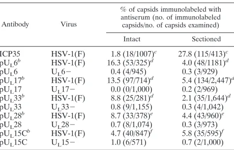
TABLE 1. Percentages of capsids immunolabeled with monospecific antiseraa
Antibody Virus
% of capsids immunolabeled with antiserum (no. of immunolabeled capsids/no. of capsids examined)
Intact Sectioned
ICP35 HSV-1(F) 1.8 (18/1007)c 27.8 (115/413)c
pUL6b HSV-1(F) 16.3 (53/325)d 4.0 (48/1181)d
pUL6 UL6⫺ 0.4 (4/945) 0.3 (3/929)
pUL17b HSV-1(F) 13.5 (97/714)d 5.4 (134/2,447)d
pUL17 UL17⫺ 0.0 (0/1,000) 0.2 (2/969)
pUL33b HSV-1(F) 8.8 (25/281)d 2.1 (35/1,644)d
pUL33 UL33⫺ 0.8 (9/1,155) 0.3 (4/1,042)
pUL28b HSV-1(F) 8.7 (33/378)e 4.4 (43/960)e
pUL28 UL28⫺ 0.7 (8/1,074) 0.3 (3/973)
pUL15Cb HSV-1(F) 4.7 (40/847)f 5.8 (35/595)f
pUL15C UL15⫺ 1.0 (6/571) 0.7 (2/1,000) aCapsids were purified from cells infected with wild-type viruses or viruses lacking the indicated open reading frames (⫺). In some experiments (intact), capsids were attached to grids and reacted with the indicated antibodies. In other experiments, capsids were embedded and sectioned, followed by reaction of the thin sections with the indicated antibodies. The number of immunolabeled cap-sids versus the number of capcap-sids examined is indicated in parentheses, and the resulting percentage of labeled capsids is shown. AllPvalues were obtained with Fisher’s exactttest.
bThe amount of immunoreactivity with a given antibody was greater (P⬍ 0.001) in wild-type capsids than in the corresponding deletion mutant capsids.
cThe immunoreactivity of sectioned capsids was greater than that of intact capsids (P⬍0.001).
dThe immunoreactivity of intact capsids was greater than that of sectioned capsids (P⬍0.001).
eThe immunoreactivty of intact capsids was greater than that of sectioned capsids (P⫽0.01).
fThe immunoreactivity of sectioned capsids compared to that of intact capsids was not statistically different (P⫽0.06).
on November 8, 2019 by guest
http://jvi.asm.org/
Tris-HCl (pH 7.8), 150 mM NaCl, and 1 mM EDTA (TNE). Capsids were either attached to Formvar carbon-coated elec-tron microscopic grids or placed into microdialysis tubes (200-m diameter), subsequently embedded in LRWhite, and sliced with a diamond knife into 60-nm sections that were then placed onto electron microscopy grids.
Previously described rabbit antisera directed against the C terminus of pUL6, the C terminus of pUL15, full-length pUL28,
and full-length pUL33 were prepared by adsorption against
capsids purified from Vero cells infected with 5.0 PFU/ml of the appropriate viral null mutant (4, 5, 27, 30, 38–40). The adsorbed antisera were then diluted 1:50 in PBS supplemented with 1% Triton-100 and 1% fish gelatin and applied directly to the electron microscopy grids, followed by extensive washing. Experiments performed with the pUL17-specific chicken IgY
were similar except that the antibody was not preadsorbed and was diluted 1:5,000 for reactions with capsids. As a control, the capsid samples were also reacted separately with a polyclonal antiserum directed against the internal scaffold protein ICP35 (10) (NC 3-4) (kindly provided by Roselyn Eisenberg and Gary Cohen). Bound immunoglobulins remaining after the washing were recognized by goat anti-rabbit immunoglobulin conju-gated to 12-nm gold beads or goat anti-chicken IgY conjuconju-gated to 12-nm gold beads. After further washing, the grids were viewed using a Philips 201 electron microscope after counter-staining with 2% aqueous uranyl acetate and 0.5% Reynold’s lead citrate. Only capsids that were visually verified as intact B capsids were included in the data. The B capsids were scored as positively immunolabeled only when a gold bead was ob-served in direct association with the capsid shell or interior.
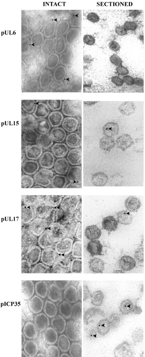
The results are summarized in Table 1, and representative examples of immunostained capsids are shown in Fig. 2.
Examination of at least 400 capsids in each treatment group and data from multiple experiments revealed the following information.
(i) Background levels of immunostaining with the pUL15-,
pUL17-, pUL28-, and pUL33-specific antisera, as revealed by
the number of appropriate mutant capsids bearing gold beads, were significantly below similarly stained wild-type HSV-1(F) capsids. (AllPvalues were⬍0.001 as assessed by Fisher’s exact
ttest.)
(ii) As shown previously (22), pUL6-specific epitopes were
recognized on the surface of the capsid inasmuch as signifi-cantly more (P ⬍ 0.001) gold beads were present in intact wild-type capsids reacted with the pUL6-specific antiserum
than in capsids lacking pUL6. These epitopes were detected
more frequently in intact capsids than in sectioned capsids (P
[image:3.585.48.279.69.637.2]⬍0.001), suggesting that the bulk of the epitopes were avail-able primarily for reaction at the capsid surface rather than internal to the capsid shell. Approximately 16.3% (53 of 325) of capsids were labeled, suggesting either that the immunogold staining was insensitive and did not detect portal protein in many capsids or that many capsids lacked portals. Biochemical studies showing that populations of B capsids average 14.8⫾
FIG. 2. Digital image of representative electron micrographs of capsids labeled with various antibodies. Capsids were purified from cells infected with HSV-1(F). These were attached to copper mesh grids (left column) or were embedded in Lowicryl and sectioned (right column). Each row shows intact capsids and thin sections that reacted with antisera directed against the indicated proteins. Bound immuno-globulin was identified by reactions with appropriate antisera conju-gated to 12-nm gold beads. Arrowheads indicate gold beads associated with capsids. Electron micrographs of immunogold analyses
per-formed with pUL33- and pUL28-specific antibodies were similar to
those of pUL17 (data not shown). A comprehensive analysis of the data
is presented in Table 1.
on November 8, 2019 by guest
http://jvi.asm.org/
2.6 copies of pUL6 per capsid (22), coupled with the high
likelihood that the portal ring contains 12 copies of pUL6 (44),
argue against the latter possibility.
(iii) As expected, the ICP35-specific antiserum did not rec-ognize the external surface of capsids to an appreciable extent inasmuch as only 18 capsids of 1,007 capsids examined (0.018%) were labeled with the NC 3-4 antibody. Upon sec-tioning of the capsids, however, ICP35-specific epitopes were rendered significantly more immunoreactive with the anti-serum (P⬍ 0.001), as revealed by increased labeling of sec-tioned capsids (115 [28%] of 413 secsec-tioned capsids). These observations indicated that, as expected, ICP35 was present in the capsid interior rather than the capsid surface and verified that the inner surfaces of unsectioned capsids were seques-tered from the applied antibodies under the experimental con-ditions used.
(iv) Epitopes from pUL17, pUL28, and pUL33 localized at
the surface of the capsid, as revealed by immunoreactivity of intact capsids, which was significantly above background levels obtained upon reaction with the corresponding deletion virus capsids. (AllPvalues were less than 0.001 by Fisher’s exactt
test.) In all three cases, although immunoreactivity was present in sectioned HSV-1(F) capsids, the level of immunoreactivity was significantly lower than that obtained using intact capsids, presumably because a given thin section contains only a limited portion of the capsid surface. Another possibility is that em-bedding capsids could reduce the immunoreactivity of pUL17,
pUL28, and pUL33. More capsids (13%) were labeled with the
pUL17-specific antibody than with either the pUL28- or
pUL33-specific antibody (8.7% and 8.9%, respectively). This
could be a consequence of increased affinity of the pUL
17-specific antibody relative to the other antibodies or increased amounts of pUL17 in association with capsids. Given the
ob-servation that only around two copies of pUL28 are present per
B capsid, and the observation that pUL17 can localize to
mul-tiple vertices, it seems likely that more pUL17 is associated
with capsids than pUL28 (7, 41). In any case, these data are
consistent with other studies of pUL17 in HSV capsids showing
that the protein is on the external capsid surface but are in contrast with the localization of the pUL17 homolog of
pseu-dorabies virus that has been reported to associate with pack-aged DNA (16, 41).
(v) Antisera directed against C-terminal epitopes of pUL15
were recognized on the external surface of capsids, as revealed by the increased immunoreactivity of intact capsids compared to that of pUL15-negative capsids. Unlike the case with pUL6-,
pUL33-, pUL28-, and pUL17-specific antibodies,
immunoreac-tivity of the pUL15-specific antibody remained high in
tioned capsids. A slight increase in immunoreactivity in sec-tioned capsids compared to that obtained with unsecsec-tioned capsids was not statistically significant (P⫽0.06). On the other hand, because less external surface area of the capsid is rep-resented in a 40- to 60-nm thin section, the preservation of immunoreactivity in sectioned capsids suggests that more pUL15 C-terminal epitopes were present within the capsid
interior than were epitopes of pUL28, pUL17, or pUL33. It is
unclear whether these observations represent the possibilities that (a) pUL15 extends from the external surface to the
inter-nal surface of the capsid, (b) the pUL15 epitopes are masked
less efficiently upon embedding than pUL28, pUL17, or pUL33
epitopes, or (c) multiple copies of pUL15 are present at
dif-ferent locations within the capsid. Assuming that all B capsids are biochemically identical (an assumption that has not been tested), the observation that each B capsid contains only 1.2 copies of pUL15 (7) argues against the latter possibility.
(vi) Very few capsids that reacted with any of the anti-bodies contained more than one gold bead. This is in con-trast to results reported previously by others (41) and may reflect the respective affinities of the different antibodies in the two studies.
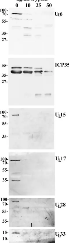
Comparative resistance of capsid-associated proteins to
protease digestion.To confirm the results obtained by
immu-nogold labeling, B capsids were purified on continuous sucrose gradients as described above and divided into four equal pools of 250l. The aliquots were incubated in the absence of trypsin or in the presence of 10, 25, or 50g/ml trypsin (MP Biomecals) for 45 min at 37°C. The digested capsids were then di-luted into 14 ml of ice-cold TNE containing protease inhibitors (1 tablet Complete protease inhibitors [Roche] per 50 ml TNE), and the diluted capsids were pelleted in an SW41 rotor at 35,000 rpm for 2 h. Pelleted capsids were solubilized in approximately 50l denaturing buffer containing SDS, mer-captoethanol, and bromophenol blue, and 25l of each sample was electrophoretically separated on a single lane of an SDS-polyacrylamide gel, followed by immunoblotting with the indi-cated antibodies as described above, except that the primary antibodies were diluted as follows: rabbit anti-pUL6, 1/1,000;
rabbit anti-ICP35, 1/1,000; rabbit anti-pUL15, 1/1,000; chicken
anti-pUL17, 1/50,000; rabbit anti-pUL28, 1/500; and rabbit
anti-pUL33, 1/500. Anti-rabbit and anti-chicken secondary antibodies
conjugated to horseradish peroxidase were diluted 1/5,000, and bound immunoglobulin was revealed by ECL (Amersham). The results are shown in Fig. 3.
Unlike all the other proteins examined, ICP35 was not sig-nificantly affected by incubation with 10g/ml trypsin. Upon digestion with 25g/ml trypsin, however, the amounts of full-length ICP35 species were decreased, and a band that ran faster than the 27,000-Mr marker became apparent. In
con-trast, upon incubation with 10g/ml trypsin, pUL6 was
par-tially cleaved to proteins with approximateMrs of 40,000 and
30,000, and these bands remained detectable even upon diges-tion with up to 50g/ml trypsin.
In contrast to the results obtained with ICP35 and pUL6,
immunoreactivity of pUL15 was completely eliminated upon
digestion with 10g/ml trypsin, whereas digestion at this con-centration significantly reduced but did not eliminate reactivity with pUL17-specific and pUL33-specific antibodies. Incubation
with concentrations higher than 10g/ml of trypsin completely eliminated pUL17 and pUL33 immunoreactivity. Digestion of
capsids with 10g/ml trypsin cleaved pUL28 into a prominent
band containing a protein with an apparent Mr of 60,000,
whereas concentrations of trypsin higher than 10g/ml pre-cluded the detection of any pUL28-specific immunoreactivity.
These data indicate that ICP35, a protein located within the capsid interior, is more resistant to tryptic digestion than pUL6,
pUL15, pUL17, pUL28, or pUL33.
Taken together, the data presented herein indicate that at least some epitopes of pUL6, pUL15, pUL17, pUL28, and
pUL33 are located at the external surface of the viral capsid.
These observations are consistent with the hypotheses that
on November 8, 2019 by guest
http://jvi.asm.org/
pUL15, pUL28, and pUL33 represent the viral terminase
inas-much as their external location would facilitate an interaction with DNA as it is being packaged. One model that is also supported by the presence of some pUL15 epitopes in
sec-tioned capsids (Table 1) is that pUL15 is more intimately
associated with the capsid, whereas pUL28 is located more
peripherally. This is consistent with the observation that empty capsids that are believed to have engaged but not retained DNA (A capsids) contain approximately 12 copies of pUL15
but less than 1 copy of pUL28 on average (7). Thus, consistent
with its DNA binding activity (2), pUL28 may associate with
DNA as it is expelled and may thereby become lost from the A capsid. In contrast, pUL17 may be located externally to either
stabilize the capsid or capsomeres or engage molecular motors for capsid transport in the nucleus or cytoplasm. As shown previously by others, the presence of pUL17 at multiple
verti-ces is consistent with these possibilities (41).
These studies were supported by Public Health Service grant R01 GM 50740 from the National Institutes of Health.
We are grateful to Fred Homa, Arvind Patel, and Andrew Davison for recombinant viruses, Gary Cohen and Roselyn Eisenberg for an-tibody to ICP35, and the Cornell Integrated Microscopy Center.
REFERENCES
1.Abbotts, A. P., V. G. Preston, M. Hughes, A. H. Patel, and N. D. Stow.2000. Interaction of the herpes simplex virus type 1 packaging protein UL15 with full-length and deleted forms of the UL28 protein. J. Gen. Virol.81:2999– 3009.
2.Adelman, K., B. Salmon, and J. D. Baines.2001. Herpes simplex DNA packaging sequences adopt novel structures that are specifically recognized by a component of the cleavage and packaging machinery. Proc. Natl. Acad. Sci. USA98:3086–3091.
3.Al-Kobaisi, M. F., F. J. Rixon, I. McDougall, and V. G. Preston.1991. The herpes simplex virus UL33 gene product is required for the assembly of full capsids. Virology180:380–388.
4.Baines, J. D., C. Cunningham, D. Nalwanga, and A. J. Davison.1997. The UL15 gene of herpes simplex virus type 1 contains within its second exon a
novel open reading frame that is translated in frame with the UL15 gene
product. J. Virol.71:2666–2673.
5.Baines, J. D., A. P. W. Poon, J. Rovnak, and B. Roizman.1994. The UL15
gene of herpes simplex virus encodes two proteins and is required for cleav-age of viral DNA. J. Virol.68:8118–8124.
6.Beard, P. M., and J. D. Baines.2004. The DNA cleavage and packaging protein encoded by the UL33 gene of herpes simplex virus 1 associates with
capsids. Virology324:475–482.
7.Beard, P. M., C. Duffy, and J. D. Baines.2004. Quantification of the DNA cleavage and packaging proteins UL15 and UL28 in A and B capsids of
herpes simplex virus type 1. J. Virol.78:1367–1374.
8.Beard, P. M., N. S. Taus, and J. D. Baines.2002. The DNA cleavage and packaging proteins encoded by genes UL28, UL15, and UL33 of herpes
simplex virus 1 form a complex in infected cells. J. Virol.76:4785–4791. 9.Catalano, C. E., D. Cue, and M. Feiss.1995. Virus DNA packaging: the
strategy used by phage lambda. Mol. Microbiol.16:1075–1086.
10.Cohen, G. H., M. Ponce de Leon, H. Diggelmann, W. C. Lawrence, S. K. Vernon, and R. Eisenberg.1980. Structural analysis of the capsid polypep-tides of herpes simplex virus types 1 and 2. J. Virol.34:521–531. 11.Cunningham, C., and A. J. Davison.1993. A cosmid-based system for
con-structing mutants of herpes simplex virus type 1. Virology197:116–124. 12.Davison, A. J.1992. Channel catfish virus: a new type of herpesvirus.
Virol-ogy186:9–14.
13.Forest, T., S. Barnard, and J. D. Baines.2005. Active intranuclear movement of herpesvirus capsids. Nat. Cell Biol.7:429–431.
14.Gibson, W., and B. Roizman.1972. Proteins specified by herpes simplex virus. VIII. Characterization and composition of multiple capsid forms of subtypes 1 and 2. J. Virol.10:1044–1052.
15.Goshima, F., D. Watanabe, H. Takakuwa, K. Wada, T. Daikoku, H. Yamada, and Y. Nishiyama.2000. Herpes simplex virus UL17 protein is associated with B capsids and colocalizes with ICP35 and VP5 in infected cells. Arch. Virol.145:417–426.
16.Klupp, B. G., H. Granzow, A. Karger, and T. C. Mettenleiter.2005. Identi-fication, subviral localization, and functional characterization of the pseudo-rabies virus UL17 protein. J. Virol.79:13442–13453.
17.Koslowski, K. M., P. R. Shaver, J. T. Casey II, T. Wilson, G. Yamanaka, A. K. Sheaffer, D. J. Tenny, and N. E. Pedersen.1999. Physical and functional interactions between the herpes simplex virus UL15 and UL28 DNA cleav-age and packaging proteins. J. Virol.73:1704–1707.
18.McNab, A. R., P. Desai, S. Person, L. L. Roof, D. R. Thomsen, W. W. Newcomb, J. C. Brown, and F. L. Homa.1998. The product of the herpes simplex virus type 1 UL25 gene is required for encapsidation but not for cleavage of replicated DNA. J. Virol.72:1060–1070.
19.Mettenleiter, T. C.2002. Herpesvirus assembly and egress. J. Virol.76:1537– 1547.
20.Mitchell, M. S., S. Matsuzaki, S. Imai, and V. B. Rao.2002. Sequence analysis of bacteriophage T4 DNA packaging/terminase genes 16 and 17 reveals a common ATPase center in the large subunit of viral terminases. Nucleic Acids Res.30:4009–4021.
[image:5.585.103.222.76.493.2]21.Newcomb, W. W., F. L. Homa, D. R. Thomsen, F. P. Booy, B. L. Trus, A. C. Steven, J. V. Spencer, and J. C. Brown.1996. Assembly of the herpes simplex
FIG. 3. Digital images of immunoblots of capsids incubated in the presence and absence of trypsin. B capsids were purified and incubated with the indicated concentrations of trypsin for 45 min at 37°C. The reaction was stopped by immersion in an excess volume containing protease inhibitors, and the capsids were pelleted in an ultracentrifuge, denatured in SDS, and electrophoretically separated, followed by im-munoblotting with antisera directed against the products of the genes indicated to the right of the figure. Positions of size standards and their Mrs (in thousands) are indicated.
on November 8, 2019 by guest
http://jvi.asm.org/
virus capsid: characterization of intermediates observed during cell-free cap-sid formation. J. Mol. Biol.263:432–446.
22.Newcomb, W. W., R. M. Juhas, D. R. Thomsen, F. L. Homa, A. D. Burch, S. K. Weller, and J. C. Brown.2001. The UL6 gene product forms the portal for entry of DNA into the herpes simplex virus capsid. J. Virol.75:10923– 10932.
23.Newcomb, W. W., B. L. Trus, F. P. Booy, A. C. Steven, J. S. Wall, and S. C. Brown.1993. Structure of the herpes simplex virus capsid. Molecular com-position of the pentons and the triplexes. J. Mol. Biol.232:499–511. 24.Newcomb, W. W., F. L. Homa, and J. C. Brown.2006. Herpes simplex virus
capsid structure: DNA packaging protein UL25 is located on the external surface of the capsid near the vertices. J. Virol.80:6286–6294.
25.Ogasawara, M., T. Suzutani, I. Yoshida, and M. Azuma.2001. Role of the UL25 gene product in packaging DNA into the herpes simplex virus capsid: location of UL25 product in the capsid and demonstration that it binds DNA. J. Virol.75:1427–1436.
26.Patel, A. H., and J. B. Maclean.1995. The product of the UL6 gene of herpes simplex virus type 1 is associated with virus capsids. Virology206:465–478. 27.Patel, A. H., F. J. Rixon, C. Cunningham, and A. J. Davison.1996. Isolation and characterization of herpes simplex virus type 1 mutants defective in the UL6 gene. Virology217:111–123.
28.Perdue, M. L., J. C. Cohen, M. C. Kemp, C. C. Randall, and D. J. O’Callaghan.1975. Characterization of three species of nucleocapsids of equine herpesvirus type-1 (EHV-1). Virology64:187–204.
29.Poon, A. P. W., and B. Roizman.1993. Characterization of a temperature-sensitive mutant of the UL15 open reading frame of herpes simplex virus 1.
J. Virol.67:4497–4503.
30.Reynolds, A. E., Y. Fan, and J. D. Baines.2000. Characterization of the UL33
gene product of herpes simplex virus 1. Virology266:310–318.
31.Reynolds, A. E., B. J. Ryckman, J. D. Baines, Y. Zhou, L. Liang, and R. J. Roller.2001. UL31 and UL34 proteins of herpes simplex virus type 1 form a
complex that accumulates at the nuclear rim and is required for envelopment of nucleocapsids. J. Virol.75:8803–8817.
32.Roizman, B., and D. Furlong.1974. The replication of herpesviruses, p. 229–403.InH. Fraenkel-Conrat and R. R. Wagner (ed.), Comprehensive virology. Plenum Press, New York, N.Y.
33.Salmon, B., and J. D. Baines.1998. Herpes simplex virus DNA cleavage and packaging: association of multiple forms of UL15-encoded proteins with B
capsids requires at least the UL6, UL17, and UL28 genes. J. Virol.72:3045–
3050.
34.Salmon, B., C. Cunningham, A. J. Davison, W. J. Harris, and J. D. Baines.
1998. The herpes simplex virus 1 UL17 gene encodes virion tegument
pro-teins that are required for cleavage and packaging of viral DNA. J. Virol.
72:3779–3788.
35.Schrag, J. D., B. V. Prasad, F. J. Rixon, and W. Chiu.1989. Three-dimen-sional structure of the HSV1 nucleocapsid. Cell56:651–660.
36.Spencer, J. V., W. W. Newcomb, D. R. Thomsen, F. L. Homa, and J. C. Brown.1998. Assembly of the herpes simplex virus capsids: preformed tri-plexes bind to the nascent capsid. J. Virol.72:3944–3951.
37.Stow, N. D.2001. Packaging of genomic and amplicon DNA by the herpes simplex virus type 1 UL25-null mutant KUL25NS. J. Virol.75:10755–10765. 38.Taus, N. S., and J. D. Baines.1998. Herpes simplex virus DNA cleavage and packaging: the UL28 gene product is a minor component of B capsids.
Virology252:443–449.
39.Taus, N. S., B. Salmon, and J. D. Baines.1998. The herpes simplex virus 1 UL17 gene is required for localization of capsids and major and minor capsid
proteins to intranuclear sites where viral DNA is cleaved and packaged. Virology252:115–125.
40.Tengelsen, L. A., N. E. Pedersen, P. R. Shaver, M. W. Wathen, and F. L. Homa.1993. Herpes simplex virus type 1 DNA cleavage and capsidation require the product of the UL28 gene: isolation and characterization of two UL28 deletion mutants. J. Virol.67:3470–3480.
41.Thurlow, J. K., M. Murphy, N. D. Stow, and V. G. Preston.2006. Herpes simplex virus type 1 DNA-packaging protein UL17 is required for efficient binding of UL25 to capsids. J. Virol.80:2118–2126.
42.Thurlow, J. K., F. J. Rixon, M. Murphy, P. Targett-Adams, M. Hughes, and V. G. Preston.2005. The herpes simplex virus type 1 DNA packaging protein UL17 is a virion protein that is present in both the capsid and the tegument compartments. J. Virol.79:150–158.
43.Trus, B. L., F. P. Booy, W. W. Newcomb, J. C. Brown, F. L. Homa, D. R. Thomsen, and A. C. Steven. 1996. The herpes simplex virus procapsid: structure, conformational changes upon maturation, and roles of the triplex proteins VP19C and VP23 in assembly. J. Mol. Biol.263:447–462. 44.Trus, B. L., N. Cheng, W. W. Newcomb, F. L. Homa, J. C. Brown, and A. C.
Steven. 2004. Structure and polymorphism of the UL6 portal protein of herpes simplex virus type 1. J. Virol.78:12668–12671.
45.White, C. A., N. D. Stow, A. H. Patel, M. Hughes, and V. G. Preston.2003. Herpes simplex virus type 1 portal protein UL6 interacts with the putative terminase subunits UL15 and UL28. J. Virol.77:6351–6358.
46.Yang, K., and J. D. Baines.2006. The putative terminase subunit of herpes simplex virus 1 encoded by UL28 is necessary and sufficient to mediate
interaction between pUL15 and pUL33. J. Virol.80:5733–5739.
47.Yu, D., and S. K. Weller.1998. Genetic analysis of the UL15 gene locus for the putative terminase of herpes simplex virus type 1. Virology243:32–44. 48.Yu, D., and S. K. Weller.1998. Herpes simplex virus type 1 cleavage and
packaging proteins UL15 and UL28 are associated with B but not C capsids during packaging. J. Virol.72:7428–7439.
49.Zhou, Z. H., B. V. Prasad, J. Jakana, F. J. Rixon, and W. Chiu.1994. Protein subunit structures in herpes simplex virus A-capsid determined from 400 kV spot-scan electron cryomicroscopy. J. Mol. Biol.242:456–469.