COMPARATIVE EVALUATION OF FASCIA ILIACA COMPARTMENT BLOCK AND INTRAVENOUS FENTANYL
FOR POSITIONING DURING SPINAL ANAESTHESIA IN FRACTURE FEMUR SURGERIES - A RANDOMIZED
CONTROLLED STUDY
Dissertation submitted in partial fulfillment of the requirements for award of the degree M.D. (Anaesthesiology) Branch X
GOVT. KILPAUK MEDICAL COLLEGE CHENNAI-10
THE TAMIL NADU DR. M.G.R. MEDICAL UNIVERSITY CHENNAI, TAMILNADU
CERTIFICATE
This is to certify that this dissertation entitled “COMPARATIVE EVALUATION OF FASCIA ILIACA COMPARTMENT BLOCK AND INTRAVENOUS FENTANYL FOR POSITIONING DURING SPINAL ANAESTHESIA IN FRACTURE FEMUR SURGERIES - A RANDOMIZED CONTROLLED STUDY” submitted by Dr.DEEPA K in partial fulfillment for the award of the degree Doctor of Medicine in Anaesthesiology by The TamilNadu Dr.M.G.R. Medical University, Chennai is a bonafide work done by her at GOVERNMENT KILPAUK MEDICAL COLLEGE, CHENNAI during the academic year 2014-2017.
Prof.Dr.R.Narayana Babu,M.D.,DCH., Prof.Dr.T.Murugan,M.D.,D.A.,
Dean, Professor & HOD,
Govt. Kilpauk Medical College, Department of Anaesthesiology, Chennai-10. Govt. Kilpauk Medical College,
DECLARATION BY THE GUIDE
This is to certify that this dissertation entitled “COMPARATIVE EVALUATION OF FASCIA ILIACA COMPARTMENT BLOCK AND INTRAVENOUS FENTANYL FOR POSITIONING DURING SPINAL ANAESTHESIA IN FRACTURE FEMUR SURGERIES - A RANDOMIZED CONTROLLED STUDY” submitted by Dr.DEEPA K in partial fulfillment for the award of the degree Doctor of Medicine in Anaesthesiology by The Tamilnadu Dr.M.G.R. Medical University, Chennai is a bonafide work done by her at GOVERNMENT KILPAUK MEDICAL COLLEGE, CHENNAI during the academic year 2014-2017, under my guidance and supervision.
DECLARATION
I, Dr.DEEPA K, solemnly declare that this dissertation, entitled
“COMPARATIVE EVALUATION OF FASCIA ILIACA
COMPARTMENT BLOCK AND INTRAVENOUS FENTANYL FOR POSITIONING DURING SPINAL ANAESTHESIA IN
FRACTURE FEMUR SURGERIES - A RANDOMIZED
CONTROLLED STUDY” has been prepared by me, under the expert guidance and supervision of Prof.Dr.T.Murugan,M.D.,D.A, Professor and HOD, Department of Anaesthesiology, Government Kilpauk Medical College and Hospital, Chennai and submitted in partial fulfillment of the regulations for the award of the degree M.D.(Anaesthesiology) by The Tamil Nadu Dr. M.G.R. Medical University and the examination to be held in April 2017.
This study was conducted at Government Kilpauk Medical College Hospital and Government Royapettah Hospital, Chennai. I have not submitted this dissertation previously to any university for the award of any degree or diploma.
Place: Chennai Date:
ACKNOWLEDGEMENT
I wish to express my sincere thanks to Dr.R.Narayana Babu,M.D.,DCH., Dean, Government of Kilpauk Medical College, Chennai for having kindly permitted me to utilize the facilities of the college for the conduct of the study.
I am extremely grateful to the Professor and Head of the Department of Anaesthesiology, Govt. Kilpauk Medical College, Prof. Dr. T.Murugan, M.D.,D.A., for his motivation, valuable suggestions, inspiration, meticulous guidance, expert advice and constant encouragement in preparing this dissertation and for providing all necessary arrangement for conducting the study.
I am grateful and indebted to Prof. Dr. R. Kundhavi Devi, M.D.,D.A., Professor, Department of Anaesthesiology, Government Kilpauk Medical College, Chennai for her concern.
I also express my sincere gratitude to all other Professors of
Anaesthesiology, KMC, Prof. Dr.Valli Sathyamoorthy, MD., DA., Prof. Dr. A.Chandrasekar , M.D., Prof. Dr. S. Krishna Kumar, M.D.,
I thank all the Assistant Professors of Anaesthesiology KMCH and GRH for their keen interest and support without which this study would not have been possible.
I am thankful to the Institutional Ethical Committee for their guidance and approval of the study.
I also thank my colleague postgraduates for supporting me throughout the study.
I thank the Department of Orthopaedics, KMCH and GRH and their faculty members for their kind cooperation and permitting me to use the hospital facilities for the study.
I also thank the theatre personnel for their co-operation and assistance.
I wish to thank all the patients whose willingness and patience made this study possible.
TABLE OF CONTENTS
S.NO TITLE PAGE NO
1 INTRODUCTION 1
2 AIMS AND OBJECTIVES 3
3 ULTRASONOGRAPHY 4
4 ANATOMY OF NERVES OF LOWER LIMB 16
5 FASCIA ILIACA COMPARTMENT BLOCK 25
6 CLINICAL PHARMACOLOGY-BUPIVACAINE 29
7 CLINICAL PHARMACOLOGY-FENTANYL 34
8 REVIEW OF LITERATURE 38
9 MATERIALS AND METHODOLOGY 47
10 OBSERVATION AND RESULTS 56
11 DISCUSSION 86
12 SUMMARY 91
13 CONCLUSION 93
ANNEXURES
S.NO TITLE
1
INSTITUTIONAL ETHICAL COMMITTEE APPROVAL
2 PROFORMA
3 PATIENT CONSENT FORM
1
INTRODUCTION
Regional anaesthesia is the most widely used anaesthetic technique for orthopaedic procedures in lower limbs1. Regional anaesthesia has many advantages over general anaesthesia as it provides a good perioperative pain relief, reduces systemic analgesic requirements, decreases poly pharmacy, avoids unnecessary airway manipulation, permits early ambulation and decreases chances of deep vein thrombosis.2
Fracture Femur is a common orthopaedic injury which causes severe pain and distress to the patient as the periosteum has the lowest pain threshold of the deep somatic structures3. Anaesthesia for femur surgeries is usually provided by subarachnoid block. Proper positioning during subarachnoid block is essential for a successful procedure. However, over riding of bone ends during movement worsens pain, delays positioning which in turn increases pain further. Alleviating pain increases patient comfort and also provides better patient positioning for subarachnoid block.
2
Different techniques have been used to identify and block nerve fibres. Blockade of peripheral nerves have evolved a long way from blind approaches eliciting paresthesia initially, to the use of peripheral nerve stimulators and to the use of ultrasound off late. Earlier, nerve blocks were performed using landmark techniques and by eliciting paraesthesia. They were associated with highfailure rates and caused injury to the nerves and surrounding structures. Nerve stimulators were invented for higher success rates and to decrease the complications. It ensured a better blockade than conventional paraesthesia technique. But both these methods can cause neurovascular injuries leading to permanent nerve damage. Ultrasound is gaining importance in recent years and has provided anaesthesiologists, an effective alternative tool for the identification and safe blockade of nerve fibres.
3
AIM OF THE STUDY
To compare the efficacy of Fascia Iliaca Compartment Block under ultrasound guidance and intravenous fentanyl for positioning during spinal anaesthesia in fracture femur surgeries.
OBJECTIVES
PRIMARY OBJECTIVES:
To compare
1. The analgesia obtained for positioning during spinal anaesthesia. 2. The ease of positioning and the time taken for giving spinal
anaesthesia
SECONDARY OBJECTIVES:
To compare
1. Hemodynamic parameters
4
ULTRASONOGRAPHY
The normal human hearing is in the range of 20 -20,000HZ. Ultrasound denotes sound waves with a frequency above the audible range.ie. more than 20,000 cycles per second.
HISTORY OF ULTRASOUND GUIDANCE FOR NERVE BLOCKS:
Ting and Sivagnanaratnam4 in 1989 were among the first to use ultrasonography to perform blocks. They were 100% successful with axillary nerve blocks and visualized the nerves around the axillary artery, the needle tip and the spread of local anaesthetic at all times.
Kapral et al.5 in 1994 demonstrated that ultrasound guidance for supraclavicular blocks were safer and more effective compared to axillary nerve blocks. Later in 1997, they showed that the “three-in-one” lower limb blocks had a better success rate when done using ultrasound compared to nerve stimulation technique6. The local anaesthetic requirement to perform an effective nerve block was also reduced under ultrasound guidance7.
5
PRINCIPLE OF ULTRASONOGRAPHY8:
[image:15.595.84.512.512.685.2]Ultrasound uses sound waves to produce an image of structures through which they pass. Ultrasound waves are emitted from piezoelectric crystals present in the probe of the ultrasound transducer. When an electric current is applied to these crystals, they rapidly change shape and vibrate and emit ultrasound waves. The process converts electrical energy into mechanical energy and is called reverse piezoelectric effect. These waves travel at different rates through tissues with different densities and return the signal back to the transducer. The crystals convert the mechanical energy of the returning echoes to an electric current (piezoelectric effect) that is converted into a two dimensional grayscale image. Hence the same crystals are used to send and receive the sound waves.
6
COMPONENTS OF AN ULTRASOUND MACHINE:
Pulser - It generates high voltage required to excite the crystals.
Transducer - The conversion of electrical to mechanical energy and vice versa is done by the transducer.
Receiver - It strengthens the weak signals.
Display - It displays the received ultrasound waves in different modes.
Memory - It keeps a record of the images and videos.
TRANSDUCER PROBES:
7
A transducer with a linear array is mostly used for nerve imaging except for deeper target nerves like the sciatic nerve where a curvilinear transducer is preferred which maximizes the returning ultrasound waves and produces an optimal needle image.
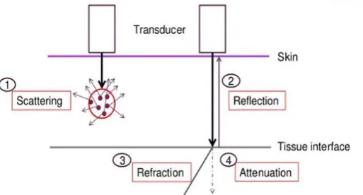
ULTRASOUND TISSUE INTERACTION9:
The ultrasound waves traverse through tissues and undergo either
REFLECTION:
The waves are reflected back as echoes.
REFRACTION:
The waves change direction after hitting an interface between two media with different velocities of sound transmission.
SCATTERING:
If the incident waves are not at right angle, then the returning echoes are scattered in all directions in a non-uniform manner
ABSORPTION:
8
DIFFRACTION:
The spreading out of the ultrasound waves as its moves further away from the source.
ATTENUATION:
[image:18.595.168.425.358.496.2]The returning echoes become weaker due to absorption, scattering or refraction.
Figure 2.- Ultrasound Tissue Interaction. ECHOGENICITY10:
Echogenicity denotes the ability of a tissue to reflect ultrasound waves in comparison with the surrounding structures.
HYPERECHOIC:
9
HYPOECHOIC:
Structures through which sound waves pass easily are called hypoechoic. Here the reflected sound waves are of less energy and appear grey on the screen. Eg: fat.
ANECHOIC:
Areas from where the sound waves are not reflected back are termed anechoic and appear black on the screen. Eg: Blood vessels
[image:19.595.79.522.351.516.2]10
MODES OF ULTRASOUND: ‘A’ MODE :
It is called ‘Amplitude Modulation” and shows amplitude spikes of varying heights. It has x axis and y axis. Amplitude is displayed along the y axis and depth along the x axis. Mostly used in ophthalmology for optic nerve imaging.
‘B’ MODE:
It is called ‘Brightness Modulation’. This is the mode used in regional anaesthesia. It is based on the brightness or the intensity of the echo and is two dimensional. It does not produce vertical spikes. The echo intensity is represented on the z axis and the depth is represented on the x axis. It does not have a y axis. The images are displayed either as large dots which denote strong echoes or as small dots, which denote weak echoes.
‘M’ MODE:
11
CONTROLS IN THE MACHINE:
GAIN-
It alters the intensity of the received signal.
TIME-GAIN COMPENSATION (TGC)-
It differentially amplifies signals from varying depths and thus provides compensation for loss of signal from deeper tissues.
FOCUS –
The beam is adjusted so that it is narrowest at the required depth to image the target structure.
DEPTH-
Varying the depth helps to keep the target structure in the middle of the screen.
ERGONOMICS :
12
PROBE ORIENTATION:
Transducer probes have a marker at one end. The operator has to orient himself as to which side of the screen corresponds to the marker on the probe. This will help in avoiding confusion that may arise when the probe is being manipulated or rotated.
NEEDLE ADVANCEMENT APPROACHES:
The needle can be inserted either parallel to the ultrasound waves (in- plane) or not parallel ( out of plane) to the ultrasound waves. In an in-plane approach, the entry of the needle is at the side of the probe and the entire needle shaft is visualized as it approaches the target. In the out- of- plane approach, the entry of the needle is away from the probe and only the tip of the needle is seen.
13
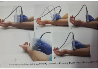
PROBE MANIPULATION11:
Understanding of the manipulation of a transducer probe is essential for regional block with ultrasound. The various probe manoeuvres are sliding, tilting, compression, rocking and rotation.
SLIDING:
Moving the transducer along the known course of the nerve helps in its identification.
TILTING:
Tilting the probe from side to side or cross plane varies the echo brightness of peripheral nerves and optimising this angle promotes nerve visibility.
COMPRESSION:
It is generally used to confirm venous structures; compression pushes air out of field, provides better contact and brings structures closer to the surface of the transducer.
ROCKING:
14
ROTATION:
[image:24.595.94.482.191.463.2]Rotating the probe produces true short axis view rather than oblique or long axis views.
Figure-5. Ultrasound probe manipulation.
ANISOTROPY:
15
ADVANTAGES OF ULTRASONOGRAPHY:
Allows direct visualization of the nerves and their relationship to other structures so reduces the volume of drug needed and vascular injury12.
The spread of the local anaesthetic can be visualized and hence decreases chances of intravascular injection
Repositioning of the needle can be done in case of misdistribution of local anaesthetic.
Improves the quality of block.
Allows for patient variability and anatomic variations.
Avoidance of side effects due to excess dose of local anaesthetic Avoidance of painful muscle contractions unlike peripheral nerve
stimulators.
16
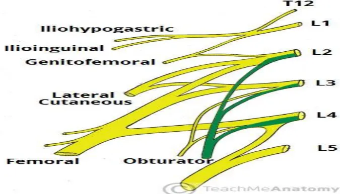
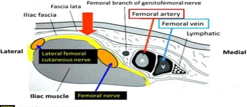
ANATOMY OF NERVES OF LOWERLIMB
13,14The four major nerves innervating the lower limb are the femoral nerve, lateral femoral cutaneous nerve, obturator nerve and the sciatic nerve. These nerves are terminal branches of the lumbosacral plexus.
LUMBOSACRAL PLEXUS:
[image:26.595.117.455.418.611.2]It is formed by the anterior rami of L1-L4 nerves. The anterior rami of L4 and L5 combine to form lumbosacral trunk which joins with anterior rami of S1 TO S3 to form sacral plexus. The lumbar plexus lies within the psoas muscle and its branches descend into the proximal thigh.
17
FEMORAL NERVE:
It carries contributions from the anterior rami of L2-L4 and is the largest branch of the lumar plexus.
[image:27.595.87.509.202.380.2]
Figure 7- Femoral Nerve course
[image:27.595.92.474.430.666.2]18
COURSE:
It descends between the psoas major and iliacus muscle behind the iliac fascia and enters the thigh lateral to the femoral artery under the inguinal ligament. It supplies the iliacus and pectineus muscle in the abdomen. The nerve splits into anterior and posterior divisions in the femoral triangle.
INNERVATION:
The anterior division gives rise to intermediate femoral cutaneous nerve and medial femoral cutaneous nerve which are sensory nerves. Nerve to Sartorius, a motor nerve is also a branch of the anterior division of femoral nerve.
19
LATERAL FEMORAL CUTANEOUS NERVE:
[image:29.595.168.432.169.321.2]It originates from L2 and L3.
Figure 9.- Lateral femoral cutaneous nerve course
COURSE:
It emerges from the lateral border of the psoas major muscle, then crosses the iliacus muscle obliquely and heads towards the anterior superior iliac spine. It is covered on its course by fascia iliaca. While passing behind the inguinal ligament, close to its lateral insertion at anterior superior iliac spine, the lateral femoral cutaneous nerve perforates fascia iliaca.
INNERVATION:
20
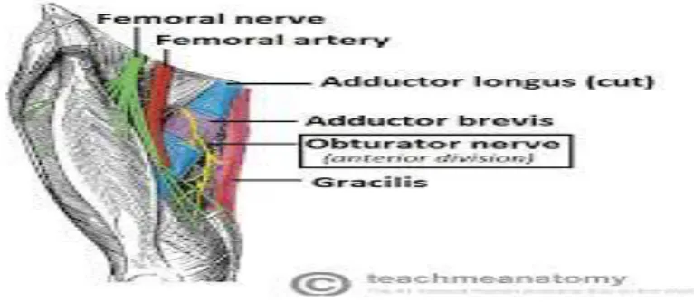
OBTURATOR NERVE:
[image:30.595.175.420.170.316.2]It is derived from L2-L4 divisions of the lumbar plexus.
Figure 10.-Obturator nerve course.
[image:30.595.106.490.385.552.2]21
COURSE:
It continues from its formation within the psoas major along the pelvic sidewall and posterior to the common iliac vessels. After passing over the pelvic brim, it enters the obturator canal and divides into anterior and posterior divisions. The psoas and pectineus muscle separate the obturator nerve from the femoral nerve along its course and so obturator nerve is not reliably blocked by fascia iliaca compartment block.
INNERVATION:
The obturator nerve supplies the adductors of thigh; gracilis and pectineus. It also gives sensory innervation to the medial aspect of thigh.
SCIATIC NERVE:
It arises from the anterior and posterior divisions of the anterior roots of L4, L5, S1, S2, S3. It supplies all the muscles below knee and also the muscles of the posterior compartment of thigh. The sensory innervation is to the entire lower leg and foot except on the medial aspect which is innervated by the saphenous nerve.
22
FASCIA ILIACA
14:
The fascia iliaca extends from the lower thoracic vertebrae to the anterior thigh. It lines the posterior abdomen and pelvis covering psoas major and iliacus muscle and also forms the posterior wall of the femoral sheath which contains the femoral vessels. It is covered by the fascia lata in the femoral triangle with which it blends distally.
ATTACHMENTS:
The fascia iliaca is attached laterally to the thoracolumbar fascia, iliac crest, anterior superior iliac spine and sartorius fascia. It is attached medially to the vertebral column, pelvic brim, and pectineal fascia and anteriorly to the posterior part of inguinal ligament and fascia lata. The space between the inguinal ligament and the hip bone is divided into a medial and a lateral compartment.
23
The fascia iliaca separates the lacuna musculorum from the lacuna vasorum with fibres that link to the capsule of the hip joint and hence forms a functional septum between the two lacunae.
[image:33.595.118.477.194.531.2].
24
FASCIA ILIACA COMPARTMENT
14 [image:34.595.96.511.343.523.2]The Fascia Iliaca Compartment is a potential space with the following limits: The space is covered above by the posterior surface of fascia iliaca; below by the iliacus and psoas major muscle. On the medial side, the space is limited by the vertebral column and is continuous craniomedially with the space between the quadratus lumborum muscle and its fascia. Craniolaterally the space is limited by the inner lip of the iliac crest.
25
FASCIA ILIACA COMPARTMENT BLOCK
Dalens et al14 first described the Fascia Iliaca Compartment Block on children using a landmark technique. It can be carried out during prehospital care, in emergency department and in the pre operative setting. It is a relatively low skill, safe and effective technique15 to provide perioperative analgesia in fracture femur patients. Ultrasound guidance will increase the success rate of the block.16
26
LANDMARK APPROACH17:
[image:36.595.131.461.310.511.2]The inguinal ligament and the femoral pulse are identified and the length of the inguinal ligament is divided into thirds. A blunt tip needle is inserted 1cm distal to the junction between the middle and the outer thirds. A blunt tip needle is inserted in a slightly cephalad direction and as the needle passes through fascia lata and iliaca, two “pops” will be felt. Then 30 to 40 ml of local anaesthetic is injected after negative aspiration.
27
ULTRASOUND GUIDED APPROACH18:
[image:37.595.77.513.423.584.2]The patient is placed in supine position. A high frequency linear ultrasound probe is placed in a horizontal direction over the anterior part of the thigh just below the inguinal ligament. The femoral artery is identified first. Then the iliacus muscle is identified lateral to the artery, covered by the fascia iliaca. The needle is then inserted either in plane or out of plane. The needle is advanced until the tip of the needle is placed beneath the fascia iliaca appreciating the pop off as the fascia is perforated. After negative aspiration, the local anaesthetic is injected and the spread of the local anaesthetic should be visible on the ultrasound machine.
28
29
CLINICAL PHARMACOLOGY
[image:39.595.98.452.177.297.2]BUPIVACAINE:19,20,21
Figure 17.- Structural formula of Bupivacaine
30
MECHANISM OF ACTION:
Bupivacaine enters the nerve’s axon membranes and accumulate within the axoplasm. It binds to the alpha subunit of the voltage gated Na+ channels on the interior of the cell membrane, prevents channel activation and inhibits the sodium influx associated with membrane depolarisation. Local anaesthetics binding to the Na+ channel doesn’t alter the resting membrane potential but increases the threshold for impulse conduction until an action potential cannot be generated and impulse propagation is abolished.
PHARMACOKINETICS:
31
Bupivacaine is a weak base with a pKa of 8.1.At physiological pH of 7.4, 17% is non-ionised. It is highly protein bound (95%), and α1 acid glycoprotein is the most important plasma protein binding site. Its volume of distribution is 73 L. The elimination half-life is 210 minutes. The Clearance is 0.47 L/min. Bupivacaine undergoes biotransformation in liver by aromatic hydroxylation, N-dealkylation, amide hydrolysis, and conjugation. The metabolites are excreted via the kidney. Less than 5% of the drug is excreted unchanged.
USES:
Bupivacaine is used in local infiltration, peripheral nerve blocks, sympathetic nerve block, epidural and caudal block. In oral and dental surgery, this drug provides excellent surgical anaesthesia and extended post operative analgesia.
The concentrations used are:
32
CONTRAINDICATIONS:
Bupivacaine is contraindicated in patients with known hypersensitivity reactions to it or other amide group of local anaesthetics; in obstetrical paracervical blocks and in intravenous regional anaesthesia as there is a chance of tourniquet failure which can lead to the drug being absorbed into systemic circulation leading to cardiac arrest. The 0.75% preparation is contraindicated in epidural anaesthesia during labour as it is known to be associated with refractory cardiac arrest.
ADVERSE EFFECTS:
The maximum dose of bupivacaine is 2-3 mg/kg. The toxic plasma concentration is >3mcg/ml. Toxicity can occur due to over dosage, accelerated absorption, intravascular injection or slow metabolic degradation. Toxicity is due to the blockade of the Na+ channels on the excitable membranes in the brain and myocardium. Bupivacaine is markedly cardiotoxic.
CNS:
33
CVS:
The marked cardiotoxicity of bupivacaine is because it binds more strongly and dissociates more slowly from the Na+ channels. Toxic doses produce cardiac dysrhythmias, atrioventricular block, ventricular tachycardia and ventricular fibrillation, bradycardia and asystole.
TREATMENT:
34
[image:44.595.180.404.128.228.2]FENTANYL:
Figure 18.-Structural formula of fentanyl
Fentanyl [N-(1-(2-Phenylethyl)-4-piperidinyl)-N-phenylpropanamide ] is a phenylpiperidine-derivative synthetic opioid agonist. It was first synthesized by Paul Janssen.22,23. Its molecular formula is C28H36N2O8. The molecular weight of free base is 336.5 and that of citrate salt is 528.59404. It is 75 to 125 times more potent as an analgesic than morphine.
MECHANISM OF ACTION:
35
endogenous ligands by binding to opioid receptors and decreasing the neurotransmission predominantly by presynaptic inhibition of the neurotransmitters .
PHARMACOKINETICS:
Fentanyl has a rapid onset of 5 minutes and a short duration of action of 30 to 60 minutes by the IV route. The rapid onset is due to its greater lipid solubility which easily permits its passage across the blood brain barrier. The short duration of action is because of its rapid redistribution into inactive tissues like fat and skeletal muscles and hence a decrease in plasma concentration. The lungs exert a significant first pass effect of upto 75% of an injected dose of the drug. When a continuous infusion or multiple IV doses of fentanyl is administered, there is progressive saturation of these inactive tissue sites, so the plasma concentration increases and hence the duration of analgesia and depression of ventilation may be prolonged.
36
concentrations are maintained by slow reuptake from the inactive tissue sites which is responsible for the persistent drug effect and the prolonged elimination half time.
Fentanyl is metabolised by N-demethylation producing norfentanyl, hydroxyproprionyl - fentanyl and hydroxyproprionyl - norfentanyl. Norfentanyl is the principal metabolite and can be detected in the urine for 72 hours after a single IV dose. Fentanyl is a substrate for hepatic P-450 enzymes and so there are possibilities of drug interactions25. Less than 10% of fentanyl is excreted unchanged in urine.
PREPARATIONS:
Fentanyl is available as pills, skin patch, lozenge, a film that dissolves in mouth, nasal sprays and as an i.v.preparation.
USES AND DOSAGE:
37
Transmucosal preparations27 like lozenge mounted on a handle or oral transmucosal preparation are designed to deliver 5 to 20 mcg/ kg of fentanyl. It is used to decrease preoperative anxiety and facilitate induction especially in children.
Transdermal patches28 delivering 75 to 100 mcg/hour are applied before the induction of anaesthesia and left in place for 24 hours. They reach peak plasma concentration in about 18 hours, tend to remain stable till the presence of the patch, followed by decreasing plasma concentration after the removal of the patch and are used for post operative analgesia.
Intranasal fentanyl is available in doses of 50, 100 and 200mcg. The bioavailability is 70 to 90%. It is relatively safe with low side effects and used especially in children.
ADVERSE EFFECTS:
38
REVIEW OF LITERATURE
A study conducted by Jadon et.al29., compared the femoral nerve block and intravenous fentanyl for analgesia obtained in surgery for femur fractures. 60 patients were divided into two groups. In one group, femoral nerve block was performed using a peripheral nerve stimulator with 20 ml of 1.5% lignocaine with adrenaline. In the other group, 1mcg/kg of fentanyl i.v. was given. Both these interventions were done 5 minutes before positioning and then both the groups received subarachnoid block.
In femoral nerve block group, during positioning, the visual analogue scale score was significantly lower (P=0.0020). The quality of positioning (P=0.000027) and the patient acceptance (P=0.000031) were significantly better when compared to intravenous fentanyl. The time required to perform subarachnoid block was also less in femoral nerve block. (P=0.000049).
39
M.J.Yun et. al.30, compared the analgesia obtained while positioning between fascia iliaca compartment block and intravenous alfentanil in the elderly who were posted for surgery for neck of femur fracture. In one group, i.v. alfentanil 10mcg/kg loading dose was given and then an infusion of 0.25mcg/kg/min was started 2 minutes prior to subarachnoid block. In the second group, fascia iliaca compartment block was done with 30ml of ropivacaine 20 minutes prior to subarachnoid block.
The visual analogue scale score was lower (P=0.001) and the acceptance of the patient was better in the block group compared to intravenous alfentanil. Also, the mean time taken to perform subarachnoid block was also significantly lower (P=0.009) in the fascia iliaca compartment group.
The study showed that fascia iliaca compartment block is more efficient compared to intravenous alfentanil for positioning in the elderly who underwent subarachnoid block for neck of femur fractures.
40
fifteen minutes before spinal block with 20 ml of 0.5% bupivacaine and 10ml of normal saline. The other 32 patients were given intravenous fentanyl 0.5mcg/kg initially followed by another 0.5mcg/kg five minutes later. Additional fentanyl 0.5mcg/kg was given in increments if the pain scores were above 4. Subarachnoid block was then performed in both the groups.
The results obtained showed that the requirement of additional fentanyl, the satisfaction of positioning and the time taken to achieve spinal block(P=0.74) did not vary significantly between the two groups.
Femoral nerve block and intravenous fentanyl were compared by SIA S.et.al.,32 for analgesia during positioning in fracture shaft of femur surgeries done under spinal block. Patients with fracture shaft of femur posted for surgery under spinal block were randomized into two groups. One group were given femoral nerve block with 15 ml of 1.5% lidocaine under the guidance of a peripheral nerve stimulator while the other group were given 3mcg/kg of intravenous fentanyl. Spinal block was done after 5 minutes in the sitting position in both the groups.
41
anaesthesia was lesser (P<0.05) in the femoral nerve block group compared to intravenous fentanyl.
The results showed that femoral nerve block is more efficacious during positioning compared to intravenous fentanyl in fracture shaft of femur surgeries done under spinal block.
42
Hence the results concluded that femoral nerve block provides better analgesia resulting in adequate positioning, rapid performance of spinal and higher acceptance among patients with femoral fracture during positioning for administration of spinal anaesthesia.
Singh AP. et.al.,34 conducted a comparative study in patients who underwent femur surgery under combined spinal epidural block. 30 patients in Group I were given FNB using nerve stimulator with 0.2% ropivacaine (15 ml) and in 30 patients in Group II, i.v. fentanyl 0.5 mcg/kg was given as preemptive analgesia.
VAS at 2 min in Group I was 5.63 and in Group II it was 8.00. Satisfaction score was better in Group I as compared to Group II patients. Time to administer subarachnoid block was 17.80 min in patients of Group I as compared to 25.03 min in Group II patients. Postoperatively, VAS scores were lower in Group I than Group II patients. The frequency of epidural top‑ups was higher in Group II than in Group I patients.
FNB is comparatively better in comparison to I.V. fentanyl when used as preemptive and postoperative analgesic in patients being operated for fracture femur.
43
Kumar D. et. Al35 performed a study in 50 patients undergoing surgery for hip fracture. All 50 patients received an USG guided Fascia Iliaca Compartment Block (FICB) in the premedication room with 30 mL of 0.5% ropivacaine by 23G spinal needle. Sensory blockade was evaluated at 5, 10 and 20 minutes after giving ropivacaine using loss of perception to cold in the lateral, anterior and medial part of the thigh. Visual analogue scale scores were noted before the block, 20 minutes after block and during positioning for spinal anaesthesia. Patient’s acceptance for FICB was evaluated 24 hour after arriving back to the orthopaedics ward using a two point score.
44
Before FIC block average VAS was 7.5 which was decreased to average of 2.94 at 20 minutes after block which was statistically significant (p<0.01). During positioning for spinal anaesthesia, 46 patients had VAS less than 4. Positioning during spinal anaesthesia was assessed unsatisfactory in 2 cases, satisfactory in 5 cases, good in 25 cases and excellent in 18 cases.
It was concluded that Ultrasound guided FICB can be performed safely without complications in controlling pain for patients with hip fracture.
45
The VAS scores before the procedure at rest (median values)were statistically comparable in both the group i.e. 8 (7-8) in FICB and 7.5 (7-8) in diclofenac group (p=0.756). After 10 minutes of intervention, the median VAS score was same in both the groups i.e. 7 (p=0466). After 20 minutes, in group F, median VAS score was 3 (2-3) and 5 (5-6.2) in diclofenac group. There was a statistically significant difference when these groups were compared (P=0.01). After 30 minutes the VAS score was 2 (1-2) in group F and 3 (1.8-3.6) in group D with P-value <0.01. After 60 minutes, median VAS score for group F was 1 (1-1.3) and 3 (2-3) for group D with P<0.01. Pain assessed by VAS score with passive hip flexion before the procedure was same in both the groups i.e. (8-9). After 30 minutes, it was 2 (2-3) in group F and 6 (5-7) in group D. The difference was a statistically significant when these groups were compared (P <0.01). The duration of analgesia was significantly longer in group F than diclofenac group when both the groups were statistically compared P <0.01
46
Foss NB et.al.,37 did a study comparing the analgesia obtained between fascia iliaca compartment block and i.m. morphine in hip fractures. 48 patients were allocated into 2 groups. One group were given fascia iliaca compartment block with 1% mepivacaine while the other group were given 0.1mg/kg i.m. morphine. Intravenous morphine was used as rescue analgesic. The results obtained showed that the analgesia obtained was higher in the block group at rest (P<0.01) and on movement. (P=0.02).
Lopez et.al.,38 conducted a study to assess the analgesia obtained with fascia iliaca compartment block in femur fractures in pre-hospital care. In 27 patients, fascia iliaca compartment block was done with 20 ml of 1.5% lidocaine with adrenaline using landmark technique. The simplified verbal score was significantly lower (P<0.05) before the block, 10 minutes later and on arrival at the trauma care center.
The study concluded that fascia iliaca compartment block provides effective preoperative analgesia in femur fractures.
47
MATERIALS AND METHODOLOGY
60 patients posted for femur surgeries at Government Kilpauk Medical College Hospital and Government Royapettah hospital from February 2016 to July 2016 were assessed for the inclusion and exclusion criteria and were included in the study after obtaining written informed consent.
SAMPLE SIZE CALCULATION: Sample size was determined based on Study :
Comparative evaluation of femoral nerve block and intravenous fentanyl for positioning during spinal anaesthesia in surgery of femur fracture
Authored by : Ashok Jadon,et al29 Published in:
Indian Journal of Anaesthesia | Vol. 58 | Issue 6 | Nov-Dec 2014
48
Description:
The formula for determining sample size is given as:
Where n = Sample size
σ = Population standard deviation
e = Margin of error
Z = The value for the given confidence interval •The confidence level is estimated at 95%
•Standard deviation 3.09
•With a z value of 1.96
•The confidence interval or margin of error is estimated at +/-0.80
• Assuming that 80 percent as power of the study, minimum sample size required for the study was calculated to be 58.
In our study 60 subjects were chosen
49
STUDY DESIGN:
Prospective, Randomized, single-blind, Controlled study.
INCLUSION CRITERIA:
1) Patients belonging to ASA grade I and II.
2) Patients of either sex, between the age group 18 to 55 years.
3) Patients with fracture femur, posted for surgery under sub-arachnoid block.
4) Patients who give a valid informed consent.
EXCLUSION CRITERIA:
1) Patients not satisfying inclusion criteria.
2) Patients belonging to ASA grade III or IV.
3) Patients with hemorrhagic diathesis, neurological disorders, psychiatric disorders.
4) Previous femoral bypass surgery.
5) Patients with allergy to local anaesthetics or opioids.
50
7) Patients on previous opioid therapy.
8) Morbid obesity.
9) Patients who will be administered with supplementary epidural or general anaesthesia. (In patients with prolonged surgeries when conversion is required).
10) Patients with spinal deformities.
11) Patients who decline consent
12) Patients with language barrier.
MATERIALS:
1) Boyle’s machine
2) Laryngoscope with different blade sizes
3) Endotracheal tubes
4) Other airway gadgets used in case of difficult intubation
5) Mind Ray ultrasound machine with linear transducer probe
6) Ultrasound jelly
51
8) Disposable 10ml, 5ml syringes, sterile gloves
9) 18G venflon needle for Fascia Iliaca Compartment Block
10) 25 G Quincke needle for sub arachnoid block
DRUGS:
1) Bupivacaine 0.5% available as 20ml vial
2) Inj. Fentanyl available as 2ml ampoule
3) Preservative free bupivacaine available as 4 ml ampoule for spinal block
4) Emergency drugs needed for resuscitation
5) Distilled water.
MONITORS:
NIBP
ECG
52
METHODOLOGY:
Patients satisfying the inclusion criteria were selected, counselled about the risks and benefits involved in the study. After obtaining informed consent, patients who were willing to be included in the study were enrolled. They were preoperatively evaluated, clinically examined and assessed. A total of 60 patients were included in the study. They were randomly allocated into two groups.
Group FICB : were administered ultrasound guided Fascia Iliaca Compartment Block preoperatively.
Group FENT: were administered intravenous fentanyl preoperatively.
53
Group FICB patients were placed in supine position. The local anaesthetic solution was prepared with 15 mL of 0.5% bupivacaine and 15ml of distilled water and hence 30ml of 0.25% bupivacaine. The Ultrasound Machine was powered on and the linear array probe was covered with sterile dressing after applying ultrasound gel. The probe was placed in a horizontal direction over the anterior part of thigh just below the inguinal ligament. The ultrasound setting used to visualise was at a frequency of 10 MHz and a depth of 3-4cm. The gain and focus were adjusted according to the image scanned. Femoral artery was identified first .Then the iliacus muscle covered by fascia iliaca was identified lateral to the artery. An 18G needle was then inserted in plane to the ultrasound beam. The needle was advanced until the tip of the needle was placed beneath the fascia iliaca (appreciating the give as the fascia is perforated) and after negative aspiration, the local anaesthetic was injected and its spread visualized on the ultrasound screen. The fascia iliaca compartment block was done 15 minutes before the sub arachnoid block.
54
Hemodynamic variables like heart rate, non invasive blood pressure, saturation of oxygen, respiratory rate were recorded after the block/ iv fentanyl and at five minutes intervals till positioning .
[image:64.595.106.502.307.447.2] The analgesia provided by either of the modes was assessed by using Visual analogue scale scores 15 minutes(ie. during positioning) after the block/ I.V. Fentanyl.
Figure 19.- Visual Analogue Scale score
55
The quality of patient positioning for administering spinal anaesthesia was recorded by another anaesthesiologist blinded to the mode of analgesia with scores of 0-332.
0-Not satisfactory 1-satisfactory 2-good 3-optimal
Time to perform spinal anaesthesia will be recorded (time from beginning of positioning to end of spinal)32.
Patient satisfaction was also recorded
1- satisfactory
2- not satisfactory
Post-operative analgesia was standardized in all patients of both groups with Inj.Tramadol 50 mg I.V. 8th hourly; first dose was given whenever patient complained of pain.
56
OBSERVATION AND RESULTS
GROUPS:
GROUP INTERVENTION NUMBER
FICB Group Fascia Iliaca
Compartment Block 30
FENT Group Intravenous fentanyl 30
NULL HYPOTHESIS :
Null Hypothesis : H0
Fascia iliaca compartment block with
bupivacaine is equal in effect to intravenous fentanyl for positioning in patients undergoing spinal anaesthesia in fracture femur surgery Alternate Hypothesis
: H1
Fascia iliaca compartment block with
bupivacaine is better in effect to intravenous fentanyl for positioning in patients undergoing spinal anaesthesia in fracture femur surgery
DATA ANALYSIS:
57
AGE:
Age - Groups
FICB
Group %
FENT
Group %
21-30 years 5 16.67 4 13.33
31-40 years 2 6.67 2 6.67
41-50 years 9 30.00 12 40.00
51-60 years 14 46.67 12 40.00
Total 30 100 30 100
2 2
9 12 14 12 0 2 4 6 8 10 12 14 16
FICB Group FENT Group
Age - Groups
58
Age Distribution FICB Group FENT Group
N 30 30
Mean 46.07 45.53
SD 10.76 9.27
P value
Unpaired t Test 0.8378
Among the patients undergoing spinal anaesthesia in fracture femur surgery, there was no statistically significant difference in relation to age distribution between FICB group (mean=46.07, SD=10.76) and FENT group (mean=45.53, SD=9.27) with a p value of >0.05 as per unpaired t test. Therefore we fail to reject the null hypothesis that there is no difference in age distribution between the intervention groups.
46.07 45.53
10.76 9.27
0.00 5.00 10.00 15.00 20.00 25.00 30.00 35.00 40.00 45.00 50.00
FICB Group FENT Group
Age Distribution
59
GENDER:
Gender -
Groups FICB Group %
FENT
Group %
Male 19 63.33 20 66.67
Female 11 36.67 10 33.33
Total 30 100 30 100
P value
Chi Squared Test 0.7866
Among the patients undergoing spinal anaesthesia for fracture femur surgery, there was no statistically significant difference in relation to gender status between FICB group (male-63.33%, female-36.67%) and FENT group (male-66.67%, female-33.33%) with a p value of >0.05 as per chi squared test. Therefore we fail to reject the null hypothesis that there is no difference in gender status between the intervention groups.
19 20
11 10
0 5 10 15 20 25
FICB Group FENT Group
Gender - Groups
60
WEIGHT:
Weight - Groups
FICB
Group %
FENT
Group %
51-60 kg 14 46.67 11 36.67
61-70 kg 15 50.00 17 56.67
71-80 kg 1 3.33 2 6.67
Total 30 100 30 100
14
11 15
17
1 2
0 2 4 6 8 10 12 14 16 18
FICB Group FENT Group
Weight - Groups
61
Weight Distribution FICB Group FENT Group
N 30 30
Mean 62.77 63.20
SD 5.46 5.10
P value
Unpaired t Test 0.7520
Among the patients undergoing spinal anaesthesia in fracture femur surgery, there was no statistically significant difference in relation to weight distribution between FICB group (mean=62.77, SD=5.46) and FENT group (mean=63.20, SD=5.10) with a p value of >0.05 as per unpaired t test. Therefore we fail to reject the null hypothesis that there is no difference in weight distribution between the intervention groups.
62.77 63.20
5.46 5.10
0.00 10.00 20.00 30.00 40.00 50.00 60.00 70.00
FICB Group FENT Group
Weight Distribution
62
NUMBER OF DAYS SINCE FRACTURE:
Number of Days Since Fracture - Groups
FICB
Group %
FENT
Group %
1-2 days 2 6.67 3 10.00
3-4 days 14 46.67 10 33.33
5-6 days 11 36.67 14 46.67
7-8 days 3 10.00 3 10.00
Total 30 100 30 100
2 3
14
10 11
14
3 3
0 2 4 6 8 10 12 14 16
FICB Group FENT Group
Number of Days Since Fracture - Groups
63
Number of Days Since
Fracture FICB Group FENT Group
N 30 30
Mean 4.53 4.67
SD 1.50 1.54
P value
Unpaired t Test 0.7354
Among the patients undergoing spinal anaesthesia in fracture femur surgery, there was no statistically significant difference in relation to duration since fracture distribution between FICB group (mean=4.53, SD=1.50) and FENT group (mean=4.67, SD=1.54) with a p value of >0.05 as per unpaired t test. Therefore we fail to reject the null hypothesis that there is no difference in duration since fracture distribution between the intervention groups.
4.53 4.67
1.50 1.54
0.00 0.50 1.00 1.50 2.00 2.50 3.00 3.50 4.00 4.50 5.00
FICB Group FENT Group
Number of Days Since Fracture
64
VAS DURING POSITIONING:
VAS During Positioning -
Groups
FICB
Group %
FENT
Group %
VAS 0 15 50.00 5 16.67
VAS 2 13 43.33 18 60.00
VAS 4 2 6.67 5 16.67
VAS 6 0 0.00 2 6.67
Total 30 100 30 100
15 5 13 18 2 5 0 2 0 5 10 15 20
FICB Group FENT Group
VAS During Positioning - Groups
65
VAS During Positioning FICB Group FENT Group
N 30 30
Mean 1.13 2.27
SD 1.25 1.55
P value Unpaired t Test
0.0029
1.13
2.27
1.25
1.55
0.00 0.50 1.00 1.50 2.00 2.50
FICB Group FENT Group
VAS During Positioning
66
Among the patients undergoing spinal anaesthesia in fracture femur surgery, there was a statistically significant difference in relation to VAS score during positioning between FICB group (mean=1.13, SD=1.25) and FENT group (mean=2.27, SD=1.55) with a p value of <0.05 as per unpaired t test. Therefore we reject the null hypothesis that there is no difference in VAS score during positioning distribution between the intervention groups.
67
QUALITY OF PATIENT POSITIONING:
0-
NOT SATISFACTORY
1-
SATISFACTORY
2-
2-GOOD
3-
OPTIMAL
QOP
Positioning –
Groups
FICB
Group
%
FENT
Group
%
QOPP 0
0
0.00
2
6.67
QOPP 1
2
6.67
5
16.67
QOPP 2
13
43.33
18
60.00
QOPP 3
15
50.00
5
16.67
Total
30
100
30
100
0 2 2 5 13 18 15 5 0 2 4 6 8 10 12 14 16 18 20
FICB Group FENT Group
QOP Positioning - Groups
68
Quality of Patient
Positioning
FICB Group
FENT Group
N
30
30
Mean
2.43
1.87
SD
0.63
0.78
P value
Unpaired t Test
0.0024
2.431.87
0.63 0.78
0.00 0.50 1.00 1.50 2.00 2.50 3.00
FICB Group FENT Group
Quality of Patient Positioning
69
Among the patients undergoing spinal anaesthesia in
fracture femur surgery, there was a statistically significant
difference in relation to quality of patient positioning between
FICB group (mean=2.43, SD=0.63) and FENT group
(mean=1.87, SD=0.78) with a p value of <0.05 as per unpaired t
test. Therefore we reject the null hypothesis that there is no
difference in quality of patient positioning distribution between
the intervention groups.
The mean quality of patient positioning score was
significantly higher in FICB group compared to FENT
group
by
a mean difference of 0.57 scoring points (23% higher). This
difference is significant with a p-value of 0.0024 as per unpaired
70
PATIENT SATISFACTION:
1-SATISFACTORY
2- N0T SATISFACTORY
Patient
Satisfaction -
Groups
FICB
Group
%
FENT
Group
%
Yes
29
96.67
23
76.67
No
1
3.33
7
23.33
Total
30
100
30
100
P value
Fishers Exact Test
0.0284
29 23 1 7 0 5 10 15 20 25 30 35FICB Group FENT Group
Patient Satisfaction - Groups
71
Among the patients undergoing spinal anaesthesia in fracture
femur surgery, there was a statistically significant difference in
relation to patient satisfaction status between FICB group
(yes=96.67%, no=3.33%) and FENT group (yes=76.67%,
no=23.33%) with a p value of <0.05 as per unpaired t test.
Therefore we reject the null hypothesis that there is no difference
in patient satisfaction status between the intervention groups.
The positive patient satisfaction status was significantly
higher in FICB group compared to FENT group by a percentage
difference of 20.00 (21% higher). This difference is significant
72
TIME TO PERFORM SAB:
Time to
Perform SAB -
Groups
FICB
Group
%
FENT
Group
%
≤ 5.00 mins
17
56.67
3
10.00
5.01-6.00 mins
13
43.33
16
53.33
6.01-7.00 mins
0
0.00
8
26.67
7.01-8.00 mins
0
0.00
3
10.00
Total
30
100
30
100
17 3 13 16 0 8 0 3 0 2 4 6 8 10 12 14 16 18
FICB Group FENT Group
Time to Perform SAB - Groups
73
Time to Perform SAB
FICB Group
FENT Group
N
30
30
Mean
4.90
5.86
SD
0.55
0.83
P value
Unpaired t Test
<0.0001
4.90
5.86
0.55 0.83
0.00 1.00 2.00 3.00 4.00 5.00 6.00 7.00
FICB Group FENT Group
Time to Perform SAB
74
Among the patients undergoing spinal anaesthesia in
fracture femur surgery, there was a statistically significant
difference in relation to time to perform subarachnoid block
between FICB group (mean=4.90, SD=0.55) and FENT group
(mean=5.86, SD=0.83) with a p value of <0.05 as per unpaired t
test. Therefore we reject the null hypothesis that there is no
difference in time to perform subarachnoid block distribution
between the intervention groups.
The mean time to perform subarachnoid block was
significantly shorter in FICB group compared to FENT group by
a mean difference of 58 seconds (16% shorter). This difference is
75
HEART RATE:
Heart Rate (beats
per min)
Before
Block
5 mins
10
mins
15 mins
(During
Positioning)
FICB
Group
Mean
86.93
87.67
86.90
86.13
SD
8.77
8.18
8.41
8.44
FENT
Group
Mean
88.70
85.63
82.37
79.67
SD
7.55
7.29
7.19
7.13
P value
Unpaired t Test
0.4065
0.3138 0.0287
0.0022
87
88 87
86 89 86 82 80 74 76 78 80 82 84 86 88 90
Before Block 5 mins 10 mins 15 mins (During
Positioning)
Heart Rate (beats per min)
76
Among the patients undergoing spinal anaesthesia in
fracture femur surgery, there was a statistically significant
difference in relation to heart rate at 10 -15 minutes between
FICB group (mean=86.52, SD=8.39) and FENT group
(mean=81.02, SD=7,10) with a p value of <0.05 as per unpaired
t test. Therefore we reject the null hypothesis that there is no
difference in heart rate distribution at (10-15 mins) between the
intervention groups.
The mean heart rate was significantly lower in FENT group
compared to FICB group by a mean difference of 6 bpm (6%
lower). This difference is significant with a lowest p-value of
77
MEAN ARTERIAL PRESSURE:
Mean Arterial
Pressure
(mm Hg)
Before
Block
5 mins
10
mins
15 mins
(During
Positioning)
FICB
Group
Mean
100.17
99.83
98.20
97.73
SD
7.34
5.97
6.06
5.61
FENT
Group
Mean
101.87
100.80
99.20
98.17
SD
6.77
5.77
5.36
5.56
P value
Unpaired t Test
0.3548
0.5262 0.5010
0.7649
100 100 98 98 102 101 99 98 95 96 97 98 99 100 101 102 103
Before Block 5 mins 10 mins 15 mins (During
Positioning) M e an Val u e s
Mean Arterial Pressure (mm Hg
)
78
Among the patients undergoing spinal anaesthesia in
fracture femur surgery, there was no statistically significant
difference in relation to mean arterial pressure between FICB
group (mean=98.98, SD=5.96) and FENT group (mean=100.01,
SD=5.75) with a p value of >0.05 as per unpaired t test.
Therefore we fail to reject the null hypothesis that there is no
difference in mean arterial pressure distribution between the
79
SPO2:
Peripheral Capillary
Oxygen Saturation
(%)
Before
Block
5 mins
10
mins
15 mins
(During
Positioning)
FICB
Group
Mean
98.03
98.73
99.13
99.27
SD
0.76
0.69
0.63
0.52
FENT
Group
Mean
98.10
98.97
99.17
98.94
SD
0.76
0.67
0.70
0.89
P value
Unpaired t Test
0.7359
0.1892
0.8467
0.1118
98
99
99
99
98
99 99
99 97 98 98 99 99 100
Before Block 5 mins 10 mins 15 mins (During
Positioning) M e an Val u e s
Peripheral Capillary Oxygen Saturation
(%)
80
Among the patients undergoing spinal anaesthesia in
fracture femur surgery, there was no statistically significant
difference in relation to peripheral capillary oxygen saturation
between FICB group (mean=98.79, SD=0.51) and FENT group
(mean=98.71, SD=0.65) with a p value of >0.05 as per unpaired t
test. Therefore we fail to reject the null hypothesis that there is no
difference in peripheral capillary oxygen saturation distribution
81
RESPIRATORY RATE:
Respiratory Rate
(breaths per min)
Before
Block
5 mins 10 mins
15 mins
(During
Positioning)
FICB
Group
Mean
17.40
17.40
17.17
16.70
SD
1.38
1.19
0.91
1.12
FENT
Group
Mean
17.33
16.97
15.57
14.57
SD
0.96
1.16
1.17
1.48
P value
Unpaired t Test
0.8287
0.0873 <0.0001
<0.0001
17 17
17 17 17 17 16 15 13 14 14 15 15 16 16 17 17 18 18
Before Block 5 mins 10 mins 15 mins (During
Positioning) M e an Val u e s
Respiratory Rate (breaths per min
)
82
Among the patients undergoing spinal anaesthesia in
fracture femur surgery, there was a statistically significant
difference in relation to respiratory rate at (10 -15 minutes)
between FICB group (mean=16.93, SD=0.93) and FENT group
(mean=15.07, SD=1.27) with a p value of <0.05 as per unpaired t
test. Therefore we reject the null hypothesis that there is no
difference in respiratory rate distribution (10-15 mins) between
the intervention groups.
The mean respiratory rate was significantly lower in FENT
group compared to FICB group by a mean difference of 2 breaths
per minute (11% lower). This difference is significant with a
83
FIRST RESCUE ANALGESIC POSTOPERATIVE:
First rescue
analgesic
Postoperative -
Groups
FICB
Group
%
FENT
Group
%
≤ 3.00 hrs
0
0.00
30
100.00
3.01-5.00 hrs
3
10.00
0
0.00
5.01-7.00 hrs
26
86.67
0
0.00
7.01-9.00 hrs
1
3.33
0
0.00
Total
30
100
30
100
0 30 3 0 26 0
1 0
0 5 10 15 20 25 30 35
FICB Group FENT Group
First rescue analgesic Postoperative - Groups
84
First Rescue Analgesic
Postoperative
FICB
Group
FENT
Group
N
30
30
Mean
5.90
1.65
SD
0.80
0.60
P value
Unpaired t Test
<0.0001
5.901.65
0.80 0.60
0.00 1.00 2.00 3.00 4.00 5.00 6.00 7.00
FICB Group FENT Group
First Rescue Analgesic Postoperative
85
Among the patients undergoing spinal anaesthesia in
fracture femur surgery, there was a statistically significant
difference in relation to time of first postoperative analgesic need
between FICB group (mean=5.90, SD=0.80) and FENT group
(mean=1.65, SD=0.60) with a p value of <0.05 as per unpaired t
test. Therefore we reject the null hypothesis that there is no
difference in time of first postoperative analgesic need between
the intervention groups.
The mean time of first postoperative analgesic need was
significantly delayed in FICB group compared to FENT group by
a mean difference of 4 hours and 15 minutes (72% more
delayed). This difference is significant with a p-value of <0.0001
86
DISCUSSION
Spinal anaesthesia is the most commonly used anaesthetic technique of choice in orthopaedics for lower limb fractures. While regional anaesthesia has been shown to be more beneficial compared to general anaesthesia, patient positioning for neuraxial blockade may cause severe pain in patients with femur fractures. Various systemic analgesics are being used to provide pain relief during positioning in these patients. Among the systemic analgesics, opioids are widely used but they are known to be associated with side effects like cognitive impairment, vomiting, urinary retention , respiratory depression especially in the elderly. Nerve blocks like the 3 in 1 block, femoral nerve block, fascia iliaca compartment blockhave all come up as an alternative approach to provide pain relief and improve positioning in these patients39,40.
87
beneath it lateral to the femoral nerve increases the success rate of block and further reduces the risk of neurovascular injury.
In this prospective, randomized study, the efficacy of fascia iliaca compartment block