SEVERITY OF ACUTE ISCHEMIC STROKE IN
CORRELATION WITH MICROALBUMINURIA
DISSERTATION SUBMITTED FOR
MD DEGREE ( BRANCH 1 ) GENERAL MEDICINE MAY 2018
CERTIFICATE FROM THE DEAN
This is to certify that this dissertation entitled “SEVERITY OF
ACUTE ISCHEMIC STROKE IN CORRELATION WITH
MICROALBUMINURIA” is the bonafide work of DR.C.RAJ RAHMAN,
in partial fulfillment of the universityregulations of the Tamil Nadu Dr. M.G.R. Medical University, Chennai, for M.D General Medicine, Branch I
examination to be held in May 2018.
Dr. D.MARUTHU PANDIAN M.S.FAIS.,FICS Dean,
CERTIFICATE FROM THE HOD
This is to certify that this dissertation entitled “SEVERITY OF
ACUTE ISCHEMIC STROKE IN CORRELATION WITH
MICROALBUMINURIA” is the bonafide work of DR.C.RAJ RAHMAN,
in partial fulfillment of the university regulations of the Tamil Nadu Dr. M.G.R. Medical University, Chennai, for M.D General Medicine, Branch I
examination to be held in May 2018.
PROF. DR. V.T. PREMKUMAR M.D.,
Professor and HOD, Department Of Medicine, Government Rajaji Hospital,
CERTIFICATE FROM THE GUIDE
This is to certify that this dissertation entitled “SEVERITY OF
ACUTE ISCHEMIC STROKE IN CORRELATION WITH
MICROALBUMINURIA” is the bonafide work of DR.C.RAJ RAHMAN, in partial fulfillment of the university regulations of the Tamil Nadu Dr. M.G.R. Medical University, Chennai, for M.D General Medicine, Branch I
examination to be held in May 2018.
PROF DR.R.PRABHAKARAN,M.D.,
Professor of Medicine,
Department Of Medicine,
Government Rajaji Hospital,
Madurai Medical College,
DECLARATION
I, DR.C.RAJ RAHMAN, solemnly declare that this dissertation titled
“SEVERITY OF ACUTE ISCHEMIC STROKE IN CORRELATION
WITH MICROALBUMINURIA” is a bonafide record of work done by me at
the Department Of General Medicine, Government Rajaji Hospital, Madurai,
under the guidance of Dr.R.PRABHAKARAN.M.D, Professor, Department of
General Medicine , Madurai Medical college , Madurai.
This dissertation is submitted to The Tamil Nadu Dr. M.G.R Medical
University, Chennai in partial fulfillment of the rules and regulations for the
award of M.D Degree General Medicine Branch- I; examination to be held in
May 2017.
Place: Madurai DR.C.RAJ RAHMAN
ACKNOWLEDGEMENT
Above all I thank the Lord Almighty for His grace and guidance.
I wish to express my sincere thanks to our
Prof.DR.D.MARUTHUPANDIAN MS., Dean, Madurai Medical College and
Government Rajaji Hospital, for permitting me to utilize the clinical materials
from this hospital to conduct the study.
I wish to express my respect and sincere gratitude to my beloved teacher
Prof. Dr. V. T. PREMKUMAR,M.D., Head of the Department of Medicine,
Government Rajaji Hospital, Madurai Medical College for his valuable
guidance and encouragement during the study and also throughout my course
period.
I would like to express my gratitude and sincere thanks to my beloved
teacher, my guide and my Unit Chief Prof. Dr.R.PRABHAKARAN,M.D., for
his valuable suggestions, patience, guidance and support throughout the study
and also throughout my course period.
I am greatly indebted to my beloved Professors,
Dr. R. BALAJINATHAN, M.D., Dr. M. NATRAJAN, M.D.,
Dr. G. BAGYALAKSHMI, M.D.,DR.J.SANGUMANI.MD., Dr. C.
DHARMARAJ, M.D., for their valuable suggestions throughout the course of
I am extremely thankful to the Assistant Professors of Medicine of my
Unit, Dr.N.RAGAVAN,M.D.,Dr.R.SHRIDHARAN,M.D., for their valid
guidance, encouragement and suggestions.
I extend my sincere thanks to Prof. Dr. SRIDHAR, M.D,D.M ., HOD
Department of neurology, Government Rajaji Hospital and Madurai Medical
College for his unstinted support and valuable guidance throughout the study
period.
I am extremely thankful to Prof. Dr.MOHAN KUMARESAN. MD.,
Head of the department of Biochemistry for their constant support, guidance,
cooperation and to complete this study.
I am grateful to my family, colleagues and friends who have encouraged
me during my times of need. Their help and support have made this possible.
Finally, I thank all the patients, the most integral part of the work,
who were always kind and cooperative. I pray for their speedy recovery,
CONTENTS
S.NO CONTENTS PAGE.NO.
1. INTRODUCTION 1
2. AIMS AND OBJECTIVES 4
3. REVIEW OF LITERATURE 5
4. MATERIALS AND METHODS 42
49
5. RESULTS AND INTERPRETATION
6. DISCUSSION 63
7. CONCLUSION 68
8 SUMMARY 69
9 LIMITATIONS OF THE STUDY 71
ANNEXURES
1. BIBLIOGRAPHY
2. KEY TO MASTER CHART 3. PROFORMA
4. MASTER CHART
1
INTRODUCTION
Microalbuminuria is thought to be a marker of wide spread vascular damage and also reflect the systemic trans capillary leakage of albumin.
The significance of microalbuminuria in relation to diabetes has been extensively studied and documented in literature. The structural, functional and biochemical aspects contributing to Microalbuminuria is the basis of several past and ongoing studies.
The prevalence of progression and recession of Microalbuminuria in various disease processes like diabetes, hypertension and coronary arterial disease was repeatedly investigated in several land mark trials and have confirmed its investigated its significance in various studies.
There are few published studies on Microalbuminuria in cerebrovascular diseases. Observations made out of these studies confirm the relationship of the Microalbuminuria in cerebrovascular disease similar to those in vascular disorder of heart and kidney.
2
hampered which leads to arterial vasoconstriction. This causes increase arterial as well as glomerular pressure and permeability.
In endothelial dysfunction glomerular basement membrane loses normal negative charges and loss of heparin like proteoglycan promoting thrombus formation and enhance atherosclerosis. Hence microalbuminuria is gaining recognition as a marker of atherogenic milieu and ischaemic stroke. Microalbuminuria and atherosclerosis found in endothelial dysfunction is manifested as increased intima media thickness of common carotid arteries .
Microalbuminuria (MA) can be defined as urinary albumin excretion rate of 20-300 mg/L in spot sample or 30-300 mg/24 hrs urine collection or urine albumin creatinine ratio in first voided morning sample is 30-300 micro g/mg . In this study, microalbuminuria was defined as 20-300 mg/L.
Microalbuminuria is also a factor that predispose hemorrhagic transformation of Ischemic stroke and poor outcome.
3
Stroke is a major public health problem all over the world. It plays an important role in the morbidity and mortality in late middle age and in the elderly. Stroke is the third most common cause of death in the world after coronary artery disease and cancer. It is the single most important case of severe debility and also the most common life threatening neurological disease.
The burden of stroke is likely to increase substantially in the future because of the aging population. Apart from employing effective stroke prevention programs, identification of factors associated with more severe stroke may help to ease the burden of this coming epidemic.
4
AIMS AND OBJECTIVES
1.To assess the severity of Acute Ischemic stroke in correlation with Micro albuminuria.
2.To assess the duration of presence of microalbuminuria after acute ischemic stroke.
5
REVIEW OF LITERATURE
DEFINITION OF STROKE
A stroke or CVAis a rapidly developing clinical symptoms and / or signs of focal and at times global cerebral function deterioration, with symptoms lasting for more than 24 hours or leading to death, with no identifiable cause other than that of vascular origin.
Cerebral ischemia is usually caused by a reduction in blood flow that lasts longer than several seconds. Neurologic symptoms manifested within seconds because neurons lack glycogen, so energy failure is rapid in this process. If the interruption of flow lasts for more few minutes, infarction or death of brain tissue results
ANATOMY OF CEREBRAL CIRCULATION
ARTERIES OF THE BRAIN
“There are Two internal carotid arteries and 2 vertebral arteries supply the brain and surrounding areas”.The 4 arteries lie within the substance of subarachnoid space and their branches joint on the inferior surface of the brain to form the circle of Willis.
INTERNAL CAROTID ARTERY:
6
towards the neck and perforates the base of the skull by passing through the carotid canal of the temporal bone and then continue its course.
The artery then runs horizontally goesforward through the cavernous sinus and appears on the medial side of the anterior clinoid process and then by perforating the duramater. “After entering the arachnoid matter it now enters the subarachnoid space and goes posteriorly to the region of the medial end of the lateral cerebral sulcus and split into the anterior and middle cerebral arteries”.
BRANCHES OF THE CEREBRAL PORTION
OPHTHALMIC ARTERY:
The ophthalmic artery emerges from the internal carotid artery and enters the orbit via the optic canal below and lateral to the optic nerve. It supplies the eye and surrounding orbital structures . ‘Its terminal branches supply the frontal area of the scalp, the ethmoid sinus and frontal sinuses and the dorsum of the nose’.
POSTERIOR COMMUNICATING ARTERY:
7 CHOROIDAL ARTERY:
The choroidal artery is a small branch that runs posteriorly, close to the optic tract, passes the inferior horn of lateral ventricle and terminate in the choroids plexus. “Choroidal artery gives off small branches to the crus cerebri, lateral geniculate body, optic tract and to the the internal capsule”.
ANTERIOR CEREBRAL ARTERY:
“It runs forward and medially and go through the longitudinal fissure of cerebrum superior to the optic nerve”. Here it communicate with its fellow on the opposite side by means of the anterior communicating artery. It curves backward over the corpus callosum. The cortical branches supply the whole of the surface of the cerebral cortex as far back as the parieto-occipital sulcus. ”.
MIDDLE CEREBRAL ARTERY:
9 BRANCHES OF THE BASILAR ARTERY
PONTINE ARTERIES.
“Pontine arteries are numerous small arteries that enter the substance of the pons along the pontine surface”.
BASILAR ARTERY
The basilar artery, formed by the union of the 2 vertebral arteries, ascends in the groove along the anterior surface of the pons. “At the upper border of the pons, basilar artery split into 2 posterior cerebral arteries”.
BRANCHES OF THE BASILAR ARTERY
PONTINE ARTERIES.
Pontine arteries are numerous small arteries that enter the substance of the pons.
LABRINTHINE ARTERY
10
11
ANTERIOR INFERIOR CEREBELLAR ARTERY
“AICA is the artery passes posteriorly and laterally and supplies the anterior and inferior part of the cerebellum, a few branches pass through the pons and upper part of the medulla of the brain”.
SUPERIOR CEREBELLAR ARTERY
Superior cerebellar artery arises near to the termination of the basilar artery. It wraps around the cerebral peduncle and supplies the superior surface of the cerebellum. “it also supplies the pons, the pineal gland and the superior medullary velum of the brain”.
POSTERIOR CEREBRAL ARTERY
12 CIRCLE OF WILLS
13
14
Lacunar Stroke
ARTERIES TO SPECIFIC BRAIN AREAS
“They are the medial and lateral striate central branches of the middle cerebral artery supply the corpus striatum and the internal capsule; the central branches of the anterior cerebral artery supply the remainder of the structures of the brain”.
The posterior cerebral, posterior communicating and basilar arteries supply the thalamus part of the brain.
15
The basilar and anterior, inferior branches and superior cerebellar arteries supply the pons of the brain’.
The vertebral, anterior and posterior spinal, posterior inferior cerebellar and basilar arteries supply the medulla oblongata.
EPIDEMIOLOGY OF CEREBROVASCULAR DISEASES IN INDIA
The burden of stroke on the community is best reflected by its morbidity.Vellore was surveyed to detect cases of hemiplegia. In the second phase (1969 - 197 I) the population was kept under surveillance for two years and attempts were made to record all cases of hemiplegia. An incidence of 13/1,00,000 population per year was observed. The second study was carried out as a part of WHO collaborative study in Rohtak. Halyana between 1972- 1974. An annual incidence of : 34/1,00,000 population was noted.
In India prevalence vary from region to region. Vellore, Gowribidanur, and Rohtak had a prevalence of 40-60/1,00,000 population, whereas the Eastern part of India showed a prevalence of 100-270/1,00,000.
Stroke incidence varies among different countries. In WHO Monica project has shown high prevalence and mortality among Finnish population. According to this study incidence of stroke varies from 106/1,00,000 in Germany to 290/1,00,000 population of men in Finland.
CLASSIFICATION OF STROKE
16
stroke
1. ANATOMICAL CLASSIFICATION.
A. By vascular supply
a. Carotid
b. Vertebrobasilar
B. By location
a. Supratentorial i. Lobar
ii. Ganglionic /thalamic b. lnfratentorial
i. Cerebel1ar ii.
ii. Brain stern
2. ETIOLOGICAL CLASSIFICATION.
A. By result.
a. Cerebral infarct
b. Cerebral haemorrhage
17
ii.“Subarachnoid”.
RISK FACTORS FOR STROKE
They can be grouped into modifiable and non-modifiable. Some of them are as follows.
Age
“The occurrence of stroke increases dramatically with advancing age, and increasing age is an important risk factor for stroke”.
Sex
Male sex is linked with increased risk of stroke. Race
The rate of cerebral infraction is higher in blacks than in whites. This could be further explained by the higher prevalence of diabetes and hypertension in them.
BLOOD PRESSURE
18
populations. They found that 3mm of Hg reduction in diastolic blood pressure should decrease the number of stroke by about a third. The SHEP study has shown significant reduction in non-fatal stroke events over 5 years in the age group 60 years and above with isolated systolic hypertension treated with active medication.
Diabetes Mellitus
“Diabetes mellitus increases the risk of cerebrovascular disease 2 – 4 fold compared with same risk in nondiabetics. Stroke secondary to diabetes may be caused by cerebrovascular atherosclerosis, cardiac embolism, and othrer abnormalities”. Diabetic patients with retinopathy and autonomic neuropathy are specifically at high risk for atherosclerosis.
Cardiovascular diseases
19
third. The SHEP study has shown a 36% reduction in non-fatal stroke events over 5 years in the age group 60 years and above with isolated systolic hypertension treated with active medication.
Claudication
Claudication is a high risk factor for both myocardial infarction and stroke, presumably reflecting atheromatous disease in various part of the brain and vascular system.
Cigarette smoking
The relative risk is about 1.5. There is a dose dependent relationship between smoking and stroke. Cigarette smoking is important factor both in men and women. ICMR has shown that smoking is a risk factor both in the young and the elderly that predisposes to stroke.
Atrial fibrillation
Most frequent cardiac source of embolism to brain is atrial fibrillation either rheumatic or non rheumatic. Atrial fibrillation is associated with increased risk for stroke accounting for 5 -24 % of all ischaemic strokes and cause for major debility.
Carotid artery disease
20
supraclavicular arterial bruit are strongly related to stenosis of underlying arteries so the examination is important.
Transient ischemic attacks
A TIA patient has an excess risk of stroke by about 5 - 10 times greater than a non TIA patient.
Lipid abnormalities
Increasing levels of total plasma cholesterol, LDL and decreased levels of HDL are strong risk factors for coronary artery disease than stroke. In a study conducted in NIMHANS they found that low HDL cholesterol is a risk factor in stroke group among various lipid fractions. Lipoprotein 'a' has been found to be an independent risk factor for stroke and it is a inflammatory marker.
Some of the risk factors are Abdominal obesity, plasma fibrinogen, homocysteine, ‘dietary measures, snoring, cornel arcus, psychological factors, diagonal ear lobe crease, physical inactivity, maternal history of stroke, left ventricular hypertrophy’, peripheral vascular disease, consumption of alcohol, oral contraceptives, plasma factor seven factor coagulant activity, hematocrit, are some of the other risk factors of stroke.
PATHOPHYSIOLOGY OF ACUTE ISCHEMIC STROKE:
21
1. Secondary to vascular occlusion a loss of supply of oxygen and glucose and their consequences.
2. “An array of changes in cellular metabolism consequent to the collapse of energy producing processes, with a disintegration of cell membranes and loss of function”.
Normal cerebral blood flow at rest in normal adult brain is 55ml/l00g/minute. When the blood flow decreases to 18ml/100g/mt brain reaches the threshold for electrical failure. When blood decreases to 8ml/100g/mt cell death can result. These thresholds mark the upper and lower limits for the ischaemic penumbra. Ischaemic penumbra is the condition of the ischaemic brain between these 2 flow threshold in which there are some neurons that are functionally silent but are structurally intact and potentially salvageable and in this period intervention can be useful.
The recognition of penumbra has focused attention on the development of treatment to minimize or to even reverse the damage effects of the ischaemic brain by drugs when started within a short period after the occlusion (therapeutic window).
22
“Ischaemia impairs cellular energy production and depletion of ATP, which is followed by a failure of ATP dependent ion transport system leading to cellular efflux of potassium and influx of sodium and water. This ischaemic depolarization causes presynaptic voltage sensitive calcium channels to open and allow calcium to enter the cell. Intracellular calcium stimulates neural transmitters, most notably excitatory amino acid glutamate”. Extra cellular glutamate then activates 2 postsynaptic receptors NMDA receptor and AMPA receptors. “Massive release of glutamate causes depolarization of adjacent neurons, triggering further neurotransmitter release increasing their energy requirement and tripping them into energy failure. This is the basis for the so-called excitotoxic cell injury”. Intracellular calcium triggers activation of enzymes, which affects the structure and function of cellular constituents and leads to damage. Stimulation of phospholipase leads to an inflammatory reaction through leucotriene production and stimulation of NO synthase, which causes build up of nitric oxide and free radicals which further damages the cell.
CLINICAL FEATURES OF STROKE.
It depends upon the site of lesion and type of stroke, ischaemic or hemorrhagic event. Distinction between types of stroke is possible by history taking physical examination and investigation.
23
The typical acute ischaemic stroke presents with abrupt onset of focal neurological deficit and is characterized by clinically by the mode of onset and subsequent course of the events that takes place.
A transient ischaemic attack is arbitrarily defined as a neurological deficit lasting less than 24 hours (usually 5-20 minutes). It often portends an impending stroke. The pathophysiological mechanisms for TIA are a low flow in an artery due to a tight stenosis or occlusion or an embolism.
“A completed stroke or cerebral infarction typically evolves into maximum deficit in a few hours. It is sometimes heralded by TIA's.
In stroke in evolution the focal ischaemia worsens from minute to minute or hour to hour. There are usually stepwise incremental increases in neurological deficit occuring over several hours after admission. The commonest sites of affection are
a. The origin of the ICA within its first 2cms b. Siphon of ICA c. Proximal segments of MCA and ACA
Occlusion of ICA - Patient may have amaurosis fugax before the occurrence of contralateral hemiparesis.
24
“Occlusion of inferior division MCA - Wernickes aphasia without weakness often with superior quadrantanopia. If nondominant hemisphere, hemi neglect and spatial agnosia will be seen”.
Occlusion of ACA- Occlusion of Precommunal segment is well tolerated because of collateral flow. If both post communal segments arise from a single ACA patient suffers profound abulia (delayed motor and verbal response), bilateral pyramidal signs and paraplegia of the extremities.
Occlusion of PCA- Occlusion of PCA causes serious neurological deficits as important structure are involved (brain stem). Thalamic syndrome, Thalamoperforate syndrome, Weber's syndrome are some of the known syndr mes of this occlusion.
Vertebral artery and posterior inferior cerebellar artery lesions - This includes the well-known Lateral medullary syndrome. Medial medullary syndrome causes 12th nerve lesion and contra lateral hemiplegia.
Basilar artery- Complete basilar syndrome results in bilateral long tract signs with variable deformities of cerebellar, cranial nerves and other segmental abnormalities of brain stem.
“Occlusion of Superior cerebellar artery - Ipsilateral cerebellar ataxia, contralateral hemianaesthesia, ipsilateral Horner's etc”.
25
DIAGNOSIS OF STROKE SUBTYPE USING IMAGING
The basic aims of imaging, in patients who have symptoms of stroke are
To document the presence or absence of haemorrhage. This information is critical as the treatment of the two types vary. To determine the location and extend of brain damage and to also assess the current and impending herniation of brain.
To exclude other entities, which may mimic stroke syndrome. To find out the cause of stroke.
“Acute infarcts are more frequently visible on MRl than on CT scans. On admission approximately 90% of the MRI are positive compared to 60% in CT scans in acute infarcts of the brain”.
COMPUTERISED TOMOGRAPHY OF ISCHAEMIC STROKE
CT radiographic images identify or exclude hemorrhage asthe cause of stroke, and they identify extraparenchymal hemorrhages,brain,neoplasms, abscesses, and other conditions mimicking as stroke.Brain CT scans obtained in the first several hours after an infarctiongenerally show nil abnormality, and the infarct may not be seen reliably for 24–48 h.
26
Although the findings may be detected within 6 -8 hours of onset of the symptoms CT may be normal up to 24 hours”.
Hyper acute infarct (less than 12 hours) • Normal 55 -60%
• “Hyper dense artery 28 -50%” • Obscuration of lentiform nuclei
Acute (12 - 24 hours) • Loss of grey white interfaces (insular ribbon sign, obscuration of cortex medullary white matter border)
• “Low density basal ganglia”. • Sulcal effacement
1 – 3 days
• Increasing mass effect of brain.
• Wedge shaped low-density area that involves both grey and white matter of the brain
• Hemorrhagic transformation can occur. 4 - 8 days
• “Gyral enhancement
• Mass effect, edema persists in the brain”. 2 - 8 weeks
• Contrast enhancement persists. • Mass effect resolves
27
• Encephalomalacic change • Volume loss and
• Rarely calcification of brain
As mentioned above early finding on CT may be sudden loss of grey White matter contrast and effacement of adjacent subarachnoid spaces of brain. However by 24 hours the abnormal low attenuating areas become obvious. Specifically insular ribbon sign has been defined as an early accurate sign of MCA infarction. The early findings on noncontrast CT are the result of development of cytotoxic edema surrounding of brain. Occasionally CT scan through the supra sellar cistern may show hyper dense MCA indicative of thrombus within the cytotoxic and vasogenic edema.
“Mass effect from cytotoxic edema is maximum between 3 - 10 days, and may lead to herniation. Mass effect I completely resolves by 3 weeks of duration”.
28
TREATMENT OF ACUTE ISCHAEMIC STROKE
a. Early status evaluation and supportive treatment care.
b.” Reperfusion strategies directed at arterial recanalization”
c. Cytoprotective strategies aimed at cellular and metabolic targets levels.
29
During the first few hours after the onset of symptoms of stroke, treatment of severe hypertension is problematic, because, a precipitous decline in arterial pressure maylead to harmful decrease in local perfusion. There is no evidence that antihypertensive therapy is beneficial in patients with stroke even above the Blood Pressure treatment thresholds recommended by various consensus panels – systolic > 200 to 220 mm of Hg, diastolic >110 to 120 mm of Hg. Therapy is indicated in patients with stroke who have aortic dissection, acute myocardial infarction, heart failure, acute renal failure or hypertensive encephalopathy; and for patients receiving thromobolytic therapy in whom the systolic pressure is 180 mm of Hg higher or the diastolic pressure is 105 mm of Hg or higher. Sudden decline in arterial perfusion may cause harmful decrease in local perfusion
THROMBOLYSIS
The use of thrombolytic agents in acute cerebral infarction has been studied extensively in studies. Three early intravenous streptokinase trials were stopped because of higher death rate mainly due to intracerebral bleedings.
30
‘Overall thrombolysis was not beneficial because of excess of cerebral haemorrhage’. However in those patients who had no signs of major infarction on initial CT scan, the functional outcome was improved in the initial period.
The National Institute of Neurological Disorder and Stroke (NINDS) rtPA stroke study showed a good benefit for rtPA in selected patients with acute stroke. ‘The NlNDS study used rtPA versus placebo in patients with acute ischaemic stroke within 3 hours of onset. Symptomatic intracerebral haemorrhage occurred in 7.5%patients on rtPA and 0.6% on placebo’. There was a nonsignificant reduction in mortality and a significant absolute increase in number of patients with only minimal disability (38% placebo vs. 44% on rtPA). Thus despite increase in rate of ICH, treatment with IV rtPA within 3 hours of onset of ischaemic stroke improved clinical outcome.
Finally ECASS II tested the NINDS dose of rtPA (0.9 mg/kg, max dose of 90 m) but allowed patients to receive drug up to 6th hour as in ECASS I. No significant benefit was found in this study.
31
ANTITHROMBOTIC AND ANTIPLATELET DRUGS,
Heparin - in the international stroke trial 19,435 patients with ischaemic stroke were randomly assigned to receive subcutaneous herparin at a dose of 5,000 to 12,500 IU twice daily or no heparin, with or without 300mg of aspirin per day, within 48 hours of onset of symptoms. There were no differences among the treatment groups in the primary outcome (death within 14 days or dependency at 6 months). Among the patients who received heparin, there was a significant reduction (0.9%) in the absolute risk of recurrent ischemic stroke during the first 14 days, an effect that was counter balanced by a significant increase (0.8%) in the absolute risk of hemorrhagic stroke.
Kay Richard et al used low molecular weight heparin within 48 hours of onset of symptoms and found that there was significant improvement in the outcome at 6 months though others do not share this thought. A meta analysis of data from trials of early treatment with anticoagulant drugs for patients with acute ischaemic stroke suggest no clinical benefit with such treatment.
32
few weeks. Both trials suggest that aspirin should be started as soon as possible after the onset of ischaemic stroke.
In the international stroke trial, secondary analysis revealed a significant decrease in the rate of recurrence of ischaemic stroke at two weeks among the patients treated with aspirin (2.8 vs. 3.9%) among those not treated with aspirin. However, there were no differences among the groups, in the combined end point of severe disability and death. Combined analysis of the results of the lnternational stroke trial and the Chinese acute stroke trial suggest that early death, recurrent stroke or late death can be prevented in 1 patient with acute stroke by giving aspirin to 100 patients with acute stroke.
“Cytoprotective drugs - The recognition of penumbra has focused attention on the development of treatment to minimize or even reverse the damaging effects of ischaemic brain damage and such drugs are effective when they are started within a short period of time after the occlusion of the artery (therapeutic window)”.
33
Citicoline, intermediary in the biosynthesis of phosphatidyl choline was found to improve functional outcome when started within 24 hours of onset of stroke signs and symptoms.
Non-pharmacological therapy – Avoidance of atheroma risk factors as well as life style modification is important in primary and secondary stroke prevention.
Surgical therapy – Carotid atherosclerosis can be surgically removed(endarterectomy) or mitigated with endovascular stenting with or without balloon angioplasty. Anticoagulation has not been directly compared with antiplatelet therapy for carotid disease.
34
In summary, a patient with recent symptomatic hemispheric ischemia, high-grade stenosis in the internal carotid artery, and an institutional perioperative morbidity and mortality rate of ≤6% generally should undergo carotid endarterectomy. If the perioperative stroke rate is >6% for any particular surgeon, however, the benefits of carotid endarterectomy are questionable.
ENDOVASCULAR THERAPY
Balloon angioplasty coupled with stenting is being used with increasing frequency to open stenotic carotid arteries and maintain their patency of the artery. These techniques can treat carotid stenosis not only at the bifurcation but also near the skull base and in the intracranial segments. The Stenting and Angioplasty with Protection in Patientsat High Risk for Endarterectomy (SAPPHIRE) trial randomized highriskpatients with symptomatic carotid stenosis >50% or asymptomatic stenosis >80% to either stenting combined with a distal emboli-protection device or endarterectomy.
35
untreated particularly for the asymptomatic patients group, and much of the benefit seen in the stenting group was due to a reduction in periprocedure MI. Two randomize trials comparing stents to endarterectomy in lowerrisk patients have been published.
BYPASS SURGERY
Extracranial-to-intracranial (EC-IC) bypass surgery has been proven not of benefit for atherosclerotic stenoses that are inaccessible to conventional carotid endarterectomy. In patients with recent stroke, an associated carotid occlusion, and evidence of inadequate perfusion of the brain as measured with positron emission tomography, no benefit from EC-IC bypass was found in a trial and therefore stopped.
PROTEINURIA
36
rate (AER), where ACR (mg/g) ≈AER (mg/24 h). Proteinuria that is not predominantly due to albumin will be missed by dipstick screening method. This information is particularly important for the detection of Bence-Jones proteins in the urine of patients suffering multiple myeloma. Tests to measure total urine protein concentration accurately rely on precipitation with sulfosalicylic or trichloracetic acid .
The magnitude of proteinuria and its composition in the urine depend on the mechanism of renal injury that leads to protein losses. Both charge and size selectivity normally prevent all plasma albumin, globulins, and also other high-molecular-weight proteins from crossing the glomerular wall; however, if this barrier is disrupted, plasma proteins may leak into the urine (glomerular proteinuria; Smaller proteins (<20 kDa) are freely filtered but are readily reabsorbed by the proximal tubule. Traditionally, healthy individuals excrete <150 mg/d of total protein and <30 mg/d of albumin. However, even at albuminuria levels <30 mg/d, risk for progression to overt nephropathy or subsequent cardiovascular disease.
37
This situation most commonly occurs with plasma cell dyscrasias, such as multiple myeloma, amyloidosis, and lymphomas, that are associated with monoclonal production of immunoglobulin light chains .
The normal glomerular endothelial cell forms a barrier composed of pores of ~100 nm that retain blood cells but offer little barrier to passage of most proteins. The glomerular basement membrane traps most large proteins (>100 kDa), and the foot processes of epithelial cells (podocytes) cover the urinary side of the glomerular basement membrane and produce a series of narrow channels (slit diaphragms) to allow molecular passage of small solutes and water but not proteins. Some glomerular diseases, such as minimal change disease, cause fusion of glomerular epithelial cell foot processes, resulting in predominantly “selective” loss of albumin. Other glomerular diseases can present with disruption of the basement membrane and slit diaphragms (e.g., by immune complex deposition), resulting in losses of albumin and other plasma proteins. The fusion of foot processes causes increased pressure across the capillary basement membrane, resulting in areas with larger pore sizes.
38
can occur without the other features of the nephrotic syndrome in a variety of other renal diseases, including diabetes.
Plasma cell dyscrasias (multiple myeloma) can be associated with large amounts of excreted light chains in the urine, which may not be detected by dipstick. The light chains are filtered by the glomerulus and overwhelm the reabsorptive capacity of the proximal tubule. Renal failure from these disorders occurs through a variety of mechanisms, including proximal tubule injury, tubule obstruction (cast nephropathy), and light chain deposition . All excreted light chains are not nephrotoxic.
39
of antithrombin III, reduced serum levels of proteins S and C, hyperfibrinogenemia, and enhanced platelet aggregation. Hypercholesterolemia may be severe and results from increased hepatic lipoprotein synthesis. Loss of immunoglobulins contributes to an increased risk of infection in this population.
MICROALBUMINURIA IN ISCHEMIC STROKE:
MA is more prevalent in patients with increased risk of vascular events . It predicts cardiovascular events and all-cause mortality in the general population and is a plausible risk factor for ischemic stroke . Furthermore, MA occurs transiently in acute diseases with different etiologies and predicts greater short- and long-term mortality in stroke patients . Although MA is clearly associated with clinical risk factors for stroke, there is little information regarding its possible role as an independent risk factor for stroke or as a predictor of stroke outcome.
Slowik et al. were the first to report a significant correlation between MA and severity of stroke. Beamer et al. described MA as an independent predictor of vascular end-point.
40
appreciated in the early 1980s when two landmark studies in London and Denmark independently reported that it was predictive of development of overt diabetic nephropathy and progressive renal failure.
MA is thought to be a marker of widespread vascular damage . It reflects the systemic transcapillary leakage of albumin , and is frequently accompanied by an increased activity of the von Willebrand factor and/or factor VII, both known to be markers of endothelial damage . Epidemiological studies suggest that MA can be regarded as a predictor of ischemic stroke in diabetic and non-diabetic subjects. The thickness of the intima-media complex in carotid arteries, which reflects the progression of atherosclerosis in these vessels, correlates
well with the presence of MA, as shown in a study by Mykkanen et al. Beamer et al. have recently proved that the prevalence of microalbuminuria is three-fold greater in patients with recent stroke when compared to controls with the same profile of cardiovascular risk factors .
41
42
MATERIALS AND METHODS
STUDY POPULATION:
SOURCE OF DATA:
The study will be conducted on 60 stroke patients admitted to Government Rajaji Hospital , Madurai and they are compared with 60 age and sex matched controls.
Inclusion criteria:
All patients with clinical diagnosis of acute Ischemic stroke involving MCA and ACA Territory confirmed by non contrast CT scan of brain after excluding the patients present in exclusion criteria were included in the study
Exclusion criteria:
1.known Diabetic and Hypertensive patients,
2.Patients without history of hypertension but with fundus changes of hypertensive retinopathy,
3.Intracranial haemorrhage,
4.Patients who are not a known case of diabetes but with HbA1c >6.5,
5.Patients with CKD and AKI,
43
7.Neoplasm,
8.Females during menstrual period and pregnancy,
9.Patients found to have macroalbuminuria at admission
ANTI CIPATED OUTCOME:
Presence of Microalbuminuria associated with increased severity of acute ischemic stroke assesed by NIHSS Stroke scale.
Incidence of microalbuminuria is increased in patients with acute ishemic stroke when compared with normal control population.
DATA COLLECTION:
Informed consent will be obtained from all patients to be enrolled for the study. In all the patients relevant information will be collected in a predesigned proforma.
The patients are selected based on clinical examinations, Non contrast CT scan. Data collected over a period of six months .
LABORATORY INVESTIGATIONS
a) Complete blood count,
b) Renal function test,
c) Urine routine
44
e) Serum electrolyte
f) Electrocardiogram
g) Non contrast CT scan of Brain
h) FBS
i) Serum Lipid profile
j) HbA1C
k) Renal artery doppler
l) NIHSS(National Institute of Health Stroke Scale) at admission and discharge
46 STUDY PROTOCOL:
Patients with clinical diagnosis of acute ischemic stroke involving MCA and ACA territory after excluding the patients present in exclusion criteria confirmed by noncontrast CT brain were included in the study.
These patients were tested for the presence of Microalbuminuria in urine within 24 hours of admission and then one month after stroke.
47
Severity of stroke assessed for all the patients within 24 hours of admission and aftrer one month using NIHSS scale is calculated.
DESIGN OF STUDY
Prospective analytical study
PERIOD OF STUDY
March 2017 TO August 2017
COLLABORATING DEPARTMENTS:
Department of general Medicine, Department of Neurology , Department of Biochemistry,
CONSENT:Individual written and informed consent.
ANALYSIS:Statistical analysis
CONFLICT OF INTEREST:Nil
Method of study:
48
This study was conducted in 60 patients admitted with clinical diagnosis of acute ischemic stroke involving MCA and ACA territory after excluding the patients present in exclusion criteria confirmed by noncontrast CT brain were included in the study.
These patients were tested for the presence of Microalbuminuria in urine within 24 hours of admission and then one month after stroke.
Severity of stroke assessed for all the patients within 24 hours of admission and after one month using NIHSS scale.
49
[image:57.595.82.517.304.715.2]RESULTS AND INTERPRETATION
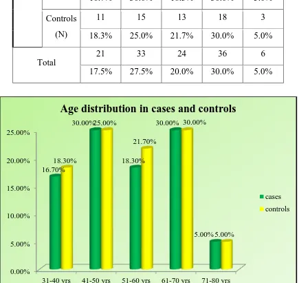
Table 1. Age distribution in cases and controls
0.00% 5.00% 10.00% 15.00% 20.00% 25.00%
31-40 yrs 41-50 yrs 51-60 yrs 61-70 yrs 71-80 yrs 16.70% 30.00% 18.30% 30.00% 5.00% 18.30% 25.00% 21.70% 30.00% 5.00%
Age distribution in cases and controls
cases controls
Age group
31-40 yrs 41-50 yrs 51-60 yrs 61-70 yrs 71-80 yrs
Cases (N)
10 18 11 18 3
16.7% 30.0% 18.3% 30.0% 5.0%
Controls
(N)
11 15 13 18 3
18.3% 25.0% 21.7% 30.0% 5.0%
Total
21 33 24 36 6
50
Gender distribution in cases and controls
0.00% 10.00% 20.00% 30.00% 40.00% 50.00% 60.00% 70.00%
male female
61.70%
38.30% 55.00%
45.00%
Gender distribution in cases and controls
cases controls
Gender
male Female
Cases(n) 37 23
% within grp 61.7% 38.3%
Count 33 27
51
Distribution of spot microalbuminuria among cases and controls
Spot microalbuminuria Chi-Square test
+ ve cases - ve cases p value
Cases(n) 42 18
0.000*
% within grp 70.0% 30.0%
Control 6 54
% within grp 10.0% 90.0%
* Significant p value
0.00% 10.00% 20.00% 30.00% 40.00% 50.00% 60.00% 70.00% 80.00% 90.00% cases controls 70.00% 10.00% 30.00% 90.00%
Distribution of spot microalbuminuria among
cases and controls
Spot microalbuminurea + ve cases
52
Distribution of One month microalbuminuria among spot
microalbuminuria positive cases:
One month
microalbuminuria
-ve cases +ve cases
cases 26 16
% within grp 62% 38%
Predisposing factors
H/O Smoking habit H/O alcohol habit
non smoker smoker non
alcoholic Alcoholic
cases 50 10 51 9
% within grp 83.3% 16.7% 85.0% 15.0%
controls 47 13 47 13
% within grp 78.3% 21.7% 78.3% 21.7%
53
Correlation between spot Microalbuminuria and NIHSS stroke scale:
NIHSS scale
Spot microalbuminuria Chi-Square test
+ ve cases -ve cases P value
no stroke (N) 0 5
0.000*
% of Total .0% 8.3%
minor stroke(N) 0 8
% of Total .0% 13.3%
moderate stroke(N) 0 5
% of Total .0% 8.3%
moderate to severe(N) 20 0
% of Total 33.3% .0%
severe(N) 22 0
% of Total 36.7% .0%
TOTAL(N) 42 18
% of Total 70% 30.0%
*significant p value
0.00% 10.00% 20.00% 30.00% 40.00% 50.00% 60.00% 70.00%
no stroke minor stroke moderate stroke moderate to severe severe TOTAL 0.00% 0.00% 0.00%
33.30% 36.70% 70% 8.30% 13.30% 8.30% 0.00% 0.00% 30.00%
Correlation between microalbuminuria and NIHSS stroke scale:
54
Distribution of spot microalbuminuria based on age groups:
age_group Chi
square test P value 31-40 yrs 41-50 yrs 51-60 yrs 61-70 yrs 71-80 yrs Positive cases
(n) 11 20 13 4 0 0.000*
% 22.9% 41.7% 27.1% 8.3% .0%
*significant p value
0.00% 5.00% 10.00% 15.00% 20.00% 25.00% 30.00% 35.00% 40.00% 45.00%
31-40 yrs 41-50 yrs 51-60 yrs 61-70 yrs 71-80 yrs 22.90%
41.70%
27.10%
8.30%
0.00%
55
Distribution of spot microalbuminuria based on gender
Spot
microalbuminuria gender P value
male female
Positive
cases (n) 28 20 0.546
% 58.3% 41.7%
ChiSquare test No significant p value
0.00% 10.00% 20.00% 30.00% 40.00% 50.00% 60.00%
male female 58.30%
41.70%
Distribution of spot microalbuminuria based
on gender
56
Distribution of one month NIHSS scale among study group
one month NIHS scale Frequency (n) Valid Percent
no stroke 11 18.3
minor stroke 12 20.0
moderate stroke 18 30.0
moderate to severe 4 6.7
severe 15 25.0
Total 60 100.0
0 10 20 30 40 50 60 70 80 90 100 18.3 20 30 6.7 25 100
Distribution of one month NIHSS scale among
study group
no stroke
minor stroke moderate stroke moderate to severe severe
57 Distribution of stroke type among study group
Distribution of stroke type among study group
stroke type Frequency Valid Percent
Valid
ACA 28 46.7
MCA 14 23.3
Lac stroke 18 30.0
0 5 10 15 20 25 30 35 40 45 50
ACA MCA Lac stroke
46.7
23.3
30
Distribution of stroke type among study
group
58
Association between one month NIHSS scale and Spot microalbuminuria in case group
Spot microalbuminuria
Total
NIHSS Scale1month - Ve
cases + ve cases P value
no stroke Frequency (n) 9 2 11
% within Spot
microalbuminuria 50.0% 4.8% 18.3%
0.000*
minor stroke Frequency (n) 8 4 12
% within Spot
microalbuminuria 44.4% 9.5% 20.0%
moderate stroke Frequency (n) 1 17 18
Spot microalbuminuria % 5.6% 40.5% 30.0%
moderate to severe
Frequency (n) 0 4 4
% within Spot
microalbuminuria .0% 9.5% 6.7%
severe
Frequency (n) 0 15 15
% within Spot
microalbuminuria .0% 35.7% 25.0%
Total
Frequency (n) 18 42 60
% within Spot
microalbuminuria 100.0% 100.0% 100.0%
* Shows significant p value chi square test
0.00% 10.00% 20.00% 30.00% 40.00% 50.00%
no stroke minor stroke moderate stroke moderate to severe severe
Association between one month NIHSS scale
and Spot microalbuminuria in case group
negative cases
59
Association between stroke size and Spot microalbuminuria in case group
Spot microalbuminuria
Total
P value
- Ve
cas e +Ve cases strke_size SI
Frequency (n) 18 2 20
0.000* % within Spot
microalbuminuria 100.0% 4.8% 33.3%
LI
Frequency (n) 0 40 40
% within Spot
microalbuminuria .0% 95.2% 66.7%
Total Frequency (n) 18 42 60
0.00% 10.00% 20.00% 30.00% 40.00% 50.00% 60.00% 70.00% 80.00% 90.00% 100.00%
negative cases positive cases
Association between stroke size and Spot microalbuminuria in case group
60
Association between stroke type and Spot microalbuminuria in case group
Crosstab Spot microalbuminuria Total -Ve cas e +Ve
cases P value
type_strke
ACA
Frequency (n) 0 28 28
% within Spot
microalbuminuria .0% 66.7% 46.7%
0.000* MCA
Frequency (n) 0 14 14
% within Spot
microalbuminuria .0% 33.3% 23.3%
Lac stroke
Frequency (n) 18 0 18
% within Spot
microalbuminuria 100.0% .0% 30.0%
Total
Frequency (n) 18 42 60
% within Spot
microalbuminuria 100.0% 100.0% 100.0%
* Shows significant p value chi square test
0.00% 10.00% 20.00% 30.00% 40.00% 50.00% 60.00% 70.00% 80.00% 90.00% 100.00%
ACA MCA Lac stroke
Association between stroke type and Spot microalbuminuria in case group
61
Association between smoking status and Spot microalbuminuria in case group
spot_ma_cases
Total
Smoking status -ve csses +ve cases P value Non smokers Frequency (n) 16 34 50
% within Spot
microalbuminuria 88.9% 81.0% 83.3%
0.571a smokers Frequency (n) 2 8 10
% within Spot
microalbuminuria 11.1% 19.0% 16.7% Total Frequency (n) 18 42 60
% within Spot
microalbuminuria 100.0% 100.0% 100.0%
a Not significant chi square test
0.00% 10.00% 20.00% 30.00% 40.00% 50.00% 60.00% 70.00% 80.00% 90.00%
negative cases positive cases
Association between smoking status and Spot microalbuminuria in case group
62
Association between mean cholesterol and Spot microalbuminuria in case group
N Mean Std. Deviati on t Std. Error Mean 95% Confidence Interval of the
Difference
Un paired
test
Lower Upper P value
Cases 60 193.35 30.842 3.865 3.982 8.291 25.709 0.000*
controls 60 176.35 14.469 3.865 1.868 8.254 25.746
193.35 176.35 165 170 175 180 185 190 195 cases controls
Association between mean cholesterol and
Spot
microalbuminuriain case group
63
DISCUSSION
60 cases of acute ischemic stroke within 24 hours of onset were admitted in the department of Medicine, Madurai Medical College and Government Rajaji Hospital during the period of March 2017 to August 2017.
Age distribution in cases and controls:
In the study group,among cases 16% present between the age group of 31 – 40yrs,30%between 41-50 yrs,18%between
51-60yrs,18%between61-70yrs,3%between71-80yrs.
Among controls 18%between31-40yrs,25%between41-50yrs,24%between51-60yrs,30%between61-70yrs,5%between71-80yrs.
Overall 17%between 31-40yrs,27%between41-50yrs,20%between51-60 yrs,30%between61-70yrs,5%bteween71-80yrs.
Gender distribution in cases and control:
Among cases 61% were males,38% were females.
Among control 55% were males,27% were females.
Overall 55% were Males 45%females.
Distribution of spot microalbuminuria among cases and controls:
64
30% were spot microalbuminuria negative.
Among controls 10% were spot microalbuminuria negative,
90% were spot microalbuminuria positive.
So among cases spot microalbuminuria was significantly positive and p value is significant.
Distribution of one month microalbuminuria among spot
microalbuminuria positive cases:
Among spot microalbuminuria positive cases one month microalbuminuria positive in 38% ,negative in 62% .
Smoking and alcohol distribution:
Among cases 83% were non smokers ,16% were smokers,
Among controls 78% were non smoker.
Among cases 85% were non alcoholic,15% were alcoholic,
Among controls 78% were non alcoholic,21% were alcoholic.
Correlation between spot microalbuminuria positive cases and NIHSS
scale:
65
Among spot microalbuminuria negative cases 8.3% had no stroke,13.3% had minor stroke,8.3% had moderate stroke.
Distribution of spot microalbuminuria positive cases based on age group:
Among spot microalbuminuria positive cases 22% in 30-40yrs,41% in 41-50 yrs ,27% in 51-60 yrs,8.3% in 61-70 yrs.
Distribution of spot microalbuminuria positive cases based on gender:
Among spot microalbuminuria positive cases 58% were males ,41% were females.
Distribution of one month NIHSS scale among study group:
Among spot micro albuminuria positive cases 18% had no stroke,20% had minor stroke,30% had moderate stroke,6% had moderate to severe stroke,25% had severe stroke.
Distribution of stroke types among study group:
66
Association between one month NIHSS scale and spot microalbuminuria
positive in case group:
Among spot microalbuminuria positive cases 18% had no stroke,20% had minor stroke ,30% had moderate stroke,6% had moderate to severe stroke,25% had severe stroke.p value is significant.
Association between stroke size and spotMA in case group:
Among spot MA +ve cases 4.8% had small infarct,95% had large infarct.p value is significant.
Among spot MA –ve cases no stroke was seen.
Association between stroke type and spot MA :
Among spot MA + ve cases 66% had ACA territory stroke,33% had MCA territory stroke.
Among spot MA –ve cases 18% had lacunar stroke.
Association between smoking status in spot MA cases:
Among spot MA +ve cases 81% were non smokers ,19% were smokers.
Among spot MA –ve cases 88% were non smokers ,11% were smokers.
Association between cholesterol in cases and control:
67
Among controls cholesterol value is between 176.3±14.4.
In this study spot microalbuminuria were measured within 24 hrs of stroke onset and severity of stroke was also assessed at the same time using NIHSS scale and then microalbuminuria also measured at the end of one month and severity of stroke also assessed during this time.
In this study majority of cases 30% are between 61-70 yrs,males are more than females,70% of cases were spot MA positive, one month MA was positive in 38% of spot MA positive cases.
Majority of cases and control included in this study were non smokers and non alcoholic.In spot MA positive cases 33% had moderate to severe stroke,36% had severe stroke assesed by using NIHSS scale done within 24 hrs and 20% were present in between age 41-50 yrs and 58% were males.
Among spot MA positive cases 46% had ACA territotry stroke,23% had MCA Territory stroke and according to one month NIHSS Scale 18% had moderate stroke,25% had severe stroke.
68
CONCLUSION
Microalbuminuria increases the ischaemic stroke risk because of association between microalbuminuria with carotid intima media thickness and reflecting increased vascular endothelial permeability leading to endothelial dysfunction.
69
SUMMARY
MA is more prevalent in patients with increased risk of vascular events . It predicts cardiovascular events and all-cause mortality in the general population and is a plausible risk factor for ischemic stroke . Furthermore, MA occurs transiently in acute diseases with different etiologies and predicts greater short- and long-term mortality in stroke patients .
Microalbuminuria increases the ischaemic stroke risk because of association between microalbuminuria with carotid intima media thickness and reflecting increased vascular endothelial permeability leading to endothelial dysfunction.
The present study found microalbuminuria in 70% of non diabetic nonhypertensive acute ischaemic stroke and degree of MA increases with severity of stroke as assessed by NIHSS stroke scale at the time of admission and after one month. When one month MA was positive it was associated with more severity of stroke.
70
establish whether the correction of increased urine albumin excretion rate in its own right modifies the risk of adverse vascular events.
Presence of Microalbuminuria associated with increased severity of acute ischemic stroke assesed by NIHSS Stroke scale.
71
LIMITATIONS OF STUDY
1.Number of patients included were relatively small.
2. As the study was cross sectional in design, it is not easy to predict exactly whether MA preceded stroke or vice versa.
3.Intra-individual variability in MA had to be considered.
72
BIBLIOGRAPHY
1. Hankey GJ. Stroke: How large a public health problem and how can the neurologist help: Arch Neurol 1999;56:748-54.
2. Fiona C. Taylor, Suresh Kumar K. Stroke in India factsheet (updated 2012). 3. Basi Seema, Albert Mimran, Pierre Fesler, Julia B Lewis.Microalbuminuria in type 2 diabetes and hypertension. A marker treatment target or innocent bystander? Diabetes Care. 2008;31:194-201.
4. Gosling P, Hughes EA, Reynolds TM, Fox J. Microalbuminuria is an early response following acute myocardial infarction. Eur Heart J. 2001;12:508-13. 5. Ochodnicky P, Henning RH, Van Dokkum RP, De Zeeuw. Microalbuminuria and endothelial dysfunction: Emerging targets for primary prevention of end organ damage. J Cardiovasc Pharmacol 2006;47(Supple 2):S151-62.
6. Deckert T, Feldt-Rasmussen B, Borch- Johnsen K, Jensen T, Kofoed. Albuminuria reflects widespread vascular damage. The Steno Hypothesis. Diabetologia. 1989; 32:219-26.
7. Davignon J, Ganz P. Role of endothelial dysfunction in atherosclerosis. Circulation. 2004;109:27-32.
73
9. Gumbinger C, Sykora M, Diedler J, Ringeb P, Rocco A. Microalbuminuria: A potential prognostic marker for acute stroke. Nervenarzt. 2012;83(10):1357-60.
10. Slowik A, Turaj W, Iskra T, Strojny J, Szczudlik A. Microalbuminuria in nondiabetic patients with acute ischaemic stroke: Prevalence, clinical correlates and prognostic significance. Cerebrovacular Disease Basel Switz. 2002;14(1):15-21.
11. Beamer NB, Coull BM, Clark WM, Wynn M. Microalbuminuria in ischaemic inischaemic stroke. Arch Neurol. 1999; 56(6):699-702
12. Lee M, Saver JL, Chang KH, Liao HW, Chang SC, Ovbiagele B. Impact of microalbuminuria on incident stroke: A meta analysis. Stroke J Cerebral Circulation. 2010;41(11):2625-31.
13. Mathur PC, Punckar Prashant, Muralidharan R. Microalbuminuria in Nondiabetic Acute Ischaemic Stroke-An Indian Perspective Annals Indian Academy of Neurology. 2005;1,8(4):237-42.
74
15. David BS. Carbohydrate. In: Burtis HC, Ashwood RE, editors. Tietz text book of clinical chemistry.3rd edi: Philadelphia: Saunders. 1999;798-801.
16. Toto RD. Microalbuminuria: Definition, detection and clinical significance. J Clin Hypertension. 2004;1(6):2-7.
17. Scandinavian stroke study group. Multicentre trial of hemodilution in iscaemic stroke: Background and study protocol. Stroke. 1985;16:885-90.
18. Luvizotto Gustavo J, Gabriel Maicon Goncalves, Gabriel Pariera Braga, Thiago Dias Fernades. Aspects correlates with Scandinavian stroke scale for predicting early neurological impairment. Arquivos de Neuro-Psiquiatria. 2015;73(5).
19. Chowdhury J, Sultana N, Ahmed S, Rahman M, Akter M, Rafique T.
Microalbumunuria as predictor of short term mortality in acute ischaemic stroke. Bangladesh Journal Medical Biochem. 2012;5(1):16 19.
75
21. Muhammad Ahsan F, M Sohail Anjum, Malik Fayyaz A, Naila Kalsoom. Frequency of Microalbuminuria in patients with ischaemic stroke. Rawal Medical Journal. 2013;38(2):97-98.
22. Haffner SM, Stern MP, Gruber KK, Hazuda MP, Mitchell BD, Patterson JK. Microalbuminuria. Potential marker for increased cardiovascular risk factors in non diabetic subjects? Atherosclerosis. 1990;10:727-31.
23. Mykkanen L, Zaccaro DJ, O’Leary DH, Howard G, Robbins DC, Haffner SM. Microalbuminuria and carotid intima–media thickness in nondiabetic and NIDDM subjects. The Insulin Resistanc Atherosclerosis Study (IRAS). Stroke. 1997;28:1710-1716.
24. Viberti GC, Hill RD, Jarrett RJ, Argyropoulos A, Mahmud U, Keen H: Microalbuminuria as a predictor of clinical nephropathy in insulin-dependent diabetes mellitus. Lancet, 1982; 1: 1430-1432
25. Mogensen CE: Microalbuminuria predicts clinical proteinuria and early mortality in maturity-onset diabetes. N Engl J Med, 1984; 310: 356-360
26. Schmitz A, Vaeth M: Microalbuminuria: a major risk factor in non-
-insulin-dependent diabetes. A 10-year follow-up study of 503 patients.
76
27. Allawi J, Jarrett RJ: Microalbuminuria and cardiovascular risk factors in type 2 diabetes mellitus. Diabetic Med, 1989; 7: 115-118
28. Mattock MB, Keen H, Viberti GC et al. : Coronary heart disease and urinary albumin excretion rate in type 2 (non-insulin dependent) diabetic patients. Diabetologia, 1988; 31: 82-87
29. Cerasola G, Cottone S, Mule G et al. : Microalbuminuria, renal dysfunction and cardiovascular complication in essential hypertension. J Hypertens, 1996; 14: 915-920
30. Haffner SM, Stern MP, Gruber KK, Hazuda HP, Mitchell BD, PattersonnJK: Microalbuminuria. Potential marker for increased cardiovascular risk factors in nondiabetic subjects ? Arteriosclerosis, 1990; 10:727-731
31. Jensen JS, Feldt-Rasmussen B, Borch-Johnsen K, Clausen P, Appleyard M, Jensen G: Microalbuminuria and its relation to cardiovascular disease and risk factors. A population-based study of 1254 hypertensive individuals. J Hum Hypertens, 1997; 11: 727-732
77
33. Metcalf P, Baker J, Scragg RK, Dryson E, Scott A, Wild C: Albuminuria in a middle-aged workforce: effect of alcohol, regular exercise and smoking. Clin. Chem, 1993; 39: 1793-1797
34. Yudkin JS, Forrest RD, Jackson CA: Microalbuminuria as predictor of vascular disease in non-diabetic subjects. Islington Diabetes Survey. Lancet, 1988; 2: 530-533
35. Damsgaard EM, Froland A, Jorgensen OD, Mogensen CE: Microalbuminuria as predictor of increased mortality in elderly people. BMJ, 1990; 300: 297-300
78
KEY TO MASTER CHART
MA - microalbuminuria
NIHSS - National Institute Of Health And Stroke Scale P - Present
A - Absent LI - large Infarct SI - Small Infarct
ACA - Anterior Cerebral artery MCA - Middle Cerebral artery Lac - Lacunar stroke
79
PROFORMA
PROFORMA
Name:
Age / Sex:
Occupation:
Presenting complaints:
Past History:
H/o DM, HT, CKD, CAD,
Clinical Examination:
General Examination:
Consciousness,
Pallor,
Jaundice,
Clubbing,
Lymphadenopathy,
80 Vitals:
PR
BP
RR
SpO2
Systemic examination:
CVS:
RS:
ABDOMEN: