“ROLE OF BRONCHOSCOPY TO DETERMINE THE ETIOLOGY OF NON
RESOLVING PNEUMONIA IN A TERTIARY CAREINSTITUTE”
DISSERTATION
SUBMITTED TO THE TAMILNADU DR.M.G.R. MEDICAL UNIVERSITY
CHENNAI
In partial fulfillment of the requirements for the degree of
M.D. (TUBERCULOSIS &RESPIRATORY MEDICINE)
BRANCH – XVII
DEPARTMENT OF THORACIC MEDICINE
TIRUNELVELI MEDICAL COLLEGE HOSPITAL
CERTIFICATE
CERTIFICATE BY THE DEAN
I hereby certify that this dissertation entitled “ROLE OF
BRONCHOSCOPY TO DETERMINE THE ETIOLOGY OF NON
RESOLVING PNEUMONIA IN A TERTIARY CARE INSTITUTE” is a
record of work done by Dr.T.PUDHUMALAR, in the Department of
THORACIC MEDICINE , Tirunelveli Medical College, Tirunelveli, during her
postgraduate degree course period from 2014- 2017. This work has not formed
the basis for previous award of any degree.
The DEAN
Date : Tirunelveli Medical College,
CERTIFICATE BY THE GUIDE
This is to certify that the dissertation entitled “ROLE OF
BRONCHOSCOPY TO DETERMINE THE ETIOLOGY OF NON
RESOLVING PNEUMONIA IN A TERTIARY CARE INSTITUTE” , is a
record of work done by Dr.T.PUDHUMALAR, in partial fulfillment for the
award of the degree of Doctor of Medicine in THORACIC MEDICINE for the
April 2017 examination by the Tamilnadu Dr.M.G.R. Medical University,
Chennai , this is a bonafide original research work done by her in the department
of TUBERCULOSIS AND RESPIRATORY MEDICINE , Tirunelveli Medical
College, under my guidance and supervision.
Date :
Place : TIRUNELVELI
Prof. Dr.K.KRISHNAMOORTHY, M.D.
PROFESSOR,
DEPARTMENT OF THORACIC MEDICINE, TIRUNELVELI MEDICAL COLLEGE,
DECLARATION BY THE CANDIDATE
I solemnly declare that this dissertation titled “ROLE OF
BRONCHOSCOPY TO DETERMINE THE ETIOLOGY OF NON
RESOLVING PNEUMONIA IN A TERTIARY CARE INSTITUTE”
submitted by me for the degree of M.D., is the record work carried out by me
during the period of 2014-2017 under the guidance of Prof.
Dr.K.KRISHNAMOORTHY, M.D, Professor and Head of the Department,
Department of Thoracic Medicine, Tirunelveli Medical College, Tirunelveli. The
dissertation is submitted to The Tamilnadu Dr. M.G.R. Medical University,
Chennai, towards the partial fulfilment of requirements for the award of
M.D.(Branch XVII) Tuberculosis and Respiratory Medicine examination to be
held in April 2017.
Place: Tirunelveli Dr. T.PUDHUMALAR,
Date: Department of Thoracic medicine,
ACKNOWLEDGEMENT
Though only my name appears on the cover of this dissertation, a great many people have been behind this task and I take this opportunity with immense pleasure to place on record my heartfelt gratitude and respect to all my distinguished resources.
I thank the DEAN Dr. SITHY ATHIYA MUNAVARAH, M.D for permitting me to conduct this study and to avail the resources of the hospital.
I am greatly thankful to my Professor and Head of the Department of Thoracic Medicine, Dr.KRISHNAMOORTHY. K M.D, who has always provided me the necessary help, guidance and support. His valuable suggestions and concern bring the successful completion of this dissertation work.
I am extremely thankful to the respected Assistant Professors of my Department, Dr. MATHAN.E, Dr. RAHMAN SHAHUL HAMEED.O.M.
and Dr.SENTHIL ARASU.P, and for their concern, contributions, suggestions,
support and co-operation during the study.
I also thank all my senior and junior postgraduate colleagues for their cooperation and support for this study. Without their cooperation, this study would not have been possible.
I shall be failing in my duty, if I do not acknowledge the contributions of my Patients who were involved in this study.
I express my sincere gratitude to my husband Dr. C.KARTHIKEYAN
Finally, I thank LORD AND MY PARENTS, K.THAMODHARAN
AND G.RAJESWARI , the Creator and the Guardian, without their blessings,
ABBREVIATIONS
1. AFB Acid fast bacilli
2. aPTT Activated partial thromboplastin time 3. ATS American Thoracic Society
4. BTS British thoracic society 5. BAL Broncho alveolar lavage
6. CAP Community acquired pneumonia 7. COP Cryptogenic organizing pneumonia 8. COPD Chronic obstructive Pulmonary disease 9. CBNAAT Catridge based nucleic acid amplification test
10. CT Computed tomography
11. ELISA Enzyme linked immunosorbent assay 12. FOB Fiberoptic bronchoscopy
13. FNAC Fine needle aspiration cytology 14. HIV Human immunodeficiency virus 15. IDSA Infectious disease society of America 16. LRI Lower respiratory infection
17. LPA Line probe assay
18. MGIT Microscopic growth indicator tube 19. MTB Mycobacterium tuberculosis
CONTENTS
S.NO TITLE PAGE.NO
1.
2.
3.
4.
5.
6.
7.
INTRODUCTION
REVIEW OF LITERATURE
MATERIALS AND METHODS
OBSERVATION AND RESULTS
DISCUSSION
SUMMARY
CONCLUSION
BIBILIOGRAPHY
ANNEXURES
MASTER CHART
1
2
55
60
74
81
INTRODUCTION
Pneumonia is defined as inflammation of the pulmonary parenchyma
caused by an infectious agent(1).. Inappropriate or delayed treatment leads to morbidity, mortality and drug resistance in significant number of population.
Non resolving pneumonia is one of the common clinical problem.
Infection with drug resistant organisms, Misdiagnosis of pathogen, presence
of co morbid conditions, development of complications, non infective
etiology are some reasons for non resolution. Selection of patients and
appropriate timing of further evaluation can be challenging. There is no
uniform diagnostic or treatment approach for patients with non resolving
pneumonia.
Along with other routine investigations, Fiberoptic bronchoscopy
(FOB), Computed Tomography scan of the thorax and CT-guided fine
needle aspiration cytology (FNAC) may be helpful in the evaluation of
non-resolving or slowly non-resolving pneumonia. FOB is one of the most useful
procedure in the evaluation of patients with non resolving pneumonia.
This study is to establish the etiology of non resolving pneumonia by
REVIEW OF LITERATURE:
Pneumonia:
Pneumonia is defined as inflammation of the pulmonary
parenchyma caused by an infectious agent (1).Pneumonitis reflects inflammation due to both infectious and non infectious cause. Various
terminologies are used to describe various forms of pneumonia which
reflects the possible etiology .These include aspiration pneumonia,
community acquired pneumonia, nosocomial pneumonia,ventilator
associated pneumonia etc.
Pneumonia is one of the leading cause for mortality and morbidity
worldwide. Inappropriate or delayed treatment leads to mortality,morbidity
and drug resistance in significant number of population. Community
acquired pneumonia (CAP) is one of the common clinical problem
characterized by cough, fever, chills, fatigue, dyspnoea, rigors, and pleuritic
chest pain—with or without new infiltrate on chest radiography. Majority of patients it responds well with initial empirical antibiotics. Only few
patients respond poorly, resulting in non resolving pneumonia or death.
ETIOLOGY:
Streptococcus pneumoniae is the most common cause of community
acquired pneumonia(CAP).Hemophilus influenza and Moraxella catarrhalis,
are more common in patients who have underlying chronic lung disease like
COPD. Staphylococcus aureus, occurs more commonly during an influenza
outbreak. Individuals on long term steroids, or alcoholics, frequent
exposure to antibiotic and those with severe underlying bronchopulmonary
disease are at risk of infection with Enterobacteriaceae species and P.
aeruginosa. Less common causes of pneumonia include Streptococcus
pyogenes, Neisseria meningitidis, Pasteurella multocida and H. influenzae.
The “atypical” organisms, which include Mycoplasma pneumoniae, Chlymadia pneumoniae, Legionella are other causes. These are more
prevalent among outpatients.
Viral causes of CAP in Adults : RSV, adenovirus, and parainfluenza virus,
Human meta pneumovirus, herpes simplex virus, Varicella-zoster virus,
SARS-associated coronavirus, and measles virus. Viral etiology(18%)
identified as a cause of community acquired pneumonia in a recent study
among immunocompetent adult patients .Respiratory Syncytial Virus was
the most common pathogen identified in this study . They concluded viral
Chlamydia psittaci [psittacosis],Coxiella burnetti[Q fever],Francisella
tularemia[tularemia],Bordetella pertussis[whooping cough].
Fungai- Histoplasma capsulatum, Cryptococcus neoformans, Blastomyces
hominis, Coccidioides immitis.
They are less common and only 2 to 3 % of total.
Anerobes -common cause of aspiration pneumonia.
Risk Factors:
Causative organisms from the upper airways or less commonly from
hematogenous spread or direct spread from a contiguous focus find their way
to lung parenchyma. Conditions such as altered sensorium, stroke facilitate
aspiration of contents into lungs. Loss of upper airway reflexes such as
cough, impairment of local defense mechanism such as mucosal blanket
which is lost due smoking and irritants ,factors that destroy alveolar
epithelium such as tobacco smoking, bronchial metaplasia or neoplasia,
pulmonary edema and congenital causes such primary ciliary dyskinesia
contribute to occurrence of pneumonia
PATHOGENESIS:
them. Pneumonia occurs when there is impaired host defense ,or as a
result of infection with virulent organisms, a large “dose” of bacteria.
Microbes gain access to the lung by any of the following routes:
Micro aspiration from the upper respiratory tract
Droplet infection from infected person
Spread from contiguous infected sites
Hematogenous spread
Once the pathogen or agent enters the bronchi and bronchioles, it
firmly fixes with the wall and causes cascade of inflammation in the host
body. There are two main types of acute pneumonia : bronchopneumonia
(with lobular topography) and lobar pneumonia (lobar topography). Lobar
pneumonia causes exudative inflammation of an entire pulmonary lobe. If
not treated, lobar pneumonia evolves in four stages.
1.exudative phase2.red hepatizaton3.gray hepatization 4.resolution
Common to all stages is the enlargement of the affected lobe with loss of its
spongy appearance.
EVALUATION OF PATIENTS WITH SUSPECTED PNEUMONIA:
a)Detailed clinical history which includes age, onset of symptoms, resident
area, recent travel, prior hospital admission, antimicrobial treatment, co
b)General examination and respiratory system examination is vital.Physical
examination may not be reliable in immuno compromised individual.
c)Investigations:
complete blood count,C reactive protein,Blood culture:polymorpho
leucocytosis is observed in bacterial etiology.
Chest X ray:Pneumonia may present with various radiological patterns
which includes consolidation, bronchopneumonia, miliary pattern,
nodules, abscess, effusion, interstitial pattern, lymph adenopathy
Noninvasive microbiological testing which includes sputum examination
for cytology, Gram stain, KOH mount, Ziehl Neelson or flurochrome
stain,various special stain to the specific organisms,sputum for culture and
sensitivity are used.
Bacterial and viral antigen detection by using ELISA ,latex particle
agglutination, PCR . For example, studies show Steptococcus pneumonia is
better identified by molecular and urinary test rather than sputum culture
examination.
To identify M.TB CBNAAT,LPA,MGIT like rapid diagnostic methods
TREATMENT:
According to 2007 IDSA guidelines, initial treatment for most of the
patients remains empirical.Selection of antibiotics is based on likely
pathogen and knowledge of local susceptibility patterns. We should consider
individual risk factor for each of the patients and must treat accordingly.
For sick patients admitted, the first antibiotic dose should be administered
as early as possible. Patients with CAP should be treated for a minimum of
5 days , should be afebrile for 48–72 h, and should have no more than 1 CAP-associated sign of clinical instability before discontinuation of
therapy.A longer duration of therapy may be needed if initial therapy was
not active against the identified pathogen or if it was complicated by
extrapulmonary infection.
Out patients with no risk factors a macrolide or doxycyline is
recommended.
Outpatients with co morbidities such as heart, lung, liver
or renal disease; diabetes mellitus; alcoholism; malignancies;asplenia;
immunosuppressing conditions or use of immunosuppressing drugs, a
respiratory fluoroquinolone or combination of Beta lactam antibiotic with
In areas with a high rate of infection with high-level (MIC _16
mg/mL) macrolide-resistant Streptococcus pneumoniae use of
alternative drugs is recommended.
Hospitalised patients who are stable can be treated with either
respiratory fluoroquinolones or combination of β- lactam with macrolides.
Inpatients requiring intensive care: β-lactam with azithromycin or a respiratory fluoroquinolone (strong recommendation) (for
penicillin-allergic patients, a respiratory fluoroquinolone and
aztreonam are recommended)
Special concerns:
If Pseudomonas infection is suspected, An antipseudomonal β-lactam (Piperacillin tazobactam,cefepime, imipenem, or meropenem) plus either
ciprofloxacin or levofloxacin should be used.Other options are anti
Pseudomonal β-lactam plus an aminoglycoside and azithromycin Or The above b-lactam plus an aminoglycoside and an antipneumococcal
fluoroquinolone (for penicillin-allergic patients, substitute aztreonam for
above b-lactam)
NON RESOLVING PNEUMONIA:
Non resolving pneumonia is one of the common clinical problems
encountered. The problems may range from simple delay in recovery to
life-threatening progressive pneumonia.
Definition of non resolving pneumonia :
Defining non resolving pneumonia is difficult, because of lack of a clear
cut and validated definition in the literature. In 1943, the term unresolved
organizing or protracted pneumonia was first described by Amberson(5).
Non resolving pneumonia is a clinical syndrome in which focal infiltrates
begin with some clinical association of acute inflammation and despite
minimum of 10 days antibiotics patient either don't improve or worsen or
radiographic opacities fail to resolve within 12 weeks(6).
According to IDSA/ATS 2007 guidelines, non resolving or
slow-resolving pneumonia refers to patients who present with persistence of
pulmonary infiltrates more than 30 days duration after initial
pneumonia-like syndrome.
Kirtland and Winterbauer defined non resolving pneumonia is less
than complete clearance of radiological infiltrates in 4 weeks or less than
symptomatically or non resolution of radiological infiltrates in an expected
period of time based on initial diagnosis and at least 10 days of antibiotics(7).
NORMAL VERSUS DELAYED RESOLUTION:
Resolution of pneumonia is not defined easily and it depends upon the
underlying etiology. Clinical recovery of the patients occur earlier when
compared to radiological resolution. Radiological resolution can take up to
six weeks. Microbiological resolution occurs with appropriate treatment.
After treatment, inflammation of lung parenchyma decreases and patients
[image:21.595.104.536.379.687.2]Empirical treatment is mainstay of treatment in CAP but studies show
6 to 15 % of inpatients are not cured by initial treatment(10,11).At the same time, treatment failures among outpatients unit is so far not well studied. The
overall mortality was around 49% in hospitalised patients with severe CAP
who were not responding to the treatment(11). In another study the mortality rate was 27%(10).
According to the IDSA 2007 guidelines, treatment failure in CAP is
classified systematically in the following ways. The first entity is progressive
pneumonia or actual clinical deterioration, with acute respiratory failure
requiring ventilatory support and/or septic shock, usually occurring within
the first three days of hospital admission. The second pattern is that of
persistent pneumonia which was defined as absence of or delay in achieving
clinical stability, using theHalm’s criteria.. The Infectious Diseases Society of America/ American Thoracic Society (IDSA/ATS) 2007 guidelines
recommend the use of Halm’s criteria to define clinical stability. These clinical criteria are reliable.They are applicable in various healthcare
systems .
Halm's criteria consist of seven variables: 1.Temperature ≤37.8 °C,
2.Heart rate ≤100 beats/minute, 3.Respiratory rate ≤24 breaths/minute,
6.normal mental status 7. normal oral intake(12). 2001 ATS guidelines mentioned clinical stability, which consists of only four criteria.
1. Improvement in symptoms(cough and dyspnoea),2. afebrile (temperature
<37.8 °C) for more than 8 hours, 3.normalisation of white blood cell count
by 10% from the previous day 4.adequate oral intake. Minimum duration to
achieve this criteria is 3 days.but in 1/4 th of patients required more than 6
days.
The final entity is non resolving or slow-resolving pneumonia used
to describe patients with persistence of pulmonary infiltrates more than 30
days duration after initial pneumonia-like syndrome(13). Around 20% of these patients have diseases other than CAP when carefully evaluated(14).
Most of the time, normal resolution of pneumonia mainly correlates
with radiological resolution. Persistance of radiological abnormalities with
improvement in clinical status is defined as slow resolution(15).
Complete radioglogical clearing occurs by one to three months in
nonbacteremic cases and three to five months in bacteremic cases. Residual
radiographic abnormalities are rare in nonbacteremic cases but are present
Factors affecting resolution of pneumonia are:
a) development of complications
b) infection with drug resistant organisms
c) misdiagnosis of pathogen
d) presence of comorbid conditions
e) non infective etiology
a) Complications:
Infectious complications which include empyema, complicated
parapneumonic effusion, and lung abscess. Sequestered foci of infection can
prevent adequate concentrations of antibiotics to reach the particular site and
prevent resolution
b) Virulence and drug resistance:
Streptococcus pneumoniae is the most common cause of community
acquired pneumonia and also responsible for majority of non resolving
pneumonia especially in patients with comorbidities.In pneumococcal
pneumonia, clinical symptoms subsided in majority of patients within 2 to 3
days of appropriate treatment. Around 6 % of patients remain febrile beyond
changes followed by slow resolution begin only after 2 to 3 weeks. Around
50 % of infected patients will show radiological resolution by 10 weeks.
Residual fibrosis was noted in one fourth of patients. In case of Mycoplasma
pneumoniae rapid resolution is a rule.40% of patients will show complete
resolution at 4 weeks and 90% of patients at 8 weeks.
Like Mycoplasma,Chlamydia pneumonia infection is moreover
milder form of disease.50% of radiological changes resolve within 4
weeks.In 20% of patients it may take 9weeks.Residual abnormalities are
seen in 20 to 30%..After the introduction of conjugate vaccine, the incidence
of Hemophilus infection has drastically reduced in children worldwide.In
adults the course of Hemophilus disease not well studied. Available reports
show slow resolution and prolonged hospitalization in adults with
Hemophilus influenza pneumonia.
Apart from this ,resistant organisms like MRSA,MDR TB, multidrug
resistant gram negative bacilli, pencillinase resistant Streptococcus
pneumonia are responsible for delay in resolution.
c) Misdiagnosis of organism:
infections like Histoplasmosis, Coccidioidomycosis, Blastomycosis,
Mucormycosis, Cryptococcus,
Aspergillus infection and Nocardia, Actinomycosis like organisms are also
responsible for non resolving pneumonia. Studies show Aspergillus may
mimic as bacterial pneumonia and are treated with multiple antibiotics
before the diagnosis is established(19).
d) Comorbid factors:
Associated co morbid conditions delay the resolution of pneumonia.
Only 20 o 30 % of patients with comorbid conditions will show radiological
resolution by 4 weeks(20). Extremes of age will show delay in resolution. 30% of patients who were older than 50 years will show slower radiological
resolution.
e) Non infective cause:
Noninfectious causes are responsible for non resolution of
pneumonia in 20 % of patients . In a study performed by Arancibia et al, in
patients hospitalised for CAP, 19 patients had progressive pneumonia and
30 had non resolving pneumonia. Out of this, around 65% cases were due
to infections . Persistance of primary infection was noted in 23 patients, 11
neoplasm, foreign body, interstitial lung disease were present in 9
patients(17).
i) Neoplasms :
Primary Lung cancer or metastasis are responsible for non
resolving pneumonia .The possible mechanisms are endobronchial
obstruction or extrinsic compession which mimic pneumonia, secondary
postobstructive pneumonia or abscess, or by mimicking an infiltrative
process. Bronchogenic carcinoma and carcinoid tumors are the most
common cause of endobronchial obstruction leading to pneumonia.
Bronchoalveolar cell carcinoma and lymphoma are the most common causes
of an alveolar infiltrate mimicking pneumonia. The frequency of
endobronchial carcinoma as a cause of nonresolving pneumonia is Low,
ranging from 0 to 8 percent in most series(21).Bronchoscopy, CT guided biopsy etc. are needed to diagnose malignancy.
ii) Inflammatory disorders
Cryptogenic organising pneumonia, diffuse alveolar damage, alveolar
haemorrhage, eosinophilic pneumonia, hypersensitivity pneumonitis,
vasculitis lipoid pneumonitis, pulmonary alveolar proteinosis, acute
iii) Drugs
Drug induced lung disease can be confused with infectious
pneumonia. Examples include Amiodarone, Bleomycin, Methotrexate,
Nitrofurantoin. Amiodarone toxicity is an important mimic of NRP and can
present as focal alveolar infiltrates.
There is no clear guidelines regarding appropriate antimicrobial
therapy for non responding pneumonia . Majority of cases of non resolution
are due to the severity of disease at presentation, associated co morbid
factors. For severe pneumococcal pneumonia, combination therapy was
found to be useful(22). In case of NRP, rather than using multiple broad spectrum antibiotic course, other etiologies should be considered and
proceeded with necessary investigations This will help the patients to get
appropriate treatment in appropriate time which in turn will reduce
BRONCHOSCOPY
The ease and safe access to visualise bronchial tree has been a long
standing desire . After various evolution, today’s flexible bronchoscope utilises modern technology for better differentiation and contrast
enhancement.
Bronchoscopy is a valuable diagnostic and therapeutic tool in
Pulmonary Medicine. It plays an important role in the diagnosis of non
resolving pneumonia.
HISTORY:
Gustavkillian is considered as the father of modern bronchoscopy.
Chevalier Jackson developed a rigid esophagoscope . Derivatives of this
device are now called rigid bronchoscopes. Shigeto Ikeda , a thoracic
surgeon, developed flexible fiberoptic bronchoscope in the year 1968, which
is now known as the year of “second revolution” in bronchoscopy(23)
Andersen was the first to perform bronchoscopic transbronchial lung biopsy
(TBLB) via the rigid bronchoscope in 1965. In 1958, Eduardo Schieppati
originally proposed transbronchial needle aspiration biopsy (TBNA) (24). Inspired by the initial experiences of Oho and his colleagues(16) , Ko-Pen Wang (1978) published the first report on the successful bronchoscopic
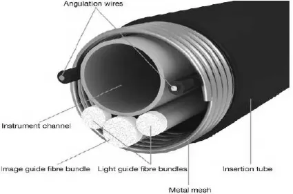
BRIEF REVIEW OF FLEXIBLE BRONCHOSCOPY DESIGN
The parts of FOB are eye piece, suction valve, angulation knob,
[image:31.595.112.574.248.602.2]working channel, insertion tube, light guide connector (fig:3.)
Fig 3:Parts of Flexible Bronchoscopy.
Bronchoscope is designed in such a way to accommodate adequate
bronchoscope can be bent only in two directions 180°and 130°. The
imaging fibre bundle in flexible fibreoptic bronchoscope is an important
component which determines the quality of image. Clearer the image, better
is the chance of diagnosis so that therapeutic options can be planned
accordingly. Each single fibre has the same dimension which is accurate .It
is positioned at similar position at both ends of the fibre bundle. If this is not
[image:32.595.95.520.335.616.2]achieved, artefacts impair the the quality of image.(Figure 4)
There are various channel ports in the bronchoscope.
a) The suction valve in control section is used for aspiration of mucus. It is
connected to an external suction apparatus . Using this valve, suction can be
done by the physician .
b) The working channel or instrument channel port is located bit deep at
the lower part of the control section.(Figure 3). Endotherapy instruments can
be inserted into the bronchoscope using this port to reach the site of interest..
e.g. to guide biopsy forceps and to take sample for further investigation. It
is also called biopsy channel. In order to save space, the instrument channel
is positioned to join the suction channel at the lower part of the control
section.
Endobronchial ultrasound (EBUS) , Navigation bronchoscopy
(NB), Ultrathin bronchoscopes(To visualise 6th to 8th generation bronchi in adults and helps to detect more peripheral lesions(26)), Autofluorescence bronchoscopy ( To detect central intraepithelial moderate or severe
dysplasia) are advanced technique used nowadays for diagnostic purpose.
Bronchoscopy can be used both for diagnostic and therapeutic
purposes.
Diagnostic Uses: Following procedures used to obtain diagnostic material.
1.Bronchoalveolar lavage(BAL)
2.Bronchial wash
3.Brushing
4.Transbronchial or (TBLB)
5.Endobronchial lung biopsy. (EBB)
6.Transbronchial needle aspiration.(TBNA)
Therapeutic Uses: Following therapeutic procedures can be done.
Balloon dilatation
Argon plasma coagulation
Laser Electrocautery
British Thoracic society (2001) recommends the following guidelines for
bronchoscopy.
BEFORE BRONCHOSCOPY:
Verbal and informed consent should be obtained before the procedure
Solid diet can be allowed upto 4 hours prior to procedure
Liquid intake upto 2 hours before procedure
Prophylactic antibiotics should be given before bronchoscopy to patients
with asplenia, prosthetic heartvalve, or previous history of endocarditis.
FOB should be avoided for atleast 6 weeks following myocardial infarction.
Atropine is not required routinely prior to the procedure.
Oral anticoagulants should be stopped at least 3 days before. Or their effect
be reversed with vitamin K injection.
DURING BRONCHOSCOPY:
During procedure, patients vitals should be monitored using pulse oximetry.
SpO2should be maintained atleast 90% . Supplemental oxygen should be
administered if necessary.
The maximal total dose of lignocaine for topical anaesthesia should be
Equipments necessary for emergency resuscitation should be kept ready.
AFTER BRONCHOSCOPY:
Oxygen supplementation may be required in patients, particularly those
with impaired lung function and those who have been sedated.
A chest radiograph should be taken at least one hour after transbronchial
biopsy to exclude air leak.
CLEANING AND DISINFECTION:
Immersion time of 20 minutes is recommended for bronchoscopes at the
beginning and end of a session and between patients.
Longer immersion time of 60 minutes is recommended in known or
suspected atypical mycobacterial infections and in HIV positive individuals
with respiratory system complaints. Mycobacterium avium intracellulare
and other atypical mycobacteria are more resistant to glutaraldehyde.
Patients with suspected tuberculosis should undergo bronchoscopy at the
end of the list.
PROCEDURE:
Figure 5 shows, step by step procedure of bronchoscopy. Gross
examination of upper respiratory tract, vocal cord movements,
MONITORING:
Patient should monitored for any fall in saturation and hemodynamic
instability. vitals which includes heart rate, oxygen saturation(>92%) ,blood
pressure, ecg monitoring are monitored throughout the procedure.
Intravenous access necesssary for all the patients. resuscitation equipments
and supplemental oxygen should be available.
Gross examination of respiratory tract during the procedure can give
valuable information. Endobronchial tuberculosis, mass or mucus plug
occluding the lumen are some of the causes of non resolving pneumonia
[image:39.595.68.562.229.610.2]which can be diagnosed by gross examination of tracheobronchial tree.
Fig 7:Tuberculous Granuloma occluding the anterior segment of Right
Upper Lobe.
[image:40.595.135.451.393.600.2]Fig 9:Irregular,narrowed lumen due to infiltrative malignant lesion
occluding right main bronchus.
[image:41.595.140.499.403.643.2]BRONCHO ALVEOLAR LAVAGE:
Bronchoalveolar lavage(BAL) is a minimally invasive important
diagnostic tool in pulmonary medicine(27,28). It is particularly useful in patients with diffuse lung abnormalities. It is also called liquid lung biopsy.
BAL fluid contains both cellular and noncellular components of the
alveoli and epithelial surface of the lower respiratory tract. Components of
the BAL fluid represent the inflammatory and immune status of the lower
respiratory tract and the alveoli(29). BAL procedure is a better option when compared to TBLB &TBNA in patients who are at risk of bleeding
TECHNIQUE:
Bronchoalveolar lavage is performed following general inspection of
the tracheobronchial tree and before biopsy or brushing(30). Middle lobe or lingula is the preferred site for lavage in diffuse lung disease. In focal lesions,
the site with maximum radiographic changes is chosen.We should avoid
suction prior to the procedure. If suction is done before the procedure, the
suction channel should be cleaned thoroughly with saline.To avoid the
bacteriostatic effect of local anesthesia,we should limit the use of lignocaine
as much as posssible.
without losing the view of lumen. Over wedging the scope will cause trauma
to patient and diminish fluid recovery. Poor wedge position leads to fluid
leakage around the scope and will stimulate cough soon after instillation.
20 ml of sterile normal saline is used at room temperature or warmed
to 37.cto decrease the cough and increase the cellular yield. Bubbling of fluid via FOB indicates return of fluid from alveolar space. Gentle suction is
used within the range of 50 to 80 mm of Hg. We have to repeat the procedure
5 to 6 times with maximum amount of fluid 100 to 120 ml. According to
ATS guidelines,40 to 70 % of instilled fluid should be aspirated and sent
immediately to analysis. The patient should be observed for at least 1 hour
for any immediate complications following procedure. Protected broncho
alveolar lavage is another method to collect uncontaminated specimen from
lower airways.
Mini broncho alveolar lavage is another method which is used mainly
in ICU setup to avoid more invasive procedure in patients who are already
in respiratory compromise. It is performed blindly,without visualising
tracheobronchial tree.Small volume of sterile saline is injected, which is then
BAL fluid sent is immediately for various analysis.
cytology and cell count
bacterial & fungal culture and sensitivity
BAL fluid for AFB stain and CBNAAT
Normal BAL
The BAL fluid obtained from healthy, nonsmoking adults without
underlying lung disease is dominated by alveolar macrophages (>80%).
Normal content of BAL:
Alveolar macrophages -80–90%
Lymphocytes- 5–15%
Polymorphonuclear neutrophils 1–3%
Eosinophils-1%
Mast cells-<1%
BAL Cytology:
The fluid is centrifuged and the concentrate is used to make smears,
thin layer preparations, or cell blocks. Cytology of BAL is useful in
identifying various type of malignancies, diffuse alveolar hemorrhage,
Fig 11 : BAL fluid cytology showing Adenocarcinoma. It has
[image:45.595.151.474.71.278.2]columnar cells with polarized nuclei and single prominent nucleoli
Fig 12 : BAL cytology smear: squamous cell carcinoma. Picture
By identifying the organisms in the BAL specimen by various staining
and culture methods we can diagnose Pneumocystis jiroveci,Toxoplasma
gondi,Strongyloides stercoralis,Cryptocoocus neoformans,Histoplama
capsulatum, Legionella pneumoniae, Infuenza virus(A & B),Respiratory
syncitial virus.
Fig 13: BAL fluid GMS(gomori methenamine silver ) stain
Fig 14: BAL specimen showing AFB in fluorescence
staining.(0.1% Auramine stain)
DETECTION OF MYCOBACTERIUM TUBERCULOSIS:
BAL fluid subjected to fluorescence staining with 0.1% Auramine
solution can detect Mycobacterium tuberculosis. Recently, rapid diagnostic
method like gene Xpert/CBNAAT is used.
Nucleic acid amplification (NAA) test uses a disposable cartridge
with the Gene Xpert Instrument System . Mycobacterium tuberculosis
complex (MTBC) and Rifampicin resistance ( RNA polymerase beta
gene(rpoB) can be detected in less than 2 hours. Bronchial wash around
Fig 15:Various steps of gene Xpert plotted in the following diagram.
BAL analysis is a useful diagnostic tool in nonresolving pneumonia.
study by Van der Eerden and colleagues revealed FOB was additional
diagnostic value in in 49% of patients who were unable to raise sputum for
Gram stain and culture and in 52% of patients for whom treatment
failed(33).BAL fluid taken after starting antibiotic treatment may significantly reduce the yield , but Feinsilver et al reported, 86% yield in
patients with nonresolving pneumonia who were already treated with
antibiotics(29).
In immunocompromised individuals the yield is higher, around
93%.The results of BAL have been shown to change disease management in
up to 84% of immunocompromised cases(35). The diagnostic yield of BAL for peripheral cancerous lesions range from 4 to 68%(36).
According to ATS guidelines, following BAL no complications were
identified in upto 95% of patients.Transient fever was noticed in 2.5% of
patients.Transient pulmonary infiltrates are also described ,but usually
subside within 24 hours. Their incidence increases with total amount of fluid
and number of segments lavaged. Persistant fever and progressively
increasing pulmonary infiltrate indicate postbronchoscopic pneumonia
BRONCHIAL WASHINGS:
Bronchial washings is an easy procedure useful to diagnose mainly
airway diseases.
Procedure:
10 to 20 ml of sterile saline is instilled into the airways and then
aspirated immediately.The aspirated material is then subjected for analysis.
The diagnostic yield of bronchial washings in various studies vary from 27
to 90% .The yield is highest for central lesions.
TRANSBRONCHIAL LUNG BIOPSY:
Biopsy of the lung was performed by open surgical methods until
1963, when Dr. Anderson performed bronchoscopic lung biopsy with a rigid
bronchoscope.
In 1974, Levin et al, published their experience with transbronchial
biopsy using flexible bronchoscopy. Various lung pathologies can be
diagnosed using transbronchial lung biopsy. It is mostly performed with
topical anaesthesia. This procedure not required hospitalisation .
Transbronchial biopsy is employed in the setting of neoplastic disease,
interstitial lung disease, pulmonary infection, unusual and unclear lung
BEFORE PROCEDURE:
A detailed history, physical examination, Chest X-Ray, CT chest,
informed consent are essential before procedure. Lab tests include
Prothrombin time (PT-INR)
Activated partial thromboplastin time (aPTT)
Renal functions tests-serum urea, creatinine
Liver function tests are necessary in special situations like patient on
anticoagulants, uremic patients.
TBLB can be done only when:
PT-INR less than 1.5
aPTT less than 50 seconds.
Platelet counts more than 50,000 .
Clopidogrel, should be stopped one week before procedure. Warfarin
should be withold 3 days before procedure. Unfractionated heparin should
be stopped before six hours . But aPTT should be monitored before the
procedure. For patients using low moleular weight heparin(LMWH),with
hold at least 12 hours before the procedure. Patients using Aspirin or
Procedure:
After the gross examination of tracheobronchial tree, TBLB is
performed. Advance the scope until it reaches the diseased segmental
bronchus of interest. Then the cup shaped biopsy forceps should be passed
via the working channel of the FOB. It is advanced to the periphery of the
diseased region. Placing the forceps near, but not at the lung surface,
decreases the risk of pneumothorax. Next, the forceps is withdrawn
approximately 1 cm, jaws are opened and advanced slightly to obtain the
sample . The forceps is then advanced to diseased area where resistance was
encountered, and the jaws are closed. In case the patient reports pain at this
point, the forceps is gently opened and withdrawn. Only the visceral pleura
is pain sensitive.The biopsy forceps is firmly retracted to obtain the sample
.This is then placed in formalin and sent for histopathologic evaluation.
Number of Biopsy Specimens
BTS recommendation(38):
4 to 6 biopsy samples for diffuse lung disease
7 to 8 samples for focal lung disease
In patients with diffuse lung disease, single sample yield is around
53% and 33% of diagnosis were provide with second sampling. In case of
sarcoidosis stage ( 2 and 3) 4 to 6 specimen will provide adequate diagnosis
with 1–3 specimens .But if we take 6–10 specimens,the yield increases to 73%.(40).
Specimen Handling
The specimens obtained from the procedure are placed in container
filled with 10 % formalin and sent for histo pathological examination.
Whenever infections are considered, biopsy material sent to the
microbiology lab in Ringer’s lactate. The quality of biopsy specimen is difficult to asssess because the size of the tissue fragment is very tiny in the
range of 1 to 3 mm. In one study, they proposed that biopsy specimen
containing more than 20 alveoli may be considered adequate to diagnosis
infective etiology(41). But subsequent studies showed it is not reliable in all cases, and many physicians still believe diagnostic yield from TBLB
depends more on the number of the specimens obtained rather than actual
number of alveoli in each biopsy specimen. In another study, they proposed
that tissue with alveoli were more likely to float in 10% formalin rather than
tissue without alveoli (float sign) but the practical value is still unproven(42) Pneumothorax and hemoptysis are the important complications but
Fig 16: Transbronchial lung biopsy procedure.
Fig 17 :Histology section of lung biopsy showing Tuberculous
Fig 18:HPE of lung biopsy showing Mucormycosis. It has irregular branching aseptate hyphae.
TBNA:
Transbronchoscopic needle aspiration (TBNA) by using rigid
bronchoscope was first reported by Schieppati in 1958. Since 1978
,fiberoptic bronchoscopy is used for trans bronchial needle aspiration.
TBNA is a useful technique for obtaining sample from mediastinal nodes.
Hilar lymph nodes, mediastinal mass lesions close to the airways also
The needle used for TBNA should be retractable with size between 18
and 22 gauge, length between 13 and 15 mm. Lymph nodes with clear
anatomic landmarks (Eg:right lower and left paratracheal mediastinal lymph
nodes , subcarinal lymph nodes and hilar lymph nodes) can be adequately
sampled .(43)A recent meta-analysis reported a sensitivity of 78% (44).
Overall major complication rate is very less approximately 0.26%.
Complications include damage to the working channel of the bronchoscope,
fever, and minor bleeding from the puncture site(46).
BRONCHIAL BRUSH
Lesions not reachable by direct biopsy with a forceps can be accessed
using bronchial brush. This instrument contains a rigid central wire
surrounded by brushes of different size and shape. The brush can be moved
to and fro against the nearby tissue, by which samples can be obtained.
Minor trauma can occur to the tissues due to brush movement. The collected
sample specimens are used for cytological or microbiological analysis.
When bronchial brushing is combined with endobronchial biopsy of central
lesions, the diagnostic yield of FOB increases between 79% and 96%.
at distal end by a wax plug. The plug can be removed easily before obtaining
the specimen. The catheter sheath and wax plug is to prevent contamination
of the brush with nasopharyngeal flora, that remain inside the working
[image:58.595.113.511.231.528.2]channel of the bronchoscope.
Fig 22 : Brush cytology smear of small cell carcinoma showing
tightly packed cells with well preserved powdery chromatin texture .
A prospective observation study was conducted by Mohammed El
Shabrawy et al(47) in Department of Chest medicine, Zagazig University,Egypt from Sep 2013 to Feb 2015.A total of 135 patients with
NRP were included in the study.Patients were subjected to FOB and
BAL.Most common cause of NRP in their study was pyogenic infections
113 (83.7%) followed by malignancy in 18 (13.5%) and TB in 4
(2.9%)patients.BAL fluid cytology was positive in 33.3% of patients. TBLB
was positive in 55.5% , bronchial brush was positive in 16.6% of patients.
Among infections, Klebsiella pneumoniae was the most common organism
isolated in 29 (24.8%) patients followed by Pseudomonas (19.65%) and
Streptococcus pneumoniae (19.65%).Predominant site of involvement was
right upper lobe in 25.9% of patients. They reported diabetes mellitus was
most common co morbidity associated with non resolving pneumonia.
Bhupendra Kumar Jain et al(48) studied the role of FOB and CT guided FNAC in diagnostic evaluation of non resolving pneumonia.Sixty five
consecutive patients with non resolving pneumonia admitted under
respiratory medicine unit were subjected to FOB.In patients where FOB
result was inconclusive,CT guided FNAC was done. The most common
cause for non resolution found in this study was pyogenic bacterial
pneumoniae (50%)was the most common bacteria isolated Pseudomonas,
Klebsiella, Legionella, Aceitnobacter, Staphylococcus were isolated in rest
of patients. Squamous cell variety was the predominant carcinoma
detected in their study accounting for eight out of 15 malignancy cases.
Smoking, alcoholism, diabetes and bronchiectasis were significantly
associated with non resolution(p<0.5). Right upper lobe was the most
common site involved in 25% patients. The overall diagnostic yield of FOB
was 81%.In patients with inconclusive results from FOB, CT guided FNAC
was done. They concluded that FOB should be the first option in evaluating
NRP before CT guided FNAC.
Arunabha D Chaudhri et al,(49)conducted a prospective, observational study in a tertiary care hospital involving 60 patients with non resolving
pneumonia .The efficacy of FOB and CT guided FNAC in arriving at
etiological diagnosis was studied. FOB was useful as a diagnostic tool in
85.7% of patients. Pyogenic infection (53.3%) was the most common
etiology followed by bronchogenic carcinoma in 26% and TB in 16.7% of
cases. Among infections, Klebsiella species were isolated in 13 cases
followed by Pseudomonas in 11 cases.Right lung particularly, right upper
Seven out of ten cases of squamous cell cancer was diagnosed by
bronchoscopy whereas all cases of adenocarcinoma was diagnosed by CT
guided FNAC . Multilobar or bilateral involvement was common in TB
patients (80%).CT guided FNAC was done in patients with inconclusive
results from FOB and in those who did not give consent for FOB. Combined
yield of FOB and CT guided FNAC was 98.33%.They concluded that FOB
is a safe and very useful procedure and should be the first investigation of
choice before CT guided FNAC in evaluation of NRP.CT guided FNAC is
a good procedure , especially for peripherally situated lesions.
Jayaprakash et al,(50) studied the etiology and clinical outcome of NRP in tertiary care institute,Kerala. Study design was prospective
observation study.Out of 821 patients admitted with pneumonia,70 patients
with NRP were studied.Tuberculosis, 25 (35.7%) was the most common
etiology followed by malignancy in 19 (27.1% ) cases, infections with drug
resistant organism in 10 (14.3%) patients, Pneumocystis pneumonia in 7.1%
and BOOP in 5.7% of patients. Adenocarcinoma (42.1%) was the most
common among malignancies. Klebsiella species (60%) was the most
common pathogen among infections.Most common risk factor associated
with non resolution of pneumonia was smoking (60%).Other statistically
A Prospective observation study was done by Batau Bhadke et al,(51) in 2010 to study the utility of FOB as a diagnostic tool in NRP.120 subjects
who satisfied inclusion criteria underwent FOB procedure.FOB was
diagnostic in 90(75%) patients. Bacterial pneumonia were found in
32(26.6%) patients,malignancy in 28(23.3%),pulmonary TB in
20(16.6%),fungal pneumonia in 6(5%) and foreign bodies in 4(3.33%)
patients. Streptococcus pneumoniae was the most bacterial etiology found in
16(50%) patients followed by Staphylococcus in 10(31.25%) and Klebsiella
in 6 (18.75%) patients.
Etiology and clinical profile of patients with NRP attending OPD,
chest hospital in Visakhapatnam was studied by Vipparthi Surya Kumari et
al,(52).A total of 32 patients with NRP were subjected to FOB, lung FNAC and CT chest .TB (33.3% )followed by malignancy (30.3%) and infections
(16.6%) were the common etiologies for non resolution found in the study.
Squamous cell carcinoma and adenocarcinoma were the common
malignancies detected.Among infective cause, commonest organism
identified was Klebsiella (57.14%) followed by Pseudomonas (28.5%) and
E.coli (14.2%). Diabetes (23.3%) was the most common co-morbidity
Nimit V Khara et al,(53) studied the diagnostic yield of FOB in 3 common lung conditions -pneumonia, TB and lung cancer. A total of 289
patients were included in study. The overall diagnostic yield of FOB was
55.7% .The yield of FOB in diagnosing pulmonary TB was 37.7%.The
diagnostic yield was 48.7% and 68.5% in pneumonia and lung cancer
respectively. They also found FOB guided BAL fluid analysis was very
useful in diagnosis and identification of the causative organism in patients
with non-resolving and hospital acquired pneumonia.
Amit J Asari et al,(54)conducted a retrospective observation study in a tertiary care hospital, Ahamedabad. The primary objective of their study was
to study the yield of FOB in diagnosis of NRP.A total of 34 patients were
studied . Pyogenic infection was the most common etiology in 19
cases(55.88%) followed by bronchogenic carcinoma 8 cases (23.5%),TB in
6 cases(17.6%).Among infections, most frequent organism isolated was
Streptococcus pneumoniae in 8 patients (42.1%).Among malignancies, the
most common histological pattern was adenocarcinoma 4 (50%) followed
by squamous cell carcinoma 25%,small cell 12.5% and large cell carcinoma.
The importance of FOB with BAL in diagnosis of sputum smear negative
of 290 patients, BAL smear detected TB bacilli in 110 patients.Even in
patients in whom BAL smear was negative,BAL culture grew TB bacilli in
64 (35.5%) patients.Study concluded that FOB guided BAL is a rapid and
useful technique to establish definitive diagnosis in patients with sputum
smear negative TB .
The yield of FOB with BAL in association with chest CT findings
and symptoms in immune compromised patients was studied by Kyle R
Brownback et al,(56). The study included a total of 133 subjects.The study population included were those on immunosuppressant therapy,retro
positive individuals,neutropenics and hematopoietic stem cell, organ
transplant recipients. The diagnostic yield of FOB was 52.7%. Infections
particularly viral were the most common etiology found in
38(48.1%)patients followed by bacterial in 9(11%),invasive Aspergillosis in
14(17.7%) and Pneumocystis jiroveci in 6 (7.6%) patients. They concluded
that symptomatic patients were more likely to have diagnosis. Significantly
higher diagnostic yield was demonstrated in patients in whom, imaging
confirmed abnormalities within alveoli or airways .There was also better
diagnostic yield with BAL performed in lower lobes compared to middle
MATERIALS AND METHODS
STUDY TITLE
The present study ‘‘ROLE OF BRONCHOSCOPY TO DETERMINE THE ETIOLOGY OF NONRESOLVING
PNEUMONIA IN A TERTIARY CARE INSTITUTE’’ was conducted
in Department of Thoracic Medicine, Tirunelveli Medical College,
Tirunelveli after obtaining approval from Tirunelveli Medical College
Institutional Ethical Committee(TIREC).
AIMS AND OBJECTIVES
To find out the etiology of non resolving pneumonia by using
Bronchoscopy .
To study the role of bronchoscopy in non resolving pneumonia.
STUDY DESIGN:
Prospective observational study
STUDY PLACE:
Department of Thoracic Medicine, Tirunelveli medical College and
hospital
STUDY POPULATION:
Adults (≥ 12 years) admitted with non resolving pneumonia in
Thoracic medicine ward,Tirunelveli Medical College Hospital during the
study period
INCLUSION CRITERIA:
Patients who fulfill the criteria of non resolving pneumonia.(
patients who presented with pneumonia like syndrome and the radiograph
has failed to resolve by 50% in 2 weeks, or completely in 4 weeks , or does
not show significant radiographic resolution after at least 10 days of
antibiotic therapy )
EXCLUSION CRITERIA:
1.Unwilling patients.
2.Known case of lung cancer
3.Known case of Sputum positive tuberculosis
4. Patients with poor general condition, hemodynamic instability,
METHODOLOGY:
After obtaining approval from ethical committee, a total of 68 patients
who fulfilled the study criteria were enrolled for the study. Name, age , sex,
residence, occupation of all patients were noted. Detailed clinical history
was taken. Duration of symptoms, prior history of ATT ,associated co
morbities like Diabetes, Hypertension, Coronary artery disease were
recorded. History of smoking, alcoholism was noted .Laboratory
investigations like complete blood count, Random blood sugar, Renal
function test, Liver function test was done. Patients were tested for HIV,
hepatitis B, hepatitis C serology, Sputum for AFB( at least 3 samples) were
taken before procedure. Sputum for Gram stain and culture, fungal stain and
culture ,sputum cytology for malignant cells were sent. Chest xray and CT
chest were taken in all patients . Further investigations like USG chest, USG
guided FNAC, cardiac evaluation, Serological test were done as needed.
Before the procedure, all patients were treated with empirical antibiotics at
least for 10 days according to the standard guidelines .
Bronchoscopy procedure, benefits, complications were explained clearly
to the patients in their local language and consent(oral &written) was
obtained .Patients pulse rate, respirator rate ,blood pressure, oxygen
degree field of view so that it provides broader view. Tip has bending range
of 130 degree upward and 180 degree downwards. It has 2.8 mm diameter
and 600 mm length of working channel which allows better instrumentation.
For trans bronchial lung biopsy, Olympus FB -231 D type of standard oval
shaped biopsy forceps was used. It has 5 mm cup opening and a 115 cm of
working length. The Olympus BC-202D-3010 model bronchial brush with
covered sheath was used .Brush was 10 mm in length and 3mm in outer
diameter Bristle diameter is 0.064mm in length.
During Bronchoscopy, gross inspection of upper respiratory tract,
visualisation of vocal cord movements ,tracheo bronchial tree inspection
followed by FOB guided procedures were done. Vitals and oxygen
saturation was monitored throughout the procedure. All procedures were
carried out according to standard guidelines and under universal precautions.
Samples collected were sent immediately to lab.BAL fluid was
centrifuged at 1500 rpm for five minutes .Then smear stained with
hematoxylin and eosin stain and then cell count ,and cytology analysis
were made .BAL fluid was also sent for AFB, Gene Xpert, Gram stain and
After the procedure, patients were observed for at least one hour for any
complications like massive Hemoptysis or hypoxia. Post procedure chest
RESULTS
SAMPLE SIZE:
Sixty eight(N = 68) Patients who fulfilled the study criteria were
included in the study.
GENDER DISTRIBUTION:
Of the 68 Patients included in the study, majority of the patients were
males 80%(n=54). Females were 20% (n= 14). [Fig 24]
MALE 80% FEMALE
[image:71.595.120.509.316.593.2]20%
AGE DISTRIBUTION:
3%(n=2) patients were between 15-20 years, 3%(n=2) were between
21-30years, 15%(n=10) were between 31-40 years , 18% (n=12) were between
41-50 years, 25%(n=17) were between 51-60 years , 30%(n=21) were
between 61-70 years and 6% (n=4) were between 71-80 years age.[Fig 25]
In my study ,majority of patients were above 50 years of age.
3% 3% 15% 18% 25% 30% 6% 0 5 10 15 20 25
<20 21-30 31-40 41-50 51-60 61-70 71-80
N O O F PA T IE N T S
[image:72.595.101.552.278.697.2](AGE IN YEARS)
CT-CHEST PATTERN
Computerised tomography of patients with non resolving pneumonia
showed varied distribution. Majority of the patients with non resolving
pneumonia had lesions affecting right lower lobe 33% (n=23) followed by
lesion distributed in right upper lobe 26%(n=18).Non resolving
consolidation was seen in left lower lobe in 14% (n=10) of patients and left
upper lobe in 11%(n=8)of patients. Lingular segment was affected in
3%(n=2) of patients. Multi lobar distribution of consolidation were seen in
[image:73.595.102.524.361.664.2]4%(n=3).[Fig 26]
TABLE 2: CT CHEST PATTERN IN MY STUDY.
CT CHEST PATTERN
NO OF PATIENTS
(n– 68) PERCENTAGE
Right upper lobe 18 26.4%
Right middle lobe 4 5.88%
Right lower lobe 23 33.8%
Left upper lobe 8 11.7%
Lingular lobe 2 2.94%
Left lower lobe 10 14.7%
FOB findings :
FOB showed various findings during gross inspection of
tracheobronchial tree. In 25%(n=17) of patients FOB study was normal.
Inflammed mucosa along with mucopurulent secretions was noted in
32%(n=22) of patients. In 19% (n=13)of patients visible endobronchial
mass lesion or nodular lesions were seen. In 8%(n=) of patients mucoid
secretions and in few patients mucoid impaction were noticed and in
remaining 8% of patients, bronchial segments were irregular, inflammed
and narrowed.[Fig 27]
Even though 25 % of patients showed normal study during the
0 5
10 15
20 25
RIGHT UPPER LOBE RIGHT MIDDLE LOBE RIGHT LOWER LOBE
[image:74.595.102.539.74.380.2]LEFT UPPER LOBE LINGULAR LOBE LEFT LOWER LOBE MULTI LOBAR
Fig 26:CT CHEST PATTERN
DIAGNOSTIC YIELD OF FOB:
Overall diagnostic yield of FOB in my study was 94%(n=64).[Fig
28].In 3 out of 4 patients ,USG guided FNAC was carried out which
revealed diagnosis. In remaining 1 patient diagnosis could not be made.
NORMAL STUDY 25%
MUCOID SECRETIONS 12%
MUCOPURULENT SECRETIONS
32% MASS/NODULAR
19%
NARROWED &IRREGULAR LUMEN
[image:75.595.103.561.97.451.2]12%
ETIOLOGY OF NONRESOLVING PNEUMONIA:
Majority of cases diagnosed in my study was infectious diseases
followed by malignancy. In my study, infections is the cause for non
resolution in 60.29% (n=41) of patients . In 26.47% (n=18) of patients
malignancy was the etiology. Combined etiology was noted in
5.88%(n=4)and Interstitial pneumonitis was diagnosed in 1%(n=1).[Fig 29]
0% 10% 20% 30% 40% 50% 60% 70% 80% 90% 100%
[image:76.595.98.527.68.350.2]diagnosed Not diagnosed
Fig 28:YIELD OF FOB
94%
Among the infectious causes,Gram negative pyogenic bacterial
infection was diagnosed in 41.1%(n=28).Tuberculosis was
diagnosed in 25% (n=17) and fungal infection - Mucormycosis
was diagnosed in 1%(n=1).[Fig 30]
Klebsiella species is the predominant bacterial infection
identified followed by pseudomonas infection.
60%
26%
6%
[image:77.595.104.544.66.389.2]2% 6%
Fig 29:ETIOLOGY OF NRP
INFECTIOUS ETIOLOGY
MALIGNANCY
DUAL ETIOLOGY
INTERSTITIAL PNEUMONITIS
TABLE 3: YIELD OF FOB IN NRP
Yield of FOB Frequency(n=68) Percentage
Infectious etiology
Pyogenic
bacterial infection
Tuberculosis
Mucormycosis
26
14 1
60.29%
Malignancy 18 26.47%
Combined etiology 4 5.88%
Interstitial Pneumonitis 1 1.47%
Undiagnosed 4 5.88%
Among the Malignancy, Squamous cell carcinoma was the predominant type
followed by Adenocarcinoma and small cell carcinoma.
Dual etiology was diagnosed in 4 patients. Squamous cell carcinoma along
with tuberculosis was diagnosed in 2 patients. Another patient, Squamous
cell carcinoma combined with secondary bacterial (klebsiella species)
infection was diagnosed. In another patient tuberculosis and coagulase
OVER ALL DIAGNOSIS
Various etiologies diagnosed in my study is summarised in table(4).[Fig 31]
TABLE 4:DIAGNOSIS OF NRP IN MY STUDY
DIAGNOSIS OF NRP NO OF PATIENTS (n=68)
% PERCENTAGE
Klebsiella Species 17 25%
Pseudomonas Species 9 13%
Tuberculosis 14 20%
Squamous Cell Carcinoma
13 19%
Adeno Carcinoma 4 6%
Small Cell Carcinoma 1 1%
Squamous Cell Carcinoma & PTB
[image:79.595.101.556.76.311.2]2 3% Squamous Cell Carcinoma & Klebsiella 1 1% · PYOGENIC BACTERIAL INFECTION 38% · TUBERCULOSIS 21% · MUCORMYCOSI S 2% MALIGNANCY 26% COMBINED ETIOLOGY 6% INTERSTITIAL PNEUMONITIS 1% UNDIAGNOSED 6%
BRONCHOALVEOLAR LAVAGE ANALYSIS: BAL CYTOLOGY:
BAL fluid cytology revealed malignancy in 9%(n=6) of patients. In
remaining 91% nonspecific inflammatory changes was observed.
BAL FLUID MICROBIOLOGICAL ANALYSIS:
Out of 68 patients, in 28 patients BAL culture revealed bacterial
infection. Klebsiella pneumoniae was the predominant
organism[17%(n=12)] followed by Pseudomonas
aeruginosa(13%)(n=9).Klebsiella oxytoca was identified in 9%(n=6)
and Coagulase negative Staphylococcus aureus identified in 1%(n=1)
0 2 4 6 8 10 12 14 16 18
[image:80.595.104.560.70.354.2]KLEBSIELLA SPECIES PSEUDOMONAS SPECIES TUBERCULOSIS SQUAMOUS CELL CARCINOMA ADENO CARCINOMA SMALL CELL CARCINOMA SQUAMOUS CELL CARCINOMA & PTB SQUAMOUS CELL CARCINOMA & KLEBSIELLA PTB & CONS MUCORMYCOSIS INTERSTITIAL PNEUMONITIS UN DIAGNOSED
Fig 31:over all etiology of NRP
Pyogenic bacterial infections was the most common etiology
observed with Klebsiella species ( 26%) being the predominant
pathogen.
Diagnosis of tuberculosis
Tuberculosis was found as the etiology of non resolving pneumonia in 25%
(n=17) of patients.
Out of this , AFB smear cytology was positive in 12 patients. BAL fluid
CBNAAT analysis detected Mycobacterium tuberculosis in 5 patients.
TBLB revealed caseating granulomas in 5 patients.
OTHER DIAGNOSIS
59% KLEBSIELLA
PNEUMONIA 18% PSEUDOMONAS
9%
KLEBSIELLA OXYTOCA
13%
CONS 1%
FOB guided protected specimen brush results:
FOB guided brush cytology revealed malignancy in 10 % (n=7) of
patients. Squamous cell carcinoma was the diagnosis in 4 patients and in
remaining patients brush cytology showed probable malignancy which was
confirmed later as squamous cell carcinoma by TBLB HPE reports.
TBLB results:
Out of 68 patients, TBLB was done in 36 patients. Out of which,
TBLB diagnosed etiology of NRP in 24 patients. Malignancy was diagnosed
in 17 patients. Granulomatous pathology was diagnosed in 5 patients. one
case HPE report revealed Mucormycosis infection and another one
[image:82.595.107.525.486.733.2]diagnosed as Interstitial Pneumonitis.[Fig 33]
TABLE 5:Transbronchial lung Biopsy Results.
TBLB RESULTS No of Patients
(n=24)
Percentage
Squamous cell carcinoma
12 50%
Adeno carcinoma 4 17%
Small cell carcinoma 1 4%
Caseating granuloma 5 21%
Mucormycosis 1 4%
Interstitial pneumonitis