S H O R T R E P O R T
Open Access
Anaplasma phagocytophilum
in ticks in Slovenia
Katja Stra
š
ek Smrdel
1, Mojca Serdt
1, Darja Duh
2, Nata
š
a Knap
1, Tatjana Av
š
i
č Ž
upanc
1*Abstract
Ticks act as vectors of many pathogens of domestic animals and humans.Anaplasma phagocytophilumin Europe is transmitted by the ixodid tick vectorIxodes ricinus.A. phagocytophilumcauses a disease with diverse clinical signs in various hosts. A great genetic diversity of thegroESLoperon ofA. phagocytophilumhas been found in ticks elsewhere. In Slovenia, the variety of thegroESLoperon was conducted only on deer samples. In this study, the prevalence of infected ticks was estimated and the diversity ofA. phagocytophilumwas evaluated. On 8 locations in Slovenia, 1924 and 5049 (6973)I. ricinusticks were collected from vegetation in the years 2005 and 2006, respectively. All three feeding stages of the tick’s life cycle were examined. The prevalence of ticks infected withA. phagocytophilumin the year 2005 and in the year 2006 was 0.31% and 0.63%, respectively, and it did not differ considerably between locations. The simi-larity among the sequences ofgroESLranged from 95.6% to 99.8%. They clustered in two genetic lineages along with A. phagocytophilumfrom Slovenian deer. One sequence formed a separate cluster. According to our study, the preva-lence ofA. phagocytophilumin ticks is comparable to the findings in other studies in Europe, and it does not vary considerably between locations and tick stages. According togroESLoperon analysis, two genetic lineages have been confirmed and one proposed. Further studies on other genes would be useful to obtain more information on genetic diversity ofA. phagocytophilumin ticks in Slovenia.
Findings
Ticks and tick-borne diseases affect animal and human health worldwide. A vector of many diseases in Europe and Slovenia isIxodes ricinus [1]. It can be found in the forest, in shrubby or wooded pastures and on surfaces with low vegetation [2]. Ticks’ feeding cycle includes three stages: larva, nymph and adult. I. ricinusfeeds on livestock, deer, dogs and a wide variety of other species, including humans [2].I. ricinusis a confirmed vector of the bacteriumAnaplasma phagocytophilum[3]. The tick becomes infected as it feeds on an infected host. Ana-plasmae are transmitted from stage to stage as the tick moults (trans-stadially), but not transovarially. No ana-plasmae have been detected in unfed larvae so far [4].
A. phagocytophilum, the agent of granulocytic anaplas-mosis, was formerly known as human granulocytic ehrli-chiosis agent (HGE agent),Ehrlichia phagocytophilaand
E. equi[5]. A. phagocytophilum causes a disease with diverse clinical signs in various hosts from asymptomatic to life-threatening [4]. No fatal infection in humans has been documented in Europe so far. On the contrary, in
the USA, the fatality rate in humans is 1% [4]. Impor-tant reservoir hosts of the bacterium are small mammals and deer. Humans, dogs, horses represent accidental hosts [4]. The wild boar is suggested as a reservoir host for a variant that infects humans [6]. The prevalence of infected ticks in Europe ranges from 0.4% - 66.7% [7]. To describe the diversity of A. phagocytophilum, the
groESLoperon is widely used as the16 S rRNAgene is too conservative [8]. It has been shown that, based on this operon, anaplasmae among deer in Slovenia cluster in two genetic lineages [9]. An immense diversity of
groESL sequences of A. phagocytophilum in ticks has also been described in Germany [10]. The variants matched to the sequences found in a German and a Swedish horse and in a Slovenian patient [10]. In a pre-vious study in Slovenia, the estimated prevalence of infected ticks from one location was 3.2% [11]. The ticks in the present study were being collected every month for two years from several locations. The preva-lence of ticks infected withA. phagocytophilumwas esti-mated and the diversity of the groESL operon of detected DNA ofA. phagocytophilumwas evaluated.
The study was performed in the years 2005 and 2006 at 8 locations in Slovenia. The criteria for selecting the loca-tions were the tick-borne pathogens’presence in human * Correspondence: tatjana.avsic@mf.uni-lj.si
1
Institute of Microbiology and Immunology, Faculty of Medicine, Zaloška 4, SI-1000 Ljubljana, Slovenia
Full list of author information is available at the end of the article
patients or a higher altitude of the location compared to others. Ticks were collected at forest edges by dragging a flag with a surface of 1 m2over 100 m of vegetation [1]. Every 2.5 m, the flag was examined for ticks. The species, stage and sex of ticks were determined by a professional entomologist. Ticks were decontaminated in 70% ethanol and sterile double distilled water and pooled in groups of 30 larvae, 10 nymphs or 5 adults. Adult ticks were first cut in half and a half of each adult tick was used for pool-ing. The remaining half of the dissected adult tick was frozen and stored separately. Pools of ticks were stored at -20°C until further analysis. The pooled samples were used for DNA extraction. First, they were homogenized using TissueLyser (Retsch for Qiagen, Hilden, Germany). DNA was extracted with BioSprint 15 DNA Blood Kit according to the manufacturer’s instructions (Qiagen, Hilden, Germany). To assess the efficiency of DNA extraction, tick mitochondrial16 S rRNAwas examined [12]. For the initial screening of samples, primer pair Ehr521 and Ehr790, specific for the16 S rRNAof genus
Anaplasmasp. andEhrlichiasp., was used [11]. All posi-tive samples were additionally tested for thegroESL
operon. A nested PCR would amplify a 1296-bp fragment of groESLoperon ofA. phagocytophilumvariants [8].
A. phagocytophilum, grown in a HL-60 cell culture, was used as a PCR positive control. If a pool of adult ticks was positive, the stored half of each dissected tick from a pool was used for DNA extraction and further amplifica-tions. All amplicons of groESL operon were further analyzed by sequencing on both strands with the BigDye Terminator Cycle Sequencing Ready Reaction Kit (Applied Biosystems, Foster City, CA, USA). The sequences were analyzed with computer programs of the Lasergene 1999 software package (DNASTAR, Madison, WI, USA) based on Clustal W algorithm [13]. The dis-tance matrices were calculated using Kimura two-para-meter method 1980 [14] and the Neighbor-joining method [15] was used for the construction of a phyloge-netic tree with TreeCon software (Yves Van de Peer, Department of Biochemistry, University of Antwerp, Antwerpen, Belgium). The stability of inferred topology was assessed with 1000 bootstrap replicates. The preva-lence of infection was calculated using the program Poo-ledInfRate version 3.0 (a Microsoft® Excel Add-In, developed by Brad Biggerstaff, CDC, Fort Collins, CO). Statistical analyses were performed using SPSS version 17.0 (SPSS Inc., Chicago, IL). P values of 0.05 or less were considered statistically significant.
On 8 locations in Slovenia, 1924 and 5049 (6973)
I. ricinusticks were collected by flagging vegetation in the years 2005 and 2006, respectively. Ticks were sepa-rated into pools: 252 pools in 2005 and 442 pools in 2006 (Table 1). At the location of MurskaŠuma, other tick species were collected, namelyDermacentor reticulatus
andHaemaphysalis concinna. AsA. phagocytophilumis transmitted byIxodesspp. ticks [16], onlyI. ricinuswas examined.
The16 S rRNAof Anaplasmasp. was detected in 26 pools of adult and nymphal stages of ticks (Table 1). None of the pool of larvae were positive in the year 2005, and were not tested in the year 2006 asA. phago-cytophilum is not transovarially transmitted in ticks (Table 1). One adult tick from each pool was positive. The prevalence of infection in the year 2005 and in the year 2006 was 0.31% and 0.63%, respectively (Table 1). No statistically significant differences were found between the prevalences at various locations and in both years (p > 0.05).
The sequences of 26 PCR amplicons of the groESL
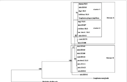
operon matchedA. phagocytophilum. The similarity var-ied from 95.6% to 99.8%. Twenty-five sequences of the
groESLoperon clustered in two genetic lineages, A and B (Figure 1). In the genetic lineage A, most of the sequences gathered in two clusters, 1 and 2. Ten sequences of thegroESLoperon were 100% identical to each other and grouped together with a reference sequence from a Slovenian dog [GenBank:EU381151] in cluster 2. Three 100% identical sequences clustered together with a sequence from a Slovenian patient [Gen-Bank:AF033101] in cluster 1 of the same lineage. In the lineage B, three sequences were 100% identical to each other and were identical to a reference sequence from a German tick [GenBank:AY281794] (Table 2). One sequence (tick EU341) from a pool of nymphs showed 95.6% similarity to other sequences from Slovenian ticks and 96% similarity with a reference sequence [GenBank: AY281848] from a German tick and did not cluster in any of the lineages A or B. Only the sequences that were not 100% identical were included in the phylogenetic study.
Table 1A. phagocytophilumin pools of ticks and its prevalence in Slovenian I. ricinus
Adult
Year Location Larvae Nymphs Male Female Total
Črni Kal 3 13 2 2 20
Sodražica 1 14/1 (0.80) 5 4 24/1 (0.63)
Murskašuma 0 2 4 7 13
Rakovnik 2 15 12 10 39
2005 Mozirje 3 23/3 (1.45) 10 8/1 (3.5) 44/4 (1.24)
Kamniška Bistrica 9 22 9 7 47
Štefanja Gora 3 27 9/1 (2.97) 6 45/1 (0.28)
Osolnik 1 10 5 4 20
Total 22 126/4 (0.34) 56/1 (0.53) 48/1 (0.69) 252/6 (0.31)
Črni Kal NT 43/5 (1.31) 4 2 49/5 (1.24)
Sodražica NT 24/1 (0.43) 8 6 38/1 (0.34)
Murskašuma NT 1 10 22 33
Rakovnik NT 32/2 (0.65) 17 14 63/2 (0.44)
2006 Mozirje NT 41/3 (0.79) 14/2 (3.66) 13/1 (1.97) 68/6 (1.24)
Kamniška Bistrica NT 34/2 (0.60) 17 18 69/2 (0.41)
Štefanja Gora NT 50/1 (0.21) 13 12 75/1 (0.17)
Osolnik NT 25/3 (1.28) 12 10 47/3 (0.90)
Total NT 250/17 (0.73) 95/2 (0.47) 97/1 (0.23) 442/20 (0.63) In parenthesis in %. NT - not tested. Ticks were sorted into pools of 10 nymphs, 30 larvae or pools of 5 halves of male or female adult ticks.
[image:3.595.57.540.372.684.2]the presence of appropriate hosts [7], as well as different screening methods used. The infection rate in Slovenia did not differ considerably between the tick stages (Table 1). In the year 2005, no larvae were found posi-tive, which is in concordance with the fact that anaplas-mae are not transovarially transmitted [4].
Only a few reports describe the diversity of A. phago-cytophilum in infected ticks sampled from vegetation [10]. The variety of groESLoperon sequences has been determined and some matched human and horse cases of anaplasmosis [10]. In this study, a high diversity of sequences of thegroESLoperon in ticks has been found. As discussed in a previous study of deer sequences of
A. phagocytophilum[9], the sequences from the ticks in Slovenia are also delineated in two genetic lineages (Figure 1). The similarity between these sequences var-ied from 97.8% to 99.8%. In the lineage A (dog, human, wild boar, tick, deer samples), the similarity ranged from 99.5% to 99.9%, and in the lineage B (tick and deer sam-ples), it varied from 99.0% to 99.8%. Different genetic lineages represent also differences in the amino acid sequence of GroESL protein (an amino acid serine (line-age A) is substituting alanine (line(line-age B) at the position 242). It is suggested that the strains ofA. phagocytophi-lumthat possess a variant of the protein with the serine might be pathogenic to humans [18].
A greater diversity of thegroESL operon was found at the nymphal stage of ticks. For this, many reasons are possible. The groESL genetic variants other than those that cause the disease in humans and dogs in Slovenia might not be pathogenic to aforementioned hosts since they have not been found in them yet [11,19]. It is nevertheless possible that they circulate only among small mammals and deer. However, the main reason could be the number of collected ticks: more nymphs than adult ticks were collected at all locations and, con-sequently, higher diversity was found.
In one pool of nymphs from Mozirje, a groESL
sequence showed only a 95.6% similarity with other
groESLoperon sequences from the ticks in Slovenia. It did not cluster within the lineages A and B. Moreover, after the translation into amino acid sequence, no differ-ence from the lineage B was found (alanine at the posi-tion 242). Probably, a novel genetic lineage of the
groESL operon of A. phagocytophilum was found. To
obtain more information about genetic diversity of
A. phagocytophilum inI. ricinus in Slovenia, additional genetic markers, such as ankA and msp4, should be analyzed.
I. ricinusnymphal and adult stages are responsible for the transmission of the pathogen asA. phagocytophilum
[image:4.595.57.538.111.401.2]was present in both stages. There was no significant
Table 2 Similarity ofgroESLoperon sequences betweenA. phagocytophilumin Slovenian ticks and GenBank reference sequences
GenBank accession number Source Similarity Location Ticks developmental stage Slovenian sequences from ticks
EU381151 dog, Slovenia 100% Mozirje Nymphs EU107
Female EU112, EU412
Male EU418
Štefanja Gora Nymphs EU367
Male EU132
Črni kal Nymphs EU254, EU277
Kamniška Bistrica Nymphs EU433
Rakovnik Nymphs EU652
AY281794 tick, Germany 100% Mozirje Nymphs EU111
Sodražica Nymphs EU329
Črni kal Nymphs EU622
AY281844 tick, Germany 100% Mozirje Nymphs EU108
99% Osolnik Nymphs EU379
99% Sodražica Nymphs EU136
AF478560 roe deer, Slovenia 100% Črni kal Nymphs EU260
AY281848 tick, Germany 96% Mozirje Nymphs EU341
AY281851 tick, Germany 99% Mozirje Nymphs EU343
AF033101 human, Slovenia 100% Osolnik Nymphs EU383, EU381
Rakovnik Nymphs EU295
AY281771 tick, Germany 100% Mozirje Nymphs EU422
AY220470 tick, Austria 100% Črni kal Nymphs EU322
AF548386 sheep, Norway 100% Kamniška Bistrica Nymphs EU431
difference in the prevalences ofA. phagocytophilumat different locations and in both years. The prevalence of infection in ticks did not differ considerably from the reports from elsewhere in Europe. With this study, we have confirmed thatI. ricinusis a vector of a variant of
A. phagocytophilumthat causes the disease in humans and dogs. The sequencing of the groESL operon has demonstrated a great diversity ofA. phagocytophilumin Slovenia. With phylogenetic analysis, two genetic lineages have been confirmed and another has been proposed. Further phylogenetic studies of several other genes, such asankAandmsp4, might be useful to obtain more infor-mation about genetic diversity.
Acknowledgements
This study was partially funded by EU grant GOCE-2003-010284 EDEN and the paper is catalogued by the EDEN Steering Committee as EDEN0193 http://www.eden-fp6project.net/. The contents are the sole responsibility of the authors and do not necessarily reflect the views of the European Commission.
Author details
1Institute of Microbiology and Immunology, Faculty of Medicine, Zaloška 4,
SI-1000 Ljubljana, Slovenia.2Public Health Institute Maribor, Prvomajska 1,
2000 Maribor, Slovenia.
Authors’contributions
KSS conducted the laboratory study and drafted the manuscript. MS was involved in laboratory study. DD facilitated the molecular laboratory study. NK conducted the field study and did the statistical analysis. TAZ was involved in the project design and participated in drafting the manuscript. All co-authors have read the manuscript.
Competing interests
The authors declare that they have no competing interests.
Received: 6 July 2010 Accepted: 4 November 2010 Published: 4 November 2010
References
1. Knap N, Durmiši E, Saksida A, Korva M, Petrovec M, AvšičŽupanc T: Influence of climatic factors on dynamics of questingIxodes ricinusticks in Slovenia.Vet Parasitol2009,164:275-281.
2. Sonenshine DE: InBiology of ticks. Volume 2.New York: Oxford University Press; 1993:488.
3. Petrovec M, LotričFurlan S,Županc TA, Strle F, Brouqui P, Roux V, Dumler JS:Human disease in Europe caused by a granulocytic Ehrlichia species.J Clin Microbiol1997,35:1556-1559.
4. Parola P, Davoust B, Raoult D:Tick- and flea-borne rickettsial emerging zoonoses.Vet Res2005,36:469-492.
5. Dumler JS, Barbet AF, Bekker CPJ, Dasch GA, Palmer GH, Ray SC, Rikihisa Y, Rurangirwa FR:Reorganization of genera in the familiesRickettsiaceae andAnaplasmataceaein the orderRickettsiales: unification of some species ofEhrlichiawithAnaplasma,CowdriawithEhrlichiaandEhrlichia withNeorickettsia, descriptions of six new species combinations and designation ofEhrlichia equiand‘HGE agent’as subjective synonyms of Ehrlichia phagocytophila.Int J Sys Evol Microbiol2001,51:2145-2165. 6. Strašek Smrdel K, Bidovec A, Malovrh T, Petrovec M, Duh D, Avsic Zupanc T:
Detection ofAnaplasma phagocytophilumin wild boar in Slovenia.Clin Microb Infect2008,15:50-52.
7. Blanco JR, Oteo JA:Human granulocytic ehrlichiosis in Europe.Clin Microb Inf2002,8:763-772.
8. Sumner JW, Nicholson WL, Massung RF:PCR amplification and
comparison of nucleotide sequences from thegroESLheat shock operon ofEhrlichiaspecies.J Clin Microbiol1997,35:2087-2092.
9. Petrovec M, Bidovec A, Sumner JW, Nicholson WL, Childs JE, Avšič
Županc T:Infection withAnaplasma phagocytophilain cervids from Slovenia: evidence of two genotypic lineages.Wien Klin Wochenschr2002, 114:641-647.
10. von Loewenich FD, Baumgarten BU, Schröppel K, Geißdörfer W, Röllinghoff M, Bogdan C:High diversity ofankAsequences ofAnaplasma phagcytophilumamongIxodes ricinusticks in Germany.J Clin Microbiol 2003,41:5033-5040.
11. Petrovec M, Sumner JW, Nicholson WL, Childs JE, Strle F, BarličJ, Lotrič -Furlan S, AvšičŽupanc T:Identity of ehrlichial DNA sequences derived fromIxodes ricinusticks with those obtained from patients with human granulocytic ehrlichiosis in Slovenia.J Clin Microbiol1999,387:209-210. 12. Black WC, Piesman J:Phylogeny of hard- and soft-tick taxa (Acari: Ixodida)
based on mitochondrial 16 SrRNAsequences.Proc Natl Acad Sci USA 1994,91:10034-10038.
13. Thompson JC, Higgins DG, Gibson TJ:CLUSTAL W: improving the sensitivity of progressive multiple sequence alignment through sequence weighting positions, specific gap penalties, and weigh matrix choice.Nucleic Acid Res1994,22:4673-4680.
14. Kimura M:A simple method for estimating evolutionary rate of base substitutions through comparative studies of nucleotide sequence.J Mol Evol1980,16:111-120.
15. Saitou N, Nei M:The Neighbour-joining method: a new method for reconstructing phylogenetic trees.Med Biol Evol1987,4:406-425. 16. Foley J, Nieto NC, Foley P, Teglas MB:Co-phylogenetic analysis of
Anaplasma phagocytophilumand its vectors,Ixodesspp. ticks.Exp Appl Acarol2008,45:155-170.
17. Jongejan F, Uilenberg G:The global importance of ticks.Parasitology2004, 129:S3-S14.
18. Rymaszewska A:Divergence within the marker region of thegroESL operon inAnaplasma phagocytophilum.Eur J Clin Microbiol Infect Dis2008, 27:1025-36.
19. Strašek Smrdel K, Tozon N, Duh D, Petrovec M, AvšičŽupanc T:Diversity of groESLsequences ofAnaplasma phagocytophilumamong dogs in Slovenia.Clin Microb Infect2009,15:79-80.
doi:10.1186/1756-3305-3-102
Cite this article as:Smrdelet al.:Anaplasma phagocytophilumin ticks in Slovenia.Parasites & Vectors20103:102.
Submit your next manuscript to BioMed Central and take full advantage of:
• Convenient online submission
• Thorough peer review
• No space constraints or color figure charges
• Immediate publication on acceptance
• Inclusion in PubMed, CAS, Scopus and Google Scholar
• Research which is freely available for redistribution