REVIEW
Methods for the field evaluation
of quantitative G6PD diagnostics: a review
Benedikt Ley
1*, Germana Bancone
2,3, Lorenz von Seidlein
4, Kamala Thriemer
1, Jack S. Richards
5,6,7,
Gonzalo J. Domingo
8and Ric N. Price
1,3Abstract
Individuals with glucose-6-phosphate dehydrogenase (G6PD) deficiency are at risk of severe haemolysis following the administration of 8-aminoquinoline compounds. Primaquine is the only widely available 8-aminoquinoline for the radical cure of Plasmodium vivax. Tafenoquine is under development with the potential to simplify treatment regi-mens, but point-of-care (PoC) tests will be needed to provide quantitative measurement of G6PD activity prior to its administration. There is currently a lack of appropriate G6PD PoC tests, but a number of new tests are in development and are likely to enter the market in the coming years. As these are implemented, they will need to be validated in field studies. This article outlines the technical details for the field evaluation of novel quantitative G6PD diagnostics such as sample handling, reference testing and statistical analysis. Field evaluation is based on the comparison of paired samples, including one sample tested by the new assay at point of care and one sample tested by the gold-standard reference method, UV spectrophotometry in an established laboratory. Samples can be collected as capillary or venous blood; the existing literature suggests that potential differences in capillary or venous blood are unlikely to affect results substantially. The collection and storage of samples is critical to ensure preservation of enzyme activity, it is recommended that samples are stored at 4 °C and testing occurs within 4 days of collection. Test results can be visually presented as scatter plot, Bland–Altman plot, and a histogram of the G6PD activity distribution of the study population. Calculating the adjusted male median allows categorizing results according to G6PD activity to calculate standard performance indicators and to perform receiver operating characteristic (ROC) analysis.
© The Author(s) 2017. This article is distributed under the terms of the Creative Commons Attribution 4.0 International License (http://creativecommons.org/licenses/by/4.0/), which permits unrestricted use, distribution, and reproduction in any medium, provided you give appropriate credit to the original author(s) and the source, provide a link to the Creative Commons license, and indicate if changes were made. The Creative Commons Public Domain Dedication waiver (http://creativecommons.org/ publicdomain/zero/1.0/) applies to the data made available in this article, unless otherwise stated.
Background
Glucose-6-phosphate dehydrogenase (G6PD) is an essen-tial enzyme in the pentose phosphate pathway (PPP), the only pathway for human red blood cells (RBC) to maintain the cells’ redox potential by reducing NADP+
to NADPH [1, 2]. The enzyme consists of two dimers encoded by a gene on the long arm of the X chromosome. Non-synonymous mutations in the gene can decrease enzyme activity or reduce the stability of the enzyme, resulting in different degrees of G6PD deficiency. Because the gene is on the X chromosome, males are hemizygous and are either classified as G6PD deficient or normal by phenotype. Females have two gene copies which are
expressed alternately in RBCs. Females therefore can be homozygous deficient (two gene copies with a deleteri-ous mutation), heterozygdeleteri-ous with one gene copy encod-ing a normal G6PD variant and one gene copy encodencod-ing a deficient G6PD variant (and a phenotype ranging from normal to deficient), or homozygous normal with both gene copies expressing G6PD variants with normal G6PD activity. Through the process of X-chromosome inactiva-tion (also called lyonizainactiva-tion), females express only one of their two copies of the gene in each cell. This occurs ran-domly in the precursors of RBCs early in the embryonic stage and results in different ratios of gene expression in mature RBCs amongst females but is constant within an individual [1]. As a consequence females heterozygous for G6PD can manifest a range of intermediate G6PD activities between typical normal and deficient G6PD activities that reflects the average enzymatic activity of these two cellular populations.
Open Access
*Correspondence: [email protected]; [email protected]
1 Global and Tropical Health Division, Menzies School of Health Research
and Charles Darwin University, Darwin, Australia
Glucose-6-phosphate dehydrogenase deficiency (G6PDd) is the most common enzymopathy with at least 400 million individuals affected worldwide [3, 4]. More than 185 clinically relevant variants of G6PDd have been reported with a spectrum of associated enzyme deficien-cies [2]. The high prevalence of these mutants is likely to have been driven by their ability to provide some degree of protection from malaria [5–9]. A case control study conducted in The Gambia looking at three major poly-morphisms associated with G6PD deficiency found that G6PDd provided significant protection against severe malaria (OR = 0.83) [10]. A cross sectional survey from the Brazilian Amazon reported a very strong protective effect against all species of malaria for the G6PD A-var-iant (OR = 0.12) and even stronger protection with the Mediterranean variant (OR = 0.01) [7]. A Thai study showed that patients with the Mahidol variant (confer-ring moderate G6PDd) had reduced Plasmodium vivax, but not Plasmodium falciparum parasitaemia [5]. More recently, a study conducted in Papua found detectable parasitaemia to be significantly lower among patients with G6PDd (OR = 0.44) with a greater protective effect
for P. vivax compared to P. falciparum [6].
The presence of G6PDd in patients with P. vivax infec-tion results in significant challenges to achieving radi-cal cure. The only class of anti-malarial drugs currently available to eliminate the dormant liver stages of P. vivax (hypnozoites) are the 8-aminoquinolone compounds and these cause haemolysis in G6PDd individuals [11]. The degree of haemolysis depends on total dose of pri-maquine (PQ), underlying G6PD variant and age of the RBC population (with lower G6PD activities in older cells); depending on conditions, haemolysis can be slow and self-limited or result in acute and potentially fatal anaemia [11, 12]. Previous studies suggest that individu-als with non-severe G6PD variants who are exposed to PQ experience an initial drop in haemoglobin (Hb) when old RBCs with low G6PD activities lyse [12]. As a response erythropoiesis increases and lysed cells are replaced by younger cells with higher G6PD activity ulti-mately stabilizing and reversing anaemia. Subsequent doses of PQ will not result in a further drop in Hb as the younger population of RBCs has higher G6PD activi-ties and at the same time the rate of erythropoiesis has increased to replace lysing RBCs in due time, resulting in a so-called “resistance phase” [12]. In contrast G6PD activities in RBCs of patients with severe G6PD variants are very low even in young RBCs, therefore, sustained exposure to an oxidizing agent will result in haemolysis irrespective of the age of the RBC. Continuous treatment with PQ in these patients will result in severe and ulti-mately fatal anaemia. There are additional factors that
contribute to risk of haemolysis at an individual level and at a specific event that are not well understood.
A number of 8-aminoquinoline compounds are under development, however to date PQ is the only drug which is licensed and widely available. Primaquine-based radi-cal cure in G6PD normal patients is usually adminis-tered over 14 days at a total dose of either 3.5 or 7 mg/ kg bodyweight depending on the assumed susceptibility of dominant local parasite strains [13]. Although either PQ regimen is usually well tolerated in G6PD normal subjects it is often not prescribed due to fears of drug-induced haemolysis; when PQ is prescribed the long treatment course, which is normally 14 days, can be asso-ciated with poor adherence and thus effectiveness [14– 16]. Tafenoquine (TQ), a related 8-aminoquinoline, is at the end of phase 3 clinical trials and is anticipated to be licensed by 2018 [17–19]. The half-life of TQ is 14 days compared to 4–6 h for PQ [20], and this allows adequate cure with a single 300 mg dose [17]. A single dose regi-men of TQ is likely to improve treatregi-ment adherence significantly, however may also raise the potential for sus-tained haemolysis in G6PDd patients and for this reason TQ is likely to be licensed for use only after G6PD nor-mal status has been confirmed.
Accessbio (USA) has recently developed a Biosensor™, a quantitative handheld device that measures G6PD activity based on the electrochemical properties of G6PD in blood samples [32, 33]. Other new quantitative tests are also in the development pipeline. This new class of diagnostic tools aims to address the diagnostic needs of heterozygous women as well as being flexible to differ-ent threshold activities for differdiffer-ent 8-aminoquinoline regimens. Whilst the WHO has published recommended guidelines on the clinical evaluation of quantitative G6PD assays required for pre-qualification [34], these do not cover technical details such as sample handling, reference testing and statistical analysis. A set of recommendations and guidelines have been published earlier on the evalu-ation of qualitative G6PD diagnostics [24], aim of this article is to summarize suitable approaches for the field evaluation of quantitative G6PD assays.
Challenges in the field
Field staff performing experimental G6PD testing at field level need to be appropriately trained on the experimen-tal assay and receive training on the requirements for sample handling for later reference testing. Undertak-ing evaluation studies of diagnostics assays and collect-ing appropriate samples in malaria endemic areas can be logistically challenging. Current G6PD reference meth-ods require significant laboratory infrastructure and well trained laboratory staff, which are lacking at many field sites. Accordingly samples collected in the field may have to be shipped to a local reference centre and this should be done in a timely manner and with a reliable cold chain.
G6PD enzyme activity and its degradation is tempera-ture dependent, hence whole EDTA blood should not be stored at room temperature. Freezing samples and maintaining a respective cold chain in a field setting is demanding and often impossible. Whole blood sam-ples can be stored at 4–8 °C for later analysis, however the duration at which samples can be stored and reli-ably assayed at a later time point is unknown. In one study conducted on 9 EDTA blood samples with nor-mal (n = 7) and intermediate G6PD activity (n = 2) and stored at 4 °C, G6PD activity remained stable for up to 21 days with a total drop in activity of less than 5% by day 21 [35]. In contrast a study of 100 EDTA blood samples from Nigeria reported a 15–21% drop in G6PD activity after 21 days of storage at 4 °C [36]. In Malaysia, a study on 188 EDTA neonatal cord blood samples stored at 4 °C reported a minimal (0.8%) fall in G6PD activity among 172 normal individuals within the first 24 h. The decrease rose to 1.2% at 48 h, 4.7% at 4 days and 5.6% at 7 days of storage [37]. Another study from Turkey, observed G6PD activity decreasing by 11% between days 1 and 3 and 40%
between days 1 and 7 when samples were stored at 4 °C [38].
Reference testing may also include flow cytometry to determine the fraction of G6PDd RBCs in heterozygous females. In these cases samples must be processed prior to shipment. Ideally samples should be cryopreserved by adding a cryopreservant such as Glycerolyte 57™ and stored at −80 °C [39]. An alternative to storing and shipping samples at very low temperatures is to replace plasma with an additive (2.5% glucose, 0.9% sodium chlo-ride, 0.027% adenine, 0.75% mannitol) which maintains G6PD activity for longer periods than EDTA in individ-ual cells at temperatures of 4–8 °C [35]. When the addi-tive was added within 48 h of collection and samples were stored at 4 °C there was no significant decrease in the proportion of RBCs with high G6PD activity for up to 21 days [35]. If there is the possibility to freeze whole blood samples at −80 °C and ship on dry ice, this may also be done, however RBCs will lyse during thawing. The consequences of this lysis are several fold: (1) specimen integrity is compromised, (2) the G6PD enzyme is less stable in lysed blood than in intact RBCs and the sam-ples should be used within 1 h of thawing and kept on ice during that period of time, and (3) the evaluation cannot include an assessment of RBC lysis (as this has already occurred in the freeze-thaw process) but only enzyme activity.
In most field studies experimental point of care tests (PoCs) are likely to be tested directly on a finger prick using capillary blood, whereas reference testing is more likely to be undertaken on venous samples; this has the potential to introduce a bias [40]. A study in Thailand assessed relevant blood parameters on 139 paired capil-lary and venous blood samples and found significantly lower values for red blood cell count, haemoglobin con-centration, haematocrit, mean corpuscular volume and significantly higher values in platelet count in capil-lary compared to venous blood; with the exception of the platelet count the difference was less than 5% for all parameters [40]. The G6PD activity of paired sam-ples collected from venous and capillary sampling were similar when measured by spectrophotometry (Trinity, UK) (median 7.51 and 7.61 U/gHb, respectively) [40]. Similarly, an evaluation of qualitative tests by FST and Carestart G6PD RDT (Accessbio, USA) showed 100% concordance between paired capillary and venous sam-ples [41].
Reference method and universal cut off activities
underlying G6PD variants and heterozygosity. Several quantitative assays have emerged that either directly or indirectly measure NADPH formation as a result of G6PD activity [2], however the majority of test evalu-ations have used spectrophotometry as the reference method. Spectrophotometry measures the formation of NADPH from G6PD activity by measuring the difference in absorbance (Δ absorbance) of the sample at 340 nm over time [42] and requires a kinetic and temperature controlled spectrophotometer, trained laboratory staff and a well-functioning laboratory infrastructure. Spec-trophotometry measurements are usually run in dupli-cate, and the inclusion of at least commercial G6PD deficient and G6PD normal controls is highly desirable. Prior to being considered as reference method spectro-photometry must be fully validated, including perfor-mance of operators, reproducibility and internal as well as external quality assurance procedures must have been established.
The WST8/1-methoxy PMS test is a robust and cost effective alternative to estimate G6PD activity that can be performed on dried blood spots and whole blood sam-ples. The WST8/1 test is based on the formation of an orange formazan dye generated by G6PD enzyme activ-ity and facilitated by the 1-methoxy PMS as an electron carrier [43]. The assay has been adapted for quantitative batch testing in a 96 well microplate and can be read by ELISA reader [44, 45]. A preliminary field evaluation showed discrepancies between spectrophotometry and the methoxy test, although the assays were not run simul-taneously [32], additional evaluation studies are under way.
Quantification of G6PD activity requires normaliza-tion of the result to account for RBC mass and the tem-perature of the assay. Whenever possible an RBC count is preferable to Hb measurement, since the low Hb con-centrations in anaemic patients may result in an artificial increase of G6PD activity, whereas this is not the case when the RBC count is used for normalization [42, 46].
Absolute values can vary between different assays [23] and hence results need to be transformed into population specific values relative to the population specific adjusted male median (see below) [24]. Presentation of the results without the corresponding adjusted male median (AMM) value significantly confounds the comparison of the assay results across different studies. Even then per-formance criteria are likely to differ if different reference assays are used. To the extent possible a single reference assay should be used [24].
To date no universal cut-off and corresponding method that defines G6PDd has been clearly adopted. The degree of G6PD activity can help to guide clinical management, but the relevant threshold varies with the
intended use. For example the threshold currently used to guide PQ radical cure is 30% of the AMM [2], whereas the proposed threshold activity for TQ treatment will be approximately 70–80% to ensure safe use in females with intermediate activity [22, 31]. The great difference in absolute values measured among different spectro-photometric assays does not permit the definition of the optimal threshold if different assays are used. The prob-lem is well illustrated by the package insert of a standard G6PD normal control supplied by Trinity Biotech (Cat. No. G6888) which provides an assigned activity range of 12.6–23.4 U/gHb. Although it would be highly desirable to use a standard that provides a constant Δ absorbance with which to normalize results, to date none has been produced.
Sample size and statistical analysis of results
In order to achieve reliable results, all studies need a sam-ple size sufficient to ensure precise and reliable results, but small enough to be feasible in an endemic setting. Unless the aim of an evaluation study is WHO prequalifi-cation where static specific sample sizes are required (see below) [34], the sample size can be adjusted to reflect the local epidemiology and demographics of the population. There are numerous sample size calculators available online [47–50] that can perform sample size calcula-tions, as well as a range of statistical software packages. Three approaches are generally considered to calculate the sample size for evaluating novel quantitative G6PD diagnostics.
If the intended setting and use of the novel diagnostic is known, a decision on whether specificity or sensitivity is of greater interest can be made and the most relevant threshold activity defined. Following these considera-tions sample size can be calculated based on the assumed performance of the novel diagnostic at a specific thresh-old activity following recommendations from the special programme for research and training in tropical diseases (TDR) [51].
standard and experimental assay is less relevant, than the correlation between the two measures. Hence a third approach is to determine the minimal correlation coeffi-cient between gold standard and experimental assay to be detected and calculate sample size accordingly [53, 54]. Irrespective of the method applied the calculated sam-ple size should be increased by 10–20% to accommodate for losses due to errors in study procedures and other problems.
Since different quantitative assays can have different absolute values [23], a direct comparison of results of the experimental assay and reference method is not informa-tive. The most immediate comparison that can be made between the new G6PD test and the gold standard is a direct correlation of results and calculation of the cor-relation coefficient. Plotting results of the experimental assay and reference method in a scatter plot including a line of equality is a simple and clear way to display the relation between experimental and reference method test results (Fig. 1). This can be augmented by the inclusion of thresholds for 10, 30, 60 and 80% enzyme activity to show the relative performance of the assay at low, intermediate and high G6PD activity.
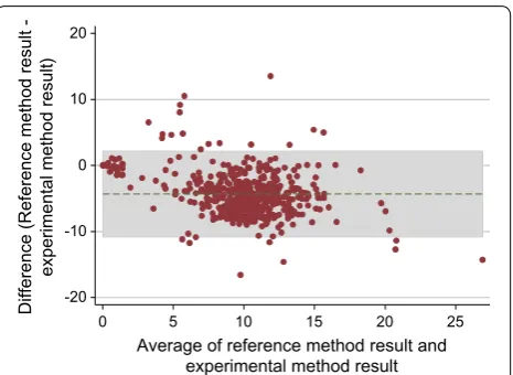
Absolute quantitative values can be compared using Bland and Altman plots which show the average of two paired results on the x-axis against the difference of the same results on the y-axis [55] (Fig. 2). From this plot the mean difference between both assays and a 95% limit of agreement (LoA) (mean difference ± 1.96 standard devi-ations) can be calculated. The standard deviations should also lie within clinically acceptable ranges of difference in G6PD activity. A mean difference deviating from zero suggests a systematic difference between absolute
readings of experimental and reference method, and potentially a systematic error in the experimental assay. The relevance of the detected mean difference is depend-ent on the intended use of the experimdepend-ental assay. The clinical relevance of any difference and LoA is made by the investigator and is guided by practical considerations.
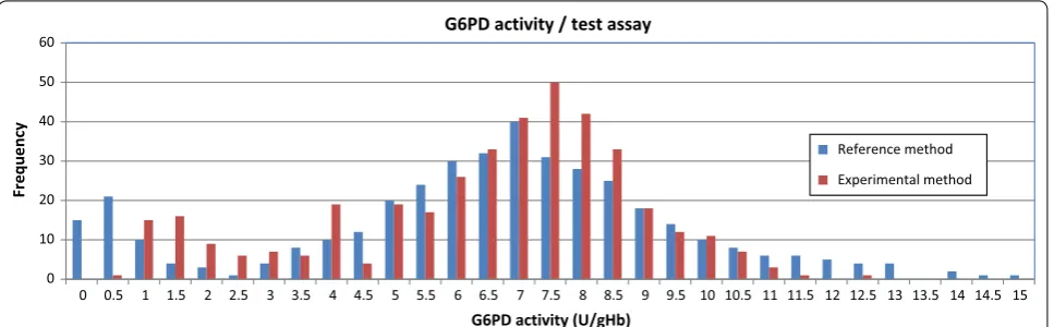
A histogram of G6PD activity, stratified by gender within a study population is a simple way to present the distribution of G6PD activity within a population. A respective histogram allows visual representation of the absolute values of different assays and whether an assay underdiagnoses samples beyond a specific G6PD cut-off activity. Within a non-skewed population of suf-ficient size the majority of individuals will have G6PD activities normally distributed around a population spe-cific median. A second smaller peak at the low end of the G6PD activity axis represents G6PDd individuals. Individuals in between these two peaks are intermedi-ate G6PD deficient and will mostly consist of heterozy-gous females. Major differences in the form of the G6PD activity distribution of experimental assay and refer-ence method are an indicator of short comings of the experimental assay (Fig. 3), a non-bimodal distribution of results among the male population further indicates short comings in the underlying method (Fig. 4).
Defining assay specific absolute values represent-ing 100% G6PD activity allows measured quantitative G6PD results to be categorized which in turn enables performance indicators to be calculated at various cut-off activities [24, 56]. The current standard approach for the definition of a study population specific 100% G6PD activity is to calculate the adjusted male median (AMM) (Fig. 5) [24], alternatively if results from geno-typing are available at time point of analysis then the 0
10 20 30 40
G6
PD
re
su
lt
from
ex
pe
rime
nt
al
assa
y
0 5 10 15 20
G6PD result from reference method (in U/gHb)
(in
U/
gH
b)
Fig. 1 Example of a scatter plot with line of equality. Starting from origin, red vertical and horizontal lines correspond to 10, 20, 30, 60 and 100% G6PD activity of the adjusted male median. The black dot-ted line = line of equality
-20 -10 0 10 20
Difference (Reference method result
-0 5 10 15 20 25
Average of reference method result and experimental method result
experimental method result)
[image:5.595.307.540.86.256.2] [image:5.595.58.291.515.677.2]median activity of all wild type participants (wt) can be calculated [40]. Cut-off activities for categories of G6PD activity can then be defined as fractions of the AMM or median of all wt as required [24, 27, 57] (Fig. 1). In order to assess the threshold activity at which the experimen-tal assay performs best, results of the reference method are transformed into a dichotomous outcome accord-ing to defined cut-off activities. Calculataccord-ing areas under the reactor operating curve (ROC) allow for a direct
comparison of performance of the experimental assay at different cut-off activities [58]. However, this procedure is unnecessary for experimental assays intended for a spe-cific use such as guiding PQ treatment, where a desired threshold activity is already known.
Once appropriate threshold activities have been estab-lished, test results of experimental assay and reference method can be categorized into true negative, true posi-tive as well as false posiposi-tive and false negaposi-tive results to
0 10 20 30 40 50 60
0 0.5 1 1.5 2 2.5 3 3.5 4 4.5 5 5.5 6 6.5 7 7.5 8 8.5 9 9.5 10 10.5 11 11.5 12 12.5 13 13.5 14 14.5 15
Frequenc
y
G6PD activity (U/gHb)
G6PD activity / test assay
Reference method Experimental method
Fig. 3 Example of a histogram to show G6PD activity distribution/test assay
0 10 20 30 40 50 60
0 0.5 1 1.5 2 2.5 3 3.5 4 4.5 5 5.5 6 6.5 7 7.5 8 8.5 9 9.5 10 10.5 11 11.5 12 12.5 13 13.5 14 14.5 15
Frequenc
y
G6PD activity (U/gHb)
G6PD activity / gender
Females males
Fig. 4 Example of a histogram to show G6PD activity distribution/gender
Calculating the adjusted male median (AMM)
The adjusted male median is calculated from male participants only.
1. Calculate median activity from all male participants
2. Exclude all samples / results with activity < 10% of the male median
3. Re-calculate the median activity from all remaining samples and define as 100% G6PD activity (= AMM)) 4. Form categories based on the AMM as desired (standard is 10%, 30%, 60% and 100%)
[image:6.595.58.542.232.382.2] [image:6.595.59.539.434.582.2]calculate standard performance indicators [51]. In this context it is desirable to change the nomenclature to avoid confusion and refer to test results as G6PD defi-cient rather than positive and G6PD normal rather than negative results. Results of this analysis can be summa-rized in an extended 2 × 2 table (see Table 2 in [32]).
WHO recommendations to establish clinical performance for assay pre‑qualification
Following field evaluation studies, successful quantita-tive G6PD diagnostics may undergo procedures for pre-qualification with the WHO. The WHO has published a guideline for WHO prequalification of in vitro diag-nostic medical devices (IVDs) to identify G6PD activ-ity [34]. The guideline provides “technical guidance to in vitro diagnostic medical device (IVD) manufacturers that intend to seek WHO prequalification of IVDs for the detection of glucose-6-phosphate dehydrogenase (G6PD) deficiency” [34], however does not list procedures to demonstrate clinical utility.
Discussion and outlook
This review presents a number of recommendations to facilitate standardized evaluation of quantitative G6PD activity test assays (Table 1). Standardized evaluation studies are necessary to provide a comparable basis for subsequent pooled analyses. As novel quantitative G6PD diagnostics become available their potential to provide robust quantitative G6PD results suitable for field deployment needs to be assessed. The most recent
generation of instruments contain an integrated temper-ature correction factor and Hb measurement and G6PD activity is displayed normalized as U/gHb. Some have argued that a quantitative G6PD result is too complex for translation into treatment at field level [59]. However, if universal cut-off activities can be established, training materials can be developed to link quantitative values to treatment decisions.
Although current biosensors are not as precise as spec-trophotometry [32], performance is likely to improve and their versatility and ability to discriminate individu-als with intermediate enzyme activity has the potential to provide a convenient platform to facilitate G6PD testing and TQ use across different endemic settings.
Authors’ contributions
BL wrote the first draft, GB, LvS, KT, JSR, GJD and RNP contributed to the con-tent. All authors read and approved the final manuscript.
Author details
1 Global and Tropical Health Division, Menzies School of Health Research
and Charles Darwin University, Darwin, Australia. 2 Shoklo Malaria Research
Unit, Mahidol-Oxford Tropical Medicine Research Unit, Faculty of Tropical Medicine, Mahidol University, Mae Sot, Thailand. 3 Centre for Tropical Medicine
and Global Health, Nuffield Department of Clinical Medicine, University of Oxford, Oxford, UK. 4 Mahidol-Oxford Tropical Medicine Research Unit
(MORU), Bangkok, Thailand. 5 Malaria Elimination Program, Burnet Institute,
Melbourne, VIC, Australia. 6 Department of Medicine, University of Melbourne,
Parkville, VIC, Australia. 7 Victorian Infectious Diseases Service, Peter Doherty
Institute for Infection and Immunity, Melbourne, VIC, Australia. 8 Diagnostics
Global Program, PATH, Seattle, WA, USA.
Competing interests
The authors declare that they have no competing interests.
Availability of data and materials Not applicable.
Consent for publication Not applicable.
Ethics approval and consent to participate Not applicable.
Funding statement
This work was supported by the Asia Pacific Malaria Elimination Network (APMEN), funded by the Australian Government Department of Foreign Affairs and Trade, and The Bill and Melinda Gates Foundation (OPP1164105). BL is funded by the Australian Department of Foreign Affairs and Trade, Grant Agreement Number 72904, BL and KT are funded by the Bill & Melinda Gates Foundation (OPRA-OPP1054404). GB is supported by the Wellcome Trust (Pro-gramme Grant 089179). SMRU is part of the Wellcome Trust Mahidol University Oxford Tropical Medicine Research Programme funded by the Wellcome Trust. JSR is supported by the Ian Gust Translational Research Fellowship. The Burnet Institute is supported by the NHMRC Independent Research Institutes Infrastructure Support Scheme, and a Victoria State Government Operational Infrastructure Support grant. GJD is funded by the Bill and Melinda Gates Foundation, Grant Number OPP1034534; and the UK Department for Inter-national Development (DFID), Grant Number 204139. RNP is funded by the Wellcome Trust (Senior Fellowship in Clinical Science, 200909).
Publisher’s Note
Springer Nature remains neutral with regard to jurisdictional claims in pub-lished maps and institutional affiliations.
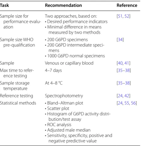
Table 1 Overview of recommendations for field evaluation of quantitative G6PD assays
Task Recommendation Reference
Sample size for performance evalu-ation
Two approaches, based on: • Desired performance indicators • Minimal difference in means
measured by two methods
[51, 52]
Sample size WHO
pre-qualification • 200 G6PD specimens• 200 G6PD intermediate speci-mens
• 1000 G6PD normal specimens [34]
Sample Venous or capillary blood [40, 41] Max time to
refer-ence testing 4–7 days [35–38]
Sample storage
temperature At 4–8 °C [35–38]
Reference testing Spectrophotometry [24, 42] Statistical methods • Bland–Altman plot
• Scatter plot
• Histogram of G6PD activity distri-bution/test assay
• ROC analysis • Adjusted male median
• Sensitivity, specificity, positive and negative predictive value
[image:7.595.57.292.493.736.2]Received: 7 June 2017 Accepted: 6 September 2017
References
1. Cappellini MD, Fiorelli G. Glucose-6-phosphate dehydrogenase defi-ciency. Lancet. 2008;371:64–74.
2. von Seidlein L, Auburn S, Espino F, Shanks D, Cheng Q, McCarthy J, et al. Review of key knowledge gaps in glucose-6-phosphate dehydroge-nase deficiency detection with regard to the safe clinical deployment of 8-aminoquinoline treatment regimens: a workshop report. Malar J. 2013;12:112.
3. Galappaththy GN, Tharyan P, Kirubakaran R. Primaquine for preventing relapse in people with Plasmodium vivax malaria treated with chloro-quine. Cochrane Database Syst Rev. 2013;10:CD004389.
4. Howes RE, Dewi M, Piel FB, Monteiro WM, Battle KE, Messina JP, et al. Spatial distribution of G6PD deficiency variants across malaria-endemic regions. Malar J. 2013;12:418.
5. Louicharoen C, Patin E, Paul R, Nuchprayoon I, Witoonpanich B, Peerapit-tayamongkol C, et al. Positively selected G6PD-Mahidol mutation reduces
Plasmodium vivax density in Southeast Asians. Science. 2009;326:1546–9. 6. Pava Z, Burdam FH, Handayuni I, Trianty L, Utami RA, Tirta YK, et al.
Submicroscopic and asymptomatic Plasmodium parasitaemia associ-ated with significant risk of anaemia in Papua, Indonesia. PLoS ONE. 2016;11:e0165340.
7. Santana MS, Monteiro WM, Siqueira AM, Costa MF, Sampaio V, Lacerda MV, et al. Glucose-6-phosphate dehydrogenase deficient variants are associated with reduced susceptibility to malaria in the Brazilian Amazon. Trans R Soc Trop Med Hyg. 2013;107:301–6.
8. Khim N, Benedet C, Kim S, Kheng S, Siv S, Leang R, et al. G6PD deficiency in Plasmodium falciparum and Plasmodium vivax malaria-infected Cambo-dian patients. Malar J. 2013;12:171.
9. Kwiatkowski DP. How malaria has affected the human genome and what human genetics can teach us about malaria. Am J Hum Genet. 2005;77:171–92.
10. Shah SS, Rockett KA, Jallow M, Sisay-Joof F, Bojang KA, Pinder M, et al. Het-erogeneous alleles comprising G6PD deficiency trait in West Africa exert contrasting effects on two major clinical presentations of severe malaria. Malar J. 2016;15:13.
11. John GK, Douglas NM, von Seidlein L, Nosten F, Baird JK, White NJ, et al. Primaquine radical cure of Plasmodium vivax: a critical review of the literature. Malar J. 2012;11:280.
12. Ashley EA, Recht J, White NJ. Primaquine: the risks and the benefits. Malar J. 2014;13:418.
13. Chu CS, White NJ. Management of relapsing Plasmodium vivax malaria. Expert Rev Anti Infect Ther. 2016;14:885–900.
14. Maneeboonyang W, Lawpoolsri S, Puangsa-Art S, Yimsamran S, Thanya-vanich N, Wuthisen P, et al. Directly observed therapy with primaquine to reduce the recurrence rate of Plasmodium vivax infection along the Thai-Myanmar border. Southeast Asian J Trop Med Public Health. 2011;42:9–18.
15. Pereira EA, Ishikawa EA, Fontes CJ. Adherence to Plasmodium vivax
malaria treatment in the Brazilian Amazon Region. Malar J. 2011;10:355. 16. Takeuchi R, Lawpoolsri S, Imwong M, Kobayashi J, Kaewkungwal J,
Pukrit-tayakamee S, et al. Directly-observed therapy (DOT) for the radical 14-day primaquine treatment of Plasmodium vivax malaria on the Thai-Myanmar border. Malar J. 2010;9:308.
17. Rajapakse S, Rodrigo C, Fernando SD. Tafenoquine for preventing relapse in people with Plasmodium vivax malaria. Cochrane Database Syst Rev. 2015;4:CD010458.
18. Beck HP, Wampfler R, Carter N, Koh G, Osorio L, Rueangweerayut R, et al. Estimation of the antirelapse efficacy of tafenoquine, using Plasmodium vivax genotyping. J Infect Dis. 2016;213:794–9.
19. Llanos-Cuentas A, Lacerda MV, Rueangweerayut R, Krudsood S, Gupta SK, Kochar SK, et al. Tafenoquine plus chloroquine for the treatment and relapse prevention of Plasmodium vivax malaria (DETECTIVE): a multicen-tre, double-blind, randomised, phase 2b dose-selection study. Lancet. 2014;383:1049–58.
20. Crockett M, Kain KC. Tafenoquine: a promising new antimalarial agent. Expert Opin Investig Drugs. 2007;16:705–15.
21. MPAC. Point-of-care G6PD testing to support safe use of primaquine for the treatment of vivax malaria. Geneva: WHO; 2015.
22. WHO. Guidelines for the treatment of malaria. Geneva: World Health Organization; 2015.
23. LaRue N, Kahn M, Murray M, Leader BT, Bansil P, McGray S, et al. Com-parison of quantitative and qualitative tests for glucose-6-phosphate dehydrogenase deficiency. Am J Trop Med Hyg. 2014;91:854–61. 24. Domingo GJ, Satyagraha AW, Anvikar A, Baird K, Bancone G, Bansil P, et al.
G6PD testing in support of treatment and elimination of malaria: recom-mendations for evaluation of G6PD tests. Malar J. 2013;12:391. 25. Beutler E, Blume KG, Kaplan JC, Lohr GW, Ramot B, Valentine WN.
Inter-national Committee for Standardization in Haematology: recommended screening test for glucose-6-phosphate dehydrogenase (G-6-PD) defi-ciency. Br J Haematol. 1979;43:465–7.
26. PATH. A guide to fluorescent spot testing for G6PD deficiency. Seatle: Programme for Appropriate Technology in Health; 2014.
27. Ley B, Luter N, Espino FE, Devine A, Kalnoky M, Lubell Y, et al. The chal-lenges of introducing routine G6PD testing into radical cure: a workshop report. Malar J. 2015;14:377.
28. Brito MA, Peixoto HM, Almeida AC, Oliveira MR, Romero GA, Moura-Neto JP, et al. Validation of the rapid test Carestart(tm) G6PD among malaria vivax-infected subjects in the Brazilian Amazon. Rev Soc Bras Med Trop. 2016;49:446–55.
29. Espino FE, Bibit JA, Sornillo JB, Tan A, von Seidlein L, Ley B. Comparison of three screening test kits for G6PD enzyme deficiency: implications for its use in the radical cure of vivax malaria in remote and resource-poor areas in the Philippines. PLoS ONE. 2016;11:e0148172.
30. Osorio L, Carter N, Arthur P, Bancone G, Gopalan S, Gupta SK, et al. Perfor-mance of BinaxNOW G6PD deficiency point-of-care diagnostic-infected subjects. Am J Trop Med Hyg. 2015;92:22–7.
31. Green J. Tafenoquine & G6PD. http://sites.path.org/dx/files/2012/11/01_ Green_Tafenoquine.pdf: Glaxo Smith Kline; 2011. Power Point Presentation.
32. Ley B, Alam MS, O’Donnell JJ, Hossain MS, Kibria MG, Jahan N, et al. A Comparison of three quantitative methods to estimate G6PD activity in the Chittagong Hill tracts, Bangladesh. PLoS ONE. 2017;12:e0169930. 33. Weppelmann TA, von Fricken ME, Wilfong TD, Aguenza E, Philippe TT,
Okech BA. Field trial of the CareStart biosensor analyzer for the deter-mination of glucose-6-phosphate dehydrogenase activity in Haiti. Am J Trop Med Hyg. 2017. doi:10.4269/ajtmh.16-0714.
34. WHO. Technical specifications series for submission to WHO prequali-fication: diagnostic assessment: in vitro diagnostics medical devices to identify glucose-6-phosphate dehydrogenase (G6PD) activity. Geneva: World Health Organization; 2016.
35. Kahn M, Ward WH, LaRue N, Kalnoky M, Pal S, Domingo GJ. Maintaining specimen integrity for G6PD screening by cytofluorometric assays. J Histochem Cytochem. 2015;63:454–8.
36. Ufelle SA, Neboh EE, Ocheni S, Ikekpeazu EJ, Maduka IC. The activity of glucose-6-phosphate dehydrogenase (G6PD) in stored blood. Orient J Med. 2014;26:5.
37. Jalil N, Azma RZ, Mohamed E, Ithnin A, Alauddin H, Baya SN, et al. Evalu-ation of glucose-6-phosphate dehydrogenase stability in stored blood samples. EXCLI J. 2016;15:155–62.
38. Yuregir GT, Aksoy K, Arpaci A, Unlukurt I, Tuli A. Studies on red cell glucose-6-phosphate dehydrogenase: evaluation of reference values. Ann Clin Biochem. 1994;31:50–5.
39. Kahn M, LaRue N, Bansil P, Kalnoky M, McGray S, Domingo GJ. Cryopreser-vation of glucose-6-phosphate dehydrogenase activity inside red blood cells: developing a specimen repository in support of development and evaluation of glucose-6-phosphate dehydrogenase deficiency tests. Malar J. 2013;12:286.
40. Bancone G, Chu CS, Chowwiwat N, Somsakchaicharoen R, Wilaisrisak P, Charunwatthana P, et al. Suitability of capillary blood for quantitative assessment of G6PD activity and performances of G6PD point-of-care tests. Am J Trop Med Hyg. 2015;92:818–94.
• We accept pre-submission inquiries
• Our selector tool helps you to find the most relevant journal
• We provide round the clock customer support
• Convenient online submission
• Thorough peer review
• Inclusion in PubMed and all major indexing services
• Maximum visibility for your research
Submit your manuscript at www.biomedcentral.com/submit
Submit your next manuscript to BioMed Central
and we will help you at every step:
42. Betke K, Beutler E, Brewer GJ, Kirkman HN, Luzzatto L, Motulsky AG, Ramot B, Siniscalco M. Standardization of procedures for the study of glucose-6-phosphate dehydrogenase. Report of a WHO Scientific Group. World Health Organ Tech Rep Ser. 1967;366:1–53.
43. De Niz M, Eziefula AC, Othieno L, Mbabazi E, Nabukeera D, Ssemmondo E, et al. Tools for mass screening of G6PD deficiency: validation of the WST8/1-methoxy-PMS enzymatic assay in Uganda. Malar J. 2013;12:210. 44. Tantular IS, Kawamoto F. An improved, simple screening method for
detection of glucose-6-phosphate dehydrogenase deficiency. Trop Med Int Health. 2003;8:569–74.
45. Kuwahata M, Wijesinghe R, Ho MF, Pelecanos A, Bobogare A, Landry L, et al. Population screening for glucose-6-phosphate dehydrogenase defi-ciencies in Isabel Province, Solomon Islands, using a modified enzyme assay on filter paper dried bloodspots. Malar J. 2010;9:223.
46. Oo NN, Bancone G, Maw LZ, Chowwiwat N, Bansil P, Domingo GJ, et al. Validation of G6PD point-of-care tests among healthy volunteers in Yangon, Myanmar. PLoS ONE. 2016;11:e0152304.
47. Mackinnon A. A spreadsheet for the calculation of comprehensive statistics for the assessment of diagnostic tests and inter-rater agreement. Comput Biol Med. 2000;30:127–34.
48. Michael Kohn MSJ, Josh Senyak. Sample size calculators. University of California San Francisco; 2017. http://www.sample-size.net/. Accessed 17 Mar 2017.
49. Rajasekhar Ramakrishnan SH. Biomath. Columbia University Medical Center, USA; 2017. http://biomath.info/. Accessed 17 Mar 2017. 50. Dean AG, Sullivan KM, MM Soe, RA Mir. OpenEpi: open source
epide-miologic statistics for public health. Emory University; 2003. http://www. openepi.com. Accessed 17 Mar 2017.
51. Banoo S, Bell D, Bossuyt P, Herring A, Mabey D, Poole F, et al. Evaluation of diagnostic tests for infectious diseases: general principles. Nat Rev Microbiol. 2006;4(12 Suppl):S20–32.
52. Dell RB, Holleran S, Ramakrishnan R. Sample size determination. ILAR J. 2002;43:207–13.
53. Fischer RA. Frequency distribution of the values of the correlation coefficient in samples from an indefinitely large population. Biometrika. 1915;10:507–21.
54. Lachin JM. Introduction to sample size determination and power analysis for clinical trials. Control Clin Trials. 1981;2:93–113.
55. Bland JM, Altman DG. Statistical methods for assessing agreement between two methods of clinical measurement. Lancet. 1986;1:307–10. 56. Panel TDRDEE, Banoo S, Bell D, Bossuyt P, Herring A, Mabey D, et al.
Evalu-ation of diagnostic tests for infectious diseases: general principles. Nat Rev Microbiol. 2010;8(12 Suppl):S17–29.
57. Glucose-6-phosphate dehydrogenase deficiency. WHO Working Group. Bull World Health Organ. 1989;67:601–11.