RESEARCH
Interleukin 32 expression in human
melanoma
Helicia Paz
1†, Jennifer Tsoi
3,5†, Anusha Kalbasi
1,4,6, Catherine S. Grasso
5, William H. McBride
4,
Dörthe Schaue
4, Lisa H. Butterfield
7,8,9,10, Deena M. Maurer
7, Antoni Ribas
1,5,6, Thomas G. Graeber
3,6and James S. Economou
1,2,3,6*Abstract
Background: Various proinflammatory cytokines can be detected within the melanoma tumor microenvironment. Interleukin 32 (IL32) is produced by T cells, NK cells and monocytes/macrophages, but also by a subset of melanoma cells. We sought to better understand the biology of IL32 in human melanoma.
Methods: We analyzed RNA sequencing data from 53 in-house established human melanoma cell lines and 479 melanoma tumors from The Cancer Genome Atlas dataset. We evaluated global gene expression patterns associated with IL32 expression. We also evaluated the impact of proinflammatory molecules TNFα and IFNγ on IL32 expression and dedifferentiation in melanoma cell lines in vitro. In order to study the transcriptional regulation of IL32 in these
cell lines, we cloned up to 10.5 kb of the 5′ upstream region of the human IL32 gene into a luciferase reporter vector.
Results: A significant proportion of established human melanoma cell lines express IL32, with its expression being highly correlated with a dedifferentiation genetic signature (high AXL/low MITF). Non IL32-expressing differentiated melanoma cell lines exposed to TNFα or IFNγ can be induced to express the three predominant isoforms (α, β, γ) of
IL32. Cis-acting elements within this 5′ upstream region of the human IL32 gene appear to govern both induced and
constitutive gene expression. In the tumor microenvironment, IL32 expression is highly correlated with genes related to T cell infiltration, and also positively correlates with high AXL/low MITF dedifferentiated gene signature.
Conclusions: Expression of IL32 in human melanoma can be induced by TNFα or IFNγ and correlates with a
treat-ment-resistant dedifferentiated genetic signature. Constitutive and induced expression are regulated, in part, by cis
-acting sequences within the 5′ upstream region.
Keywords: Interleukin 32 (IL32), Melanoma dedifferentiation, IL32 transcriptional regulation, Immune infiltration, Myeloid polarization
© The Author(s) 2019. This article is distributed under the terms of the Creative Commons Attribution 4.0 International License (http://creat iveco mmons .org/licen ses/by/4.0/), which permits unrestricted use, distribution, and reproduction in any medium, provided you give appropriate credit to the original author(s) and the source, provide a link to the Creative Commons license, and indicate if changes were made. The Creative Commons Public Domain Dedication waiver (http://creat iveco mmons .org/ publi cdoma in/zero/1.0/) applies to the data made available in this article, unless otherwise stated.
Background
Though first described in 1992 as NK4, interleukin 32
(IL32) [1, 2] was recharacterized in 2005 as a
proinflam-matory cytokine differentially expressed in IL18 respon-sive cells. Since then, its expression has been implicated in various pathologies, including rheumatoid arthritis, pathogen responses, atherosclerosis, and several
malig-nancies [3–12]. IL32 is broadly expressed in immune
cells, including natural killer cells [13–15], T
lympho-cytes [16–18], macrophages, and dendritic cells [19, 20].
Functionally, IL32 can activate NF-κB and p38 mitogen activated protein kinase pathways and induce expres-sion of proinflammatory cytokines including TNFα, IL8 and CCL2. In addition to its presence in immune tis-sues, IL32 expression can also be induced in human epi-thelial tissues. IL32 is also expressed in a broad range
of human epithelial cancers–gastric [3], lung [5], breast
[4], colon [6–9], pancreas [10], and thyroid [11, 12]—as
well as hematologic malignancies such as lymphoma and
leukemia [16, 21]. IL32 expression in cancer is linked to
Open Access
*Correspondence: jeconomou@mednet.ucla.edu
†Helicia Paz and Jennifer Tsoi are co-first authors
1 Department of Surgery, University of California, Los Angeles, 10833 Le
Conte Ave, Los Angeles, CA 90095, USA
features associated with worse prognosis, including
angi-ogenesis, invasion, and metastasis [4].
However, the role of IL32 in human melanoma cells is less well understood. Initial studies suggested that IL32 expression was related to a more invasive, meta-static phenotype marked by a loss of e-cadherin
expres-sion [22]. Others reported that IL32 isoforms alpha and
gamma were highly enriched in PD-L1 expressing
mela-noma specimens [23].
Herein we report an unbiased analysis of IL32
expres-sion in melanoma using RNA sequencing data from: [1] a
large in-house panel of melanoma cell lines and [2] tumor
tissues from The Cancer Genome Atlas (TCGA) dataset. IL32 expression correlated with a dedifferentiated pheno-type which has been characterized by resistance to tar-geted therapies, escape from immune recognition, and a
high AXL/low MITF genetic signature [24–27].
Expres-sion of IL32 in human melanoma can be transcription-ally regulated in the context of a proinflammatory tumor microenvironment with either induced or constitutive IL32 expression strongly, but not invariably, correlating with a dedifferentiated genetic signature.
Methods Cell culture
The human melanoma cell lines were established from patient biopsies. These cells were grown in RPMI 1640 media supplemented with 10% FBS, 1% l-glutamine and 1% penicillin/streptomycin/actinomycin D (Life Tech-nologies, Grand Island, NY). Melanoma cell lines were treated with 1000 IU/mL TNFα and 100 IU/mL IFNγ (PeproTech US, Rocky Hill, NJ) for the indicated time points. M397 was cultured with recombinant 100 ng/ mL IL32α, -β, or -γ (R&D Systems, Minneapolis, MN) for 7 days, with fresh IL32 being added every 2–3 days. Cells were counted using Trypan blue. Jurkat T lymphocytes were cultured in RPMI 1640 supplemented with 10% FBS.
Gene expression analysis
Gene expression FPKM values for the melanoma cell line panel was obtained from the Gene Expression Omnibus (GEO) accession number GSE80829. The hallmark gen-eset for NF-κB and TNFα signaling (TNFA_SIGNAL-ING_VIA_NFKB) was downloaded from the Molecular Signatures Database (MSigDB). Gene set enrichment analysis of the full ranked list of IL32 correlations was performed using the pre-ranked tool and the GO Biologi-cal Process Ontology gene sets from MSigDB v6.1. Gene expression FPKM values of skin cutaneous melanoma tumor biopsies from The Cancer Genome Atlas (TCGA SKCM) was downloaded from the Genomic Data Com-mons (portal.gdc.cancer.gov). For all analyses, FPKM
values were log2 transformed with an offset of 1. Calcula-tion of the enrichment overlap of the top 100 correlated genes with IL32 was performed using the MSigDB inves-tigate gene sets tool (software.broadinstitute.org/gsea/ msigdb/annotate.jsp) and the hallmark gene sets.
Retroviral vectors
IL32 isoforms were cloned into an MSCV retroviral vec-tor with a T2A-GFP using the HiFi DNA Assembly Clon-ing Kit (NEB, Ipswich, MA). The retroviral vector was kindly provided by A. Ribas. Retroviruses were produced using GP2-293 cells (Clontech, Mountain View, CA) and human melanoma cells were transduced with IL32 expressing retroviral vectors with 4ug/mL Polybrene. GFP positive cells were sorted 48 h after transduction. Cells maintained approximately 95% GFP positivity over the course of the experiment. IL32 expression was con-firmed by real time PCR and Western blot.
Real time PCR
RNA was extracted using PureLink RNA Mini Kit (Ther-moFisher, Waltham, MA) and cDNA was synthesized using the High-Capacity RNA-to-cDNA kit (Ther-moFisher, Waltham, MA). Real time PCRs were per-formed using the Applied Biosystems 7500 using the following primers designed using NCBI Primer-BLAST. GAPDH was used a housekeeping gene.
IL32 Primers:
IL32a Fwd: 5′-GAG GCA ACA GAT CCC CTG TC-3′.
IL32a Rev: 5′-GGC TCC GTA GGA CTT GTC AC-3′.
IL32b Fwd: 5′-TCT CTC GGC TGA GTA TTT GTGC-3′.
IL32b Rev: 5′-ATG ACC CAG CTC CAC TGA GA-3′.
IL32 g Fwd: 5′-TAC TTC TGC TCA GGG GTT GG-3′.
IL32 g Rev: 5′-TGG GTG CTG CTC CTC ATA AT-3′.
Differentiation gene set:
NGFR Fwd: 5′-TCA TCC CTG TCT ATT GCT CCA-3′.
NGFR Rev: 5′-TGT TCT GCT TGC AGC TGT TC-3′.
MLANA Fwd: 5′-GCT CAT CGG CTG TTG GTA TT-3′.
MLANA Rev: 3′-TTC TTG TGG GCA TCT TCT TG-5′.
MITF Fwd: 5′-ATC AGC AAC TCC TGT CCA GC-3′.
MITF Rev: 5′-GCC AGT GCT CTT GCT TCA GA-3′.
AXL Fwd: 5′-ACC TAC TCT GGC TCC AGG ATG-3′.
AXL Rev: 5′-CGC AGG AGA AAG AGG ATG TC-3′.
GAPDH Fwd: 5′-TGC ACC ACC AAC TGC TTA GC-3′.
GAPDH Rev: 5′-GGC ATG GAC TGT GGT CAT GAG-3′.
5′ rapid amplification of cDNA ends (RACE)
To determine the transcriptional start site of IL32, 5′
RACE was performed according the manufacturer’s
directions for the SMARTer RACE 5′/3′ kit (Takara,
synthesis was done according the manufacturer’s
direc-tions. The 5′ RACE reaction using the following gene
specific primer and a universal primer (Universal Primer Short, UPM, Takara) using 25 cycles of 94° for 30 s, 68° for 30 s, and 72° for 3 min. RACE products were run on an agarose gel and purified using the NucleoSpin Gel and PCR Clean-Up kit (Takara, Mountain View, CA). RACE products were cloned into the pRACE vector using the In-Fusion HD Cloning kit (Takara, Mountain View, CA) and sequenced using the M13F and M13R sequencing primers.
Luciferase constructs
The promoter region of IL32 were pcr’d from the Human BAC clone RP11 (RPC1-11 clone 473M20) using Pfusion DNA polymerase (NEB, Ipswich, MA). Promoter region greater than 3 kb were cloned into pGL3 using HiFi DNA Assembly Cloning Kit (NEB, Ipswich, MA). The pro-moter regions were derived using the following primers.
− 10.5 kb Fwd: 5′-ACC GAG CTC TTA CGC GTG CTA
GCC CGC AGG GAA GAA GGT GAG AGATG-3′.
− 7.5 kb Fwd: 5′-ACC GAG CTC TTA CGC GTG CTA
GCC CCT CTT TAG CGG TGA GTG GGG-3′.
− 5.5 kb Fwd: 5′-ACC GAG CTC TTA CGC GTG CTA
GCC CCA GTA GCT GGC TGT TTC GTG-3′.
− 2.5 kb Fwd: 5′-CTA GCC CGG GCT CGA GAT CTA
GAA AGT AGA TGA GGC CAG-3′.
− 2 kb Fwd: 5′-CTA GCC CGG GCT CGA GAT CTA AAA
CAG GGT ACA TAC AGT CTG -3′.
− 1.5 kb Fwd: 5′-CTA GCC CGG GCT CGA GAT CTG
GAG TGC AGT GGC ACC AT-3′.
− 1.0 kb Fwd: 5′-CTA GCC CGG GCT CGA GAT CTG
TTT GGC CCC AGG AAA CC-3′.
− 0.5 kb Fwd: 5′-CTA GCC CGG GCT CGA GAT CTC
CCC AGC CAG CTG TCC CG-3′.
− 2.5 kb to − 0.5 kb Rev: 5′-CTC GAG CTC GAG TGG
CGG CCA AAA GTT CAA GGAGC-3′.
− 10.5 kb to − 3 kb Rev: 5′-GAA TGC CAA GCT TAC
TTA GAT CGC ATG GCG GCC AAA AGT TCA AG-3′.
Promoter regions were cloned into the SmaI/XhoI (less
than − 3.0 kb) or HindIII (greater than − 3.0 kb) sites of
pGL3 Luciferase reporter vector (Promega, Madison, WI). Promoter constructs were confirmed by Sanger sequencing. Promoter constructs were transfected into
1.8–2 × 10⁴ cells human melanoma cells in equal molar
concentrations using Lipofectamine LTX with PLUS Rea-gent (ThermoFisher, Waltham, MA). pGL4.74 (hRluc/TK, Promega, Madison, WI) was used for transfection nor-malization. Firefly and Renilla luciferase expression was assessed 48 h after transfection using the Dual-Glo Lucif-erase Assay system (Promega, Madison, WI). Cells were treated with 1000 IU/mL TNFα and 100 IU/mL IFNγ (PeproTech US, Rocky Hill, NJ) 24 h prior to assessing
luciferase activity. All values were normalized to the pGL3 empty vector control.
Myeloid cell polarization
PBMCs were collected using a ficoll gradient (Ficoll-Paque Plus, GE Healthcare) and monocytes were isolated by a CD14 positive selection (Miltenyi, Bergisch Gladbach, Germany). Monocytes were split into five experimental groups: (1) Negative Control (no cytokines), (2) GM–CSF
(Sanofi) + IL-4 (Cell Genix) at 1000 U/ml, 3)
Recombi-nant IL32α (R&D Systems) at 100 ng/ml, 4) RecombiRecombi-nant IL32β (R&D Systems) at 100 ng/ml, 5) Recombinant IL32γ (R&D Systems) at 100 ng/ml, and cultured using Cell Genix Media to yield immature DCs at day 5. Immature DC were harvested and surface stained for flow cytom-etry analysis. Cell surface markers were observed on the double positive, HLA-DR and CD86 population of cells. Antibodies used included CD80 FITC (BD, Clone L307.4), Mouse IgG1 FITC (Beckman Coulter PN IM0639U), CD86 Pe-Cy7 (BD, Clone FUN-1), HLA-DR PerCpCy5.5 (BD, Clone G46-4), CD1B APC (BioLegend, Clone SN13), CD14 APC-Cy 7 (BD, Clone MφP9), CD68 BV 711 (BD, Clone Y1/82A), Mouse IgG2B BV 711 (BD, Clone 27-35), and Zombie Aqua Viability Dye BV 510 (BioLegend).
Statistical analyses
Gene expression bar graphs are shown as mean and standard differentiation. Gene expression was normal-ized to GAPDH and expressed as Delta Ct values. Com-parison between the reference control (untreated M397, M398, and M249) was performed with one-way analysis of variance (ANOVA, 95% CI) with a Dunnetts’ multiple comparisons test. All calculations were done using Prism software. p-values less than 0.05 were considered statis-tically significant. For luciferase activity, data was nor-malized to Renilla luciferase expression and fold changes were calculated against the pGL3 empty control vector. Error bars represent standard deviation.
Results
IL32 expression in human melanoma
We examined RNA sequencing data from a panel of 53 established human melanoma cell lines derived from
resected metastatic deposits [25, 26]. Across this
data-set, IL32 expression was detectable in a majority of the
melanoma cell lines (Fig. 1a). IL32 expression in a
sub-set of melanoma cell lines was confirmed by real time PCR (data not shown). Immunohistochemical staining of IL32 in melanoma tissues, pulled from The Human Protein Atlas, indicates a range of expression within
tumor samples (Additional file 1: Figure S1A) [28, 29].
NCI-60 Cancer Cell Line database or the Cancer Cell
Line Encyclopedia (Additional file 1: Figures S1B, C,
respectively) [30, 31].
IL32 expression in human melanoma cell lines and tissues correlates with a dedifferentiated gene signature
We globally assessed IL32 co-expression patterns across the 53 cell lines by calculating the Pearson correlation
between the expression of IL32 to all genes in the dataset. Among the top 75 anti-correlated genes with IL32 expres-sion, we found classical melanocyte and pigment
asso-ciated genes such as MITF, TYR, PMEL, and MLANA
(Fig. 1b). Gene expression scatterplots of MITF and TYR
vs. IL32 show that melanoma cells with high IL32 expres-sion are typically dedifferentiated, whereas differentiated melanoma are associated with absent or lower levels of
012345
M263 M297 M308
M3
95AR M397
M3
97AR M398 M230 M249 M412
b
M375 M311 M421 M376 M412
a
M238 M262 M406 M245 M207 M285 M403
M2
49AR M202 M229 M244 M402 M410
M4
09AR M399 M243 M417 M423 M416 M408 M368 M395 M409 M407 M296
M2
29AR PB M370 M255 M420 M257 M381 Sbcl
2
M233
M2
38AR M411 M318 M418
IL32
a
0.0 −0.2 −0.4 −0.6
ZNF704 KLHL24 LRRC8B MTUS1 TMCC2 KLF15 FAM69B ANK2 NR4A1 PARL NAT8L ID4 SGK1 PDE3B PPP1R3D GPR137B ESRP1 LONRF1 PCDH7 MITF IGSF11 THNSL1 COX5A HEY2 TYR CELF2 ALKBH7 NRTN ULK3 LAMA1 DSTYK MAP6D1 LGALSL BDH2 ST3GAL6 ETV5 APOE MESDC1 ANKRD6 TMEM177 POLR3K USP6NL CTTNBP2 BEST1 MPPED2 ACP5 GJB1 CAPN3 TTYH2 PPFIA3 SCARB1 PRR5 CPN1 PFKM CDK2 SLC24A5 WDR91 ANKRD44 TMC6 CEACAM1 PLEKHH2 CA14 PLA2G4A IVNS1ABP PTP4A3 MXI1 PDE8A MLANA PRTG PMEL TRIM63 TRAPPC2L MFGE8 ATP7B ATP1A1 Correlation R= −0.578 p= 5.94e−06 2.5 5.0 7.5
0 2 4
IL32 MITF R= −0.572 p= 7.68e−06 0 4 8
0 2 4
IL32
TY
R
Melanocytic Genes
b c 0.0 0.2 0.4 0.6 0.8
IL32 BIRC3 IL31RA PDCD1LG2 HRH1 ROBO4 TNFRSF9 SCNN1G ADRA1B MAP3K14 WNT5B ARNTL2 CSF2 IRAK2 CCL2 SH3RF1 TEKT1 GPR39 SLC14A1 TSLP COL5A1 FOXC2 TRIM22 DCBLD1 DNAH5 PDGFC TNIP1 ARHGEF28 SLC2A1 ITGA2 PAPPA RRAS CCDC85B APOL3 BDKRB1 MYPN TRAM2 FAM167A ITGA3 BASP1 FLNB IRF1 CARD10 CLCF1 NFKBIA ZC3H12A EDN1 AOX1 JUN CALB2 LYZL6 SOCS5 TNFRSF12A SDK1 RAB27B MARCH4 XDH ZMIZ2 CTSW SYNJ2 EGFR IL11 FADS3 IL6 JUND COL13A1 PLEC GLIS1 PVRL3 TMEM59L CRISPLD2 SAMD9L B3GALT5 TMEM154 SPIN1 Correlation R= 0.735 p= 3.57e−10 0 2 4 6
0 2 4
IL32 BIRC 3 R= 0.626 p= 5.23e−07 1 2 3 4 5
0 2 4
IL32
IRF1
NFκB and TNFα Genes
[image:4.595.60.540.83.556.2]IL32 (Fig. 1b). Furthermore, among the top 20 differen-tially expressed genes in melanoma cell lines in the lowest quartile of IL32 expression (compared to top quartile), we identified enrichment of genes related to
pigmenta-tion and melanogenesis (TYR, MLANA, PMEL, TYRP1,
DCT, and MITF; hypergeometric p-value = 7.9 × 10⁻¹⁵).
Previous studies have shown that a dedifferentiated MITF-low transcriptional state is associated with an NF-κB-high inflammatory cell state and that TNFα
treatment can promote dedifferentiation [24, 27, 32].
Therefore, we investigated if genes positively corre-lated to IL32 were involved in NF-κB or TNFα sign-aling. To assess this, we obtained NF-κB and TNFα signaling genes from the well-curated hallmark
collec-tion [33]. Within the top 75 genes highly correlated to
IL32 expression, we observed a strong enrichment for the NF-κB and TNFα hallmark genes (hypergeometric
p-value = 2.1 × 10⁻¹¹) (Fig. 1c). Scatterplots for
repre-sentative genes are shown in Fig. 1c. TNFα and IL32
interactions have previously been described in the
lit-erature [16].
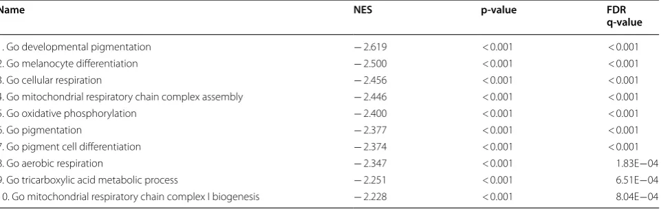
As a confirmatory step, we performed Gene Set Enrichment Analysis on the full ranked list of IL32 expression correlations. Supportive of our earlier findings, the top gene sets highly enriched for IL32 anti-correlation include pigmentation and metabolic gene sets, and the top most highly enriched for IL32 expression include NF-κB and immune related gene
sets (Tables 1, 2). Together, this suggests that IL32 is
among several hundred melanoma genes whose expres-sion is affected by exposure to these proinflammatory cytokines.
TNFα and IFNγ induced IL32 expression in general synchrony with dedifferentiation in melanoma cell lines
To evaluate if IL32 is induced with inflammatory sign-aling, three non-IL32-producing melanoma cell lines (M397, M398, M249) were exposed to TNFα or IFNγ
for 72 h (Fig. 2). We selected these three melanoma cell
Table 1 Gene set enrichment analysis (GSEA) of genes correlated with IL32
Top ten enriched gene sets correlated with IL32 include NF-κB and immune-related gene sets
Name NES p-value FDR
q-value
1. Go I kappab kinase NF kappab signaling 2.461 < 0.001 < 0.001
2. Go cellular response to mechanical stimulus 2.456 < 0.001 < 0.001
3. Go positive regulation of chemotaxis 2.302 < 0.001 2.008E−03
4. Go tumor necrosis factor mediated signaling pathway 2.286 < 0.001 1.506E−03
5. Go modification by symbiont of host morphology or physiology 2.266 < 0.001 1.449E−03
6. Go response to mechanical stimulus 2.257 < 0.001 1.601E−03
7. Go positive regulation of response to external stimulus 2.223 < 0.001 2.745E−03
8. Go toll like receptor signaling pathway 2.217 < 0.001 2.698E−03
9. Go pattern recognition receptor signaling pathway 2.215 < 0.001 2.533E−03
10. Go NIK NF kappab signaling 2.212 < 0.001 2.280E−03
Table 2 Gene set enrichment analysis (GSEA) of genes anti-correlated with IL32
Top ten enriched gene sets negatively correlated with IL32 include pigmentation and metabolic gene sets
Name NES p-value FDR
q-value
1. Go developmental pigmentation − 2.619 < 0.001 < 0.001
2. Go melanocyte differentiation − 2.500 < 0.001 < 0.001
3. Go cellular respiration − 2.456 < 0.001 < 0.001
4. Go mitochondrial respiratory chain complex assembly − 2.446 < 0.001 < 0.001
5. Go oxidative phosphorylation − 2.400 < 0.001 < 0.001
6. Go pigmentation − 2.377 < 0.001 < 0.001
7. Go pigment cell differentiation − 2.374 < 0.001 < 0.001
8. Go aerobic respiration − 2.347 < 0.001 1.83E−04
9. Go tricarboxylic acid metabolic process − 2.251 < 0.001 6.51E−04
[image:5.595.55.547.362.518.2] [image:5.595.59.540.564.716.2]lines to observe the parallel impact of TNFα and IFNγ on IL32 and the melanoma differentiation state because they exhibited a low AXL/high MITF differentiated genetic signature. Both TNFα and IFNγ induced expression of the three predominant isoforms of IL32 (α, β, γ) in all three cell lines at varying levels. Overall, the impact of TNFα on IL32 expression was greater than the impact of IFNγ. Induction of IL32γ by TNFα was the most notable response in all three cell lines. The relative level of IL32 isoform transcripts in these three cell lines, after TNFα induction, is generally lower than the constitutive expres-sion in the two high IL32-producing, dedifferentiated cell
lines M318 and M418 (Fig. 3a). For reference, we also
evaluated IL32 transcript levels in the Jurkat T cell line, a recognized high IL32 expressing leukemia cell line. In general, with the exception of M318, the γ isoform is the most abundant and when induced by cytokine stimula-tion has significantly higher and more prolonged
induc-tion (Figs. 2, 3a, b).
To test the parallel impact of IL32 induction with TNFα or IFNγ treatment on differentiation, we also
evaluated expression of melanocytic markers MITF and
MLANA, neural crest marker NGFR and the receptor
tyrosine kinase AXL. A large and growing body of
litera-ture has shown the expression of AXL strongly inversely correlated with the expression of MITF, with a high AXL/
MITF ratio associated with therapy resistance [24, 25]. At
baseline, these three melanoma cell lines exhibited a low AXL/high MITF differentiated genetic signature. In two of three cell lines (M397 and M398) we observed inver-sion of the AXL/MITF ratio towards a dedifferentiated genotype in parallel with induction of IL32. However, one of three cell lines (M249) did not dedifferentiate in response to TNFα or IFNγ, despite induction of IL32. The lack of correlation between IL32 induction and inverted AXL/MITF ratio in M249 suggests that one event is not invariably accompanied by the other. Furthermore, enforced expression of recombinant IL32α, -β, or -γ in M397 did not impact expression of melanoma
differenti-ation genes MITF, AXL, NGFR or MLANA, strongly
sug-gesting that IL32 is not a causal or upstream event in the
melanoma dedifferentiation process (Additional file 2:
Figure S2).
M397
M398
M249
α β γ α β γ
γ α α γ α γ α β γ 0 40 500 1500 2500 IL32 IFN TNF -**** ** *** ***
α β γ α β γ α β γ
0 10 20 8000 13000 2500 IL32 IFN TNF -**** **** ****
α β γ α β γ α β γ
0 4 8 30 60 90 IL32 IFN TNF -*
- IFNγ TNFα 0.0 0.5 1.0 1.5 2.0 Re la tive Gene Express io n MITF **
- IFNγ TNFα 0.0 0.5 1.0 1.5 2.0 MLANA ***
- IFNγ TNFα 0.0 0.5 1.0 1.5 NGFR * *
- IFNγ TNFα 0.0 0.5 1.0 1.5 Re la tive Gene Expressi on MITF *** ****
- IFNγ TNFα 0.0 0.5 1.0 1.5 MLANA ** ***
- IFNγ TNFα 0 10 20 30 40 AXL *
- IFNγ TNFα 0 10 20 30 NGFR ** ****
- IFNγ TNFα 0.0 0.5 1.0 1.5 Re la tive Ge ne Express io n MITF ***
- IFNγ TNFα 0.0 0.5 1.0 1.5 MLANA **** ****
- IFNγ TNFα 0 2 4 6 8 AXL **** ****
- IFNγ TNFα 0 10 20 30 40 NGFR ****
- IFNγ TNFα 0
1 2 3
AXL
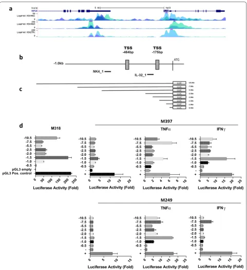
[image:6.595.61.541.376.666.2]Proinflammatory cytokines TNFα and IFNγ induce IL32 promoter activity in melanoma cell lines
We characterized the transcriptional regulation of IL32 in human melanoma, both in constitutive and cytokine-induced cells. CHIP-seq analysis of active enhancer and promoter elements, as defined by H3K4Me1, H3K27Ac,
H3K3Me3 marks [34–37] in HUVEC and K562, cell
lines known to express IL32, indicate the presence
of regulatory elements 5′ relative to the IL32
trans-lational start site (ATG) (Fig. 4a, ENCODE data set;
University of California, Santa Cruz (UCSC) genome
browser). Using 5′ RACE we determined the location
of the transcription start site (TSS) in the IL32
posi-tive cell line M318; two TSS were identified at − 464 bp
and − 175 bp (chr16: 3,115,349 and chr16: 3,115,638)
relative to the ATG (Fig. 4b). The Eukaryotic Promoter
Database (EPD; http://epd.vital .it.ch) identified two
putative IL32 promoters EPD NK4_1 and EPD IL32_1,
which are − 558bps and − 221bps relative to the ATG
(see “Discussion” Fig. 4b). Based on this information,
we cloned various lengths of the IL32 5′ upstream
genomic region (− 10.5, − 7.5, − 5.5, − 2.5, − 2, − 1.5,
− 1, − 0.5) into a luciferase reporter construct; these
constructs encompassed the putative EPD
promot-ers, both identified TSS sites, and the upstream (5′)
ENCODE histone marks. These constructs were tran-siently transfected into IL32 negative (M249, M397)
and IL32 positive (M318) cell lines (Fig. 4d). Strong
promoter activity was noted in the IL32 positive but not the negative cell lines, with the highest level in the
− 10.5 and − 1.5 constructs. When both IL32 negative
cell lines were exposed to TNFα or IFNγ for 24 h, there was a significant induction of promoter activity. TNFα consistently induced stronger IL32 promoter activ-ity than IFNγ. M249, which cannot be dedifferentiated with TNFα or IFNγ, also showed strong IL32 promoter induction consistent with IL32 transcript expression. The promoter construct profiles were similar in both constitutive and induced settings.
IL32 in the melanoma microenvironment is associated with immune infiltration and melanoma dedifferentiation
Given the impact of TNFα and IFNγ on IL32 sion in melanoma cell lines, we investigated the expres-sion of IL32 in the chemokine-rich melanoma tumor microenvironment using RNA sequencing data from b
a
0 24 48 72
0 500 1000 1500 2000 5000 10000 15000 20000
Hours
R
el
ati
ve
Ge
ne
Expr
essio
n M397 Time Course
IL32 α
α
α α α
IL32γ
γ
IL32β β
Jurka t
M318 M418 M397 M398 M249
0.0 0.5 1.0 1.5 2.0
Re
la
tiv
e
G
ene
Express
io
n IL32
+TNF
Jurka t
M318 M418 M397 M398 M249
0.0 0.5 1.0 1.5 2.0
2.5 IL32
+TNF
Jurka t
M318 M418 M397 M398 M249 0
1 2 3 4 30 40 50
60 IL32
+TNF
[image:7.595.58.540.89.381.2]a
b
c
d
M249 M397
0 50 100 150 200 250
pGL3 Pos. controlpGL3 empty -0.5 -1.0 -1.5 -2.0 -2.5 -5.5 -7.5 -10.5
Luciferase Activity (Fold) M318
0 5 10 15 20
+ --0.5 -1.0 -1.5 -2.0 -2.5 -5.5 -7.5 -10.5
Luciferase Activity (Fold)
0 5 10 15 20 25
+ --0.5 -1.0 -1.5 -2.0 -2.5 -5.5 -7.5 -10.5
Luciferase Activity (Fold) IFN
0 2 4 6 8 10
+
--0.5 -1.0 -1.5 -2.0 -2.5 -5.5 -7.5 -10.5
Luciferase Activity (Fold) TNF
0 5 10 15
+
--0.5 -1.0 -1.5 -2.0 -2.5 -5.5 -7.5 -10.5
Luciferase Activity (Fold)
0 5 10 15 20 25
+
--0.5 -1.0 -1.5 -2.0 -2.5 -5.5 -7.5 -10.5
Luciferase Activity (Fold) TNF
0 5 10 15 20
+
--0.5 -1.0 -1.5 -2.0 -2.5 -5.5 -7.5 -10.5
Luciferase Activity (Fold) IFN
-10.5kb -7.5kb -5.5kb -2.5kb
-1.5kb -2.0kb
-1.0kb -0.5kb
α γ
α γ
[image:8.595.58.540.85.613.2]tumor biopsies as part of The Cancer Genome Atlas (TCGA) dataset. We observed strong Pearson correla-tion coefficients between IL32 expression, found within the melanoma tumor and the surrounding microenvi-ronment, and genes related to immune cell infiltration, such as CD3E (0.94), CD8A (0.89), IFNγ (0.75), and
GZMB (0.86) (Additional file 3: Figure S3). Additionally,
we observed a strong correlation between IL32 expres-sion and several checkpoint receptors and ligands, such as PD-1 (0.87), PD-L1 (0.69), Tim-3 (0.82), Lag3 (0.85),
and TIGIT (0.86) (Additional file 4: Figure S4). We also
observed correlations between IL32 and genes related to other immune subsets, including myeloid cells (ITGAM, 0.71; CD68, 0.61; CD14, 0.74), dendritic cells (ITGAX, 0.71; ITGAE, 0.47) and B cells (CD19, 0.67). Consistent with this, the top 100 genes that correlated with IL32 expression in the TCGA dataset were enriched with gene sets related to allograft rejection (hypergeometric
p-value = 4.3 × 10−36), inflammatory response
(hypergeo-metric p-value = 3.3 × 10⁻¹⁰), and IFNγ signaling
(hyper-geometric p-value = 8.3 × 10⁻⁹).
In light of the impact of a proinflammatory microen-vironment on melanoma differentiation, as well as the association between IL32 and dedifferentiation in mela-noma cell lines, we investigated if this phenomenon was also present in the TCGA dataset. We observed a correla-tion between IL32 expression in melanoma tumor biop-sies with a high AXL/low MITF drug-resistant signature, consistent with data from melanoma cell lines
(Addi-tional file 3: Figure S3). However, IL32 did not inversely
correlate with signature melanocyte genes (MLANA,
PMEL, TYR ) as we observed in the cell line data (data not shown). Additionally, IL32 expression did not correlate with genes associated with disease progression, tumor
cell invasion, and migration, such as CD155/PRV and
MMP2 (Additional file 4: Figure S4).
IL32 isoforms have differential impact on myeloid polarization
IL32 from melanoma cells may influence non-melanoma cells in the tumor microenvironment, such as myeloid cells, which can play both pro- and anti-tumor roles depending on their phenotypic state. We preliminar-ily assessed of the impact of IL32 on the polarization of myeloid cells. It has previously been reported that IL32γ induces the differentiation of monocytes into phagocytic macrophage-like cells (though they discordantly express
CD14 and CD1a) [19]. However, the effect of IL32γ, or
other IL32 isoforms (IL32α and IL32β), on myeloid cell polarization is not well understood. We observed that monocytes cultured with either IL32β or IL32γ express both CD68 and CD80, markers associated with M1
proinflammatory macrophages thought to play an
anti-tumor role (Additional file 5: Figure S5). In contrast,
exposure to IL-32α resulted in a CD68+ but CD80−
population. Likewise, IL32β and IL32γ, but not IL32α, resulted in higher CD14 and CD1b expression.
Discussion
Human melanomas produce a number of cytokines
generally associated with the immune system [38]. In
an analysis of the RNA transcripts from 53 established melanoma lines, a significant proportion express IL32 isoforms. Thus, we evaluated the biology underlying IL32 expression in the context of melanoma both in cell lines and in melanoma tumor samples.
IL32 is found on human chromosome 16p13.3, is expressed in at least 6 splice variants (α, β, γ, δ, ε, ζ) and was originally isolated from activated natural killer and T
cells [16]. Which isoforms are secreted as opposed to
act-ing intracellularly is still unresolved and hampered by an
as of yet unidentified cell surface receptor [39, 40]. The
γ isoform is the only isoform that has a leader sequence, is composed of all exons, is believed to be secreted, and
believed to be the most biologically active [41]. A rodent
IL32 homologue does not exist-raising the obvious ques-tion as to whether or not this gene product is dispensa-ble or redundant-but human recombinant and transgenic
IL32 appears to be biologically active in mice [12, 42–44].
Despite these gaps in our knowledge, much of the pub-lished literature on the biology of IL32 comes from experiments utilizing commercially available
recombi-nant protein, generally the α, β, and γ isoforms [1].
Several themes emerge regarding IL32 biology: (a) IL32 can modulate the production of various cytokines
including IL1β [45], TNFα [46], IL6 [47], IL8 [8], and
may activate cells of the immune system [15, 19]; (b)
IL32 expression can be induced by various viruses and
microbes [2, 20, 42, 48–51] and may play a role in
anti-viral immunity [52]; (c) IL32 may have direct effects on
various cancer cells in vitro; [8, 9, 22, 47, 53, 54]; (d) IL32
may play a role in tumor immunity [4, 7, 12, 14, 44]; [41];
and (e) IL32 has been associated with various
inflamma-tory diseases [43, 45], such as ulcerative colitis and
rheu-matoid arthritis [55]. It is also curious that, largely from
immunohistochemistry studies, IL32 is expressed by a broad range of epithelial and in hematopoietic malignan-cies and is generally correlated with aggressive biology, although no unifying mechanism has been identified.
phenotype results in reduced expression of melanocytic-lineage antigens, which may also provide a mechanism
for immune evasion [32]. Melanomas, which progress on
MAPK inhibitor therapies, acquire this genetic signature
of reduced MITF and RTK upregulation [25]. MITF
con-trols the expression of a broad range of genes in melano-cyte-lineage cells that govern differentiation, migration,
and proliferation [56–58]. The low MITF signature is
reg-ulated by receptor tyrosine kinases, including AXL [59].
The plasticity of these neural crest-derived cutaneous malignancies is underscored by the ability of an inflam-matory signal—TNFα or IFNγ, for example—to effect a differentiated to dedifferentiated conversion in mela-noma cell lines with only a 72 h in vitro exposure, a phe-nomenon that reverses when the cytokine is removed
from culture [26]. We found that TNFα and IFNγ, which
promote a dedifferentiated melanoma phenotype, also induce IL32 expression in non-IL32 expressing mela-noma cell lines by impacting activity at the promoter level.
Palstra et al. recently reported that the DNA ele-ment encompassing rs4349147 is a strong long distance enhancer essential for the expression of IL32 in CD4
T cells located 10 kb 3′ of the IL32 promoter [52].
Jur-kat cells are heterozygous for the rs4349147-A and rs4349147-G alleles. The A allele increases the expression of IL32α, generally viewed as anti-inflammatory, whereas the G allele promoted the expression of the proinflamma-tory IL32γ and IL32β isoforms. These proinflammaproinflamma-tory isoforms enhanced lymphocyte activation and suscepti-bility to HIV infection. This report provides considerable insight into various observations on the biology of IL32 isoforms. Two putative IL32 promoters have been
identi-fied using the Eukaryotic Promoter Database (http://epd.
vital -it.ch), which are depicted in Fig. 4. These two pro-moters, at least in Jurkat cells, designated EPD NK4_1 and EPD IL32_1, are thought to support the transcription of IL32γ and IL32α/β, respectively.
Our IL32 promoter constructs encompass both the identified EPD NK4_1 and EPD IL32_1 putative
pro-moter regions and extend 10.5 kb 5′ to the ATG. The
luciferase activity driven by these constructs would then necessarily reflect constitutive and induced gene
expres-sion of all three isoforms, as well as the putative cis-acting
elements residing within this 5′ upstream region. Palstra
et al. performed formaldehyde-assisted isolation of regu-latory elements (FAIRE) assays in Jurkat and a melanoma cell line (G361), which demonstrated significant increase in DNA accessibility in the region surrounding rs4349147
in the former but not the latter cell line [52]. We
there-fore conjecture that this long-range enhancer may not play a role in melanoma IL32 transcription. In another IL32 promoter study using Akt-activated endothelial cell
line constructs extending 2.5 kb 5′ to the ATG [60]; the
difference in cell lines and activation signals precludes any meaningful comparison.
Given the impact of proinflammatory cytokines on IL32 expression, we investigated the IL32 expression in immunologically rich melanoma tumors using RNA sequencing data available in the TCGA dataset. IL32 expression was highly correlated with a T cell dominant immune signature in these tumors, probably a result of immune cell IL32 production. We also observed a corre-lation between IL32 expression, derived from tumor cells and the surrounding microenvironment, and a high AXL/ low MITF gene signature, as in the melanoma cell lines. However, we interpret this result cautiously because, unlike in the melanoma cell line dataset, IL32 expres-sion in the melanoma tumor microenvironment did not inversely correlate with pigmentation or melanoma
dif-ferentiation genes such as MLANA, TYR or PMEL. Thus,
the high AXL/low MITF signature may be a result of increased IL32 expression by infiltrating immune cells, rather than IL32 expressing dedifferentiated melanoma cells in the tumor microenvironment.
There is substantial evidence that IL32 induces dedif-ferentiation of human monocytes towards a macrophage-like phonotype with dendritic cell-macrophage-like aspects. IL32 exposed monocytes altered their morphology within 3 days (flattening with extensive pseudopodia), which was accompanied by increased expression of CD1 and CD14
[19]. These macrophage-like cells exhibited active
phago-cytic properties and were also induced to express TNFα, IL-1β and IL6. Our preliminary in vitro studies indicated that IL32, in an IL32β and IL32γ isoform specific manner, can modulate the polarization of myeloid cells toward an M1-like macrophage phenotype with costimulatory molecule expression, lending additional support for its impact in the tumor microenvironment. It is plausible, then, that IL32 is expanded within the microenvironment as a byproduct of the feedback loop between the anti-tumor inflammatory response (marked by TNFα and
IFNγ) and dedifferentiation of melanoma [23].
Conclusions
In summary, this study shows that a significant per-centage of cell lines derived from metastatic melanoma express IL32. IL32 expression, which correlates with a dedifferentiated “invasive” genetic signature, can be induced by inflammatory molecules TNFα and IFNγ that modulate IL32 transcriptional activity at the pro-moter level. IL32 expression by dedifferentiated tumor cells may contribute to a proinflammatory tumor microenvironment.
and PD-1/PDL1 checkpoint inhibition underscores the importance of understanding potential contributions of
melanoma-elaborated cytokines such as IL32 [61].
Additional files
Additional file 1: Figure S1. Expression of IL32 in human melanoma.
(A) Immunohistochemistry analysis of IL32 expression in cutaneous melanoma in two patients with low and high IL32 expression, respectively (images and data acquired from The Human Protein Atlas, and Image available at https ://www.prote inatl as.org/ENSG0 00000 08517 -IL32/patho logy/tissu e/melan oma#img). Left panel is a cutaneous melanoma sample from a 73-year old woman (patient id: 2900). Right panel is a cutaneous melanoma sample from an 83-year old man (patient id: 2156). Staining was performed using BioLegend mouse anti-human IL32 monoclonal antibody (Cat # 513401) at a 1:4500 dilution after HIER antigen retrieval (pH = 6). (B and C) IL32 transcript expression across multiple different cancer lines organized by cancer type from the NCI-60 Cancer Cell Line database and the Cancer Cell Line Encyclopedia, respectively.
Additional file 2: Figure S2. Addition of recombinant IL32 does not
impact melanoma cell line growth or differentiation. (A) Tumor growth over time in parental M397 melanoma cell line, compared to M397 treated with recombinant IL32α, -β or -γ. (B and C) Expression of melanoma dif-ferentiation genes by quantitative RT-PCR at baseline or after treatment with TNFα, recombinant IL32α, IL32β, or IL32γ measured at day 3 (B) or day 7 (C).
Additional file 3: Figure S3. IL32 expression in the TCGA dataset. (A-B)
Scatterplot of log2 FPKM expression values between IL32 and select immune genes (A) or between the ratio of AXL and MITF log2 FPKM expression values (B) in the melanoma TCGA dataset (n = 479).
Additional file 4: Figure S4. IL32, checkpoint receptors/ligand
expres-sion, and markers of disease progression in the TCGA dataset. Scatterplot of log2 FPKM expression values between IL32 and select checkpoint receptors/ligands, as well as, select markers of disease progression (MMP2 and PVR).
Additional file 5: Figure S5. Expression of phenotypic markers on
human monocytes after exposure to stimuli. CD14 + cells, from PBMC isolation, were cultured in the presence of GM-CSF + IL-4, Cell Genix Media, recIL32α, recIL32β, or recIL32γ for 5 days. The different treatments are displayed by using a gray scale. On day 5, the phenotype of the cells was assayed using flow cytometry analysis for various surface markers. GM-CSF + IL-4 and Cell Genix Media alone were as a positive and negative control, respectively. Cells were then assessed for surface expression of CD68, CD80, CD14, CD1B by flow cytometry. Frequency of positive cells is shown in the left panels, and mean fluorescence intensity is shown in the right panels. Statistical analysis was done using a one-way repeated meas-ures ANOVA, using the GM-CSF + IL-4 treatment as a control. Each shape (circle, square, triangle) represents a different healthy donor. *P ≤ 0.05, **P ≤ 0.01, ***P ≤ 0.001.
Abbreviations
IL32: interleukin 32; TCGA : the cancer genome atlas; ATG : translational start site; RACE: rapid amplification of cDNA ends.
Authors’ contributions
HP performed, analyzed, and interpreted the human cell line and IL32 pro-moter data. JT analyzed and interpreted the sequencing and TCGA data. AK analyzed and interpreted the TCGA data. CSG analyzed the sequencing and TCGA data. DMM performed, analyzed and interpreted the myeloid cell data. WHM, DS, LHB, AR, TGG, and JSE all contributed to the design of experiments and the interpretation of results. HP, JT, AK, DMM and JSE were significantly involved in writing of this manuscript. All authors read and approved the final manuscript.
Author details
1 Department of Surgery, University of California, Los Angeles, 10833 Le Conte
Ave, Los Angeles, CA 90095, USA. 2 Department of Microbiology,
Immunol-ogy, and Molecular Genetics, University of California, Los Angeles, CA 90095, USA. 3 Department of Molecular and Medical Pharmacology, Crump Institute
for Molecular Imaging, University of California, Los Angeles, CA 90095, USA.
4 Department of Radiation Oncology, David Geffen School of Medicine,
University of California, Los Angeles, CA 90095, USA. 5 Department of
Medi-cine, David Geffen School of MediMedi-cine, University of California, Los Angeles, CA 90095, USA. 6 Jonsson Comprehensive Cancer Center, University of
Califor-nia, Los Angeles, CA 90095, USA. 7 Department of Immunology, University
of Pittsburgh, Pittsburgh, PA 15213, USA. 8 Department of Medicine, University
of Pittsburgh Cancer Institute, Pittsburgh, PA 15213, USA. 9 Department
of Surgery, University of Pittsburgh Cancer Institute, Pittsburgh, PA 15213, USA.
10 Department of Clinical and Translational Science, University of Pittsburgh,
Pittsburgh, PA 15213, USA.
Acknowledgements
We appreciate constructive input from Dr. Michael Carey (UCLA).
Competing interests
LHB declares Simpatica, Scientific Advisory Board Member, Jan. 2017-present; StemImmune Scientific and Medical Advisory Board, April 6, 2017-present; SapVax Advisory Board Nov. 15, 2017-present; NextCure, Scientific Advisory Board, 2018-present; Replimmune, Scientific Advisory Board, 2018-present; Western Oncolytics, Scientific Advisory Board, 2018-present; Torque Thera-peutics, Consultant, 2018-present. AR has received honoraria from consulting with Bristol Myers-Squibb, Amgen, Chugai, Genentech, Merck, Novartis and Roche, and is on the scientific advisory board of Advaxis, Arcus, Bioncotech, Compugen, CytomX, Five Prime, FLX-Bio, ImaginAb, Isoplexis, Merus and Rge-nix. During the conduct of this work AR was on the scientific advisory board and held stock in Kite-Pharma, and is co-founder of PACT Pharma and Tango Therapeutics. JSE is a scientific advisor to Allogene Therapeutics and Neogene Therapeutics.
Availability of data and materials
One of the datasets analyzed during the current study is included in the Gene Expression Omnibus (GEO), https ://www.ncbi.nlm.nih.gov/geo/query /acc. cgi?acc=GSE80 829 [26]. Additional datasets analyzed in this current study are available from The Cancer Genome Atlas, https ://cance rgeno me.nih.gov/.
Consent for publication
Not applicable.
Ethics approval and consent to participate
Not applicable.
Funding
Univ. Pittsburgh Skin Biology T32 Training Grant pre-doctoral Scholar (DMM); P50 CA121973-03 Pittsburgh SPORE in Melanoma and Skin Cancer (LHB); NIH NCI PO1 DCA 168585 (AR, TGG); NIH 1R01CA191234-01A1 (DS); Joy and Jerry Monkarsh Fund (JSE); Vincent Price Research Fund (JSE).
Publisher’s Note
Springer Nature remains neutral with regard to jurisdictional claims in pub-lished maps and institutional affiliations.
Received: 18 January 2019 Accepted: 27 March 2019
References
1. Heinhuis B, Netea MG, van den Berg WB, Dinarello CA, Joosten LA. Inter-leukin-32: a predominantly intracellular proinflammatory mediator that controls cell activation and cell death. Cytokine. 2012;60(2):321–7. 2. Joosten LA, Heinhuis B, Netea MG, Dinarello CA. Novel insights into the
3. Khawar MB, Abbasi MH, Sheikh N. IL-32: a novel pluripotent inflammatory interleukin, towards gastric inflammation, gastric cancer, and chronic rhino sinusitis. Mediat Inflamm. 2016;2016:8413768.
4. Wang S, Chen F, Tang L. IL-32 promotes breast cancer cell growth and invasiveness. Oncol Lett. 2015;9(1):305–7.
5. Wang Y, Yang Y, Zhu Y, Li L, Chen F, Zhang L. Polymorphisms and expres-sion of IL-32: impact on genetic susceptibility and clinical outcome of lung cancer. Biomarkers. 2017;22(2):165–70.
6. Yang Y, Wang Z, Zhou Y, Wang X, Xiang J, Chen Z. Dysregulation of over-expressed IL-32 in colorectal cancer induces metastasis. World J Surg Oncol. 2015;13:146.
7. Oh JH, Cho MC, Kim JH, Lee SY, Kim HJ, Park ES, et al. IL-32gamma inhibits cancer cell growth through inactivation of NF-kappaB and STAT3 signals. Oncogene. 2011;30(30):3345–59.
8. Park ES, Yoo JM, Yoo HS, Yoon DY, Yun YP, Hong J. IL-32gamma enhances TNF-alpha-induced cell death in colon cancer. Mol Carcinog. 2014;53(Suppl 1):E23–35.
9. Yun HM, Park KR, Kim EC, Han SB, Yoon DY, Hong JT. IL-32alpha suppresses colorectal cancer development via TNFR1-mediated death signaling. Oncotarget. 2015;6(11):9061–72.
10. Nishida A, Andoh A, Inatomi O, Fujiyama Y. Interleukin-32 expression in the pancreas. J Biol Chem. 2009;284(26):17868–76.
11. Plantinga TS, Costantini I, Heinhuis B, Huijbers A, Semango G, Kusters B, et al. A promoter polymorphism in human interleukin-32 modulates its expression and influences the risk and the outcome of epithelial cell-derived thyroid carcinoma. Carcinogenesis. 2013;34(7):1529–35. 12. Heinhuis B, Plantinga TS, Semango G, Kusters B, Netea MG, Dinarello CA,
et al. Alternatively spliced isoforms of IL-32 differentially influence cell death pathways in cancer cell lines. Carcinogenesis. 2016;37(2):197–205. 13. Gorvel L, Korenfeld D, Tung T, Klechevsky E. Dendritic cell-derived
IL-32alpha: a novel inhibitory cytokine of NK cell function. J Immunol. 2017;199(4):1290–300.
14. Cheon S, Lee JH, Park S, Bang SI, Lee WJ, Yoon DY, et al. Overexpression of IL-32alpha increases natural killer cell-mediated killing through up-reg-ulation of Fas and UL16-binding protein 2 (ULBP2) expression in human chronic myeloid leukemia cells. J Biol Chem. 2011;286(14):12049–55. 15. Park MH, Song MJ, Cho MC, Moon DC, Yoon DY, Han SB, et al.
Interleu-kin-32 enhances cytotoxic effect of natural killer cells to cancer cells via activation of death receptor 3. Immunology. 2012;135(1):63–72. 16. Kim SH, Han SY, Azam T, Yoon DY, Dinarello CA. Interleukin-32: a cytokine
and inducer of TNFalpha. Immunity. 2005;22(1):131–42.
17. Shoda H, Fujio K, Yamaguchi Y, Okamoto A, Sawada T, Kochi Y, et al. Interactions between IL-32 and tumor necrosis factor alpha contribute to the exacerbation of immune-inflammatory diseases. Arthritis Res Ther. 2006;8(6):R166.
18. Li W, Liu Y, Mukhtar MM, Gong R, Pan Y, Rasool ST, et al. Activation of interleukin-32 pro-inflammatory pathway in response to influenza A virus infection. PLoS ONE. 2008;3(4):e1985.
19. Netea MG, Lewis EC, Azam T, Joosten LA, Jaekal J, Bae SY, et al. Interleu-kin-32 induces the differentiation of monocytes into macrophage-like cells. Proc Natl Acad Sci USA. 2008;105(9):3515–20.
20. Ohmatsu H, Humme D, Gonzalez J, Gulati N, Mobs M, Sterry W, et al. IL-32 induces indoleamine 2,3-dioxygenase(+)CD1c(+) dendritic cells and indoleamine 2,3-dioxygenase(+)CD163(+) macrophages: relevance to mycosis fungoides progression. Oncoimmunology. 2017;6(2):e1181237. 21. Suga H, Sugaya M, Miyagaki T, Kawaguchi M, Fujita H, Asano Y, et al.
The role of IL-32 in cutaneous T-cell lymphoma. J Invest Dermatol. 2014;134(5):1428–35.
22. Lee J, Kim KE, Cheon S, Song JH, Houh Y, Kim TS, et al. Interleukin-32alpha induces migration of human melanoma cells through downregulation of E-cadherin. Oncotarget. 2016;7(40):65825–36.
23. Taube JM, Young GD, McMiller TL, Chen S, Salas JT, Pritchard TS, et al. Differential expression of immune-regulatory genes associated with PD-L1 display in melanoma: implications for PD-1 pathway blockade. Clin Cancer Res. 2015;21(17):3969–76.
24. Konieczkowski DJ, Johannessen CM, Abudayyeh O, Kim JW, Cooper ZA, Piris A, et al. A melanoma cell state distinction influences sensitivity to MAPK pathway inhibitors. Cancer Discov. 2014;4(7):816–27.
25. Muller J, Krijgsman O, Tsoi J, Robert L, Hugo W, Song C, et al. Low MITF/ AXL ratio predicts early resistance to multiple targeted drugs in mela-noma. Nat Commun. 2014;5:5712.
26. Tsoi J, Robert L, Paraiso K, Galvan C, Sheu KM, Lay J, et al. Multi-stage differentiation defines melanoma subtypes with differential vulner-ability to drug-induced iron-dependent oxidative stress. Cancer Cell. 2018;33(5):890–904.
27. Landsberg J, Kohlmeyer J, Renn M, Bald T, Rogava M, Cron M, et al. Melanomas resist T-cell therapy through inflammation-induced reversible dedifferentiation. Nature. 2012;490(7420):412–6.
28. Uhlen M, Bjorling E, Agaton C, Szigyarto CA, Amini B, Andersen E, et al. A human protein atlas for normal and cancer tissues based on antibody proteomics. Mol Cell Proteom. 2005;4(12):1920–32.
29. Uhlen M, Zhang C, Lee S, Sjostedt E, Fagerberg L, Bidkhori G, et al. A pathology atlas of the human cancer transcriptome. Science. 2017;357:6352.
30. Cerami E, Gao J, Dogrusoz U, Gross BE, Sumer SO, Aksoy BA, et al. The cBio cancer genomics portal: an open platform for exploring multidimen-sional cancer genomics data. Cancer Discov. 2012;2(5):401–4. 31. Gao J, Aksoy BA, Dogrusoz U, Dresdner G, Gross B, Sumer SO, et al.
Inte-grative analysis of complex cancer genomics and clinical profiles using the cBioPortal. Sci Signal. 2013;6(269):pl1.
32. Mehta A, Kim YJ, Robert L, Tsoi J, Comin-Anduix B, Berent-Maoz B, et al. Immunotherapy resistance by inflammation-induced dedifferentiation. Cancer Discov. 2018;8(8):935–43.
33. Liberzon A, Birger C, Thorvaldsdottir H, Ghandi M, Mesirov JP, Tamayo P. The molecular signatures database (MSigDB) hallmark gene set collec-tion. Cell Syst. 2015;1(6):417–25.
34. Guenther MG, Levine SS, Boyer LA, Jaenisch R, Young RA. A chromatin landmark and transcription initiation at most promoters in human cells. Cell. 2007;130(1):77–88.
35. Zhang T, Cooper S, Brockdorff N. The interplay of histone modifications— writers that read. EMBO Rep. 2015;16(11):1467–81.
36. Zhou VW, Goren A, Bernstein BE. Charting histone modifications and the functional organization of mammalian genomes. Nat Rev Genet. 2011;12(1):7–18.
37. Soares LM, He PC, Chun Y, Suh H, Kim T, Buratowski S. Determinants of histone H3K4 methylation patterns. Mol Cell. 2017;68(4):773–85. 38. Qin Y, Milton DR, Oba J, Ding Z, Lizee G, Ekmekcioglu S, et al.
Inflamma-tory IL-1beta-driven JNK activation in stage III melanoma. Pigment Cell Melanoma Res. 2015;28(2):236–9.
39. Heinhuis B, Koenders MI, van den Berg WB, Netea MG, Dinarello CA, Joosten LA. Interleukin 32 (IL-32) contains a typical alpha-helix bundle structure that resembles focal adhesion targeting region of focal adhe-sion kinase-1. J Biol Chem. 2012;287(8):5733–43.
40. Novick D, Rubinstein M, Azam T, Rabinkov A, Dinarello CA, Kim SH. Proteinase 3 is an IL-32 binding protein. Proc Natl Acad Sci USA. 2006;103(9):3316–21.
41. Choi JD, Bae SY, Hong JW, Azam T, Dinarello CA, Her E, et al. Iden-tification of the most active interleukin-32 isoform. Immunology. 2009;126(4):535–42.
42. Bai X, Shang S, Henao-Tamayo M, Basaraba RJ, Ovrutsky AR, Matsuda JL, et al. Human IL-32 expression protects mice against a hyperviru-lent strain of Mycobacterium tuberculosis. Proc Natl Acad Sci USA. 2015;112(16):5111–6.
43. Kim SJ, Lee S, Kwak A, Kim E, Jo S, Bae S, et al. Interleukin-32gamma transgenic mice resist LPS-mediated septic shock. J Microbiol Biotechnol. 2014;24(8):1133–42.
44. Qu Y, Taylor JL, Bose A, Storkus WJ. Therapeutic effectiveness of intratu-morally delivered dendritic cells engineered to express the pro-inflam-matory cytokine, interleukin (IL)-32. Cancer Gene Ther. 2011;18(9):663–73. 45. Khawar B, Abbasi MH, Sheikh N. A panoramic spectrum of complex
interplay between the immune system and IL-32 during pathogenesis of various systemic infections and inflammation. Eur J Med Res. 2015;20:7. 46. Kim MS, Kang JW, Jeon JS, Kim JK, Kim JW, Hong J, et al. IL-32theta gene
expression in acute myeloid leukemia suppresses TNF-alpha production. Oncotarget. 2015;6(38):40747–61.
47. Kang JW, Park YS, Lee DH, Kim JH, Kim MS, Bak Y, et al. Intracellular interac-tion of interleukin (IL)-32alpha with protein kinase Cepsilon (PKCepsilon) and STAT3 protein augments IL-6 production in THP-1 promonocytic cells. J Biol Chem. 2012;287(42):35556–64.
•fast, convenient online submission
•
thorough peer review by experienced researchers in your field
• rapid publication on acceptance
• support for research data, including large and complex data types
•
gold Open Access which fosters wider collaboration and increased citations maximum visibility for your research: over 100M website views per year
•
At BMC, research is always in progress.
Learn more biomedcentral.com/submissions
Ready to submit your research? Choose BMC and benefit from: THP-1-derived human macrophages infected with New World
Leishma-nia species. PLoS Negl Trop Dis. 2017;11(2):e0005413.
49. Montoya D, Inkeles MS, Liu PT, Realegeno S, Teles RM, Vaidya P, et al. IL-32 is a molecular marker of a host defense network in human tuberculosis. Sci Transl Med. 2014;6(250):250ra114.
50. Schenk M, Krutzik SR, Sieling PA, Lee DJ, Teles RM, Ochoa MT, et al. NOD2 triggers an interleukin-32-dependent human dendritic cell program in leprosy. Nat Med. 2012;18(4):555–63.
51. Zhou Y, Zhu Y. Important role of the IL-32 inflammatory network in the host response against viral infection. Viruses. 2015;7(6):3116–29. 52. Palstra RJ, de Crignis E, Roling MD, van Staveren T, Kan TW, van
Ijcken W, et al. Allele-specific long-distance regulation dictates IL-32 isoform switching and mediates susceptibility to HIV-1. Sci Adv. 2018;4(2):e1701729.
53. Nicholl MB, Chen X, Qin C, Bai Q, Zhu Z, Davis MR, et al. IL-32alpha has differential effects on proliferation and apoptosis of human melanoma cell lines. J Surg Oncol. 2016;113(4):364–9.
54. Yun HM, Oh JH, Shim JH, Ban JO, Park KR, Kim JH, et al. Antitumor activity of IL-32beta through the activation of lymphocytes, and the inactivation of NF-kappaB and STAT3 signals. Cell Death Dis. 2013;4:e640.
55. Heinhuis B, Koenders MI, van de Loo FA, Netea MG, van den Berg WB, Joosten LA. Inflammation-dependent secretion and splicing of IL-32{gamma} in rheumatoid arthritis. Proc Natl Acad Sci USA. 2011;108(12):4962–7.
56. Hartman ML, Czyz M. MITF in melanoma: mechanisms behind its expres-sion and activity. Cell Mol Life Sci. 2015;72(7):1249–60.
57. Yajima I, Kumasaka MY, Thang ND, Goto Y, Takeda K, Iida M, et al. Molecu-lar network associated with MITF in skin melanoma development and progression. J Skin Cancer. 2011;2011:730170.
58. Hsiao JJ, Fisher DE. The roles of microphthalmia-associated transcrip-tion factor and pigmentatranscrip-tion in melanoma. Arch Biochem Biophys. 2014;563:28–34.
59. Ahmed F, Haass NK. Microenvironment-driven dynamic heterogeneity and phenotypic plasticity as a mechanism of melanoma therapy resist-ance. Front Oncol. 2018;8:173.
60. Kobayashi H, Lin PC. Molecular characterization of IL-32 in human endothelial cells. Cytokine. 2009;46(3):351–8.