Virus
Aggregation
as
the
Cause
of the
Non-neutralizable Persistent
Fraction
CRAIG WALLIS AND JOSEPH L. MELNICK
Departmenit of VirologyanidEpidemiology, Baylor Unliversity CollegeofMedicinie, Hoasto;i, Texas77025
Received for publication6 March 1967
The non-neutralizableor persistent fraction of virus populations has been found
to be caused by aggregated virus. Detailed investigation was performed with the
prototype strain ofechovirus type 4 (Pesascek), as this virus is notorious for its
large non-neutralizable fraction. When Pesascek virus was clarified by low-speed
centrifugation, homologousantiserumhardly neutralized thevirus. However,when
the viruswas filtered through membranes havinga porosity only twice the
diam-eter of the
virus,
monodispersed virus was obtained which wasefficientlyneutral-ized. Serum titerswereupto 1,000 times higher if the neutralizationtestwascarried
out with monodispersed virus. Virus in non-neutralizable aggregates was found
to constitute 30% of the infective units of unfiltered Pesascek virus but only
0.1% of theantigenically related DuToit strain. This explains why DuToit strain
has been a more satisfactory indicator strain for detecting type 4 antibodies,
re-gardless of theecho 4 strain usedforinducingtheantibodies. Clarifiedsuspensions
and ultrafiltrates ofvirusesbelongingtothepicorna-, reo-, myxo-, adeno-,herpes-,
and poxvirus groups were studied. Clarified suspensions yielded persistent
frac-tions of0.005%G forpoliovirus, of0.1% forreovirus, of 0.6% forinfluenza virus, of
<0.001% foradenovirus, of0.06% forherpesvirus, and of 10to 30% for vaccinia
virus. Inall cases the persistent fractionswere removedby membrane filters which
hada porediameterno largerthantwice that ofthe virus undertest, andthe high
concentration ofvirusineach ultrafiltratewascompletelyneutralizedbyantiserum.
Thirty years ago, Burnet and his colleagues
(5) showed thata significant fraction ofvaccinia
and myxomatosis virus could not be neutralized
by antiserum. In 1953, working with poliovirus,
Ledinko and Melnick (14) described the
"break-through"phenomenonasthedelayedappearance
of virus infectivity in cultures that had received
virus and an equivalent amount of antibody.
Subsequently, a number of investigators
re-ported persistentfractionsforavarietyofviruses,
and attributedthis insensitivity toantibodies to:
(i) a distinct, nonsusceptible viral population
(7, 16, 20), (ii) dissociation of thevirus-antibody
complex (8), and (iii) a nonavid antibody (13).
Viruses of several different groups were
rein-vestigated, but, because the non-neutralizable
fraction is reported to be greater with strains of
echovirus type 4 than with other viruses, such
strains were investigated in greater detail. Ever
since it was first isolated (17), Pesascek virus
wasfound to be difficult to neutralize. However,
antisera that yieldedeither lowornegativetiters
by tube neutralization tests were found to yield
titers about 100-fold higher bytestsinvolving 80
to 90% plaque reduction (10). Even with only
50 to 100 plaque-forming units (PFU) as
chal-lenge virus, complete plaque reduction was
al-mostneverachieved. Intube neutralizationtests,
Pesascek antiserum that failed to neutralize
homologous virus readily neutralized a related
strain (DuToit), indicating that the antiserum
waspotent (2, 10, 12, 15, 25, 30).
Attempts to explain the echovirus type 4
breakthrough phenomenonhave been made. The
possibility of dissociation of the virus-antibody
complex was investigated, but Pesascek serum
wasfound to be boundto Pesascek virus tothe
same degree as it was to DuToit (25). Even
though antibodywas bound, scarcelyanyof the
virus was neutralized. Barron and Karzon (2)
suggested that the neutralization breakthrough
occurred because echovirus 4was presentastwo
antigenic variants, only one of which could be
neutralized. However, from the studies tobe
re-ported, itnowappearsthatthepersistent fraction
in populations of echovirus consists of virus
aggregates which are non-neutralizable. Similar
observationsweremadewithpoliovirus, reovirus,
herpesvirus, influenza virus, andvaccinia virus.
478
on November 11, 2019 by guest
http://jvi.asm.org/
NON-NEUTRALIZABLE PERSISTENT VIRUS FRACTION
MATERIALS AND METHODS
Monkey kidney (MK) cells.Kidneys from immature
rhesus and green monkeys were trypsinized. Kidney
cellswere grown in Melnick's mediumA and
main-tained inmediumB (18).
Virusesandtheir assays. Twoplaque-purifiedstrains of echovirus type 4wereused: prototype Pesascek and therelated DuToit strain. Astock of each virus was growninasinglelotof rhesus MK cells maintained in medium B. Viruses were harvested when
50%7c
of the cells in the infected cultures showed cytopathicchanges. The harvest was centrifuged at 3,000 rev/
min for 20 min, and the clear supernatant fluid was frozen at-40Cuntil used. Thetiters ofstocksusedin these studies were 106PFU/ml for both strains.
Otherviruses were plaque-purified strains of
viru-lentpoliovirus:type1, Mahoney; type 2, MEF1;and
type 3,P24. Attenuated strains were Sabin's
plaque-purifiedlinesasusedinthe oral poliovaccine (type 1,
LSc; type 2, P712; and type 3, Leon). Other virus groups wererepresented by plaque-purified stocks of vaccinia virus (WR strain),herpesvirus (KOS strain),
reovirus 1 (strain 716), adenovirus (SV15), and a
plaque-producingstrainof influenza virustypeB(23).
Theseviruses were grownand assayed in green MK cells, with theexception of the reovirus, which, like
the echoviruses, was grown and assayed in rhesus
MKcultures.
All viruses were assayed by the PFU method.
Overlay medium consisted of Earle's salt solution,
0.4%NaHCO3,
0.1%,,
skimmilk, 1:60,000neutralred,and 1.5% agar(Difco). Toenhance plaqueformation
of Pesascek virus, 1 mm cysteine was used in the
overlay (28). For other enteroviruses, 25 mm MgCl2
was included in overlay medium (28); for reovirus, 1:60 Oxoid pancreatin (27); and for adenovirus,
influenza virus,andherpesvirus, 400/ig of protamine
sulfateper ml (23, 24).
Neutralizationttests. After 1 hr ofincubation at37
C, the virus-serum mixtures were placed in an
ice-water bath (0C) until they were assayed (within 30 min). Subsequent treatments of the mixtures are
de-scribedunder Results.
Filtration
through/
membraiies (26). To filter echo-viruses through 50-mu membranes, virus adsorptionhadto beavoided; this was achieved by pretreating
the membranes with nutrient medium. However, the medium contains components which coat membranes and preventsubsequentvirus adsorption, and it must first beclarified through other membranesto prevent
clogging of thevirus-filtering membranes. To prevent
adsorption of the membrane-coating components to
theclarifyingmembranes,fetal calf serum was diluted
10-fold in distilled water (as in hypotonic solution the essential components do not adsorb to membranes) before itwasused for pretreatment of the membranes. Inpractice, 100 mlof10%,' fetal calf serum in dis-tilled water was filtered at 25 psi through a 90-mm AP 20clarifying pad andthen in seriesthrough 0.3-,
0.22-, 0.1-, and 0.05-p membranes (Millipore Corp.,
Bedford, Mass.). To the final filtrate,
lOX
Earle's saline was added in sufficient volume to restore isotonicity. This filtrate wasthen capable of coating but not clogging membranes to be used for virusfiltration. Three membranes with pore sizes of0.22,
0.1,and 0.05 Awereplaced in asingle 25-mm holder
and treated with 10 ml of the coating solution. After the solution passed through themembranes,the mem-brane was washed with 5 ml oftris(hydroxymethyl)
aminomethane(Tris) bufferto removeresidual serum. Virus was then passed through the membrane. The void volume was colorless, so that collection was started only when the phenol red of the virus suspen-sion was first observed in the filtrate.
RESULTS
Neutralization tests with unfiltered and filtered
echovirus type4 strains. Basedon previous
titra-tionsofthe stocks, Pesascek and DuToit strains
werediluted to contain 200 PFU per 0.1 ml and
mixed with equal volumes of hyperimmune
horse serum (9) that had beenserially diluted in
Tris buffer. After 1 hr of incubation at 37 C,
samples were assayed for residual virus by the
plaque method. Pesascek and DuToit viruses
were passed through a series of membranes to
yield 50-m,u filtrates, which were mixed with
serum and assayed as described above. In a
duplicate experiment, the samedilutions ofvirus
and serum were inoculated into tube cultures to
determine cytopathic end points in fluid medium. When a picornavirus with diameters of 25 to 30
m,u isfiltered through a membrane with a 50-m,u
pore diameter, only those virions in a
monodis-persedstate can pass. Thus, the ultrafiltrate will
be referred to as monodispersed virus.
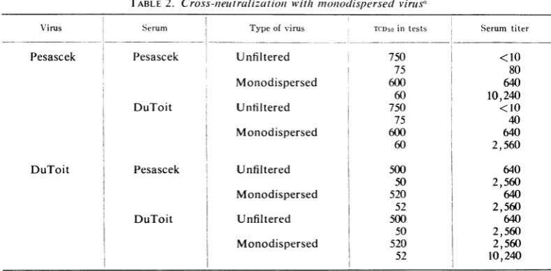
From theresults ofthese experiments (Tables
1 and 2), it is evident that the non-neutralizable
fractionofPesascek virus is intheform of
aggre-gated units of virus. Unfiltered Pesascek virus in
the small dose of 120 PFU was not completely
neutralized by homologous serum in the plaque
reduction tests at a dilution as low as 1:10,
whereas the monodispersed virus was completely
neutralizedat adilution of1:640. Whenan
80%7,
plaque reduction was used as the end point, the
serum had atiter of 640against unfiltered virus,
but a titer of 10,240 with monodispersed virus.
Ifmonodispersed virus preparations are used for
comparison, Pesascek and DuToit viruses
be-have inprecisely the samefashion. Theresults of
the tube neutralization tests presented a similar picture. The reference hyperimmune horse serum
had been reported as failing to neutralize the
Pesascek virus which had been used as
immu-nizing antigen (9). For this reason, the DuToit
strain hadto beemployed as an indicator strain
in measuring type 4 antibodies. However, once
the aggregates had been removed from the
Pesascek stock, it could be used as effectively as DuToit for measuring type 4 antibodies.
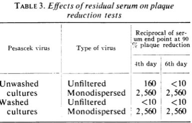
Effects of residual serum onneutralization tests.
It would appear that virus in aggregates can
479
VOL. 1,1967
on November 11, 2019 by guest
http://jvi.asm.org/
replicate even if antibody is bound to the aggre-gate. If the cell infected with such an
antibody-virus aggregate releases antibody-virus particles into a
cellular environment containing residual free
antibodies, the progeny virus would be largely
neutralized. Ledinko and Melnick (14) showed
that the breakthrough phenomenon occurred
with poliovirus only after cultures were changed
to freshmedium, thus reducing the concentration
ofresidual serum. Clark and Tyrrell (6) reported
that high dilutions of serum gave neutralization
endpoints similar to those of low dilutions (when
both concentrations were in excess), if the
cul-tures were washed after the virus-antibody
ad-sorptionperiod. If the cultures were not washed,
the lower dilutions of serum gave higher
neu-tralization end points, because the residual free
antibodies prevented progeny virus from
spread-ing
the infection inthe culture.To explore the effect of residual serum, the
following experiment
was carried out. Aneu-tralization test was performed with Pesascek virus as described for Table 1 above. After
ad-sorption
of the virus-serum mixtures onmono-layers,
half of the cultures were washed threetimes with nutrient medium, and the agar
over-lay was added to all the cultures, washed and
unwashed. Readings were made on the 4th and 6th days. The results of this test are shown in Table 3. With unfiltered virus, when residual serum was removed from cultures after
adsorp-tion of the virus-antibody complex, the
previ-TABLE 1 Cr-oss-iwelutralizatioln testswvit/h
mo,odispersed
virlusa\'ir-us activity(PFU'O.1 ml,)
Serum Typeofvirus Tris
buffer con-trol i Pesascek Unfiltered Monodispersed DuToit Unfiltered Monodispersed Pesascek Unfiltered Monodispersed
| DuToit Unfiltered
I Moniodispersed 120 92 120 92 101 IIl 101 III Plaquereduction
Inpiesenceofser-umdilutionis 1() 40 1
7 12 0
10
14 6 0 0 0 0 0 0 0 0 0160 640 2,60 10,240 21 24 62 79
0 0 2 10
19 28 37 92
0 0 12 29
0 0 8 39
0 0 3 49
0 0 14 17
0 0 0 17
a Test read on6th day.
IReciprocal ofserum end poiilt.
I
I49,960 1(00% 90%
99 <10b 40
44 640 2,560
100 <10 40
75 640 640
90 640 65 640 55 640 31 2 560
2,560 2,560 640 2,560 80( 640 10,240 160 10,240 2,560 2,560 640 10,240
TABLE2. Cross-uieltralizat joii wit/i moniodispersedvirius Virus Pesascek DuToit Serum Pesascek I DuToit Pesascek DuToit
aTest read on6th day.
Type of virus TCD50in tests
Unfiltered 750 75 Monodispersed 600 60 Unfiltered 750 75 Monodispersed 600 60 Unfiltered Monodispersed Unfiltered Monodispersed 500 50 520 52 500 50 520 52 Virus Pesascek DuToit
i Serum titer
<10 80 640 10,240 <10 40 640 2,560 640 2,560 640 2,560 640 2,560 2,560 10,240
on November 11, 2019 by guest
http://jvi.asm.org/
[image:3.461.41.436.456.650.2]NON-NEUTRALIZABLE PERSISTENT VIRUS FRACTION
ously
recognized
breakthrough of Pesascek virus(top line) was very pronounced. With
monodis-persed virus, there was no difference between the
results in washed and unwashed cultures,
indi-cating total neutralization of the original
chal-lengevirus. In fact, no neutralization was evident
inthewashed cultures even on the 4th-day read-ing, with unfilteredvirus.
Measurement of persistent fraction.The
follow-ing experiment was
performed
to determine the quantity of non-neutralizable aggregates thatwere present in the stock preparation. Undiluted
Pesascek and DuToit viruses, both unfiltered
and monodispersed preparations, were each
mixed with an equal volume of undiluted
ho-mologous serum. After incubation at 37 C for 1
hr, the virus-serum mixtures were assayed.
Cul-tures were washed three times with nutrient
medium to remove residual serum just prior to
overlay, so as to favor the detection of progeny
virions. Theresults of this experiment areshown in Table 4. Stock Pesascek virus was decreased
intiter in the presence ofhomologous serum
by
0.6 log, indicating that about 25% of the virus
population was non-neutralizable, whereas 5.6
log units of dispersed virus were
completely
neutralized. Stock DuToit virus was decreased intiter 3.0 log by homologous serum, and
again
the
monodispersed
virus wascompletely
neu-tralized. Thus, unfiltered DuToit virus contains
only 0.1 % non-neutralizable virus. This would
not bedetected when 100 PFU or 100 TCD5o are
used in
neutralization
tests.Estimation of size and number of
non-neu-tralizable Pesascek aggregates. The following
experiments
wereperformed
to determine the size of non-neutralizableaggregated
virus.They
alsoprovided
data on the level ofthepersistent
fraction after the virus waspassed
through aseries of membranes of different
porosities.
The virus was diluted to contain 200PFU/0.1
ml in Tris buffer. Part was mixed with an equal volume of Tris buffer and another part with 1:1,000 Pesascekserum. After1 hrofincubation
at 37 C, the samples were plated. A duplicate
sample of200 PFU/0.1 ml of virus inTris was
filtered through a pretreated 0.45-,u membrane; a part of the
0.45-,u
filtrate was mixed with the antiserum and another part with Tris asde-scribed above. The remainder of the 0.45-,u
fil-trate was passed through a pretreated 0.3-,u
membrane, and the 0.3-,ufiltratewas treated with
antiserumandTris.Serial filtrationswerecarried
out through pretreated 0.22-, 0.1-, and 0.05-,u
membranes.
To determine whether the decrease in PFU by
each filtration was due to adsorption ofvirus to
[image:4.461.232.425.74.198.2]the pretreated membranes, the 0.05-,u filtrate was
TABLE 3. Effects ofresidlualserumotn plaque
reductionl tests
Reciprocalof ser-um end point at 90 Pesascekvirus Type of -irus %°plaque reduction 4th day 6th day
Unwashed Unfiltered 160 <10
cultures Monodispersed 2,560 2,560
Washed Unfiltered <10 I <10
cultures Monodispersed 2, 560 2,560
serially passed through two more freshly
pre-treated membranes and pre-treated with antiserum
or Tris. The results of the assays are shown in Table 5. The unfiltered stock contained
36%'
ofits activity in the non-neutralizable fraction.
After filtration through the membranes, virus
was decreased linearly with a cumulative loss of
32%S
of the virus after filtration through the0.05-,u
membranes. The filtrates showed that, asaggregates were
removed,
thepersistent
fractionwas gradually decreased. The total amount of
virus that could be removed by filtration was
32%-,
and the original level of the persistent fraction divided by the unfiltered stock ranged from25c%-
(Table 4) to36%,
(Table 5).Recovery of non-neutralizable aggregatesfrom
membranes. The next experiment was performed
in an attempt to recover the non-neutralizable
aggregates from the membranes. Undiluted
Pesascek virus was filtered through treated
membranes of 0.22-, 0.1-, and
0.05-A
porositiesto
yield
monodispersed virus. The membraneswere washed with 5 ml of Tris buffer, and this washing was assayed. The membranes were
carefully removed from the filter holder, inverted, and
placed
in a fresh filter holder. Themem-branes were back-washed by forcing 5 ml of Tris buffer through the inverted membranes (in 0.05-, 0.1-, and 0.22-,u order) to recover virus
thathad been retained on them. Samples of the
unfiltered virus, the filtrates, the straightforward washing, and fluid from the back-washing ofthe
filtrates were treated with Tris buffer or 1:10
antiserum. After 1 hr ofincubation at 37 C, the
samples
were plated. Before overlay, cultureswere rinsed three times with nutrient medium to remove residual antibodies. The results are shown
inTable 6.
Unfiltered virus was only reduced by 0.4 log
with 1:10 antiserum, but monodispersed virus
was completely neutralized. Virus recovered by
back-washing the inverted membranes again
contained a large persistent fraction, for it was
onlydecreasedintiter by 0.6 log in the presence
of antiserum.
481
VOL. 1, 1967
on November 11, 2019 by guest
http://jvi.asm.org/
TABLE 4. Measuremenit of persistentfractionz
Virus Type of virus Treatment' LogPFU/ml Non-neutralizable fraction
Pesascek Unfiltered Tris 5.9
Pesascek serum 5.3 0.25
Monodispersed Tris 5.6
Pesascek serum 0.0 0.0000
DuToit Unfiltered Tris 6.2
DuToit serum 3.2 0.001
Monodispersed Tris 6.1
DuToit serum 0.0 0.0000
Virusplus equal volume of Tris buffer (control) or undiluted antiserum. Held 1 hr at 37 C and then
assayed.
TABLE 5. Estimationi ofsize anid numEw1ber of
lnonI-nleutralizable Pesaseek aggregates
Virus
Unfiltered ...
Filtrates
0.45 ,z...
0.3A... 0.22 , ...
0.1 A...
Filtrate, 0.05,
1st... 2nd... 3rd...
PlU/O.l ml
ITr, In
InfTris
1:1,000 bufer serum117 42
104 95 91 84
82 82 79
20 16 10 2 0 0 0
Fraction PFU re-tained by
filter
(cumula-tive)5
0.09 0.19 0.22 0.28
0.30 0.30 0.32
Non-neutra-lizable frac-tion in each filtrate
(0.36)
0.19 0.17 0.11 0.02
0.00 0.00 0.00
a Obtained from columnof data in assay of fil-tratesofvirus in Trisbuffer without serum.
Adsorption
oj
antibody to non-neutralizableaggregates. The following experiment was
per-formed to determine whether aggregated virus binds antibody, even though the virus is not
neutralized in the process. Virus-antibody
mix-tures were filtered through a 50-m, membrane,
and were assayed for free (excess) antibody in
the filtrates. Stock Pesascek virus was diluted
threefold in Tris buffer and mixed withan equal volume of Pesascek serum used in excess at a dilution of 1:10. Controls without serum were
included. After 1 hr of incubation at 37 C, part
ofeach samplewasassayed. As shownin Fig. 1,
the remainder of each of the two samples was filtered through a 50-m, membrane, and the
filtrates were assayed. To test for free antibody,
the filtrate of the stock virus-serum mixturewas
then serially diluted in Tris buffer, and each
dilution was mixed with an equal volume of
monodispersedvirustoyieldafinalconcentration
of 50 PFU/0.1 ml. To demonstrate that the
TABLE 6. Recovery ofnioni-nteutralizable aggregates
by back-washinig membraniesa
LogPFU/ml Pesascek virus
Tris Pesascek serum,1:10 Unfiltered... ... 6.3 5.9
Filtrate of 0.22-, 0.1-, and 0.05-M
membranes .. ... 6.0 0.0
Washing of membranes with
buffer .. ... 4.7 0.0
Back-washing of 0.05-, 0.1-, and
0.22-,
membranes... 5.6 5.0aIn 0.22-, 0.1-, and
0.05-Iu
series.aggregate had remained in the filter, the 50-my
membrane used to filter the stock virus-serum
mixture was removed from the filter holder,
placed in a mortar, and homogenized with
alun-dum. The homogenate was taken up in 5 ml of
10
%j
bovine fetal serum inmediumBandassayed.A similar experiment was carried out with
monodispersed virus (left part ofFig. 1).
Mono-dispersedvirus
(50-m,u
filtrate) wasmixed withanequal volume of Pesascek serum diluted
1:10;
the viruswasincubated for1 hrat37 C andthen
assayed. A control without serum was included.
The virus-serum mixture was filtered through a
freshlyprepared50-m, membrane, and the filtrate
was assayed. As above, this virus-serum filtrate
was then serially diluted in Tris, mixed with
monodispersed virus,andassayedtomeasurefree
antibody. Again,the
50-m,u
membrane used forfiltration of thevirus-serum mixturewasobtained,
homogenized, andassayed.Theamountofexcess
antibody in the stock virus-serum mixture was
one-fourth the amount of free
antibody
in thefiltrate of the monodispersed virus-serum
mix-tures. This is shown by the fact that the 100c%
plaque reduction end
point
of the stockon November 11, 2019 by guest
http://jvi.asm.org/
[image:5.461.46.241.241.419.2] [image:5.461.247.442.242.406.2]NON-NEUTRALIZABLE PERSISTENT VIRUS FRACTION
Pesascek virus: undiluted stock
filtered
+ +
Tris 1:10
(5.9)
(.B
Tris (6.0) serum
1.0)
filtered
filtrate: membranehomogenized
(0.0) andhomogenateassayed: (0.0)
L: 10serum
(5.2)
filtered
filtrate: membranehomogenized (0.0) andhomogenateassayed:
/
(4.6)
Filtratesserially diluted and testedfor freeantibody
inneutralizationtestsagainstmonodispersedvirus
AveragenumberofPFU/0.Iml when testedat indicated dilution ofvirus-serum filtrate plusequalvolume of filteredvirus:
1:10 1:20 1:40 1:80 1:160
B -A
-0 4 7 15 27
0 0 0 3 5
FIG. 1. Experimentsdescribingtheadsorption of antibody to non-neutralizable aggregates. Numerals in paren-theses indicatelog,0titer(PFU/ml).Filteredviruswasusedintheneutralizationtests(calculated dose, 50PFU/O.I ml; dosefoundintest,46PFU/O.Iml), in tests AandBatthe bottom ofthefigure.
serum
filtrates
yielded
a value of 1:10, ascom-pared
with 1:40 for themonodispersed
virus-serumfiltrate.
GeneticnatureofPesascekvirus.Thefollowing
experiment was performed todetermine whether
monodispersed virus particlesbred progeny that,
like DuToit, was neutralizable without filtration. Undiluted virus and an
0.05-A
filtrate were eachinoculated into MK cultures, and after 48 hr
(75% cytopathic effects) the progeny were
har-vested and neutralization tests were performed.
Progeny virus derived from theunfiltered parent
and fromthemonodispersed seed behaved alike.
Both harvests werenon-neutralizable unless they
were filtered through 0.05-,u membranes.
How-ever,
monodispersed
virus of both harvests (100PFU) wasreadily neutralized tothe same degree
byPesascekserum (end point about 1:10,000).
Persistentfraction of polioviruses.Another
sub-group of the picornaviruses was investigated in
viewofthethoroughstudiesthat hadbeencarried
out with them by Dulbecco et al. (7) and by
VOL. 1,1967
483
on November 11, 2019 by guest
http://jvi.asm.org/
[image:6.461.32.410.62.507.2]Mandel (16). Undiluted, unfiltered type 1
polio-virus (Mahoney) and 10-fold dilutions of the
virusweremixedwithequalvolumesofundiluted ordiluted hyperimmune horse serum (9), or with
Tris buffer. Undiluted virus was filtered through an
0.05-,u
membrane and also treated withanti-sera. Neutralization tests were carried out as
described above. The results of such a series of
testsareshown in Fig. 2.
Neutralization with monodispersed virus yielded a first-order inactivation curve, whereas the unfiltered virus manifested 3 to 4 logs of
persistent virus. In this curve, undiluted
mono-dispersed virus was compared to 10-1 unfiltered
virus in the various dilutions of immune serum,
since filtration of the virus caused the titer to
decrease to one-third of its value with unfiltered virus. Experiments with other polioviruses (type
1, LSc; type 2, P712 and MEF 1; and type 3,
Saukett and Leon) gave similarresults; however, only those for Saukett aretabulated in Table 7,
wheretheresultsonviruses
belonging
todifferentgroups arelisted.
Persistent fraction of other viruses. Similar
neutralizationtests werecarriedoutwith vaccinia
8
7'
6 ~LJL
0~~~'
O 4 UNFILTERED VIRUS
X> 3 *
0 2D D
0~~~~~~~
011 0
I- 2
TRIS 10-2 10'1
BUFFER
CONCENTRATION OF ANTISERUM
FIG. 2. Removalofthepersistentfractioll of
polio-virusbyfiltration throughamembraneof0.05IAaverage porediameter.
virus, herpesvirus, reovirus, influenza virus, and
adenovirus. The chief variation in theexperiments
withthese larger viruses was that membranes of
larger porosity were used to permit the viruses to pass through the pores (pore sizes are listed in the footnotes of Table 7).
The filtered sample of vaccinia virus was
com-pletely neutralized by 1:10 antiserum, whereas
the unfiltered virus persisted with 4.2 log virus
even in the presence of undiluted serum. The
persistent fraction of vaccinia virus was reported asearlyas 1937 (5), and the tendencyof vaccinia virusto aggregate hasalso beenwell documented
(22).
Similarly, reovirus could not be completely
neutralized unless passed through aproper-sized
membrane. Reovirus has also been examined in
an electron microscope and shown to contain a
significant fraction ofaggregates (29). Similarly, herpesvirus and influenza virus could be
com-pletely neutralizedonly iftheviruseswerefiltered through proper-sized membranes. In contrast to
the other viruses, adenovirus was completely
neutralized without prior filtration ofthe virus.
Thus, it is likely this virus strain is not
signifi-cantly aggregated, orthataggregatesof thisvirus
areinactivatedby antibody.
Separationofmixed virus suspensions. Inviewof
the results presented above, the separation of
mixtures of viruses by
neutralization
was rein-vestigated. Echovirus (type 4 Pesascek) and poliovirus (type1 Mahoney) weremixed togetherinamountsof 106and 104
PFU/ml,
respectively,since these
concentrations,
when inoculated into culturesindividually,
produced50%
cytopathic effect onthe 2nd day. Passage oftheechovirus,the poliovirus, and the mixture were made, and
firstharvests wereexamined. Tenfolddilutions of filtered and of unfiltered virus were mixed with 1:10 homotypic serum, 1:10
heterotypic
serum,and a mixture in which each serum was present
at a final
concentration
of 1:10. Tris bufferwasused as acontrol.
As shown in Fig. 3, effective neutralization occurred only when the viruses had beenfiltered
through 0.05-,u membranes. Serial passage ofthe
first-harvest filtered samples treated with both
echovirus and
poliovirus
serumfailedtoproduce
cytopathic effects evenafter blind passage. Firstharvest of the mixed virus
suspension,
whenfiltered and treated with only poliovirus serum, was serially transferred two
times,
and the thirdpassage upon filtrationwas
completely
neutralizedby echovirus serum.
Similarly,
the first-harvestmixture that was filtered and treated with only
echovirus serum
yielded
a third harvestwhich,
afterfiltration,
wascompletely
neutralizedby
polioserum. When thetests were carriedoutwithon November 11, 2019 by guest
http://jvi.asm.org/
[image:7.461.46.234.347.627.2]VOL. 1,1967 NON-NEUTRALIZABLE PERSISTENT VIRUS FRACTION
TABLE 7. Persistentfractionis
oj
other virusesLogvirus titer(PFU/'ml)after incubation with Trisbuffer or antiserum
-Non-neutralizable
Viruses Concnofvirusused fractions basedon
Antiserum undilutedserum
Tris
Undiluted 1:10 1:100
Vaccinia
Unfiltered Undiluted 6.0 5.0 5.1 5.7 0.10
10-l 5.0 4.2 4.0 4.7 0.16
Filtereda Undiluted 4.9 0.0 0.0 3.1 0.00000
Reovirus
Unfiltered Undiluted 7.2 5.1 5.3 6.8 0.01
10-1 6.2 3.9 3.8 5.6 0.005
Filteredb Undiluted 6.7 0.0 2.0 3.7 0.000000
Influenza
Unfiltered Undiluted 5.1 2.5 2.7 3.1 0.003
Filteredc I Undiluted 5.0 0.0 0.0 0.0 0.00000
Adenovirus
Unfiltered Undiluted 6.7 0.0 2.1 3.5 0.000000
10-1 5.7 0.0 0.0 0.0 0.000000
Filteredb Undiluted 6.0 0.0 0.0 0.0 0.000000
Type 3 poliovirus
Unfiltered Undiluted 8.0 3.7 3.6 4.5 0.00005
10-1 7.0 3.0 3.7 4.2 0.0001
Filteredd Undiluted 7.8 0.0 2.0 2.5 0.0000000
Herpesvirus
Unfiltered Undiluted 7.1 3.7 3.5 5.2 0.0004
Filterede Undiluted 7.0 0.0 0.0 2.1 0.0000000
a Filtrate, 0.65 ,u.
bFiltrate,0.22 ,u.
cFiltrate,0.3JA
dFiltrate, 0.05
g.
e Filtrate,0.45
y.
unfiltered virus, the results were in doubt, for even the mixture of both antisera failed to
neu-tralizetheharvest; evenwhenasingleserum was
used, onecould notbecertain that therecovered
virus was not a mixture containing heterotypic
virus and a persistent fraction of thehomotypic
virus.
Deaggregationand lossof thepersistent
fiaction.
Methodsknowntobreakupvirus aggregateswere
tested to determine whether such treatments would decrease the non-neutralizable fraction. Stock Pesascek virus was treated as follows: (i)
filtered throughan0.05-,umembraneasdescribed
above, (ii) treatedina Raytheon sonic oscillator
at 20 kc/sec for 5 min, or (iii) treated with 1 %,
trypsinfor 2 hrat 37 C.Aportionofthe
suspen-sion was also centrifuged at 12,000 x g for 2
hr-a force that is not high enough to sediment
monodispersed virus, but onethat should cause
large aggregates to be sedimented. Test material
was taken from the top third of the tube. Each
sample, includinganuntreated control, was then
[image:8.461.232.426.445.593.2]diluted 1:3 in Tris bufferand mixed withanequal
TABLE 8. Persistent
fractioni
of Pesascekstraini
of echoviruis4after variouts treatmenitsLog virus titer (PFUf'ml)after incubation with
Trisorantiserum
Non-neutra-Virus treatment lizable
fraction bfer Pesascek buffer serum
Unfiltered stock virus.. 6.0 5.5 0.30
Plus sonic treatment. 6.0 5.4 0.25 Plus centrifugation... 6.2 3.5 0.002
Plus trypsin
diges-tion 5.7 3.4 0.005
Plus filtrationthrough
0.05-, membrane... 6.1 0.0 0.000000
volume of Tris buffer or 1:10 Pesascek serum.
After an incubation period of 1 hr at 37 C, the
samples were assayed for infectious units. The
results of this test are shown in Table 8.
Asdescribed above, the stock virushada high
485
on November 11, 2019 by guest
http://jvi.asm.org/
Poliovirus(P) 4.0
1stharvest
-I
+f +
Pserum Tris
3.7 7.2
+
Pserum
5.9
Eserum
7.0
Mixture:
poliovirus(P),4.0 echovirus(E),6.0
1s
I1stharvest
filtered
+
Pserum
0.0
E + Pserum
5.6
Tris
7.0
Tris 7.1
Eserum Tris
6.1 6.4
Echovirus
(E)
6.01stharvest
filtered
+
Eserum Tris
0.0 6.0
filtered
A-Pser
5.6
+
um Eserum
6.8
2ndharvest
3rdharvest
E+ Pserum
0.0
2ndharvest: blind passage
I
.1
3rdharvest: blindpassage0.0
I
Tris 6.8
2ndharvest
I filtered
3rdharvest
+ +
Pserum Tris
0.0 7.3
+ +
Pserum Tris
4.2 8.1
filtered
+ +- +
Eserum Tris Eserum Tris
5.5 6.1 0.0 5.7
FIG. 3. Flowdiagram oftheseparationiofamixed virussuspenisiontcon2tainingecliovirustype4 (Pesascek) and poliovirus type I (Mahoniey). Numerals inidicatelog,o titer (PFU/ml).
persistentfraction of0.30, which wascompletely
removed by filtration through an O.05-A
mem-brane. Centrifugation or trypsin treatment
de-creased thepersistentfraction from 0.30to0.002
or0.005,respectively. These resultsareconsistent
with theremovalordispersionoflargeaggregates.
However, sonic treatment did not influence the
persistentfraction.
DISCUSSION
The breakthrough phenomenon and the
non-neutralizableor persistentfraction ofviruses has
not heretofore been satisfactorily explained.
Dulbecco et al. (7) demonstrated a persistent
fraction with poliovirus and western equine
encephalitis virus, and considered the
non-neu-tralizablefractions to be distinct, nonsusceptible
on November 11, 2019 by guest
http://jvi.asm.org/
[image:9.461.57.443.59.546.2]NON-NEUTRALIZABLE PERSISTENT VIRUS FRACTION
populations. This theory has been supported by
others (16, 20). Fazekas de St. Groth and his
associates (8) attributed such phenomena to the
dissoaiation of the virus-antibody complex, and Lafferty (13) suggested that nonavid antibody was
the key factor. Bradish et al. (4) suggested that
antibodies themselves aggregate virus, resulting in virus persistence. Ashe and Notkins
(1)
neu-tralizeda major portionof thepersistentfraction
of herpesvirus with anti-y-globulin, and they
believed that added globulin acted by attaching
to and forming a bridge between the antiviral
molecules boundtothe viralsurface, thus
block-ing critical sites. However, the effects they
re-ported foradded
anti-y-globulin might
wellhavebeenin
reducing
the number of infectiousaggre-gates byforminga smaller numberofaggregates
of larger size, with specific antibodies combined on different aggregates by means of a bridge of
anti--y-globulin.
Inthe current report, we have shownthat the
persistent fraction ofa number of viruses tested
is due to aggregated virus. Large aggregates of
virus areknowntoexist foranumber of
entero-viruses (3, 11,
19),
vaccinia virus(22),
andreovirus (29). The non-neutralizable aggregates maycontainvirions withsomeneutralizablesites
that are not available to antibody molecules.
Neutralizedvirusisknowntoadsorbtocells
(16,
20). Even if the surface of the aggregate were
neutralized, theaggregate
might
becomeactivatedupon cellular contact. The failure ofaggregates to be neutralized is reminiscent of the failure of
poliovirus
aggregatestobecompletely
inactivated byformaldehyde (21).
Of the 13virusestested in this
study, only
onefailed to manifest a
persistent
fraction. In a number of cases, thepersistent
fraction of thevirus tested was a small
proportion
of the virus population, and would not affect routineneu-tralization tests with
only
100 TCD50 or PFU.However, in the caseof viruses like echo 4and
vaccinia,
thelarge
fraction ofpersistent
virus markedly influences such tests, unlessmonodis-persed
virus is used.The
separation
of mixtures ofvirus by
neu-tralization is difficult if the viruses include a
significant
non-neutralizable fraction. Ifmono-dispersed virus is
used,
viruses ofsimilarsizecanbe readily separated by neutralization even
when-as in thecaseofechovirustype4Pesascek
strain-the non-neutralizable fraction contains
30%c
of theentireactivity
of the stockcontainingaggregates.
ACKNOWLEDGMENTS
We thank Lillian Brinkman, Fred Morales, and Saul Grinstein for their assistance in these
experi-ments.
This investigation was supported by Public Health Service grants AI-05382 from the National Institute
of Allergy and Infectious Diseases and HE-05435
fromtheNational Heart Institute.
LITERATURE CITED
1. ASHE,W. K., AND A. L. NOTKINS. 1966. Neutral-ization of an infectious herpes simplex virus-antibody complex by anti--y-globulin. Proc. Natl. Acad. Sci. U.S. 56:447-451.
2. BARRON, A., AND D. T. KARZON. 1961. Charac-teristics of echo 4 virus (Shropshire) isolated
during epidemic aseptic meningitis. J.
Im-munol.87:608-615.
3. BENGTSSON, S., V. HANZON, L. PHILIPSON, AND A. WESTMAN. 1960. Echo virus particles in sprayed
preparations from purified suspensions. J.
Ultrastruct. Res. 4:482-486.
4. BRADISH, C. J., J. 0. FARLEY, AND H. E. N. FERRIER. 1962. Studies on the nature of the neutralization reaction and the competition
for neutralizing antibody between components
ofthevirussystem offoot-and-mouth disease.
Virology 18:378-400.
5. BURNET,F. M., E. V.KEOGH,ANDD.LUSH. 1937. The immunological reactions of filterable viruses. Australian J. Exptl. Biol. Med. Sci. 15: 227-247.
6. CLARKE, S. K. R., AND D. A. J. TYRRELL. 1958. The neutralization of influenza and other viruses by homologous immune serum. Studies in roller tube cultures. Arch. Virusforsch. 8: 453-468.
7. DULBECCO, R., M. VOGT, AND A. STRICKLAND. 1956. Astudyof the basic aspects of neutraliza-tion with two animal viruses, Western Equine Encephalitis virus and poliomyelitis virus.
Virology2:162-205.
8. FAZEKAS DEST. GROTH, S., G. S. WATSON, AND A. F. REID. 1958. The neutralization of animal viruses. I. A model of virus-antibody inter-action. J.Immunol. 80:215-224.
9. HAMPIL, B., J. L. MELNICK, C. WALLIS, R. W. BROWN, E. T. BRAYE, AND R. R. ADAMS, JR. 1965.Preparationofantiserumtoenteroviruses in large animals. J. Immunol. 95:895-908. 10. ITOH,H.,ANDJ. L. MELNICK. 1957. Theinfection
of chimpanzees with echoviruses. J. Exptl. Med. 106:677-688.
11. JAMISON, R. M., ANDH. D. MAYOR. 1966.
Com-parative studyofseven picornaviruses of man.
J.Bacteriol. 91:1971-1976.
12. JAMISON, R. M., H. D. MAYOR, AND J. L. MELNICK. 1963. Studies on echo 4 virus (picornavirus group) and its intracellular development. Exptl. Mol. Pathol. 2:188-202. 13. LAFFERTY, J. J. 1963. The interaction between virus and antibody. II. Mechanism of the re-action. Virology 21:76-90.
14. LEDINKO, N., AND J. L. MELNICK. 1953. Polio-myelitis viruses in tissue culture. V. Reaction ofvirus andantibody;variables of the quantita-tive neutralization test. Am. J. Hyg. 58:223-247.
VOL.
19
1967 487on November 11, 2019 by guest
http://jvi.asm.org/
15. MALHERBE, H., R. HARWIN, AND A. H. SMITH.
1957. An outbreak of aseptic meningitis
as-sociated with echo virus type 4. S. African Med. J. 31:1261-1264.
16. MANDEL, B. 1962. Early stages ofvirus-cell inter-action as studied by using antibody. Cold
SpringHarborSymp. Quant. Biol.27:123-136. 17. MELNICK, J. L. 1954. Applicationoftissueculture methods to epidemiological studies of polio-myelitis. Am. J. Public Health 44:571-580. 18. MELNICK, J. L., H. A. WENNER, AND L. ROSEN.
1964. The enteroviruses, p. 194-242. In E. H.
Lennette and N. J. Schmidt
fed.],
Diagnostic procedures for viral and rickettsial diseases, 3rd ed. American Public Health Association, Inc., New York.19. RIFKIND, R. A., G. GODMAN, C. HOWE, C. MORGAN, AND H. ROSE. 1961. Structure and
development of virusesas observed in the elec-tron microscope. VI. Echovirus type 9. J. Exptl. Med. 114:1-12.
20. RUBIN, H., AND R. M. FRANKLIN. 1957. On the mechanism ofNewcastle disease virus
neutrali-zation by immune serum.Virology 3:84-95.
21. SALK, J. E. 1955. Poliomyelitisvaccineinthefall
of 1955. Am. J. Public Health46:1-14.
22. SHARP, D. G.,AND K. S.KIM. 1966. Multiplicity reactivation and radiation survival of
aggre-gated vaccinia virus. Virology29:359-366.
23. TAKEMOTO, K. K., AND P. FABISCH. 1963.
In-fluence of acid polysaccharides on plaque for-mation by influenza A2 and B viruses. Proc.
Soc. Exptl. Biol. Med.114:811-814.
24. TYTELL, A. A., H. A. TOROP, AND F. J.
MCCARTHY. 1962. Adenovirus plaque forma-tionin grivet monkey kidney cells. Proc. Soc. Exptl. Biol. Med. 109:916-918.
25. WALLIS, C.,ANDJ. L. MELNICK. 1965. Infectivity of type 4echovirus-antibody complex. Virology 26:175-179.
26. WALLIS, C., AND J. L. MELNICK. 1967.
Concen-tration of enterovirusesonmembrane filters. J.
Virol. 1:472-477.
27. WALLIS, C., J. L. MELNICK, AND F. RAPP. 1966.
Effects of pancreatinonthegrowth of reovirus. J. Bacteriol. 92:155-160.
28. WALLIS, C., F. MORALES, J. POWELL, AND J. L. MELNICK. 1966. Plaqueenhancementof
entero-viruses by magnesium chloride, cysteine, and pancreatin. J. Bacteriol. 91:1932-1935. 29. WALLIS, C., K. 0. SMITH, AND J. L. MELNICK.
1964. Reovirus activation by heating and in-activation by cooling in MgCl2 solutions. Virology 22:608-619.
30. YOHN, D. S., AND W. McD. HAMMON. 1960. ECHO 4 viruses: improved methods and strain selection for identification and serodiagnosis. Proc. Soc. Exptl. Biol. Med. 105:55-60.