Adaptive response to oxidative stress in the filamentous
fungus
Aspergillus niger
B1-D
Qiang Li, Brian McNeil, Linda M. Harvey
⁎
Strathclyde Fermentation Centre, Strathclyde Institute of Pharmacy and Biomedical Sciences, Royal College Building, University of Strathclyde, 204 George Street, Glasgow G1 1XW, UK
Abstract
In the present study, we used a recombinant filamentous fungus strain,Aspergillus niger B1 D, as a model system, and investigated the antioxidant defences in this organism. Our findings indicate that pretreatment with low concentrations of H2O2completely prevents killing by this
oxidant at high concentrations. It shows thatA. nigeradapts to exposure to H2O2by reducing growth and inducing a number of antioxidant
enzyme activities, including superoxide dismutase, catalase, glutathione peroxidase, glutathione reductase, of which the induction of catalase is the most pronounced. Moreover the decline of these antioxidant enzymes activities after H2O2detoxification, coincides with recommencement of
growth. Results from monitoring the extracellular H2O2concentration clearly indicate a very rapid detoxification rate for H2O2in adaptedA. niger
cultures. A mathematical model predicts only very low concentrations of intracellular H2O2accumulating in such cultures. Our results also show
that glutathione plays a role in the oxidative defence against H2O2inA. niger. On addition of H2O2, the intracellular pool of glutathione increases
while the redox state of glutathione becomes more oxidized.
Keywords:Oxidative stress; Filamentous fungi; Adaptive responses; H2O2; Antioxidant enzymes
Introduction
H2O2is an unavoidable by-product of all living organisms
which rely on respiration for energy production. The mitochon-drion is the main site of H2O2production[1]where continuous
production of superoxide caused by electron leakage takes place, and incomplete reduction of superoxide gives rise to H2O2[2].
H2O2 production is also involved with a number of oxidase
reactions, such as fatty acid catabolism β-oxidation. The cytotoxicity of H2O2is generally thought to be due to its ability
to damage macromolecules, including lipids, DNA, and proteins [3]. Compared with other reactive oxygen species (ROS) , H2O2
is less toxic, but is able to diffuse into different compartments from its original production sites before reaching its targets[4].
Moreover, H2O2can react with transition metal ions through the
Fenton reaction[5]to produce the highly toxic hydroxyl radical, which greatly exacerbates the cytotoxicity of H2O2.
Yeasts have become a favourite model to study oxidative stress in microbial systems, and the research carried out in a range of yeast species, including mainlySaccharomyces cerevisiae,has contributed to our knowledge on how eukaryotes defend them-selves from continuous damage caused by ROS. InS. cerevisiae, two catalases (CTT, CTA)[6]and some peroxidases, cytochrome cperoxidase (CCP)[7]and glutathione peroxidase (GPx)[8], have been shown to be involved in H2O2catabolism. Mutants in
which the genes for these catalases were deleted were signif-icantly less able to acquire H2O2tolerance compared with the
wild type[9].
In filamentous fungi, more than one catalase gene has been shown to exist, and they are differentially regulated. For exam-ple, inAspergillus nidulans, two differentially regulated genes, catA and catB, have been characterized [10–12]. CatA is preferentially expressed in spores, while catB is expressed only in mycelia, and both could be induced by oxidative stress. In Abbreviations:CAT, Catalase; GPx, Glutathione peroxidase; GR,
Aspergillus niger, intracellular catalase activities were shown to be induced by exogenous H2O2[13,14]. Despite these earlier
studies, the intrinsic role of catalase in filamentous fungi is still unclear, and to what extent catalase actually contributes to cellular oxidative stress defence via H2O2detoxification is still
unknown.
The tripeptideγ-L-glutamyl-L-cystinylglycine (glutathione—
GSH) is one of the major antioxidant molecules in yeast cells, and is thought to play a vital role in buffering the cell against ROS [15]. Glutathione can react with oxidants nonenzymically, or the reaction can be catalysed by glutathione peroxidase, and the oxidized glutathiol (GSSG) could be recycled by glutathione reductase (GR) at the expense of NADPH. Mutants of S. cerevisiaedeficient in glutathione synthesis[15–17]or glutathi-one recycling[18]showed increased sensitivity to H2O2, but were
unaffected in their ability to adapt to H2O2. Generally, studies on
the glutathione pathway involving filamentous fungi are rather scarce, and the findings are sometimes unclear and contradictory, compared with studies on yeasts. For example, GPx is thought to play only a minor role in the defence against oxidative stress inA. niger [19], In Penicillium chrysogenum, GSH concentration slightly increases in the presence of elevated concentrations of exogenous H2O2while GSSG increased significantly, resulting in
an imbalance of glutathione homeostasis. Meanwhile, GPx, GR, and glutathione cycling activities all were elevated by H2O2
addition[20].
Submerged culture is the system employed industrially for the production of a variety of metabolites of enormous economic importance produced by filamentous fungi[21]. The cultivation of these organisms in submerged culture involves vigorous agi-tation and aeration; thus it is not surprising that oxidative stress is frequently reported as occurring[22]. In the present work, we report on the physiological responses of a filamentous fungus,A. niger B1-D, to exposure to oxidative stress simulated by the addition of H2O2. We have monitored the activities of all major
antioxidant enzymes reported from previous studies in filamen-tous fungi, which should allow a more coherent and complete understanding of the antioxidant defense adopted by this orga-nism. We have examined the role of the glutathione pathway in this fungus in detail, to help clarify the antioxidant effects of GSH in the presence of elevated levels of H2O2.
Materials and methods
Strain and cultivation
A recombinant strain ofA. niger B1-D[23]has been used throughout this study. Aspergillus complete medium was used for the batch cultivation[24]. The composition of the medium is 50 g glucose, 5 g NH4Cl, 20 ml salt solution, 10 ml vitamin
solution, and water to 1.0 L. Salt solution was KCl, 26 g; MgSO4·7H2O, 26 g; KH2PO4, 76 g; trace-element solution,
50 ml; and water to 1 L. Trace-element solution was Na2B4O7·
10H2O, 40 mg; CuSO4·5H2O, 400 mg; FePO4·2H2O, 800 mg;
MnSO4·2H2O, 800 mg; Na2MoO4·2H2O, 800 mg; ZnSO4·
7H2O, 8 g; and water to 1 L. Vitamin solution:p-aminobenzoic
acid, 20 mg; thiamine hydrocholoride, 50 mg; biotin, 10μg;
nicotinic acid, 100 mg; calcium D-pantothenic acid, 200 mg; pyridoxine monohydrochloride, 50 mg; riboflavin, 100 mg; and water to 1 L.
Shake flask cultivation was carried out in 500-ml conical flasks containing 200 ml sterile medium. An inoculum of 1 × 10⁵ spores per milliliter was used. The inoculated flasks were placed in a rotary shaker (New Brunswick Scientific, Edison, USA) at 200 rpm and 25 ± 0.1°C. After 70 h incubation, a stock solution of H2O2was aseptically added to the culture to
the desired final concentration by syringe filtration through a sterile 0.2-μm cellulose acetate membrane (NALGENE, Fisher, UK). In adaptation experiments, 100 μM H2O2was added to
batch cultures of A. nigerat 70 h, followed by 10 or 20 mM H2O22 h later. For comparison, 10 or 20 mM H2O2was added
without pretreatment of 100μM H2O2.
Biochemical assays
Biomass was estimated according to the method of Bai et al. [24]. Five milliliters of fungal culture was withdrawn and filtered through a 4.25-cm-diameter GF/C filter, which has a particle retention time size of approximately 1.2 μm (Whatman Ltd., UK). The filter cake was washed twice with 5 ml distilled water, dried for 20 min in a microwave oven (650 W) on medium–low power, and cooled in a desiccator before weighing. Glucose concentration of the filtrate was determined by an enzymatic assay kit (r-biopharm, Germany).
H2O2concentration of the filtrate was assayed by the FOX
method [25]. Fifty microliters of properly diluted sample was added to 950μl FOX1 reagent (100μM Xylenol orange, 250μM ammonium ferrous sulfate, 100 mM sorbitol, and 25 mM H2SO4) in a cuvette (Fisher, UK), vortexed, and incubated at
room temperature for 5 min to allow colour development. The absorbance was read at 560 nm. The signal was read against a H2O2 standard curve measured at the same time along with
samples and was linear in the 0–100μM concentration range. For the authenticity of H2O2, the same sample pretreated with
200 U/ml catalase was used as blank. For the assay of biomass, glucose, and H2O2, all samples were analyzed in triplicate.
After fungal cells were harvested, filtered, and washed, the filter cake was resuspended in the same volume of sodium phosphate buffer (50 mM, pH 7.0). The fungal cells were dis-rupted by a high-pressure cell disrupter (Model 4000, Constant System Ltd., UK). Cell-free extracts were separated from cell debris by centrifugation at 4°C, 18,407 ×gfor 30 min. The clear supernatant was used to assay enzyme activities immediately. SOD activity was measured by its ability to inhibit the reduction of cytochrome c by the superoxide radical produced by the xanthine/xanthine oxidase system, which was describe by Crapo et al.[26]. Operating concentrations were as follows: 0.1 mM xanthine, 0.1 mM EDTA, 50 mM Na2CO3(pH 10.2), 10μM
ferricytochrome c, and enough xanthine oxidase to cause ΔA550 nm= 0.025/min. CAT activity was determined by a
recycling assay was used to measure total GSH and GSSG[30]. Protein concentrations in the cell-free extracts were assayed by the Bradford method[31]. For the enzymatic assays, triplicate samples were collected and disrupted, and the supernatants were used individually.
MTT-specific cell viability was measured by a method of Emri et al.[32]with modification. Two milliliters of broth was aseptically withdrawn, filtered, washed, and added to a vial into which 2 ml of fresh medium and 50μl of 5 mg/ml solution of 3-(4,5-dimethyl-2-thiazolyl)-2,5-diphenyl-2H-tetrazolium bro-mide (MTT) were also added. The sample was incubated for 24 h at 30°C causing the cells to turn purple, after which time 1 ml of 10% (w/v) solution of sodium dodecyl sulfate (SDS) in 20 mM HCl was added. The sample was incubated for a further 24 h at 30°C, after which time it was centrifuged at 9279 ×gfor 5 min and the MTT-formazan content of the supernatant was measured spectrophotometrically at 570 nm. A blank lacking cells was also carried out along with samples, the absorbance of which was subtracted from absorbances of samples. Results were expressed as A570 nm/g DCW. All samples were analyzed in
triplicate.
Results
Fig. 1shows the effects of H2O2addition on the viability of
A. nigerB1-D. Direct addition of high concentrations of H2O2,
either 10 or 20 mM, leads to reduced viability, indicating that these concentrations are lethal to this culture. However, pretreat-ment with a low, nonlethal concentration of H2O2(100μM) for
2 h prior to exposure to lethal concentrations completely
re-moved the deleterious effects by H2O2. These results proved
that batch cultures of A. niger can adapt to H2O2 via
preexposure to low levels of this stressor. The ability to adapt to H2O2 is compromised by 100 μg/ml cycloheximide, a
conventional protein translation inhibitor, indicating that ade novoprotein synthesis was required for this adaptive response. Cycloheximide itself has no noticeable effect on the viability of the culture for a 2-h treatment.
Fig. 2 shows the effects of H2O2addition on the dry cell
weight (DCW) and glucose concentration versus time inA. niger B1-D. From the figure, it can be seen thatA. nigergrew expo-nentially during the time between 70 and 96 h, and protein con-centration remained stable in that period. However, a temporary reduction in growth was observed after exposure to 100μM H2O2. Consequent exposure to 10 mM H2O2 led to further
inhibition. Nevertheless, glucose was still utilized, indicating that the culture was still metabolically active. The culture recovered from the effects of the oxidative stress within 4 h after addition of the H2O2.
The response in terms of intracellular protein content was slightly different from that of cell mass: protein content decreased to 67% of its original value after additions of 100μM H2O2, but
took longer to recover to prestress levels. Overall, pretreatment with low concentration of H2O2changedA. niger's subsequent
metabolism profoundly at the expense of growth.
Antioxidant enzymes have been thought of as the first line of defence in response to oxidative stress. In the present study we monitored four key antioxidant enzyme activities, SOD, CAT, GPx, and GR, previously reported as being of importance in oxidative stress defence in filamentous fungi[14,20,22], and the results are presented inFig. 3. In the control, the activities of all four enzymes remained stable during the time between 70 and 96 h. However, CAT, GPx, and GR activities were all signif-icantly induced by 100 μM H2O2, further enhanced by
sub-sequent addition of 10 mM H2O2, and then began to decline 4 h
after addition, coinciding with the recovery in growth. It is noticeable that the induction of CAT activity showed the greatest increase. Though not directly connected with H2O2
detoxifica-tion, intracellular SOD activity was also enhanced. Its maximum appeared 8 h after addition of 10 mM H2O2, and SOD activity
declined more slowly compared with the other three intracellular antioxidant enzymes. This is consistent with other reports which proposed that SOD activity is mainly induced by superoxide, but could be increased moderately by H2O2 [14]. The intrinsic
connection of these two stresses is still ambiguous, and we pro-pose that addition of H2O2to living cultures, especially in large
quantities, may lead to an overabundance of molecular oxygen when H2O2is dismutated to H2O and O2by CAT. By contrast,
addition of H2O2at lethal doses failed to induce these activities.
As shown in Table 1, activities of SOD, CAT, and GR fell significantly after addition of 10 mM H2O2to unadapted cultures,
while adding 20 mM H2O2to such cultures reduced activities to
almost unmeasurable levels. Therefore, the induction of antioxidant enzymes activities is essential for the cells to acquire the ability to adapt to H2O2.
Fig. 4 shows the changes in the concentrations of GSH and GSSG in response to H2O2addition. Compared to the control, both
Fig. 1. Effect of high concentrations of H2O2addition on the viability ofA. niger
B1-D. Seventy-two-hour-old batch cultures ofA. nigerwere incubated with 10 or 20 mM H2O2for 2 h and then sampled for the viability assay. Results were
then standardized based on prestress values. Some cultures were pretreated with 100μM H2O2for 2 h before challenge with 10 or 20 mM H2O2. Others had
100 μg/ml cycloheximide added during the pretreatment. The effect of cycloheximide was elucidated by incubating with 72-h-old batch culture for 2 h. Results were obtained from three independent experiments and are expressed as mean ± SD. (1) Control; (2) 10 mM H2O2(pre-treated with 100μM
H2O2for 2 h; (3) 20 mM H2O2(pre-treated with 100μM H2O2for 2 h). (4) 10
mM H2O2. (5) 20 mM H2O2; (6) 10 mM H2O2(pretreated with 100μM H2O2
and 100μg/ml cycloheximide for 2 h); (7) 20 mM H2O2 (pretreated with
100 μM H2O2 and 100 μg/ml cycloheximide for 2 h); (8) 100 μg/ml
[image:3.595.43.277.70.232.2]GSH and GSSG concentrations increased significantly following addition of 100μM H2O2, and then 10 mM H2O2, but the ratio of
GSH/GSSG decreased. These results indicate that glutathione is implicated in oxidative defence against H2O2inA. niger.
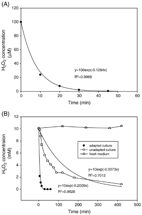
Fig. 5 shows the changes in H2O2 concentrations in the
culture fluid during the process of culture adaptation. The detoxification of 10 mM H2O2 in the adapted culture is
significantly faster than in the unadapted cultures. This is probably due to the significant induction of antioxidant enzymes activities in the adapted culture, and the marked decrease in such activities in undapted cultures. Altogether, these results strongly support the view that the acquisition of H2O2 tolerance was due to increased capacity to remove the
oxidant itself, thus avoiding prolonged exposure to H2O2and
associated deleterious intracellular events. Further, it has been proposed that the decomposition rate of exogenous H2O2by
intact viable cells is a first-order reaction[33]: this implies that the decline in H2O2 concentration in the cultures should be
exponential. In the present study, we found that the detoxifi-cation profile of 100μM H2O2in cultures ofA. nigerdid indeed
follow this trend, as supported by the high value ofR2, as did the detoxification profile of 10 mM H2O2in adapted cultures,
but not in unadapted cultures (Fig. 5B).
Discussion
Adaptation to H2O2
The intracellular concentration of H2O2 in aerobically
[image:4.595.146.464.68.491.2]growing Escherichia coli is estimated between 0.1 and 1μM [34]. In the present work, we found thatA. nigeris able to cope with up to 100μM exogenous H2O2, and adapt to up to 20 mM
Fig. 2. Effect of H2O2addition on the dry cell weight (DCW), glucose concentration, and intracellular protein content in batch cultures ofA. nigerB1-D. H2O2was
following preexposure. A 100μM H2O2leads to a temporary
growth arrest and a decrease in protein content. Previous work inA. nigershowed that protein concentrations decreased under conditions of oxidative stress[35]. InS. cerevisiae, exposure to H2O2resulted in a slowdown of protein biosynthetic processes,
and a stimulation of protein degradation pathways [36]. However, despite a general decline in protein content, some antioxidant enzymes activities could be greatly enhanced. In the present study, we showed induction of CAT, SOD, GPx, and GR activities by exogenous H2O2inA. niger. On the other hand,
direct addition of high concentrations of H2O2failed to induce
these activities. Meanwhile, we showed that detoxification of H2O2in the adapted culture is by far more rapid compared with
the unadapted cultures. Therefore, we propose that the induction of these enzymes is the mechanism by which such cultures acquire tolerance to high (potentially lethal) concentrations of H2O2inA. niger. More importantly, we have demonstrated
that this tolerance is simply due to a rapid detoxification of exogenous H2O2, avoiding prolonged exposure and associated
intracellular damage.
A number of papers have reported the induction of antioxidant enzymes activities by H2O2in microorganisms[29,37,38], but
[image:5.595.128.461.67.501.2]the time course effects of H2O2 are not discussed. However,
Fig. 3. Effect of H2O2addition on the activities of antioxidant enzymes in batch cultures ofA. nigerB1-D. H2O2was added to the culture at 70 and 72 h, to final
concentrations of 100μM and 10 mM, respectively. The results were obtained from three independent experiments, and expressed as mean ± SD.
Table 1
Effects of direct addition of H2O2(10 or 20 mM) on the activities of antioxidant
enzymes in batch cultures ofA. nigerB1-D
CAT u/mg prot SOD u/mg prot GR mu/mg prot
Control 2.61 ± 0.22 246.3 ± 10.0 88.9 ± 5.0 10 mM H2O2 1.65 ± 0.15 31.7 ± 9.6 69.1 ± 6.6
20 mM H2O2 N.D. N.D. 9.0 ± 1.4
H2O2was added at 72 h at the indicated concentrations, and incubated for 4 h.
[image:5.595.302.553.673.722.2]monitoring the changes in exogenous H2O2levels with time is a
key factor in attempting to understand the process of adaptation. Our results show that the antioxidant enzymes activities, those involved in the H2O2detoxification, including CAT, GPx, and
GR, peaked 4 h after addition of 10 mM H2O2 (Fig. 3). The
decline in activity of these enzymes coincides with the recovery from growth arrest, meaning that cells are shifting from oxidative defence back to growth. This indicates that the cells may not keep the ability to tolerate severe H2O2stress permanently.
Overpro-duction of these antioxidant enzymes requires extra energy[24], so it makes sense that these enzyme activities are decreasing after exogenous H2O2 is completely detoxified, in order to avoid
wasting energy. The mechanism through which this decline takes place is still obscure, but from our findings it is clear that activities of these antioxidant enzymes do not correlate well with endogenous H2O2concentrations, since the complete removal of
10 mM H2O2 took places less than 1 h after addition in the
adapted culture (Fig. 5), while the defensive enzyme activities remained high for much longer (Fig. 3).
CAT and GPx
In filamentous fungi, two peroxidases, CAT and GPx, have been reported to be associated with H2O2detoxification. CAT
catalyses the decomposition of H2O2to water and oxygen, while
[image:6.595.131.476.67.272.2] [image:6.595.320.555.337.693.2]GPx reduces peroxide at the expense of GSH and NADPH [13,14,20]. The relative extent to which these two enzymes contribute to H2O2 detoxification is still unclear. Our results
show the prestressed level of GPx activity is far less than that of CAT. Similar results have been reported by a number of research groups using different organisms, such asP. chrysogenum[20], Candida albicans[38], andS. cerevisiase[8,9]. In addition, in the present study CAT activity increased 8.5-fold on addition of 100μM H2O2, while GPx only rose by a factor of 1.6. Here, we
propose that CAT plays more important role in H2O2
Fig. 4. Effect of H2O2addition on the GSH and GSSG in batch cultures ofA. nigerB1-D. H2O2was added to the culture at 70 and 72 h, to final concentrations of
100μM and 10 mM, respectively.●GSH (control);○GSH (H2O2);
▪
GSSG (control);□GSSG (H2O2);▴GSH/GSSG (control);▵GSH/GSSG (H2O2). The resultswere obtained from three independent experiments and expressed as mean ± SD.
Fig. 5. H2O2detoxification in batch cultures ofA. nigerB1-D. (A) H2O2was
added to batch culture at 70 h to final concentration of 100μM. (B) H2O2was
added to batch culture, adapted (pretreatment with 100μM H2O2for 2 h) or not,
detoxification in filamentous fungi. However, GPx has been shown to be indispensable in coping with stress induced by the addition of other peroxides, such astert-butyl hydroperoxide (t -BHP), which cannot be detoxified by CAT[20]. InS. cerevisiae, a mutant with deleted CAT genes(ctt1Δ/cta1Δ) grew similarly to the wild type under nonoxidative stress conditions, and showed a similar susceptibility to H2O2, but its ability to adapt to
H2O2had been severely compromised[9]. On the other hand, a
mutant carrying deletion of a GPx homologous gene was hypersensitive to both H2O2andt-BHP[8]. In the wild type, the
basal mRNA level of that gene was high, and the product of the gene was expressed constitutively[8]. From these results and our own findings, we hypothesize that, in fungi, CAT and GPx contribute to H2O2 detoxification at the same time under
(normal) nonstress conditions, but CAT plays a more significant role in dealing with sudden bursts of H2O2, not only due to its
overwhelmingly higher capacity to remove H2O2but also the
cells ability to rapidly boost the activity of this enzyme in response to H2O2. This adaptive capability has an intrinsic,
central role in many areas of fungal life; for example, CAT has been shown to be implicated in the virulence of some fungi, in which the high capacity of the fungal cells to remove H2O2is
essential to counteract the burst of ROS generated by the host cells to kill the invading fungal pathogens[39].
Glutathione pathways (GPx, GR, GSH/GSSG)
The effect of H2O2addition on GR follows the same trend as
that of GPx. However, it is worth noting that the reductase acti-vities are much higher than the peroxidase actiacti-vities. Meanwhile, the redox state of glutathione, expressed as the ratio between the reduced from to the oxidized form, GSH/GSSG, is over 100. It has been reported that the redox state of glutathione in fungi is usually above 20[40]. Here we show that this figure is higher in thisA. niger, and the reduced state of glutathione corresponds to a high ratio of reductase activity to peroxidase activity.
Our results also clearly show that GSH does play a role in oxidative defence inA. niger. On the one hand, GSH reduces intracellular oxidants by reacting with them, enzymatically or nonenzymatically[40]. By contrast, GSH synthesis is boosted in attempting to rectify the imbalance of glutathione homeo-stasis. Emri et al. [20]showed that there were no significant changes in GSH levels in response to H2O2. The extremely high
concentration of H2O2 (0.35 and 0.7 M) used in their work
might account for this discrepancy; as we demonstrated here different concentrations of H2O2 impose distinct effects on
microorganisms.
Estimation of intracellular concentration of H2O2
According to previous studies, H2O2does not freely diffuse
across biomembranes, and the fast removal of H2O2by
intra-cellular antioxidant enzymes is able to generate a concentration gradient between extracellular and intracellular milieu[4,33]. It is this gradient which provides the driving force for H2O2entering
the cells where it is decomposed. The intracellular concentration of H2O2is usually much lower than the external one.
Under in vivo conditions, both CAT [41] and GPx [42] displayed first-order kinetics with their substrates, not exhibiting saturation effects. As such, the ratio between the concentration of intracellular and extracellular H2O2is described by the[33]
½H2O2in=½H2O2out¼R¼kapp=ðkGPxþkCATÞ; ð1Þ wherekapprefers to the first-order rate constant of the apparent
decomposition rate by intact cells, andkGPxandkCATrefer to the
first-order rate constants of GPx and CAT in the cell-free extract, respectively[33]. In the present study, we found that intracellular CAT activity is overwhelmingly higher than that of GPx. So the equation can be simplified to
½H2O2in=½H2O2out¼R¼kapp=kCAT: ð2Þ Further, kCAT could be derived from intracellular CAT
activity in the cell-free extract. In present study, one unit of CAT activity is defined as the decomposition of one micromole H2O2in one minute in the presence of 10 mM H2O2[27]. The
value of the CAT activity (UCAT, U/ml) measured in this assay
is equal to that of the initial decomposition rate of H2O2by the
CAT (mM/min).
r0¼kCAT C0
kCAT¼r0=C0¼UCAT=C0 ¼UCAT=10; ð3Þ where r0 refers to the initial rate, when the initial substrate
concentration is 10 mM (C0). Finally, the gradient could be
derived as
½H2O2in=½H2O2out¼R¼10kapp=UC: ð4Þ The estimated gradient is a lower limit, because only CAT is considered for H2O2 detoxification in our model; if other
enzymes consume H2O2, the gradient will be steeper.
Never-theless, our model gives us valuable insights into the profile of the intracellular concentration of H2O2which is distinct from
the extracellular one. kapp and kCAT from adapted and
un-adaptedA. nigerare described inTable 2. There is a relatively small gradient between intracellular and extracellular H2O2
concentrations when 100μM H2O2is added. This suggests the
rate at which exogenous H2O2 entering the cells is beyond
their capacity to remove it, and accumulation of H2O2inside the
[image:7.595.301.553.685.722.2]cells occurs quickly. By contrast, the estimated gradient in the
Table 2
First-order constants of apparent decomposition rate of H2O2by intact cells,kapp;
first order constants of decomposition rate of H2O2by CAT in cell-free extract,
kCAT; H2O2concentration gradient between intracellular and extracellular milieu
kapp kCAT [H2O2]in/[H2O2]out
100μM 0.1284 0.154 0.834
10 mM 0.2009 0.758 0.265
100μM H2O2was added to batch culture ofA. nigerB1-D at 70 h (unadapted);
10 mM H2O2 was added to batch culture ofA. nigerB1-D pre-treated with
adapted culture is much higher than in unadapted cultures. This shows thatA. nigerhas a greatly enhanced capacity to remove H2O2after adaptation, and the cells’ ability to remove H2O2
becomes comparable to the rate of H2O2 entering the cells.
Thus, accumulation of this oxidant inside the cells is avoided. By gaining new insights into the detoxification rate of exogenous H2O2 by intact adapted cells, and into the
intracellular antioxidant enzyme activities, we have shown that enhanced degradation of H2O2, which alters the
concen-tration gradient of the stressor and minimises intracellular accumulation, is the mechanism of the adaptive effect.
Conclusions
Pretreatment of A. niger with H2O2 at a nonlethal
concentration confers greatly enhanced resistance to killing by H2O2at lethal concentrations in the early exponential phase of
growth. This adaptation involves the induction of a number of antioxidant enzymes, predominantly catalase. Enhancement of the activities of these antioxidant enzymes prevents accumula-tion of H2O2 inside the cells, as supported by the increased
decomposition rate of external H2O2by intact cells, and higher
gradient of H2O2 concentration between intracellular and
extracelluar milieu estimated from a mathematical model.
Acknowledgments
Qiang Li thanks ORS and University of Strathclyde for financial support. He also thanks Dr. Zhonghu Bai for invaluable discussions.
References
[1] Turrens, J. F. Mitochondrial formation of reactive oxygen species. J. Physiol.552(2):335 344; 2003.
[2] Dalton, T. P.; Shertzer, H. G.; Puga, A. Regulation of gene expression by reactive oxygen.Annu. Rev. Pharmacol. Toxicol.39:67 101; 1999. [3] Jamieson, D. J. Oxidative stress responses of the yeastSaccharomyces
cerevisiae.Yeast14(16):1511 1527; 1998.
[4] Branco, M. R.; Marinho, H. S.; Cyrne, L.; Antunes, F. Decrease of H2O2
plasma membrane permeability during adaptation to H2O2in
Saccharo-myces cerevisiae.J. Biol. Chem.279(8):6501 6506; 2004.
[5] Imlay, J. A. Pathways of oxidative damage.Annu. Rev. Microbiol.57:395 418; 2003.
[6] Izawa, S.; Maeda, K.; Miki, T.; Mano, J.; Inoue, Y.; Kimura, A. Importance of glucose-6-phosphate dehydrogenase in the adaptive response to hy-drogen peroxide inSaccharomyces cerevisiae.Biochem. J.330(2):811 817; 1998.
[7] Minard, K. I.; McAlister-Henn, L. Antioxidant function of cytosolic sources of NADPH in yeast.Free Radic. Biol. Med.31(6):832 843; 2001. [8] Inoue, Y.; Matsuda, T.; Sugiyama, K.; Izawa, S.; Kimura, A. Genetic
analysis of glutathione peroxidase in oxidative stress response of Sac-charomyces cerevisiae.J. Biol. Chem.274(38):27002 27009; 1999. [9] Izawa, S.; Inoue, Y.; Kimura, A. Importance of catalase in the adaptive
response to hydrogen peroxide: analysis of acatalasaemicSaccharomyces cerevisiae.Biochem. J.320(1):61 67; 1996.
[10] Navarro, R. E.; Stringer, M. A.; Hansberg, W.; Timberlake, W. E.; Aguirre, J. catA, a new Aspergillus nidulansgene encoding a developmentally regulated catalase.Curr. Genet.29(4):352 359; 1996.
[11] Kawasaki, L.; Wysong, D.; Diamond, R.; Aguirre, J. Two divergent catalase genes are differentially regulated during Aspergillus nidulans
development and oxidative stress. J. Bacteriol. 179 (10):3284 3292; 1997.
[12] Navarro, R. E.; Aguirre, J. Posttranscriptional control mediates cell type-specific localization of catalase A duringAspergillus nidulans develop-ment.J. Bacteriol.180(21):5733 5738; 1998.
[13] Kreiner, M.; Harvey, L. M.; McNeil, B. Oxidative stress response of a recombinant Aspergillus niger to exogenous menadione and H2O2
addition.Enzyme Microb. Technol.30(3):346 353; 2002.
[14] Angelova, M. B.; Pashova, S. B.; Spasova, B. K.; Vassilev, S. V.; Slokoska, L. S. Oxidative stress response of filamentous fungi induced by hydrogen peroxide and paraquat.Mycol. Res.109(2):150 158; 2005.
[15] Stephen, D. W.; Jamieson, D. J. Glutathione is an important antioxidant molecule in the yeastSaccharomyces cerevisiae.FEMS Microbiol. Lett.
141(2 3):207 212; 1996.
[16] Izawa, S.; Inoue, Y.; Kimura, A. Oxidative stress response in yeast: effect of glutathione on adaptation to hydrogen peroxide stress inSaccharomyces cerevisiae.FEBS Lett.368(1):73 76; 1995.
[17] Lee, J. C.; Straffon, M. J.; Jang, T. Y.; Higgins, V. J.; Grant, C. M.; Dawes, I. W. The essential and ancillary role of glutathione inSaccharomyces cerevisiaeanalysed using a grande gsh1 disruptant strain.FEMS Yeast Res.
1(1):57 65; 2001.
[18] Grant, C. M.; Collinson, L. P.; Roe, J. H.; Dawes, I. W. Yeast glutathione reductase is required for protection against oxidative stress and is a target gene for yAP-1 transcriptional regulation.Mol. Microbiol.21(1):171 179; 1996.
[19] Bai, Z.; Harvey, L. M.; McNeil, B. Elevated temperature effects on the oxidant/antioxidant balance in submerged batch cultures of the filamentous fungus Aspergillus niger B1-D. Biotechnol. Bioeng. 83 (7):772 779; 2003.
[20] Emri, T.; Pocsi, I.; Szentirmai, A. Glutathione metabolism and protection against oxidative stress caused by peroxides inPenicillium chrysogenum. Free Radic. Biol. Med.23(5):809 814; 1997.
[21] Gibbs, P. A.; Seviour, R. J.; Schmid, F. Growth of filamentous fungi in submerged culture: problems and possible solutions.Crit. Rev. Biotechnol.
20(1):17 48; 2000.
[22] Bai, Z.; Harvey, L. M.; McNeil, B. Oxidative stress in submerged cultures of fungi.Crit. Rev. Biotechnol.23(4):267 302; 2003.
[23] Archer, D. B.; Jeenes, D. J.; MacKenzie, D. A.; Brightwell, G.; Lambert, N.; Lowe, G.; Radford, S. E.; Dobson, C. M. Hen egg white lysozyme expressed in, and secreted from,Aspergillus nigeris correctly processed and folded.Bio/Technology8:741 745; 1990.
[24] Bai, Z.; Harvey, L. M.; McNeil, B. Physiological responses of chemostat cultures ofAspergillus nigerB1-D to simulated and actual oxidative stress. Biotechnol. Bioeng.82(6):691 701; 2003.
[25] Wolff, S. P.; Lester, P. Ferrous ion oxidation in presence of ferric ion indicator xylenol orange for measurement of hydroperoxides. In: Packer, L. (Ed.), Oxygen radicals in biological systems, part C. Methods in enzymology, vol. 233. Academic Press, San Diego, pp. 182 189; 1994. [26] Crapo, J. D.; McCord, J. M.; Fridovich, I. Preparation and assay of
superoxide dismutases. In: Fleishcher, S., Packer, L. (Eds.), Biomem-branes, part D. Methods in enzymology, vol. 53. Academic Press, San Diego,pp. 382 393; 1978.
[27] Aebi, H. Catalase in vitro. In: Packer, L. (Ed.), Oxygen radicals in biological systems, methods in enzymology, vol. 105. Academic Press, San Diego,pp. 121 126; 1984.
[28] Tappel, A. L. Glutathione peroxidase and hydroperoxides. In: Fleishcher, S., Packer, L. (Eds.), Biomembranes, part C. Methods in enzymology, vol. 52. Academic Press, San Diego, pp. 506 513; 1978.
[29] Lee, J.; Dawes, I. W.; Roe, J. H. Adaptive response of Schizosaccharo-myces pombeto hydrogen peroxide and menadione.Microbiology141
(12):3127 3132; 1995.
[30] Anderson, M. E. Determination of glutathione and glutathione disulfide in biological samples. In: Meiser, A. (Ed.), Glutamate, glutamine, glutathi-one, and related compounds. Methods in enzymology, vol. 113. Academic Press, San Diego, pp. 548 555; 1985.
[32] Emri, T.; Molnar, Z.; Pocsi, I. The appearances of autolytic and apoptotic markers are concomitant but differently regulated in carbon-starving Asper-gillus nidulanscultures.FEMS Microbiol. Lett.251(2):297 303; 2005. [33] Antunes, F.; Cadenas, E. Estimation of H2O2gradients across
biomem-branes.FEBS Lett.475(2):121 126; 2000.
[34] Storz, G.; Imlay, J. A. Oxidative stress.Curr. Opin. Microbiol.2(2):188 194; 1999.
[35] Bai, Z.; Harvey, L. M.; White, S.; McNeil, B. Effects of oxidative stress on production of heterologous and native protein, and culture morphology in batch and chemostat cultures ofAspergillus nigerB1-D.Enzyme Microb. Technol.34(1):10 21; 2004.
[36] Godon, C.; Lagniel, G.; Lee, J.; Buhler, J. M.; Kieffer, S.; Perrot, M.; Boucherie, H.; Toledano, M. B.; Labarre, J. The H2O2stimulon in
Sac-charomyces cerevisiae.J. Biol. Chem.273(35):22480 22489; 1998.
[37] Davies, J. M.; Lowry, C. V.; Davies, K. J. Transient adaptation to oxidative stress in yeast.Arch. Biochem. Biophys.317(1):1 6; 1995.
[38] Jamieson, D. J.; Stephen, D. W.; Terriere, E. C. Analysis of the adaptive oxidative stress response ofCandida albicans.FEMS Microbiol. Lett.138
(1):83 88; 1996.
[39] Hamilton, A. J.; Holdom, M. D. Antioxidant systems in the pathogenic fungi of man and their role in virulence.Med. Mycol.37(6):375 389; 1999.
[40] Pocsi, I.; Prade, R. A.; Penninckx, M. J. Glutathione, altruistic metabolite in fungi.Adv. Microb. Physiol.49:1 76; 2004.
[41] Chance, B.; Sies, H.; Boveris, A. Hydroperoxide metabolism in mammalian organs.Physiol. Rev.59(3):527 605; 1979.