S H O R T R E P O R T
Open Access
Surfen and oxalyl surfen decrease tau
hyperphosphorylation and mitigate neuron
deficits in vivo in a zebrafish model of
tauopathy
Seyedeh Maryam Alavi Naini
1,2, Constantin Yanicostas
1, Rahma Hassan-Abdi
1, Sébastien Blondeel
1,
Mohamed Bennis
3, Ryan J. Weiss
4, Yitzhak Tor
4, Jeffrey D. Esko
5and Nadia Soussi-Yanicostas
1*Abstract
Background:Tauopathies comprise a family of neurodegenerative disorders including Alzheimer’s disease for which there is an urgent and unmet need for disease-modifying treatments. Tauopathies are characterized by pathological tau hyperphosphorylation, which has been shown to correlate tightly with disease progression and memory loss in patients suffering from Alzheimer’s disease. We recently demonstrated an essential requirement for 3-O-sulfated heparan sulfate in pathological tau hyperphosphorylation in zebrafish, a prominent model organism for human drug discovery. Here, we investigated whether in vivo treatment with surfen or its derivatives oxalyl surfen and hemisurfen, small molecules with heparan sulfate antagonist properties, could mitigate tau hyperphosphorylation and neuronal deficits in a zebrafish model of tauopathies.
Results:In vivo treatment of Tg[HuC::hTauP301L; DsRed] embryos for 2 days with surfen or oxalyl surfen significantly reduced the accumulation of the pThr181 tau phospho-epitope measured by ELISA by 30% and 51%, respectively. Western blot analysis also showed a significant decrease of pThr181 and pSer396/pSer404 in embryos treated with surfen or oxalyl surfen. Immunohistochemical analysis further confirmed that treatment with surfen or oxalyl surfen significantly decreased the AT8 tau epitope in spinal motoneurons. In addition, in vivo treatment of Tg[HuC::hTauP301L; DsRed] embryos with surfen or oxalyl surfen significantly rescued spinal motoneuron axon-branching defects and, as a likely consequence, the impaired stereotypical touch-evoked escape response. Importantly, treatment with hemisurfen, a surfen derivative devoid of heparan sulfate antagonist activity, does not affect tau hyperphosphorylation, nor neuronal or behavioural deficits in Tg[HuC::hTauP301L; DsRed] embryos.
Conclusion:Our findings demonstrate for the first time that surfen, a well-tolerated molecule in clinical settings, and its derivative, oxalyl surfen, could mitigate or delay neuronal defects in tauopathies, including Alzheimer’s disease.
Keywords:Tauopathy, Zebrafish, Alzheimer’s disease, Tau protein, Tau hyperphosphorylation, Surfen, Oxalyl surfen, Heparan sulfate
* Correspondence:[email protected]
1PROTECT, Inserm, Université Paris Diderot, Sorbonne Paris Cité, Paris, France
Full list of author information is available at the end of the article
Introduction
Tauopathies comprise more than 20 neurodegenerative dis-eases including Alzheimer’s disease (AD), frontotemporal dementia (FTD), Pick’s disease, progressive supranuclear palsy (PSP) and other related disorders. Tauopaties are characterized by accumulation of hyperphosphorylated iso-forms of the microtubule-associated tau protein in brain forming distinct inclusions [1].
We have recently shown that in vivo depletion of Hs3st2, an enzyme involved in 3-O-sulfation of heparan sulfate chains and predominantly expressed in neuronal cells, sig-nificantly decreased tau hyperphosphorylation and partially rescued neuronal and behaviour defects in transgenic Tg[HuC::hTauP301L; DsRed] zebrafish embryos [2]. Tg[HuC::hTauP301L; DsRed] embryos display key features of tauopathies, such as tau hyperphosphorylation [3]. These findings suggest that inhibition of heparan sulfate-related activities could have beneficial therapeutic effects for tauo-pathies. Here, we have tested the hypothesis that treatment of Tg[HuC::hTauP301L; DsRed] zebrafish embryos with small molecules displaying heparan sulfate antagonist properties could mitigate pathological tau hyperphosphorylation and rescue the induced neuronal and behavioural deficits.
Glycosaminoglycan (GAG)-protein interactions have long been recognized as therapeutic targets in various dis-ease conditions such as cancer, inflammation and AD [4, 5]. Inhibition of harmful processes mediated by endogen-ous GAGs, by treatment with exogenendogen-ous GAGs or GAG mimetics, has been developed as a therapeutic strategy in Alzheimer’s disease [6–8]. Surfen (1,3-bis (4-amino-2-methylquinolin-6-yl) urea) is a quinolone-based low MW derivative, which was initially developed for the produc-tion of depot insulin for diabetic patients [9]. This well-tolerated substance was later shown to possess antibacter-ial, trypanocidal, and anti-inflammatory properties [10– 12]. Of particular interest, surfen was later shown to ex-hibit heparin-neutralizing activity and the ability to antagonize heparan sulfate (HS)-protein interactions [5, 13]. As a first attempt to investigate the effects of surfen and its analogs, on tau hyperphosphorylation, we have examined whether surfen and two recently synthesized surfen derivatives, oxalyl surfen (N1,N2 -bis(4-amino-2-methylquinolin-6-yl)oxalamide) and hemisurfen (1-(4-amino-2-methylquinolin-6-yl)urea) [14], could reduce tau hyperphosphorylation and alleviate neuron defects in vivo in Tg[HuC::hTauP301L; DsRed] zebrafish embryos.
Results
Surfen, oxalyl surfen and hemisurfen are well tolerated in zebrafish embryos
Initially, experiments were set up to analyze potential toxicity of the surfen analogs used in this study, (Fig.1a). One set of 24 h post-fertilization (hpf ) zebrafish Tg[HuC::hTauP301L, DsRed] embryos were incubated for
2 days in E3 medium containing 1% DMSO and surfen, oxalyl surfen or hemisurfen. As negative controls, age-matched wild-type and Tg[HuC::hTauP301L, DsRed] em-bryos were incubated for 2 days in E3 medium contain-ing 1% DMSO. As positive control, age-matched Tg[HuC::hTauP301L, DsRed] embryos were incubated in E3 medium containing 1% DMSO and 1 lithium chloride (LiCl), a long known inhibitor of tau hyperphosphoryla-tion [15]. For each agent, operational concentrations were defined as the highest concentration not inducing any visible morphological abnormalities, including heart rhythm and blood flow defects (Fig.1b), nor any signifi-cant increase in embryo lethality (Additional file 1: Fig-ure S1c). Operational concentrations (3 μM for surfen, 2 μM for oxalyl surfen, and 3μM for hemisurfen) were then used for all subsequent experiments.
Surfen and oxalyl surfen reduce tau hyperphosphorylation in vivo
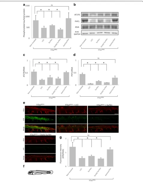
As a first attempt to determine whether treatment with surfen, oxalyl surfen or hemisurfen could decrease tau hyperphosphorylation in vivo, we quantified tau pThr181 phosphorylation by ELISA assay. Treatment of 24 hpf Tg[HuC::hTauP301L; DsRed] embryos for 2 days with sur-fen or oxalyl sursur-fen, decreased the accumulation of hyper-phosphorylated tau by 30% and 51%, respectively (surfen and oxalyl surfen: P < 0.05), when compared to embryos treated with 1% DMSO (Fig. 2a). In contrast, no signifi-cant differences in pThr181 phospho-tau accumulation could be detected in embryos incubated with hemisurfen (P = 0.83, Fig. 2a). As expected, treatment with LiCl re-sulted in a 43% decrease in hyperphosphorylated tau when compared to embryos treated with 1% DMSO (N= 5,n= 250 (number of embryos);P< 0.05, Fig.2a).
To confirm the ELISA results, we performed Western blot analysis with antibodies directed against either the pThr181 (AT270) or pSer396/pSer404 (PHF1) phospho-tau epitopes (Fig.2b). Data showed a significant decrease in accumulation of both epitopes following treatment with surfen, oxalyl surfen and LiCl (P < 0.05). In con-trast, hemisurfen did not significantly affect tau phos-phorylation on AT270 nor PHF1 epitopes, (P= 0.42) and (P= 0.12) respectively (Fig.2b, c, d).
oxalyl surfen, decreases the somato-dendritic localization of hTauP301L (Fig. 2e), a feature of tau pathology in AD and FTD [16]. As tau missorting is linked to tau hyper-phosphorylation, the decrease in somato-dendritic tau might be a consequence of the decrease in tau phosphor-ylation. In general, we observed stronger effects of oxalyl surfen than surfen (Fig.2).
Surfen and oxalyl surfen rescue motoneuron defects and behavioral deficits
Tg[HuC::hTauP301L; DsRed] larvae displayed markedly reduced motoneuron axon-branching and elongation, and, as a likely consequence, they showed an impaired escape response to touch stimuli [3]. Because surfen and
oxalyl surfen markedly decrease tau hyperphosphoryla-tion, we next investigated whether treatment with these two compounds could rescue, at least partially, neuronal deficits and promote functional recovery. First, we ana-lysed motoneuron axon morphology by immunohisto-chemistry using znp1 [17], an antibody that specifically recognizes synaptotagmin II, a synaptic protein highly expressed in primary motoneuron axons. Results showed that treatment with surfen, oxalyl surfen, and LiCl, but not hemisurfen, significantly increased primary moto-neuron branching in Tg[HuC::hTauP301L, DsRed] em-bryos (1% DMSO vs. surfen-treated emem-bryos: P < 0.001; 1% DMSO vs. oxalyl surfen-treated embryos: P < 0.05; 1% DMSO vs. LiCl-treated embryos:P< 0.01; 1% DMSO
Fig. 1Surfen and oxalyl surfen are well tolerated by Tg[HuC::hTauP301L; DsRed] embryos.aChemical structure of Surfen
[image:3.595.58.540.85.458.2]vs. hemisurfen-treated embryos: P = 0.43) (Fig. 3a and b).
Next, we quantified znp1 staining in wild-type and in treated and 1% DMSO Tg[HuC::hTauP301L; DsRed] em-bryos. As previously shown [3], a significant decrease in znp1 staining was observed in 1% DMSO Tg[HuC::h-TauP301L, DsRed] individuals when compared to age-matched wild-type embryos (P< 0.05) (Fig.3aandc). Inter-estingly, znp1 staining in Tg[HuC::hTauP301L; DsRed] em-bryos was significantly rescued following a 2 day treatment with surfen (P < 0.01), oxalyl surfen (P < 0.05), and LiCl (P< 0.01), but not hemisurfen (P =0.21) (Fig.3aandc).
We next assessed whether treatment with surfen or oxa-lyl surfen could rescue the motility defects of Tg[HuC::h-TauP301L; DsRed] zebrafish larvae in response to touch stimuli. As previously shown, Tg[HuC::hTauP301L; DsRed] zebrafish larvae showed significantly impaired motility characterized by slower movements and reduced touch-induced escape when compared to wild-type age-matched larvae (wild-type vs. 1% DMSO Tg[HuC::hTauP301L; DsRed] embryos: P < 0.05) (Fig. 3d). Interestingly, treat-ment with surfen or oxalyl surfen, fully rescued the motil-ity deficit (surfen and oxalyl surfen vs. 1% DMSO: P < 0.05; Fig.3d), while treatment with hemisurfen had no ef-fect on motility deef-fects (P = 0.27; Fig. 3d). These results provided functional evidence that surfen and oxalyl surfen not only significantly decrease tau hyperphosphorylation, but also alleviate the behavioural consequences of the neuronal deficits induced by the expression of the human mutant tauP301Lprotein.
Discussion
Here we showed that treatment with surfen, or its ana-log, oxalyl surfen, significantly decreased tau hyperpho-sphorylation and rescued motoneuron defects and
behavioral abnormalities induced by expression of mu-tant hTauP301Lin Tg[HuC::hTauP301L; DsRed] zebrafish. In contrast, hemisurfen did not affect tau phosphoryl-ation, motoneuron and behavioural deficits. Surfen has been shown to inhibit heparan sulfate-protein interac-tions [13], whereas hemisurfen does not [14]. Thus sur-fen and oxalyl sursur-fen may mediate their beneficial effects by blocking heparan sulfate-tau interactions. Taken together, these results strengthen the hypothesis of a substrate modulator effect of highly sulfated HS chains on tau, possibly through a chaperone-like activ-ity uncovering the tau residues that are phosphorylated in pathological situations. In this context, surfen and oxalyl surfen may inhibit the chaperone activity of sul-fated polysaccharide chains, thus preventing tau hyper-phosphorylation. It is also possible that the decline in tau phosphorylation mediated by surfen and oxalyl sur-fen is due to alterations of heparan sulfate biosynthesis or metabolism. Surfen may affect heparan sulfate struc-ture and stability, as it has been previously shown in vitro that surfen prevents interaction of heparin with heparin biosynthetic and degrading enzymes (Schuksz et al.). However, it should also be noted that surfen also displays Ca channel blocker [18], zinc ion binding [11] and immunomodulatory activities [10, 19]. These prop-erties can potentially affect pathophysiological mecha-nisms in tauopathies. Calcium deregulation is reported to contribute to neurodegeneration in iPSC-derived neurons from FTD patients [20]. Surfen may affect cal-cium levels by blocking calcal-cium channels or modulat-ing calcium channels through interaction with heparan sulfate [21]. Zinc ion (Zn2+) has been implicated in tau fibrillization and toxicity [22,23]. A c5a receptor antag-onist is reported to decrease tau hyperphosphorylation in 3xTg mouse model of Alzheimer’s disease [24], while
(See figure on previous page.)
Fig. 3Surfen and oxalyl surfen rescue motoneuron defects and functional deficits in Tg[HuC::hTauP301L; DsRed] embryos.aImmunohistochemical visualization of the synaptotagmin II protein (anti-znpl antibody) in 72 hpf wild-type (WT) and Tg[HuC::hTauP301L; DsRed] (hTauP301L) embryos treated for 2 days with 1% DMSO (hTauP301L+ 1% DMSO), 80 mM LiCl (hTauP301L+ LiCl), 3
μM surfen (hTauP301L+ surfen), 2
μM oxalyl surfen (hTauP301L+ oxalyl surfen) or 3
μM hemisurfen (hTauP301L+ hemisurfen), showed that surfen and oxalyl surfen markedly rescued the motorneuron axon defects seen in Tg[HuC::hTauP301L; DsRed] embryos. The imaged caudal area corresponds to the box represented in (b). Scale bar: 50
[image:6.595.60.538.83.548.2]surfen also acts as an inhibitor of c5a receptor binding [10].
Importantly, apart from rare reports of hypersensitivity reactions [25,26], surfen is well tolerated in clinical set-tings. Although one study linked high doses and pro-longed administration of surfen to lymphosarcoma and lesions reminiscent of nutritional deficiency, surfen is well tolerated in mice [13]. In good agreement, we found surfen and oxalyl surfen to be well tolerated in zebrafish embryos at the effective concentrations of 3 μM and 2μM, respectively. Oxalyl surfen shows a stronger effect on tau phosphorylation and behavioural rescue than sur-fen, suggesting the molecule to be more efficient against tau pathology. However, the derivative is toxic to zebra-fish embryos at a lower concentration, pointing out a potentially less favourable safety profile than surfen. Fur-ther investigations are needed to determine wheFur-ther the two molecules could also counteract neurodegenerative processes linked to tau alterations in humans.
Materials and methods
Animals
Zebrafish were maintained at 28 °C in our zebrafish fa-cility under standard conditions as described by Wester-field (1995) [27]. Developmental stages were determined as hours post-fertilization (hpf ) as described by Kimmel et al. [28].ABstrain was used as wild-type fish. The zeb-rafish transgenic line stably expressing the human mu-tant TauP301L protein that is associated with frontotemporal dementia with Parkinsonism linked to chromosome 17 (FTDP-17) (the Tg[HuC::hTauP301L; DsRed]), has been previously described [3], and was kindly provided by Christian Haass, Bettina Schmid, and Dominik Paquet (Deutsches Zentrum für Neurodegener-ative Erkrankungen or DZNE, Munich, Germany).
Compounds
Surfen (1,3-bis(4-amino-2-methylquinolin-6-yl)urea) was obtained from the Open Chemical Repository in the De-velopmental Therapeutic Program at the National Cancer Institute (NSC12155) or synthesized according to published methods. The synthesis and characterization of oxalyl surfen (N1,N2-bis(4-amino-2-methylquinolin-6-yl)oxalamide) and hemisurfen (1-(4-amino-2-methylqui-nolin-6-yl)urea) have been previously described [14,29].
Treatments
As surfen and oxalyl surfen bind avidly to plastic, we ei-ther pre-coated all plasticware with serum containing medium or used glass vessels. Stock solutions (surfen and hemisurfen, 30 mM; oxalyl surfen, 21.7 mM) were prepared in DMSO. Working solutions were prepared as needed by diluting stock solutions in E3 medium and adjusting the DMSO concentration to 1% (vol/vol). Final
concentrations for treatments were determined as 3μM for surfen, 2μM for oxalyl surfen, 3μM for hemisurfen and 80 mM for lithium chloride (LiCl) based on max-imal non-toxic concentrations for zebrafish embryos (Additional file 1). Embryos (24 hpf ) were manually dechorionated and incubated for 2 days in 1–2 ml of ei-ther control medium (E3 medium containing 1% DMSO) or E3 medium containing 1% DMSO and the surfen derivatives in BSA-coated 6-well microtiter plates. All solutions were changed daily.
ELISA
Embryos treated as previously described were anaesthe-tized with MS-222 in E3 medium on ice. After removal of the yolk, embryos were snap frozen on dry ice and homogenized by sonication in lysis buffer (50 mM Tris HCl, 150 mM NaCl, 10% Triton X100, 1 mM EDTA, 1X Protease Inhibitor Cocktail [Roche], 1 mM Sodium orthovanadate (NaVO4) [Sigma-Aldrich], and 1 mM So-dium fluoride (NaF), pH 8). Insoluble material was re-moved by a 30 min centrifugation (10,000g) at 4 °C and protein concentration was determined with Bradford protein assay (Bio-Rad). Accumulation of phosphory-lated tau was quantified using the INNOTEST® Phospho-Tau (181P) ELISA (Innogenetics, Gent Belgium), using mAb HT7 for coating, phospho-dependent mAb AT270 (specific for phospho-threonine-181 tau epitope) as detector antibody, and a synthetic phosphopeptide for standardization.
Western blot
Zebrafish larvae were collected, anaesthetized in MS-222, and lysed on ice with lysis buffer (50 mM Tris-HCl, 150 mM NaCl, 1% Triton X-100, 10 mM NaF, 1 mM Na3VO4, pH 8.0) supplemented with protease and phos-phatase inhibitors (Pierce). Lysates were homogenized by sonication and centrifuged at 12000 g for 15 min. The protein content in the supernatants was quantified using a Bradford protein assay (Bio-Rad).
Samples containing 10 μg proteins were subjected to SDS-PAGE in 10% acrylamide gel. Primary antibodies against phosphorylated tau, AT270 and PHF1 (Pierce, Thermo Scientific), anti-human total tau antibody K9JA (Rabbit Polyclonal Antibody, Dako Cytomation), and anti-GADPH (Abcam) were used. Blots were subse-quently incubated for 1 h at room temperature with the corresponding secondary antibodies diluted in phosphate-buffered saline containing 5% milk and re-vealed using ECL RevelBlOt® Plus (Ozyme) following manufacturer’s instructions.
Immunohistochemistry
1-phenyl-2-thiourea (PTU) in E3 medium starting at 20 hpf. After anaesthesia with MS-222, whole embryos were fixed in 4% paraformaldehyde in PBS, and preserved in methanol (MeOH) 100%. Fixed and acetone-cracked embryos were then blocked and permeabilized for 1 h at room temperature in a PBS solution containing 10% NGS, 1% DMSO and 0.1% Tween 20. Embryos were then incubated overnight at room temperature with the phosphorylation-independent primary anti-human total tau antibody K9JA (Rabbit Polyclonal Antibody, Dako Cytomation) diluted at 1:300, the phosphorylation-dependent site specific anti-PHF-Tau antibody AT8 diluted at 1:50 (gift from D. Paquet), and anti-Znp-1 (Mouse Monoclonal Antibody; Hybridoma Bank, Iowa, USA) (1:300) to investigate the morphology of primary motoneurons. After several washes, embryos were blocked as previously described [30], and incubated overnight at 4 °C with a solution con-taining CY3-coupled goat anti-rabbit (1:500) and Alexa Fluor 488-coupled goat anti-mouse antibody (1:500). Em-bryos were mounted in 1% agarose (low melting, Bio-Rad) in PBS buffer.
Image analysis
Bright field images of embryos were captured using a stereomicroscope (SteREO Lumar. V12, Zeiss) equipped with a digital camera (DXM 1200F, Nikon) controlled by the ACT-1 software (Version 2.63 Nikon). Fluorescently labelled embryos were imaged using a microscope equipped with an ApoTome system (Zeiss) fitted with an AxioCam MRm camera (Zeiss) controlled by the Axiovi-sion or ZEN software. All images were processed with Adobe Photoshop 7.0 (Adobe System, San Jose, CA). When necessary, brightness, contrast, and colour balance, were uniformly optimized. Fluorescence intensities and densimetric quantification of protein immunoblots were performed using ImageJ/Fiji (Rasband, W.S., ImageJ, U. S. National Institutes of Health, Bethesda, Maryland, USA, http://imagej.nih.gov/ij/, 1997–2012) on grayscale images. For each value, quantifications were performed using im-ages from three independent experiments.
Behavioural analysis
Larvae behaviour was analysed at 48 hpf after 1 day treatments with the different compounds. The larval es-cape response reflex was assessed by gently touching the tip of the tail with a fine plastic rod. Embryos were clas-sified as responders or responders, with non-responders failing to respond by swimming at least three times their own body length.
Statistics
Values for mean, standard deviation (SD) and standard error of mean (SEM) were calculated using Microsoft Excel, version 12.0.6683.5002. Statistical analysis was
performed using Microsoft Excel and Student’s t test. Error bars represent SEM, *P < 0.05, ** P < 0.01, ***P
<0.001.
Additional file
Additional file 1:Figure S1.Percentage of embryonic survival observed for 72 hpf wild-type (WT) and Tg[HuC::hTauP301L; DsRed] (non-treated) embryos incubated for 2 days in E3 medium containing 1% DMSO or E3 medium containing 1% DMSO with LiCl (10–150 mM)(a), surfen (0.1–10μM)(b), oxalyl surfen (0.1–10μM)(c)or hemisurfen (0.1– 10μM)(d). Note that at the selected concentrations (80 mM LiCl, 3μM for surfen and hemisurfen and 2μM for oxalyl surfen) are the maximal non-toxic concentrations (n= 250, *P< 0.05, ***P< 0.001, ns: non-significant, Student’sttest). (TIFF 55462 kb)
Abbreviations
AD:Alzheimer’s disease; DMSO: Dimethyl sulfoxide; dpf: Days post fertilization; FTD: Frontotemporal dementia; hpf: Hours post fertilization; LiCl: Lithium chloride
Acknowledgements
We thank Christian Haass, Bettina Schmid, and Dominik Paquet (DZNE, Munich, Germany) for providing us with the Tg[HuC::hTauP301L; DsRed] transgenic line. We also thank Foudil Lamari (Biochimie des Maladies Neuro-métaboliques, Hôpital de la Pitié-Salpêtrière, Paris) and Pauline Claus (Inserm UMR 1141) for technical assistance.
Funding
SMAN received a grant from Servier Research Institute. This work was supported by Institut National de la Santé et la Recherche Médicale (INSERM), the French National Research Agency (ANR-16-CE18–0010), and Fondation NRJ (Institut de France) to NSY and grants CA46462 and CA112278 from the National Institute of Health to JDE and YT. Funding sources had no involvement in study design, collection, analysis or interpretation of data, or decision to publish.
Availability of data and materials
All data generated or analysed during this study are included in this published article.
Authors’contributions
SMAN, NSY and CY designed the research; SMAN, SB and RHA performed the research; RJW, YT, and JDE provided the surfen derivatives; SMAN, SB and MB analysed the data; SMAN, CY and NSY wrote the manuscript. All authors read and approved the final manuscript.
Authors’information
SMAN, CY, SB and NSY are from the Département Hospitalo-Universitaire: Promoting Research Oriented Towards Early CNS Therapies, Inserm, Hôpital Robert Debré, Paris, France. The present address for SMAN is Laboratoire Neurosciences Paris Seine, INSERM UMRS 1130, UMR 8246, Université Pierre et Marie Curie, 75252 Paris, France. MB is from the Department of Biology, Cadi Ayyad University, Marrakesh, Morocco. RJW and YT are affiliated with the Department of Chemistry and Biochemistry, University of California, San Diego, La Jolla, CA, USA. JDE is from the Department of Cellular and Molecu-lar Medicine, University of California, San Diego, La Jolla, CA, USA.
Ethics approval
All experiments involving zebrafish handling complied with the guidelines of the French Animal Ethics Committee, and were approved by that committee in the ethics statement No. 2012–15/676–0069.
Consent for publication
All authors critically revised the manuscript and approved the final version before submission.
Competing interests
Author details
1PROTECT, Inserm, Université Paris Diderot, Sorbonne Paris Cité, Paris, France. 2Institut de Biologie Paris Seine-Laboratoire Neuroscience Paris Seine, Inserm
UMRS 1130, CNRS UMR 8246, UPMC UM 118, Université Pierre et Marie Curie, Paris, France.3Cadi Ayyad University, Marrakesh, Morocco.4Department of
Chemistry and Biochemistry, University of California, San Diego, La Jolla, CA, USA.5Department of Cellular and Molecular Medicine, University of
California, San Diego, La Jolla, CA, USA.
Received: 1 December 2017 Accepted: 28 February 2018
References
1. Spillantini MG, Goedert M. Tau pathology and neurodegeneration. Lancet Neurol. 2013;12:609–22.
2. Sepulveda-Diaz JE, Alavi Naini SM, Huynh MB, Ouidja MO, Yanicostas C, Chantepie S, et al. HS3ST2 expression is critical for the abnormal phosphorylation of tau in Alzheimer’s disease-related tau pathology. Brain. 2015;138(5):1339–54.
3. Paquet D, Bhat R, Sydow A, Mandelkow E, Berg S, Hellberg S, et al. A zebrafish model of tauopathy allows in vivo imaging of neuronal cell death and drug evaluation. J Clin Invest. 2009;119(5):1382–95. Available from:
http://www.pubmedcentral.nih.gov/articlerender.fcgi?artid=2673864&tool= pmcentrez&rendertype=abstract
4. Bishop JR, Schuksz M, Esko JD. Heparan sulphate proteoglycans fine-tune mammalian physiology. Nature. 2007;446(7139):1030–7. Available from:
http://www.nature.com/doifinder/10.1038/nature05817
5. Weiss RJ, Esko JD, Tor Y. Targeting heparin and heparan sulfate protein interactions. Org Biomol Chem. 2017;15(27):5656–68. Available from:http:// xlink.rsc.org/?DOI=C7OB01058C
6. Gervais F, Paquette J, Morissette C, Krzywkowski P, Yu M, Azzi M, et al. Targeting soluble Aβpeptide with Tramiprosate for the treatment of brain amyloidosis. Neurobiol Aging. 2007;28(4):537–47.
7. Dudas B, Rose M, Cornelli U, Pavlovich A, Hanin I. Neuroprotective properties of glycosaminoglycans: potential treatment for neurodegenerative disorders. In: Neurodegenerative Diseases; 2008. p. 200–5.
8. Sabbagh MN. Clinical effects of oral Tramiprosate in APOE4/4 homozygous patients with mild Alzheimer’s disease suggest disease modification. J Prev Alzheimers Dis. 2017;4(3):136–7.
9. Umber F, Störring FK, Föllmer W. Erfolge mit Einem Neuartigen Depotinsulin Ohne Protaminzusatz (Surfen-Insulin) - III. Mitteilung Klin Wochenschr. 1938; 17(13):443–6.
10. Lanza TJ, Durette PL, Rollins T, Siciliano S, Cianciarulo DN, Kobayashi SV, et al. Substituted 4,6-diaminoquinolines as inhibitors of C5a receptor binding. J Med Chem. 1992;35(2):252–8.
11. Panchal RG, Hermone AR, Nguyen TL, Wong TY, Schwarzenbacher R, Schmidt J, et al. Identification of small molecule inhibitors of anthrax lethal factor. Nat Struct Mol Biol. 2004;11(1):67–72.
12. Goble F. Chemotherapy of experimental trypanosomiasis; trypanocidal activity of certain bis (2-methyl-4-amino-6-quinolyl) amides and ethers. J Pharmacol Exp Ther. 1950;98(1):49–61.
13. Schuksz M, Fuster MM, Brown JR, Crawford BE, Ditto DP, Lawrence R, et al. Surfen, a small molecule antagonist of heparan sulfate. Proc Natl Acad Sci U S A. 2008;105(35):13075–80.
14. Weiss RJ, Gordts PL, Le D, Xu D, Esko JD, Tor Y. Small molecule antagonists of cell-surface heparan sulfate and heparin–protein interactions. Chem Sci. 2015;6(10):5984–93. Available from:http://xlink.rsc.org/?DOI=C5SC01208B
15. Nery LR, Eltz NS, Hackman C, Fonseca R, Altenhofen S, Guerra HN, et al. Brain intraventricular injection of amyloid-βin zebrafish embryo impairs cognition and increases tau phosphorylation, effects reversed by lithium. PLoS One. 2014;9(9):e105862.
16. Zempel H, Mandelkow E. Lost after translation: Missorting of tau protein and consequences for Alzheimer disease. Trends Neurosci. 2014;37:721–32. 17. Trevarrow B, Marks DL, Kimmel CB. Organization of hindbrain segments in
the zebrafish embryo. Neuron. 1990;4(5):669–79.
18. Rivas-Ramirez P, Gadotti VM, Zamponi GW, Weiss N. Surfen is a broad-spectrum calcium channel inhibitor with analgesic properties in mouse models of acute and chronic inflammatory pain. Pflügers Arch. 2017:1325–34.
https://doi.org/110.1007/s00424-017-2017-8.
19. Warford JR, Lamport A-C, Clements DR, Malone A, Kennedy BE, Kim Y, et al. Surfen, a proteoglycan binding agent, reduces inflammation but inhibits
remyelination in murine models of multiple sclerosis. Acta Neuropathol Commun. 2018;6(4):1–21.
20. Imamura K, Sahara N, Kanaan NM, Tsukita K, Kondo T, Kutoku Y, et al. Calcium dysregulation contributes to neurodegeneration in FTLD patient iPSC-derived neurons. Sci Rep. 2016;6:34904.
21. Garau G, Magotti P, Heine M, Korotchenko S, Lievens PMJ, Berezin V, et al. Heparin/heparan sulfates bind to and modulate neuronal L-type (Cav1.2) voltage-dependent Ca2+channels. Exp Neurol. 2015;274:156–65. 22. Huang Y, Wu Z, Cao Y, Lang M, Lu B, Zhou B. Zinc binding directly regulates tau
toxicity independent of tau hyperphosphorylation. Cell Rep. 2014;8(3):831–42. 23. Mo ZY, Zhu YZ, Zhu HL, Fan JB, Chen J, Liang Y. Low micromolar zinc
accelerates the fibrillization of human tau via bridging of 291 and Cys-322. J Biol Chem. 2009;284(50):34648–57.
24. Fonseca MI, Ager RR, Chu S-H, Yazan O, Sanderson SD, LaFerla FM, et al. Treatment with a C5aR antagonist decreases pathology and enhances behavioral performance in murine models of Alzheimer’s disease. J Immunol. 2009;183(2):1375–83.
25. Jermendy G, Szabo E. Granulomatous dermatitis caused by surfen in diabetics treated with insulin-depot-S-Richter. Orv Hetil. 1989;130(34):1825–8. 26. Goerz G, Ruzicka T, Hofmann N, Drost H, Grüneklee D. Granulomatous allergic
reaction of the delayed type to surfen. Hautarzt. 1981;32(4):187–90. Available from:http://www.ncbi.nlm.nih.gov/pubmed/7014534. [cited 2017 Oct 26] 27. Westerfield, M. The zebrafish book. A guide for the laboratory use of
zebrafish (Danio rerio). 5th ed. Eugene: Univ. of Oregon Press; 2007. 28. Kimmel CB, Ballard WW, Kimmel SR, Ullmann B, Schilling TF. Stages of
embryonic development of the zebrafish. Dev Dyn. 1995;203(3):253–310. 29. Peng TC, Daniels CT. The synthesis of some 6-N-substituted Amido
derivatives of 4, 6-Diaminoquinaldine and a study of their in vitro antibacterial activity. J Am Pharm Assoc. 1956;9:3703.
30. Yanicostas C, Barbieri E, Hibi M, Brice A, Stevanin G, Soussi-Yanicostas N. Requirement for zebrafish Ataxin-7 in differentiation of photoreceptors and cerebellar neurons. PLoS One. 2012;7(11):e50705.
• We accept pre-submission inquiries
• Our selector tool helps you to find the most relevant journal
• We provide round the clock customer support
• Convenient online submission
• Thorough peer review
• Inclusion in PubMed and all major indexing services
• Maximum visibility for your research
Submit your manuscript at www.biomedcentral.com/submit
![Fig. 1 Surfen and oxalyl surfen are well tolerated by Tg[HuC::hTauP301L; DsRed] embryos](https://thumb-us.123doks.com/thumbv2/123dok_us/8340571.305595/3.595.58.540.85.458/fig-surfen-oxalyl-surfen-tolerated-htaup-dsred-embryos.webp)
![Fig. 3 Surfen and oxalyl surfen rescue motoneuron defects and functional deficits in Tg[HuC::hTauP301L; DsRed] embryos](https://thumb-us.123doks.com/thumbv2/123dok_us/8340571.305595/6.595.60.538.83.548/surfen-oxalyl-surfen-motoneuron-defects-functional-deficits-embryos.webp)
