This is a repository copy of
Molecular epidemiology of endemic Clostridium difficile
infection
.
White Rose Research Online URL for this paper:
http://eprints.whiterose.ac.uk/1289/
Article:
Fawley, W.N. and Wilcox, M.H. (2001) Molecular epidemiology of endemic Clostridium
difficile infection. Epidemiology and Infection, 126 (3). pp. 343-350. ISSN 0950-2688
https://doi.org/10.1017/S095026880100557X
eprints@whiterose.ac.uk https://eprints.whiterose.ac.uk/
Reuse
See Attached
Takedown
If you consider content in White Rose Research Online to be in breach of UK law, please notify us by
Molecular epidemiology of endemic
Clostridium difficile
infection
W. N. F A W L E Y M. H. W I L C O X *
Department of Microbiology, Leeds General infirmary and Uniersity of Leeds, Leeds LS1 3EX, UK
(Accepted 21 January 2001)
S U M M A R Y
This is the first study to provide a comprehensive insight into the molecular epidemiology of endemicClostridium difficile and particularly that associated with a recently recognized epidemic strain. We DNA fingerprinted allC.difficile isolates from the stools of patients with symptomatic antibiotic-associated diarrhoea and from repeated samples of the inanimate ward environment on two elderly medicine hospital wards over a 22-month period. Notably,C.
difficile was not recoverable from either ward immediately before opening, but was found on both wards within 1–3 weeks of opening, and the level of environmental contamination rose markedly during the first 6 months of the study period. C. difficile infection (CDI) incidence data correlated significantly with the prevalence of environmental C. difficile on ward B (r¯0±76, P!0±05) but not on ward A (r¯0±26,P"0±05). We found that RAPD and RS–PCR typing had similar discriminatory power, although, despite fingerprinting over 200
C.difficile isolates, we identified only six distinct types. Only two distinct C. difficile strains were identified as causing both patient infection and ward contamination. Attempts to determine whether infected patients or contaminated environments are the prime source for cross-infection byC.difficile had limited success, as over 90 % ofC. difficileisolates were the UK epidemic clone. However, a non-epidemic strain caused a cluster of six cases of CDI, but was only isolated from the environment after the sixth patient became symptomatic. The initial absence of this strain from the environment implies patient-to-patient and}or staff-to-patient spread. In general, routine cleaning with detergent was unsuccessful at removing C. difficile
from the environment. Understanding the epidemiology and virulence of prevalent strains is important if CDI is to be successfully controlled.
I N T R O D U C T I O N
Clostridium difficile is the prime pathogen causing antibiotic associated diarrhoea and colitis particularly in the hospital setting [1– 4]. While it has been established that certain antibiotics, notably second and third generation cephalosporins have a high propensity to causeC.difficileinfection (CDI) [2, 5], it is important that exposure of hospitalized patients to sources ofC.difficile is minimized. This has become
* Author for correspondence.
increasingly difficult on hospital wards where sus-ceptible patients share the same living space as C.
difficile infected individuals. Clusters of cases of nosocomial CDI have been reported in a variety of hospital units, including geriatric and surgical wards, and intensive care and transplantation units [6].
indi-344 W. N. Fawley and M. H. Wilcox
viduals with CDI in comparison with asympto-matically colonized patients, presumably secondary to diarrhoea, which can often be unexpected and explosive, so increasing shedding ofC.difficile[7].C.
difficilespores are highly resistant to many commonly used disinfectants and may persist for many months in hospital ward environments [8]. Additionally, it has been shown that the frequency ofC.difficile positive healthcare personnel hand cultures was highly corre-lated with the intensity of environmental contami-nation [9]. The true significance of the environment as a potential reservoir for C. difficile and its role in subsequent patient infection remains unclear, pri-marily because it has proven difficult to determine whether environmental contamination is a cause or consequence of diarrhoea. Studies to date have been limited, however, in that they studied environmental contamination either only as a point prevalence rate or over a short period of time (!6 months).
Molecular epidemiologic analysis of C. difficile
isolates collected from geographically distinct hospitals throughout the United Kingdom has demon-strated the presence of a single predominant strain, suggesting the possibility that some strains have a greater propensity for nosocomial transmission [10]. We have previously reported that this epidemic strain was responsible for 75–80 % of CDIs encountered as part of a prospective ward crossover study examining antibiotic-related C. difficile risk [11]. Elderly in-patients are most closely associated with CDI, yet few reports have studied the epidemiology of C. difficile
amongst hospitalized patients in non-outbreak situations. Therefore, we prospectively studied allC.
difficileisolates recovered from symptomatic patients and from repeated environmental sampling on two elderly medicine hospital wards over a 22-month period, from their opening to a planned move. We aimed to investigate the molecular epidemiology of endemic C. difficile infection, particularly that associated with the UK epidemic strain, and to explore the relationship between environmental con-tamination and patient infection.
M A T E R I A L S A N D M E T H O D S
Study design
We DNA fingerprinted allC.difficileisolates from the stools of patients with symptomatic antibiotic associated diarrhoea and from systematically collected samples of the inanimate ward environment on two
elderly medicine hospital wards over a 22-month period, from ward opening to a planned move, during the period October 1995 to July 1997. The study wards were of similar design, each consisting of four six-bedded bays and containing four side rooms, and were situated on the same floor of a 10-year-old-building.
C. difficileinfection diagnosis, culture and identification
Faecal samples were tested for the presence of C.
difficilecytotoxin on request in the routine diagnostic laboratory from patients with diarrhoea suspected to be due to C. difficile. Cytotoxin was detected by a microtitre tray method using Hep-2 cells with
Clostridium sordelliiprotected controls, and a 1 in 50 final dilution of faeces in cell culture medium. Cytotoxin positive faeces were stored at ®20°C pending culture forC.difficile.
Environmental sampling was performed monthly. Sites were sampled in a systematic manner (10¬10 cm areas) with sterile cotton wool swabs moistened with 0±25 % Ringer’s solution (Oxoid, Basingstoke, UK), and then cultured immediately for C. difficile. C.
difficile isolates were recovered from environmental and frozen faecal samples by culture on cycloserine cefoxitin supplemented agar without egg yolk (modified CCEY ; Lab M Bury, UK) for 48 h in an anaerobic cabinet at 37°C. After direct inoculation onto modified CCEY, environmental swabs were incubated anaerobically in Robertson’s cooked meat broth for 48 h at 37°C. Resultant broth cultures were then inoculated onto modified CCEY medium as before. AllC.difficileisolates were recognized by their characteristic colonial morphology and odour, and in cases of doubt, RaplD ANA II System (Innovative Diagnostic Systems, GA, USA) was used. All C.
difficilestrains were stored in PBS}glycerol solution at
®20°C.
DNA fingerprinting
per-formed on DNA samples extracted from both single and multiple colonies.
RAPD primer ARB11 [13] (5«-CTA GGA CCGC-3«) and RS–PCR primers [14] L1 (5«-CAA GGC ATC CAC CGT-5«) and G1 (5«-GAA GTC GTA ACA AGG-3«), (all obtained from the Oligonucleotide Synthesis Service, Institute of Pathology, University of Leeds, UK) were used in the study. The following were added to each 25µl reaction volume : 5µl PCR
buffer (¬10 concentrate, BioLine), 8µl of
deoxy-nucleoside triphosphate premix (1±25 meach dNTP) (Pharmacia & Upjohn Inc, Herts, UK), 0±5µl
BioExtracttaqpolymerase (2 units}reaction, BioLine, UK), and 3µl DNA extract. For RAPD, 0±5µl
ARB11 primer (40 pmol) and 2µl of 50 m MgCl #
(final concentration 4 m) were added to the reaction mixture. For RS–PCR, 0±25µl L1}G1 premix
(25 pmol) and 2±25µl of 50 m MgCl
# (final
con-centration 5±5 m) were added. DNA amplification was carried out in an Ericomp Twinblock EasyCyclerTM (Lazer Laboratory Systems, Southampton, UK). RAPD reactions were subjected to 35 cycles, each lasting 1 min at 94°C, 1 min at 36°C and 2 min at 72°C, whereas RS–PCR reactions were subjected to 34 cycles, each lasting 1 min at 94°C, 1 min at 45°C and 1 min at 72°C. Amplified DNA was separated by agarose gel electrophoresis using Tris–borate EDTA (TBE) buffer pH 8±0 and 2 % MetaphorTMgels (Flowgen, Staffs, UK) for 4 h at 180 V}180 mA. DNA fingerprints were visualized, after ethidium bromide staining, with an ImageMasterTMVDS camera (Pharmacia & UpJohn Inc, Herts).
R E S U L T S
C. difficilestrains recovered from patients
During the study period there were 125 separate cases of CDI defined by routine diagnostic testing, 55 cases on ward A and 70 cases on ward B. Specimens from patients with recurrent diarrhoea were excluded. This corresponded to CDI incidences of 9±2 and 8±9 cases per 100 patient admissions for wards A and B, respectively.C.difficilewas successfully cultured from 86.4 % of stored faecal samples, providing 108 strains for DNA fingerprinting.
After visual comparison of DNA fingerprints, isolates that differed by three or more DNA fragments from other strains typed by RAPD were assigned to a new typing group. RAPD based on primer ARB11
9 8 7 6 5 4 3 2
1 10
VIII VII VI V IV III II I (a)
9 8 7 6 5 4 3 2
1 10
[image:4.612.331.524.67.389.2]VIII VII VI V IV III II I (b)
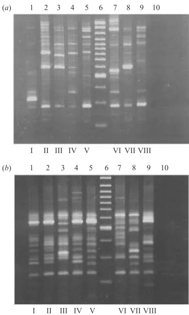
Fig. 1. Summary of DNA fingerprints from C. difficile
isolates from both patients and the environment on wards A and B using RAPD–PCR technique (a) and RS–PCR technique (b). Prints from ethidium bromide stained 2 % MetaphorTM agarose gels are shown. Assigned genotypes are indicated at bottom of lanes. On both gels : lane 6 : 100 bp ladder ; lane 10 : negative PCR control.
successfully separated 108 strains into four distinct types (Fig. 1). Strains typed by RS–PCR were considered distinguishable if "2 inter-strain band differences were present [15]. This method was found to have an equivalent discriminatory power to RAPD, separating the 108 strains into the same four groups. On ward A, only two genotypes were found (I and II). Apart from one isolate (genotype II), all strains originating from ward A were genotype I (Table 1). On ward B, only three genotypes were discovered (I, III and IV), and genotype I represented 87 % of all patient isolates examined.
C. difficilestrains recovered from ward environments
346 W. N. Fawley and M. H. Wilcox
Table 1. C. difficilegenotypes isolated from patients and their enironment on study wards A and B
Clinical isolates
Environmental isolates
Ward Genotype (number of isolates) Number of isolates Environmental site
A I 47}48 40}43 Endemic
II 1}48 NI NA†
V NI* 1}43 Commodes
VI NI 1}43 Radiators
VII NI 1}43 Toilet floor
B I 52}60 55}60 Endemic
III 1}60 4}60 Toilet floor}air vents
IV 7}60 NI NA
VIII NI 1}60 Sluice floor
* NI, Not isolated ;†NA, not applicable.
16
14
12
10
8
6
4
2
0
Number of occasions site found culture positi
v
e
Ward floors Radiators Toilet floors Air vents Sluice floor Commodes
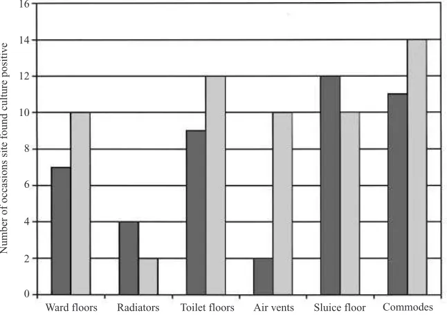
Fig. 2.Frequency ofC.difficileculture positive environmental sites on study wards A and B.+, ward A ;P, ward B.
comprised radiators (16 %), air vents (16 %), commodes (16 %) and floors (52 %) from wards, toilets and sluice rooms. Overall, 34 and 36 % of sites were C. difficile culture positive on wards A and B, respectively.C.difficilewas most frequently cultured from commodes and toilet}sluice room floors (Fig. 2). ln total, only 21 (6±8 %) sites were positive when swabs were cultured on modified CCEY medium alone, the majority (85.7 %) of which yielded !5 bacterial colonies. When swabs were enriched by culture in Robertson’s cooked meat broth, total recovery was increased markedly (35±1 % of sites were found to be
C.difficile positive). Five strains were unrecoverable after freezer storage, leaving 103 environmental isolates available for DNA fingerprinting.
[image:5.612.144.467.256.484.2]con-Number of isolations
[image:6.612.71.296.55.346.2]Number of isolations
Fig. 3. Quarterly figures : CDI and environmental culture positivity for study wards A (a) and B (b). +, Patient isolates ;E, environmental isolates.
6/10 13/10 27/10 5/12 4/1 31/1 9/4
Bay floors
Toilet floors
Sluice floors
Commodes
Radiators
Air vents
+
+ +
+ +
+
+
(a)
6/10 13/10 27/10 5/12 4/1 31/1 9/4 Bay floors
Toilet floors
Sluice floors
Commodes
Radiators
Air vents
+
+ +
+ +
+ +
+ +
+
+ +
+
(b)
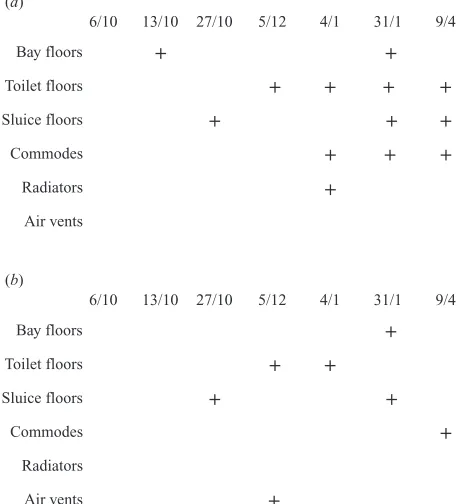
Fig. 4.Recovery ofC.difficilefrom environmental sites on wards A (a) and B (b) during months 0–6. indicates at least one of the samples from site type was C. difficile
positive.
tamination. Genotype IV was the only example of a
C. difficile type that was recovered from the en-vironment after, but not before, isolation from six symptomatic patients.
CDI and ward environmental contamination
Quarterly figures for CDI and environmental culture positivity for wards A and B are shown in Figure 3. CDI incidence data correlated significantly with the prevalence of environmental C. difficile on ward B (r¯0±76, P!0±05) but not on ward A (r¯0±26 ;
P"0±05). Figure 4 shows the month-by-month increases in the numbers of environmental site types on the study wards that were C. difficile culture positive during the first 6 months of the study. C.
difficile was not isolated from the environment of either ward before opening, but was found from both wards within 1–3 weeks of opening.
D I S C U S S I O N
We are aware of only one previous study [9] that systematically examined the relationship between environmental contamination and CDI. Samore and colleagues documented marked environmental con-tamination and transmission to personnel and patient contacts by an endemic C. difficile strain over a 6-month period [9]. Several studies have documented the presence ofC.difficilespores in areas occupied by infected patients, but these have been over short time periods, and evidence of bacterial acquisition from exposure to contaminated environmental sources is scarce. One study concluded that disparate strains responsible for causing disease were more likely to have originated from an environmental source than from cross-infection from patient to patient [16]. Elsewhere, a cluster of CDls on a surgical unit was associated with an identical strain found in the environment [17]. Conversely, Cohenet al. found no evidence to suggest environmental acquisition of C.
difficile[18].
[image:6.612.68.298.408.660.2]en-348 W. N. Fawley and M. H. Wilcox
vironmental contamination levels over the 22-month testing period (r¯0±76,P!0±05). It is interesting to speculate why such a marked difference in strength of correlation was found between wards A and B. We are unaware of marked differences, but several factors not specifically addressed in this study, including anti-biotic prescribing practice, patient type and cleaning efficiency, may have influenced either the incidence of CDI or environmental contamination. A comparative trial of CDI risk associated with treatment with cefotaxime or piperacillin-tazobactam occurred on these two wards during part of the present study period [11]. However, this was a ward crossover trial, and thus effects of antibiotic use on CDI or en-vironmental contamination should in theory have been balanced.
Endemic CDI can be controlled by reducing the use of high risk agents [19–22], However, changes in incidence of CDI following altered antibiotic pre-scribing practice have generally been seen in non-comparative settings, and}or have not been tested by reintroduction of the suspected antibiotics. Recently, Stone and Kibbler [23] reported that a fall in incidence ofC.difficilediarrhoea in an elderly medicine unit was associated with a reduction in cephalosporin use. Feedback to clinicians on C. difficile rates and antibiotic prescribing levels was relaxed leading to an increase in diarrhoea. Following re-enforcement of an antibiotic policy to limit cephalosporin prescribing,C.
difficilediarrhoea rates reduced again. The purpose of the present study was not to examine the effects of intervention in environmental cleaning, but rather to determine the baseline relationship betweenC.difficile
contamination levels and CDI. Conventional reaction to an outbreak of CDI includes enhanced environ-mental cleaning, and we have reported the success of this approach [24].
Attempts to determine whether infected patients or contaminated environments are the prime source for cross-infection byC.difficilehad limited success. Over 90 % of C. difficilestrains isolated in the study were the UK epidemic clone (genotype I). This makes assessment of the interplay of distinct C. difficile
strains between the patient and the hospital ward difficult. In addition, the undulating frequencies of both infection and ward contamination remained in phase and therefore it was not possible to determine whether patient CDIs preceded a rise in ward contamination or vice versa. We have shown since this study was performed that environmental recovery of
C. difficile is markedly increased if lysozyme is
incorporated into the selective agar used for sample culture [25]. We believe that use of this improved approach would not have substantially altered the findings of this study, as we have isolated similar limited C. difficile types seen here using lysozyme-containing media (unpublished data). The two DNA fingerprinting techniques that we employed had similar discriminatory power. RAPD typing results correlated fully with those of RS–PCR typing in this setting, although despite fingerprinting over 200 C.
difficile isolates, we identified only six distinct types. As expected, RS–PCR technique was found to be slightly more reproducible than RAPD, given the high susceptibility of the latter even to very small variations in testing conditions. DNA fingerprinting by pulsed-field gel electrophoresis was also examined initially but was abandoned as it was ineffective in producing a pulsotype for the endemic clone. DNA from these strains was repeatedly degraded, presumably by endonucleases (data not shown), as described by several other groups [9, 26, 27].
more when exposed to non-chlorine based cleaning agents compared with chlorine-containing disin-fectants [29]. Therefore, use of some detergents}
disinfectants may actually be exacerbating environ-mental persistence of the UK epidemic C. difficile
strain. It has been estimated that each CDI case costs more than £4000 [30]. This high figure could be used to justify expenditure on improved standards of hospital cleanliness [31].
The Anaerobic Reference Unit of the Public Health Laboratory Service has confirmed the endemic strain identified here as PCR ribotype 1 (J. Brazier, personal communication). This strain is known to be endemic in 33 of 58 hospitals in England and Wales [32]. It is interesting to speculate on the high prevalence of this strain in hospitalized patients [10]. Results from the present study suggest a relationship may exist between CDI incidence and the level ofC.
difficilespore contamination in the hospital environ-ment. Thus, more cases of endemic infection result in the release of more spores into the environment, creating the potential for more cases of endemic infection. However, although severalC.difficilestrains were found in the study, only genotype I was predominant. This implies that strain-specific charac-teristics have contributed to persistence. This is the first comprehensive insight into the molecular epi-demiology of endemic C. difficile, particularly that associated with a recently recognized epidemic strain, Understanding the epidemiology and virulence of prevalent strains is important if CDI is to be successfully controlled.
R E F E R E N C E S
1. Wilcox MH. Cleaning upClostridium difficileinfection. Lancet 1996 ;348: 767–8.
2. Riley TV. Clostridium difficile: a pathogen of the nineties. Eur J Clin Microbiol Infect Dis 1998 ; 17: 137–41.
3. Wilcox MH, Smyth ET. Incidence and impact of
Clostridium difficileinfection in the UK, 1993–1996. J Hosp Infect 1998 ;39: 181–7.
4. Cartmill TD, Panigrahi H, Worsley MA, McCann DC, Nice CN, Keith E. Management and control of a large outbreak of diarrhoea due to Clostridium difficile. J Hosp Infect 1994 ;27: 1–15.
5. Anand A, Bashey B, Mir T, Glatt AE. Epidemiology, clinical manifestations, and outcome of Clostridium difficile diarrhoea. Am J Gastroenterol 1994 ; 89: 519–23.
6. Samore MH. Epidemiology of nosocomialClostridium difficile. J Hosp Infect 1999 ;43(Suppl) : S183–90. 7. Kaatz GW, Gitlin SD, Schaberg DR, Wilson KH,
Kauffman CA, Seo SM. Acquisition of Clostridium difficilefrom the hospital environment. Am J Epidemiol 1988 ;127: 1289–93.
8. Struelens MJ, Maas A, Nonhoff C, Deplano A, Rost F, Serruys E, Delmee M. Control of nosocomial trans-mission ofClostridium difficilebased on sporadic case surveillance. Am J Med 1991 ;91: 138S–44S.
9. Samore MH, Venkataraman L, DeGirolami PC, Arbeit RD, Karchmer AW. Clinical and molecular epidemi-ology of sporadic and clustered cases of nosocomial
Clostridium difficile diarrhea. Am J Med 1996 ; 100: 32–40.
10. Stubbs SLJ, Brazier JS, O’Neill GL, Duerden BI. PCR targeted to the 16S-23S rRNA gene intergenic spacer region of Clostridium difficile and construction of a library consisting of 116 different PCR ribotypes. J Clin Microbiol 1999 ;37: 461–3.
11. Settle CD, Wilcox MH, Fawley WN, Corrado OJ, Hawkey PM. Prospective study of the risk of
Clostridium difficile diarrhoea in elderly patients fol-lowing treatment with cefotaxime or piperacillin-tazobactam. Aliment Pharmacol Ther 1998 ; 12: 1217–23.
12. Wilcox MH, Fawley WN, Settle CD, Davidson A. Recurrence of symptoms in Clostridium difficile in-fection – relapse or reinin-fection ? J Hosp Infect 1998 ;38: 93–100.
13. Killgore GE, Kato H. Use of arbitrary primer PCR to typeClostridium difficileand comparison of results with those by immunoblot typing. J Clin Microbiol 1994 ; 32: 1591–3.
14. Jensen MA, Webster JA, Straus N. Rapid Identification of bacteria on the basis of polymerase chain reaction– amplified ribosomal DNA spacer polymorphisms. Appl Environ Microbiol 1993 ;59: 945–52.
15. Cartwright CP, Stock F, Williams EC, Beekman SE, Gill VJ. PCR amplification of rRNA intergenic spacer regions as a method for epidemiological typing of
Clostridium difficile. J Clin Microbiol 1995 ;33: 184–7. 16. Samore MH, Bettin KM, DeGirolami PC, Clabots CR, Gerding DN, Karchmer AW. Wide diversity of
Clostridium difficiletypes at a tertiary referral hospital. J infect Dis 1994 ;170: 615–21.
17. Testore GP, Pantosti A, Cerquetti M, Babudieri S, Panichi G, Gianfrilli PM. Evidence for cross-infection in an outbreak of Clostridium difficile-associated di-arrhoea in a surgical unit. J Med Microbiol 1988 ;26: 125–8.
18. Cohen SH, Tang YJ, Muenzer J, Gumerlock PH, Silva J Jr. Isolation of various genotypes of Clostridium difficile from patients and the environment in an oncology ward. Clin Infect Dis 1997 ;24: 889–93. 19. Impallomeni M, Galletly MP, Wort SJ, Starr JM,
Rogers TR. Increased risk of diarrhoea caused by
Clostridium difficile in elderly patients receiving cefo-taxime. BMJ 1995 ;311: 1345–6.
350 W. N. Fawley and M. H. Wilcox
21. Jones EM, Kirkpatrick BL, Feeny R, Reeves DS, MacGowan AP. Hospital-acquiredClostridium difficile
diarrhoea. Lancet 1997 ;349: 1176–7.
22. Ludlam H, Brown N, Sule O, Redpath C, Coni N, Owen G. An antibiotic policy is associated with reduced risk of Clostridium difficile-associated diarrhoea. Age Ageing 1999 ;28: 578–80.
23. Stone S, Kibbler C, How A, Balestrini A. Feedback is necessary in strategies to reduce hospital acquired infection. BMJ 2000 ;321: 302.
24. Wilcox MH, Settle CD, Fawley W, Parnell P, Porter C, Keer V, Hawkey P. Isolation of patients with
Clostridium difficileinfection. J Hosp Infect 1997 ;37: 331–4.
25. Wilcox MH, Fawley WN, Parnell P. Value of lysozyme agar incorporation and alkaline thioglycollate exposure for the environmental recovery ofClostridium difficile. J Hosp Infect 2000 ;44: 65–9.
26. Kato H, Kato N, Watanabe K, Ueno K, Ushijima H, Hashira S, Abe T. Application of typing by pulsed-field gel electrophoresis to the study ofClostridium difficilein
a neonatal intensive care unit. J Clin Microbiol 1994 ; 32: 2067–70.
27. Van Dijck P, Avesani V, Delmee M. Genotyping of outbreak-related and sporadic isolates of Clostridium difficile belonging to serogroup C. J Clin Microbiol 1996 ;34: 3049–55.
28. Department of Health}Public Health Laboratory Ser-vice Joint Working Group. Clostridium difficile in-fection. Prevention and management. Heywood, UK : BAPS Health Publicaitons, 1994.
29. Wilcox MH, Fawley WN. Hospital disinfectants and spore formation byClostridium difficile. Lancet 2000 ; 356: 1324.
30. Wilcox MH, Cunnliffe JG, Trundle C. Financial burden of hospital acquired Clostridium difficile infection. J Hosp Infect 1996 ;34: 23–30.
31. Standards for environmental cleanliness in hospitals. NHS Estates. London : HMSO ; 2000.