REVIEW
The impact of cryopreservation on bone
marrow-derived mesenchymal stem cells:
a systematic review
Soukaina Bahsoun
1, Karen Coopman
2and Elizabeth C. Akam
1*Abstract
Mesenchymal stem cells (MSCs) represent an invaluable asset for the field of cell therapy. Human Bone marrow-derived MSCs (hBM-MSCs) are one of the most commonly used cell types in clinical trials. They are currently being studied and tested for the treatment of a wide range of diseases and conditions. The future availability of MSCs thera-pies to the public will require a robust and reliable delivery process. Cryopreservation represents the gold standard in cell storage and transportation, but its effect on BM-MSCs is still not well established. A systematic review was conducted to evaluate the impact of cryopreservation on BM-MSCs and to attempt to uncover the reasons behind some of the controversial results reported in the literature. Forty-one in vitro studies were analysed, and their results organised according to the cell attributes they assess. It was concluded that cryopreservation does not affect BM-MSCs morphology, surface marker expression, differentiation or proliferation potential. However, mixed results exist regarding the effect on colony forming ability and the effects on viability, attachment and migration, genomic stabil-ity and paracrine function are undefined mainly due to the huge variabilities governing the cryopreservation process as a whole and to the lack of standardised assays.
Keywords: Bone-marrow derived mesenchymal stem cells, Cell therapy, Cryopreservation, Mesenchymal stem cells,
Tissue culture, Systematic review
© The Author(s) 2019. This article is distributed under the terms of the Creative Commons Attribution 4.0 International License (http://creat iveco mmons .org/licen ses/by/4.0/), which permits unrestricted use, distribution, and reproduction in any medium, provided you give appropriate credit to the original author(s) and the source, provide a link to the Creative Commons license, and indicate if changes were made. The Creative Commons Public Domain Dedication waiver (http://creativecommons.org/ publicdomain/zero/1.0/) applies to the data made available in this article, unless otherwise stated.
Background
Bone marrow non-hematopoietic stem cells represent a fraction of the bone marrow cell population. They may arise from the constituents of the bone marrow struc-ture and they can differentiate into mesenchymal tissues such as adipose, cartilage and bone. Bone marrow non-hematopoietic stem cells were first mentioned by Julius Cohnheim in 1867 and later cultured and characterized by Freidenstein et al. in the 1970s [1–4]. Friedenstein demonstrated that bone marrow non-hematopoietic stem can be selected by adherence to culture flask and exhibit the following characteristics: fibroblast morphol-ogy, colony-forming ability and in vitro proliferation and differentiation potentials [5]; all of which were indicative
of ‘stemness’ properties [6]. With that said, it must be noted that within the scientific community, there is still an ongoing discussion about the true nature of these cells. Two names propagated for these cells “Stromal Stem Cells” [7] and “Mesenchymal Stem Cells” [8, 9].
The then newly discovered source of stem cells has attracted great interest in medical research. In addition to the characteristics listed above, isolating mesenchy-mal stem cells from bone marrow was surrounded with minimal ethical issues and could substitute embryonic
stem cells [6]. Therefore, hBM-MSCs became the
sub-ject of intense research and in 1995 the first autologous intravenous infusion of these cells in cancer patients
was performed [10]. Later, MSCs have been shown to
have widespread immunomodulatory effects [11] as well as an angiogenic induction ability [12]. Taken together these characteristics enlarged the scope of application of hMSC-based therapies. As of April 2019, a search on
Open Access
*Correspondence: e.c.akam@lboro.ac.uk
1 School of Sport, Exercise and Health Sciences, Loughborough University,
Loughborough, Leicestershire LE11 3TU, UK
the U.S. National Library of Medicine (ClinicalTrials.gov) using the term ‘bone marrow mesenchymal stem cells’ retrieved 368 clinical trials aiming to treat conditions like stroke, graft versus host disease, osteoarthritis, crohn’s disease, ischemic heart disease and multiple sclerosis.
The future availability of cell therapies to the public will be dependent on easy and quick logistics as well as robust and reliable delivery process. Abazari et al. [13] suggested that if cell therapies “cannot be delivered clinically and logistically then their benefit is irrelevant”. Cryopreserva-tion remains the cell therapy industry “standard” for bio-preservation [14] as well as the primary option of storage for hMSC-based products [15]. In fact, cryostorage has evolved from being a marginal process in the cell therapy manufacturing process to become a tool widening the availability of stem cell therapy in particular and regen-erative medicine in general. However, despite its evolving role, cryobiology is lagging behind the speed at which the cell therapy industry is growing.
Cryopreservation is particularly crucial for a successful cell therapy for various reasons. It facilitates cell trans-port, it enables the generation of cell banks with indefi-nite shelf-life thus ensuring off-the-shelf steady supply, access and availability and it gives time for quality control testing and in vitro assays [14, 16, 17]. In addition, cry-ostoring therapeutic doses of cells in hospitals and clin-ics could make cell therapy a treatment choice for many diseases and conditions including acute conditions [18]. Furthermore, cryopreserved cells are ideal for sequen-tial treatments such as the case of chronic heart failure or ischemic heart disease to ensure the consistency of the treatment [19]. Banking cells is also an appropriate option from an economical and a regulatory aspect [20]. The logistics of administration of MSC in many immu-notherapy trials were simply described as cryopreserv-ing cells, thawcryopreserv-ing them when needed and administercryopreserv-ing them within a couple of hours. This scenario would only be feasible if thawed cells preserved their viability, safety and potency [20].
Cryopreservation of cells is associated with several injuries; physical and molecular. A controversy still exists about the efficacy of fresh cells versus cryopreserved and whether viability implies functionality [21]. In early MSC-based clinical trials, using cryopreserved cells was hypothesised to be the source of failure [21]. In addition, the variability in the outcome of MSC-based clinical tri-als was proposed to mainly be due to the functional alter-ations that the freeze–thaw process provokes in MSCs rather than the freezing method itself [17].
Human Bone marrow-derived MSCs (hBM-MSCs) are the most commonly used source of MSCs in clini-cal trials [22] and have been deployed across 17
Euro-pean centres manufacturing MSCs [23]. The effects of
cryopreservation on this type of cells are not well defined. The aim of this review is to assess whether rigorous data exist regarding the impact of the freeze–thawing process on BM-MSCs phenotypic and functional traits. To our knowledge, this is the first review to factor numerous aspects of the freezing process (freezing solution compo-sition, the freezing protocol, the duration of storage, the concentration of cells at freezing, the passage number at freezing as well as the thawing method) in one analysis, for studies conducted over about 20 years. Such detailed analysis may allow firm conclusions to be drawn regard-ing BM-MSCs performance after the freeze-thawregard-ing process as well as help uncover possible reasons behind some of the controversial existing results and highlight areas which require further investigation.
Methods
The inclusion criteria for this review were: Articles or conference papers assessing the impact of cryopreserva-tion by slow freezing on BM-MSCs in suspension. There was no restriction on the species from which cells were derived. Studies where bone marrow itself was frozen, where freezing of BM-MSCs was done by vitrification or using a 3D structure and where cryopreservation impact was only assessed in vivo, were excluded. A systematic literature search was conducted using PubMed, Science direct and Google Scholar (last search performed April 2019). Two combinations of search terms were used ‘cryopreservation mesenchymal’ and ‘freezing mesenchy-mal’. The output of each search was first scanned for the relevance of title. Articles were excluded if the topic is unrelated or when an eligibility criterion is not met. The retained articles were then screened for the relevance of abstracts (and in few cases materials and methods) and retained when meeting all the eligibility criteria (Fig. 1). From the 41 retained studies, information regarding the freezing solution composition, the freezing protocol, the duration of storage, the concentration of cells at freez-ing, the passage number at freezing as well as the thawing method was extracted and tabulated. Next, studies were grouped in tables according to the “hMSC checklist” proposed in [24]. Cell surface marker expression, differ-entiation potential, proliferation and growth, attachment and migration potential, genomic stability and paracrine function were examined. In addition, post-thaw viability and morphology information was also collated because they are primary evaluators of cryopreservation.
Results
Species, freezing and thawing methods
(5 studies), monkey (3 studies), dog (3 studies), horse (2 studies), pig (2 studies), minipig (1 study), mouse (1 study), calf (1 study) and sheep (1 study) (Fig. 2).
Across the retained studies, various freezing media for-mulations were used. 20% of studies (17% human) used commercially available freezing solution such as CELL-BANKER and CRYOSTOR while the rest used “in-lab” homemade formulations. 66% of studies (41% human) used or tested various amounts of serum in the freezing media with the serum principally being animal-derived, 20% of studies (12% human) froze cells in serum-free media while 5% used freezing media containing plasma or human platelet lysates (all of which are human studies). For 17% of the studies, not enough information was included about FBS content and/or about the composition of the freezing medium (12% human). Of the serum-free studies, one study assessed the efficiency of Sericin as a substitute
Search term: ‘Cryopreservation and Mesenchymal’ Search term: ‘Freezing and Mesenchymal’
36 retained by abstract 65
retained by title 695
studies retrieved
Step1: PubMed Step 2: PubMed
1 retained by abstract 4 retained
by title Duplicates
removed 518
studies retrieved
Step 4: Science Direct Step 3: Science Direct
0 retained by abstract 13
retained by title Duplicates
removed 1939
studies retrieved
0 retained by abstract 4 retained
by title Duplicates
removed 2222
studies (research
articles) retrieved
4 additional articles found via Google Scholar and through checking the references of each retained article
When screening titles and abstracts the following criteria were checked and studies included if:
• Topic: A cryopreservation study • Aim: To assess cryopreservation impact • Cell source: Bone marrow
• Research article or conference paper
When screening titles and abstracts the following criteria were checked and studies excluded if:
• Abstract, letter, review or article not in English • Freezing of bone marrow itself rather than BM-MSCs • Freezing done by vitrification
• Freezing on a 3D structure
• Freezing impact assessed in-vivo only
Inclusion criteria Exclusion criteria
Fig. 1 Schematic representation of the Bone-marrow derived mesenchymal stem cell cryopreservation search strategy. Diagram of the current
systematic search analysis. Studies of bone-marrow derived mesenchymal stem cells aligned to cryopreservation and/or freezing were identified using a combination of two search terms ‘cryopreservation mesenchymal’ and ‘freezing mesenchymal’ using PubMed, Science direct and Google scholar. The output of each search was scanned at the title level, then at the abstract level and articles were retained when meeting eligibility criteria, both inclusion and exclusion (see boxes titled inclusion criteria and exclusion criteria in this figure). In specific, for the term ‘cryopreservation and mesenchymal’ in PubMed, 695 studies were retrieved. By checking the titles against the eligibility criteria, only 65 studies were retained. The abstracts of these 65 articles were then read and checked against the eligibility criteria and only 36 of the 65 articles were retained. For the subsequent searches, these steps of retaining and eliminating articles were followed but preceded by eliminating duplicates i.e. articles which appeared in previous searches
26
5 3 3 2
2
Human Rat Dog Monkey Pig Horse Mouse Calf Sheep Minipig
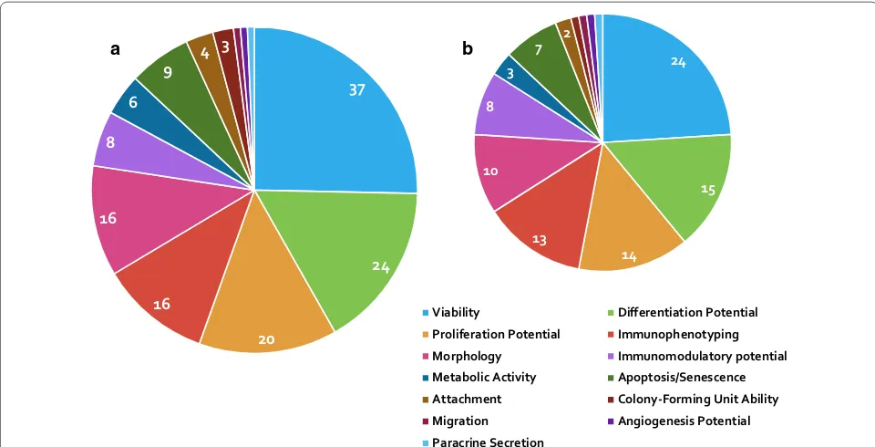
Fig. 2 Pie chart showing the number of studies per species. The
[image:3.595.61.538.90.363.2] [image:3.595.308.537.488.597.2]to FBS [25]. Across all of the studies, 13 assessed the freez-ing in xeno-free media. More than 90% of studies used dimethylsulfoxide (DMSO) at a concentration ranging from 1 to 20% with 10% being the most commonly used. Carboxylated poly-l-lysine (COOH-PLL) was investigated as a cryoprotectant to replace DMSO [26] and hydroxy-ethyl starch was added to freezing solution as a strategy to reduce the percentage of DMSO [27]. Two studies tested various freezing solutions containing polyethylene glycol (PEG), trehalose and 1,2 propanediol in order to develop a well-defined, serum-free and reduced-DMSO freez-ing solution [28, 29]. Only two studies utilized strategies to prevent post-thaw apoptosis through the addition of Rho-associated kinase inhibitor [30] and Caspase inhibitor z-VAD-fmk [31] in the freezing media.
Concerning freezing protocols, two procedures prevail. The first involves incubating the cells at a freezing rate of − 1 °C/min in a − 80 °C freezer for several hours (up to 24 h) then moving the cells to liquid nitrogen (LN2).
The second is based on a two to seven-step sequential freezing process using a programmable freezing device to freeze the cells prior to − 150 °C freezer or LN2
stor-age. Four studies reported whether cells were stored in liquid phase [32] or vapour phase [30, 33, 34] of LN2. Five
studies stored the cells at − 80 °C [26, 35–38] and 1 study stored the cells at − 70 °C [39].
Seven studies (3 human and 4 animal) did not specify the passage number at which cells were frozen. Six studies (3 human and 3 animal) used cells at passage 1, one study (monkey) used cells at passage 9 and the rest (20 human and 6 animal) used cells at passages ranging from 1 to 6. The concentration of cells at freezing was very variable ranging from 1 * 105 to 1 * 107 cells/mL with 1 * 106 cells/mL being the
most frequently used (17 studies; 9 human and 8 animal). There was only one study in which human-derived cells were frozen in cryopreservation bags at a concentration of 1.8 * 108 [40]. There was a huge variation regarding the
dura-tion of storage of cells in the frozen state; the shortest period being 1 h [30] and the longest 10 or more years [37].
Seven studies (human) did not include information about their thawing protocols. Two studies (1 human and 1 animal) just mentioned ‘quickly thawed’ [20, 39], one in α-MEM [41], one at room temperature [37] and one in a 37 °C incubator [42]. For all the rest (16 human and 13 animal), there was some consistency; cells were typically thawed at 37 °C, most likely in a water bath with or with-out gentle agitation for 1–4 min.
Post‑thaw assessment
Presented in Fig. 3 are how many of the forty-one
retained studies assessed different cellular attributes,
37
24
20 16
16 8
6
9 4 3 24
15
14 13
10 8
3
7 2
Viability Differentiation Potential
Proliferation Potential Immunophenotyping
Morphology Immunomodulatory potential
Metabolic Activity Apoptosis/Senescence Attachment Colony-Forming Unit Ability
Migration Angiogenesis Potential
Paracrine Secretion b
a
Fig. 3 a Pie chart showing the proportion of the retained studies assessing different cellular attributes in all species. b Pie chart illustrating the
[image:4.595.57.537.421.666.2]it shows that viability and differentiation potential are the only attributes assessed in over half of the retained studies.
Viability
Post-thaw viability was the most assessed cell attribute (37 studies). Table 1 lists the studies which assessed via-bility immediately post-thaw (37 studies) or after a period of post-thaw culture, which was monitored in five stud-ies (post-thaw time point from 4 h to 3 passages). Three main methods for viability assessment were used; trypan blue exclusion, flow cytometry and fluorescent micros-copy. The immediately post-thaw viability varied from about 50% to 100% which is noteworthy. Sixteen studies reported no change in viability immediately after thaw-ing while 10 studies reported significantly lower viabil-ity. In some studies where a new freezing formulation was tested, viability was compared to 10% DMSO which is still considered the gold standard for freezing BM-MSCs. The timing of viability measure is crucial due to the induction of apoptotic events some time post-thaw [43]. As mentioned above only five studies assessed the long-term effect (up to 3 passages post-thaw of freez-ing) with two reporting lower viability and three report-ing no effect. It was also noted that variability in the level of viable cells within the same study differed when using different methods of measurement such as trypan blue exclusion compared to flow cytometry [44, 45].
Morphology
Table 2 summarises the studies which assessed post-thaw cell morphology. This attribute was mainly assessed using microscopy. Irrespective of all the variables considered in this data analysis and the time post-thaw at which cell morphology was checked, 13 of the 16 studies agree that cryopreservation itself has no effect on post-thaw cell morphology. The addition of Rho-associate kinase (ROCK) inhibitor Y-27632 was reported to give BM-MSCs a web-like appearance which indicated some neu-ronal differentiation [30]. In addition, several cell shapes were observed at day 2 and day 5 post-thaw [46] and cell shrinkage was detected using flow cytometry [38].
Immunophenotyping
Marker expression is one of the International Society for Cellular Therapy (ISCT) criteria for defining MSCs [47] so it is of real importance to check BM-MSCs pheno-type before freezing and/or after thawing. Table 3 lists the studies which assessed BM-MSCs marker expression post-thaw. Despite its importance, less than half of the 41 studies retained assessed post-thaw marker expres-sion retention and despite all the variables taken into consideration in this investigation, there was a consensus
regarding the methodology used (flow cytometry) and the results; cryopreservation does not affect BM-MSCs immunophenotype.
Differentiation potential
Tri-lineage differentiation (adipogenic, osteogenic and chondrogenic) is another criterion listed by the ISCT guide for defining MSCs [47]. Hence, more than half of the studies [24] assessed BM-MSCs post-thaw differen-tiation potential and these are listed in Table 4. Osteo-genesis is the most frequently assessed differentiation pathway (20 studies) qualitatively through Alizarin red staining and/or quantitatively through measurement of alkaline phosphatase activity. There was an agreement among 18 studies that cryopreservation did not affect BM-MSCs osteogeneic potential. One study reported lower osteogenesis [39] and one reported improved oste-ogenesis post-thaw [48]. Adipogenesis was next in terms of frequency of testing using Oil Red O staining as a qual-itative assessment and no effect of cryopreservation was observed in 12 studies. Only one study provided a quan-titative assessment of adipogenesis and it was the only one reporting lower differentiation level [49].
Chondrogenesis presented as the least studied tri-linage differentiation pathway. Only five studies differentiated thawed BM-MSCs into chondrocytes with a qualitative assessment made via Alcian Blue staining. It was concluded that cryopreserved cells did not lose the ability to chondro-genic differentiation (of note thawed BM-MSCs were also able to commit to neuronal and endothelial lineages).
Proliferation potential
Table 5 lists the 20 studies which examined post-thaw BM-MSCs proliferation potential. Various methods were used to determine proliferation rate such as popu-lation doublings and DNA quantification. The major-ity of results agree that cryopreservation does not affect post-thaw BM-MSCs proliferation potential, nonethe-less lower proliferation rate was obtained by two stud-ies [34, 39] and higher proliferation rate was obtained by one study [50]. Colony-forming unit ability, a traditional measure of BM-MSCs proliferation, was assessed by three studies with mixed results [25, 32, 51].
Metabolic activity
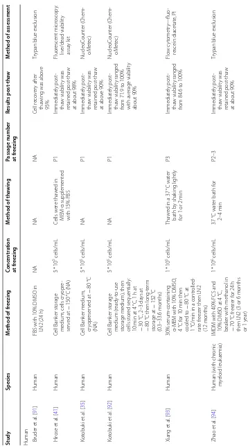
Table
1
Exp
erimen
tal studies assessing viabilit
y immedia tely p ost-tha w or af ter a p erio d of p ost-tha w cultur e Study Species M ethod of fr eezing Conc en tr ation at fr eezing M ethod of tha wing
Passage number at fr
eezing Results post ‑tha w M ethod of assessmen t
Human Bruder et
al
. [
91
]
Human
FBS with 10% DMSO in LN2 (24
h) NA NA NA Cell r eco ver y af ter tha
wing was abo
ve
95%
Tr
ypan blue ex
clusion H irose et al . [ 41 ] Human Cell Bank er st orage
medium, cells cr
yopr e-ser ved at − 150 °C (NA) 5 * 10
5 cells/mL
Cells w er e tha w ed in MEM -α supplement ed
with 15% FBS
P1 Immediat ely post -tha w viabilit y was retained post -tha w
at about 98%
Fluor escent micr oscop y: liv e/dead viabilit y assa y k it K ot obuk i et al . [ 35 ] Human Cell Bank er medium, cr yopr eser ved at − 80 °C (NA) 5 * 10
5 cells/mL
NA P1 Immediat ely post -tha w viabilit y was retained post -tha w at abo ve 90% NucleoC ount er ( Chem -oM et ec) K ot obuk i et al . [ 92 ] Human Cell Bank er st orage medium (r eady-t o-use st
orage medium), then
cells st
or
ed sequentially
:
10
min at 4
°C, 1 h at − 30 °C, 2–3 da ys at − 80
°C then long-t
er m st orage at − 152 °C (0.3–33.6 months) 5 * 10
5 cells/mL
NA P1 Immediat ely post -tha w viabilit y ranged
from 71.9 t
o 100% with a verage viabilit y about 90% NucleoC ount er ( Chem -oM et ec) Xiang et al . [ 93 ] Human 30% serum-containing
α-MEM with 10% DMSO
, 4 °C f or 10 min then cooled t o − 80 °C at 1
°C/min in a contr
olled-rat
e fr
eez
er then LN2
(12
months)
1
* 10
5 cells/mL
Tha
w
ed in a 37
°C wat
er
bath b
y shak
ing lightly
for 1 or 2
min P3 Immediat ely post -tha w viabilit y ranged
from 84.6 t
o 100% Flo w c yt ometr y—fluo -rescein diacetat e, PI Zhao et al . [ 94 ]
Human (with chr
onic
m
yeloid leuk
aemia)
IMDM with 40% FCS and 10% DMSO at 4
°C,
beak
er with methanol in
− 70 °C fr eez er f or 24 h
then LN2 (3 or 6
months or 1 y ear) 1 * 10
6 cells/mL
37
°C wat
er bath f
or 2–4 min P2–3 Immediat ely post -tha w viabilit y was retained post -tha w
at about 90%
Tr
ypan blue ex
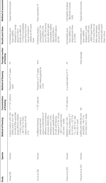
[image:6.595.104.462.82.728.2]Table
1
(c
on
tinued)
Study
Species
M
ethod of fr
eezing
Conc
en
tr
ation
at fr
eezing
M
ethod of tha
wing
Passage number at fr
eezing
Results post
‑tha
w
M
ethod of assessmen
t
Heng [
30
]
Human
Cultur
e medium with 10%
DMSO and 0, 10 or 100 micr
oM of R
ho
-associat
e
kinase (R
OCK
) inhibit
or
Y-27632, cooling t
o
−
80
°C f
or 2
h, then
vapour phase of LN2 (1 h)
1.17
*
10
5 cells/mL
Tha
w
ed in a 37
°C wat
er
bath
P5
Immediat
ely post
-tha
w viabilit
y
dr
opped t
o a range
about 91.3% t
o
89.4%; No eff
ec
t of
Y-27632 immediat
ely
post
-tha
w but ther
e
was an incr
ease in
viabilit
y at 24
h post
-tha
w
Tr
ypan blue ex
clusion
Liu et
al
. [
28
]
Human
13 diff
er
ent fr
eezing
media t
est
ed with
var
ious combinations of
diff
er
ent concentrations
of serum, DMSO
, PEG,
tr
ehalose and 1.2-P
ro
-panediol
, equilibration
of cells with fr
eezing
media at 4
°C f
or 10
min,
−
80
°C o
ver
night then
LN2 (min. 1
w
eek)
1
* 10
6 cells/mL
Tha
w
ed in a 37
°C wat
er
bath, shak
ing gently f
or
2
min
N/A
A fr
eezing solution composed of 7.5% c(v/v) DMSO
, 2.5%
(w/v) PEG, 2% bo
vine
serum albumin ga
ve
comparable viabilit
y
(about 82.9%) t
o
10% DMSO (about 82.7%)
Flo
w c
yt
ometr
y–PI
D
oan et
al
. [
95
]
Human
DMEM/F12 with 10% DMSO
, incubation, 4
°C
for 10
min,
−
20
°C f
or
1
h,
−
80
°C f
or 1
da
y
then LN2 (1
y
ear)
1
* 10
6 cells/mL
In a wat
er bath at 37
°C
P3
Immediat
ely post
-tha
w viabilit
y was
retained post
-tha
w
at 72.95%
Cell V
iabilit
y
Analyz
er
(Beck
man C
ount
er
,
USA)
F
rançois et
al
. [
45
]
Human
α-MEM with 30% FBS and 5% DMSO
,
−
80
°C f
or
24
h then LN2 (1
w
eek)
NA
NA
Ear
ly passage
Immediat
ely post
-tha
w viabilit
y
dr
opped t
o
≤
60%
(Annexin V/PI)
and
>
80% (
Tr
ypan blue);
A
t 4
h post
-tha
w via
-bilit
y was bet
w
een
44 and 61%; viabilit
y
incr
eased af
ter post
-tha
w cultur
e
Tr
ypan blue ex
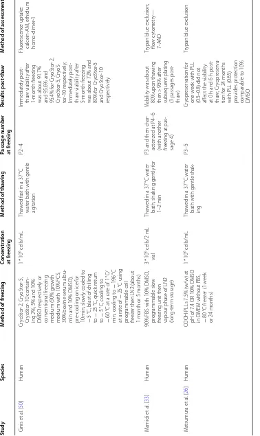
[image:7.595.107.476.93.727.2]Table 1 (c on tinued) Study Species M ethod of fr eezing Conc en tr ation at fr eezing M ethod of tha wing
Passage number at fr
eezing Results post ‑tha w M ethod of assessmen t Ginis et al . [ 50 ] Human Cr yoSt or -2, Cr yoSt or -5, Cr yoSt or -10 contain
-ing 2%, 5% and 10% DMSO r
espec tiv ely or con ventional fr eezing
medium (90% g
ro
wth
medium with 10% FCS, 30% bo
vine serum albu
-min and 10% DMSO), pr
e-cooling on ice f
or
10
min, slo
wly cooled t
o
−
5
°C, blast of chilling
to
−
25
°C, quick r
etur
n
to
−
5
°C, cooling t
o
−
60
°C at a rat
e of 1
°C/
min, cooling t
o
−
196
°C
at a rat
e of − 25 °C using pr og rammable cell freez
er then LN2 (about
1
month or 5
months)
1
* 10
6 cells/mL
Tha
w
ed fast in a 37
°C
wat
er bath with gentle
ag itation P2–4 Immediat ely post -tha w viabilit y af ter 1-month fr eezing
was about 91.7% and 95.6% and 95.4% f
or Cr yoSt or -2, Cr yoSt or -5, Cr yoS -tor -10 r espec tiv ely ; Immediat ely post -tha w viabilit y af ter 5-month fr eezing
was about 72% and 80% f
or Cr yoSt or -5 and Cr yoSt or -10 respec tiv ely Fluor escence uptak e: calcein-A M, ethidium homo -dimer -1 M amidi et al . [ 33 ] Human
90% FBS with 10% DMSO
,
pr
og
rammable slo
w
freezing unit then vapour phase of LN2 (long-t
er
m st
orage)
3
* 10
6 cells/2
mL
vial
Tha
w
ed in a 37
°C wat
er
bath, shak
ing gently f
or
1–2
min
P3 and then char
-ac
ter
iz
ed at P4–6
(with another freezing at pas
-sage 4)
Viabilit
y was about
80% upon tha
wing
then >
95% af
ter
subsequent plating (3 passages post
-tha
w)
Tr
ypan blue ex
clusion; flo w c yt ometr y— 7-AAD M atsumura et al . [ 26 ] Human
COOH-PLLs 7.5% (w/w) at pH of 7.4 OR 10% DMSO in DMEM without FBS, −
80 °C fr eez er (1 w eek or 24 months) 1 * 10
6 cells/mL
Tha
w
ed in a 37
°C wat
er
bath with gentle shak
-ing P3–5 Cr yopr eser vation f or one w
eek with PLL
(0.5–0.8) did not affec
t the viabilit
y
at 0
h and 6
h post -tha w ; Cr yopr eser va -tion f or 24 months
with PLL (0.65) provides pr
ot ec tion comparable t o 10% DMSO Tr
ypan blue ex
[image:8.595.104.482.90.725.2]Table
1
(c
on
tinued)
Study
Species
M
ethod of fr
eezing
Conc
en
tr
ation
at fr
eezing
M
ethod of tha
wing
Passage number at fr
eezing
Results post
‑tha
w
M
ethod of assessmen
t
Chinnadurai et
al
.
[
20
]
Human
Fr
eezing media,
−
80
°C
then LN2 (NA)
5
* 10
6 cells/mL
Quick
ly tha
w
ed (1–2
min)
P3–5
Immediat
ely post
-tha
w viabilit
y
dr
opped t
o about
87% (tr
ypan blue)
and 71.5% (flo
w
cyt
ometr
y)
Tr
ypan blue ex
clusion;
flo
w c
yt
ometr
y—
Annexin V
, PI
Holubo
va et
al
. [
69
]
Human
60% α-MEM medium with 30% pHPL and 10% DMSO
, pr
og
rammable
contr
olled rat
e fr
eez
er at
rat
e 1
°C/min t
o
−
80
°C
then LN2 (1,3,6,7 and 8 months)
1
* 10
6 cells/mL
NA
P3
Immediat
ely post
-tha
w viabilit
y is
70–90%
Flo
w c
yt
ometr
y—
7-AAD staining
M
oll et
al
. [
38
]
Human
4
°C human blood t
ype AB
plasma containing 10% DMSO
, fr
oz
en t
o
−
80
°C
using rat
e-contr
olled
cell fr
eezing de
vice (NA)
1–2
*
10
6 cells/mL
NA
P2–4
Viabilit
y r
educed t
w
o-fold b
y cr
yopr
eser
va
-tion when exposed to human serum (cell count and PI incor
poration)
Cell count
er and ana
-lyser syst
em (
CASY
-TT
); flo
w c
yt
ometr
y–
Annexin V
, PI
V
er
dano
va et
al
. [
25
]
Human
15 diff
er
ent fr
eezing
solutions containing var
ious concentrations
of DMSO (0, 1, 5, 10 and 100%) in the pr
esence or
absence of ser
icin at 1 or
5%, cooling t
o
−
80
°C
at a rat
e 1
°C/min in a
CoolC
ell container then
LN2 (72
h)
1.4
*
10
5 cells/mL
In a 37
°C wat
er bath as
quick
ly as possible
P1–3
H
ighest viabilit
y (24
h
post
-tha
w) was
obtained using standar
d fr
eezing
medium (10% DMSO and 25% FBS in cultur
e medium);
Viabilit
y of cells (24
h
post
-tha
w) fr
oz
en in
cultur
e medium
con
-taining 10% DMSO and 1% ser
icin was
not sig
nificantly dif
-fer
ent fr
om standar
d
freezing medium
Fluor
escent micr
os
-cop
y—D
[image:9.595.116.467.87.727.2]Table 1 (c on tinued) Study Species M ethod of fr eezing Conc en tr ation at fr eezing M ethod of tha wing
Passage number at fr
eezing Results post ‑tha w M ethod of assessmen t Al-Saqi et al . [ 66 ] Human
10% DMSO in M
esencult
-XF or STEM
-CELLBANKER
at 4
°C, cr
yo
vials on ice
then mo ved t o − 80 °C
with a cooling rat
e − 1 °C/min f or 24 h then
then LN2 (NA)
0.5–1
*
10
6 cells/mL
Tha
w
ed in a 37
°C wat
er
bath f
or 1 or 2
min P3 No diff er ence in viabilit y immediat ely post -tha w bet w een tw o fr eezing media;
CELLBANKER (85.6%) and 10% DMSO (86%); No sig
nificant
diff
er
ence in viabilit
y bet w een non-cr yopr eser ved and cr yopr eser ved using both media Fluor escence -based liv e/dead assa y imme -diat ely post -tha w ; flo w c yt ometr y—PI (t w
o passages post
-tha w) L uetzk endor f et al . [ 40 ] Human
5% human albumin and 10% DMSO
, aut
omatiz
ed
pr
ocess in a pr
og
ram
-mable fr
eez
er then LN2
(21–51 da ys) 1.8 * 10
8 in cr
yo -pr eser vation bags Tha w ed at P3–4 Immediat ely post -tha w viabilit y was
retained at >
90%
viabilit
y using both
methods f
or 4
donors out of 5
Tr
ypan blue ex
clu -sion; flo w c yt ometr y: 7-AAD P ollock et al . [ 67 ] Human 60% plasmalyt
e A, 20%
of 25% HAS and 20% DMSO (final concentra
-tion of DMSO was 10% by v
olume), contr
olled
rat
e fr
eez
er then LN2
(30–45 da ys) 1–10 * 10
6 cells/mL
Tha
w
ed quick
ly in a 37
°C P1–6 Immediat ely post -tha w viabilit y was
retained at >
80% f
or
almost all samples
Fluor escent micr os -cop y—A cr idine orange , PI Chinnadurai et al . [ 68 ] Human IFN ɣ
, caspase inhibit
or
Z-VAD
-F
MK or 3-meth
yl adenine pr e-licensing 48 h pr ior t o cr yopr eser
-vation, 5% human serum albumin, 5%, 20%, 40%, 90% hPL in aMEM with 10% DMSO OR Cr
yoSO -free DMSO -fr ee cr yo -pr eser vation medium, cooling rat e 1 °C/min then st ep -do wn fr eezing
using a 7-st
ep pr og ram in Cr yoM ed contr olled-rat e fr eez
er then LN2
(NA)
5–10
*
10
6 cells/mL
In a 37
°C wat
er bath f
or
1
min
P2–6
The addition of var
ious
concentrations of human plat
elet
lysat
e did not
sig nificantly enhance MSC r eco ver y and viabilit y; IFN ɣ p re -licensing pr ior t o cr yopr eser vation enhances tha w ed MSC sur vival Tr
ypan blue ex
[image:10.595.78.498.89.726.2]Table 1 (c on tinued) Study Species M ethod of fr eezing Conc en tr ation at fr eezing M ethod of tha wing
Passage number at fr
eezing Results post ‑tha w M ethod of assessmen t Gramlich et al . [ 18 ] Human Cr yoSt
or CS5 media,
−
80
°C f
or 90
min then
vapour phase of LN2 (7–30
da
ys)
1
* 10
6 cells per mL
In a 37
°C wat er bath P3–5 Immediat ely post -tha w viabilit y was
retained at >
95%
(viabilit
y only mar
-ginally r educed af ter tha wing)
TUNEL staining; F
luo -rescent micr oscop y— Hoechst, PI L echant eur et al . [ 34 ] Human 40% PBS +
40% of HSA
solution (20%)
+
20%
DMSO added under agitation at 4
°C, aut omat ed cr yofr eez er
with a 9-st
ep pr og ram to − 160
°C then vapour
phase of LN2 (NA)
2
* 10
6 cells/mL
Fr
eezing bag is pr
ot
ec
ted
in st
er
ile plastic bag and
tha
w
ed in a 37
°C wat
er
bath f
or a f
ew min P3 Immediat ely post -tha w viabilit y ranged
from about 50% t
o
90% with about 14% decr
ease in viabilit
y
Tr
ypan blue ex
clusion Y uan et al . [ 52 ] Human (BM -MSC eng i-neer ed t o expr ess TRAIL) 5% DMSO
, 30% FBS in
alpha-MEM OR human albumin with 0.5–20% DMSO
, isopr opanol freezing bo x o ver night in, − 80 °C fr eez er then LN2 (1–3 w eeks) 1 * 10
6 cells/mL or
5
* 10
6 cells/mL or
10
*
10
6 cells/mL
In a wat
er bath at 37
°C
with gentle shak
e f or 2 min P5 Sig nificantly r educed immediat ely post -tha w viabilit y
with 0% DMSO (5.16%); I
mmediat ely post -tha w viabilit y incr eased with incr eased DMSO%
in the fr
eezing; 15%
and 20% DMSO ga
ve
reduced viabilit
y
(about 70.6% and 64.1% r
espec -tiv ely) immediat ely post -tha w solution up t
o 10%; at 5%
DMSO same viabilit
y
obtained f
or diff
er
-ent cell conc-entra
-tions Flo w c yt ometr y— Annexin V , D API O
ther species Car
valho et al . [ 44 ] Rat
DMEM with 10% FBS and 5% DMSO
, cells incubat
e at r oom t emperatur e f or 15
min then vials cooled
at 3
°C/min, 5
°C/min,
10
°C/min dur
ing 15, 45,
10 min r espec tiv ely until − 80
°C using pr
og ram -mable fr eezing de vice
then LN2 (1
month)
1
* 10
7 cells/mL
Tha
w
ed in a 37
°C wat
er
bath with constant gentle shak
ing Fr oz en do wn af ter 4 w eeks in cultur e Immediat ely post -tha w viabilit y dr opped t o about 90.58% (tr ypan blue)
and 66.25% (flo
w
cyt
ometr
y)
Tr
ypan blue; flo
w
cyt
ometr
y—Annexin
[image:11.595.75.524.72.729.2]Table
1
(c
on
tinued)
Study
Species
M
ethod of fr
eezing
Conc
en
tr
ation
at fr
eezing
M
ethod of tha
wing
Passage number at fr
eezing
Results post
‑tha
w
M
ethod of assessmen
t
Liu et
al
. [
29
]
Rat, mouse and calf
14 diff
er
ent fr
eezing
solutions t
est
ed with
var
ious combinations of
diff
er
ent concentrations
of serum, DMSO
, PEG,
tr
ehalose and 1.2-P
ro
-panediol
, equilibration
for 15
min at 4
°C,
−
80
°C o
ver
night then
LN2 (min. 1
w
eek)
1
* 10
6 cells/mL
Tha
w
ed in a 37
°C wat
er
bath with gentle shak
-ing f
or 2
min
NA
Ther
e w
er
e var
iations
bet
w
een species
with r
espec
t t
o cell
viabilit
y—M
ouse
MSCs w
er
e mor
e
robust than rat and bo
vine MSCs;
Reduced DMSO (5%) with 2% PEG, 3% tr
ehalose and
2% albumin ga
ve
higher immediat
ely
post
-tha
w viabilit
y
(91.5% [mouse]) t
o
10% DMSO (75.3% [mouse])
Tr
ypan blue ex
clusion
Naaldijk et
al
. [
27
]
Rat
Cr
yopr
ot
ec
tant consist
ed
of h
ydr
ox
yeth
yl star
ches
of diff
er
ent mean molec
-ular w
eights [MW
=
109,
209, 309, 409, 509, 609
kDa] and/or DMSO
,
then cells w
er
e fr
oz
en
accor
ding t
o one of
se
ven diff
er
ent fr
eezing
pr
ot
ocols (NA)
1
* 10
5 cells/0.5
mL
Tha
w
ed in a 37
°C wat
er
bath
P1–3
Immediat
ely post
-tha
w viabilit
y was
appr
oximat
ely 85%;
viabilit
y af
ter 3
da
ys
of tha
wing was
lo
w
er
Tr
ypan blue ex
clusion
Da
vies et
al
. [
42
]
Rat
10% DMSO in 90% FBS, then vials incubat
ed
for 1
h at 4
°C, 2
h at
−
20
°C, o
ver
night at
−
80
°C then LN2 (NA)
1
* 10
6 cells/mL
Tha
wing in a 37
°C RS
G
alax
y S
+
incubat
or
for
about 5
min
P1
Immediat
ely post
-tha
w viabilit
y was
retained post
-tha
w at >
90%;
But lo
w
er viabilit
y
was obtained af
ter
in
vitr
o expansion of
cr
yopr
eser
ved cells
Tr
ypan blue ex
[image:12.595.85.428.90.724.2]Table 1 (c on tinued) Study Species M ethod of fr eezing Conc en tr ation at fr eezing M ethod of tha wing
Passage number at fr
eezing Results post ‑tha w M ethod of assessmen t R enzi et al . [ 31 ] Sheep
, horse and rat
13 diff er ent fr eezing media t est ed with var ious combinations of diff er ent concentra
-tions of FBS, DMSO
, Tr ehalose , h ydr ox yeth yl star ch, bo vine serum
albumin and C
aspase inhibit or z-VAD -fmk , 4 °C f or 60 min, g radual reduc
tion of t
em -peratur e − 1 °C/min t o − 40 °C, − 10 °C/min t o − 70
°C in a contr
olled
rat
e fr
eez
er then vapour
phase of LN2 (5
da
ys)
1
* 10
6 cells/mL
Tha
w
ed in a 37
°C wat
er
bath
P4
No DMSO or lo
w DMSO ga ve v er y poor viabilit y; T he best viabilit y was
obtained when using FBS with 10% DMSO
Tr
ypan blue ex
clusion
(e
valuat
ed at 0, 24 and
48 h post -tha w) Li et al . [ 96 ] D og
DMEM with 10% FBS and 10% DMSO
, 4 °C f or 1 h, − 20 °C f or 2 h, − 80 °C for 10.5
h then LN2
(1
month)
1
* 10
6 cells/mL
Tha
w
ed at 37
°C P4 Immediat ely post -tha w viabilit y was retained post -tha w at 90.1% Tr
ypan blue ex
clusion Zhu et al . [ 46 ] D og
DMEM containing 10% FBS and 10% DMSO
, 4 °C for 1 h, − 20 °C f or 2 h, − 80 °C f or 10.5 h then LN2 (3 y ears) 1 * 10
6 cells/mL
Tha
w
ed in at 37
°C P4 No sig nificant diff er
-ence in cell viabilit
y
Tr
ypan blue ex
clusion E damura et al . [ 36 ] D og Cr yopr ot ec tant solution
with or without 10% DMSO and 10% FBS, biofr
eezing v
essel at
−
80
°C in a fr
eez er (7 da ys) 1 * 10
6 cells/mL
Tha
w
ed in a 37
°C wat er bath f or 1 min P1
DMSO and FBS-fr
ee
freezing ga
ve higher
viabilit
y (about 99%);
DMSO and FBS containing fr
eezing media ga ve lo w er viabilit y (about 89.7%) Tr
ypan blue ex
clusion N itsch et al . [ 97 ] M onk ey Fr
eezing medium contain
-ing 0,1,5,10 or 15% DMSO (v/v), contr
olled
rat
e fr
eez
er using an
optimised fr eezing rat e then − 150 °C fr eez er (1 w eek) 1 * 10
6 cells/mL
In a 37
°C wat er bath P9 Immediat ely post -tha w viabilit y was
about 80% f
or the
diff
er
ent DMSO
concentrations; Highest viabilit
y 24 h post -tha w f or cells fro
zen with 5 or 10%
DMSO
Tr
ypan blue ex
[image:13.595.84.516.75.728.2]Table
1
(c
on
tinued)
Study
Species
M
ethod of fr
eezing
Conc
en
tr
ation
at fr
eezing
M
ethod of tha
wing
Passage number at fr
eezing
Results post
‑tha
w
M
ethod of assessmen
t
Laut
er
boeck et
al
.
[
49
]
M
onk
ey
Thr
ee diff
er
ent fr
eezing
solutions t
est
ed (2 of
them x
eno
-fr
ee) con
-taining diff
er
ent concen
-trations of DMEM, DMSO and/or FBS, meth
ylcel
-lulose
, polo
xamer
-188,
α-t
ocopher
ol
, cell
suspension equilibrat
ed
for 10, 30 or 60
min then
placed in contr
olled
rat
e fr
eez
er using one
-st
ep fr
eezing pr
ot
ocol
or t
w
o-st
ep fr
eezing
pr
ot
ocol then
−
150
°C
(at least 24
h)
1
* 10
6 cells/mL
In a 37
°C wat
er bath f
or
90
s
NA
Viabilit
y maintained
af
ter tha
wing
A
ut
omatic cell count
er
O
ck and R
ho [
51
]
Pi
g
ADMEM solution sup
-plement
ed with 10%
FBS and 1% penicil
-lin–str
ept
om
ycin with
40%, 20% or 10% DMSO
,
contr
olled rat
e pr
og
ram
-mable fr
eezing de
vice at
−
1
°C/min fr
om 25
°C t
o
−
80
°C then then LN2
(<
1
month)
2
* 10
6 cells/mL
In a 37
°C wat
er bath f
or
1
min
P5
Ther
e was a sig
nificant
diff
er
ence bet
w
een
fresh and cells cryopr
eser
ved with
10% (about 77.6%) or 20% DMSO (about 67%); No sig
nificant
diff
er
ence bet
w
een
fresh and cells cryopr
eser
ved with
5% DMSO (about 83.9%)
Tr
ypan blue ex
clusion
R
omanek et
al
. [
98
]
Pig (BM
-MSC tr
eat
ed
with a high h
ydr
o-static pr
essur
e (HHP)
bef
or
e fr
eezing)
10% DMSO
, 2
h at
−
20
°C then LN2 (up t
o
4
w
eeks)
NA
37
°C wat
er bath with
gentle shak
ing
NA
Sig
nificant diff
er
-ence bet
w
een cells
tr
eat
ed with HHP
and contr
ol imme
-diat
ely post
-tha
w
(about 75.2%– 81.7%); No diff
er
ence
in viabilit
y at 8
da
ys
post
-tha
w (about
81.6%–82.1%)
Tr
ypan blue ex
[image:14.595.83.465.83.731.2]Table
1
(c
on
tinued)
Study
Species
M
ethod of fr
eezing
Conc
en
tr
ation
at fr
eezing
M
ethod of tha
wing
Passage number at fr
eezing
Results post
‑tha
w
M
ethod of assessmen
t
M
itchell et
al
. [
32
]
Horse
Six diff
er
ent fr
eezing solu
-tions t
est
ed (20% serum
[aut
ologous equine
serum, commer
cial
equine serum or FBS], 10% DMSO and 70% media OR 95% serum and 5% DMSO),
−
80
°C
freez
er f
or 24
h then
liquid phase of LN2 (2–5
da
ys)
10
*
10
6 cells/mL
In a 35
°C wat
er bath with
gentle ag
itation
P3–6
Immediat
ely post
-tha
w viabilit
y was
retained at about 80–90% r
egar
dless
of
the cr
yopr
eser
vation
for
mulation
Flo
w c
yt
ometr
y—F
luo
-rescein diacetat
e, PI
D
etails on the e
xper
imen
tal cr
yopr
eser
va
tion pr
oc
esses taken b
y diff
er
en
t r
esear
ch g
roups
. T
his table aims t
o pr
ovide the individual fr
eezing pr
ot
oc
ols outlined in the e
xtr
ac
ted papers alongside the c
onc
en
tr
ation and
passage of c
ells a
t the poin
t of cr
yopr
eser
va
tion and the pr
oc
ess of tha
[image:15.595.225.363.97.723.2]Apoptosis and senescence levels
Typically assessed using flow cytometry, the induction of apoptosis is evident when considering the six studies entered in Table 7. However, cryopreservation does not seem to induce senescence (refer Table 7) although more studies are needed to draw a firm conclusion.
Attachment and migration
Only four studies assessed BM-MSCs attachment ability post-thaw, and these are recorded in Table 8. This table indicates that frozen cells have lower adherence capa-bility post-thaw. Only one study assessed post-thaw cell migration [52]. It concluded that cryopreservation has no effect on post-thaw cell migration ability.
Paracrine function
Paracrine function is related to two main MSCs activities namely immunomodulation and angiogenesis. In total, 10 studies (Refer Table 9) assessed BM-MSCs paracrine function with immunomodulation being the most fre-quently assessed with eight studies. The results of these studies are equally balanced with four of them reporting no effect of cryopreservation on BM-MSCs post-thaw
immunomodulatory potential and four reporting an impaired potential. Angiogenesis potential and secretion of growth factors were only assessed by one study each with no effect of cryopreservation reported.
Discussion
A recent analysis of MSC-based clinical trials showed that although no safety concerns surround MSC infusion, the translation from bench to bedside is still confronted by what the authors called the ‘Achilles heel’; donor heterogeneity, ex vivo expansion, immunogenicity and cryopreservation [53]. There is no doubt that cryopreser-vation is essential for MSC therapy translation, both autologous and allogeneic, and is still one the limitations to be addressed.
Cryopreservation by slow freezing can cause two types of cell damage; physical and molecular. Physical injuries were the first to be identified and include ice nucleation, solution effects, osmotic shock, cold shock as well as cry-oprotectant toxicity [14]. Molecular injuries encompass the effect of cryopreservation on gene expression, protein levels, cell functionality, the induction of stress response as well as post-thaw epigenetic changes [54, 55].
Table 2 Bone-marrow derived mesenchymal stem cell studies assessing post-thaw cell morphology
The key results on bone-marrow derived mesenchymal stem cell morphology are presented in this table. For further details on the cryopreservation experimental details refer to either Table 1 or Additional file 2 which provide the individual freezing protocols outlined in the extracted papers alongside the concentration and passage of cells at the point of cryopreservation and the process of thawing
Study Species Results post‑thaw Method of assessment
Human
Kotobuki et al. [92] Human No effect on morphology Microscopy (fluorescent/phase contrast) Haack-Sorensen et al. [19] Human No effect on morphology NA
Xiang et al. [93] Human No effect on morphology Microscopy (light) at cell confluency post-thaw Zhao et al. [94] BM-MSC (human with
chronic myeloid leukemia)
No effect on morphology NA
Heng [30] Human The addition of Y-27632 altered the morphol-ogy of the cells (web-like appearance) NA
Liu et al. [28] Human No effect on morphology Microscopy (fluorescent) Doan et al. [95] Human No effect on morphology Microscopy (light) 7 days post Mamidi et al. [33] Human No effect on morphology NA
Moll et al. [38] Human Effect of cryopreservation seen on forward scatter but not side scatter when exposed to human serum
Microscopy; cell counter and analyser system (CASY-TT); Flow cytometry
Al-Saqi et al. [66] Human No effect on morphology Microscopy (light) two passages post Other species
Liu et al. [29] Rat, mouse and calf No effect on morphology Microscopy (light) Naaldijk et al. [27] Rat No effect on morphology Microscopy (light) Davies et al. [42] Rat No effect on morphology Microscopy (phase contrast) Zhu et al. [46] Dog Cells had several shapes such as long fusiform,
polygon and astroid Checked at days 2 and 5 after thawing NA Edamura et al. [36] Dog No effect on morphology Microscopy (light)
[image:16.595.58.534.101.395.2]In the case of MSCs, studying the effect of molecular injuries and how to mitigate them is a twofold problem. Firstly, investigating molecular injuries is still a develop-ing branch of cryobiology. In fact, immediately post-thaw cell viability has always been the most assessed cell attrib-ute in cryopreservation studies. However, it has been shown that signs of cellular damage may take some time to manifest (cryopreservation-induced delayed-onset cell death [54]). Leading to viability and functional losses which are compounded by a lack of detection and report-ing in immediate post-thaw analysis. Approaches to tackle molecular injuries, intracellular-like freezing solu-tions and anti-apoptotic compounds, can be deployed yet research in this area is still at an early stage [54].
Secondly, establishing and standardising potency markers and assays to characterise MSCs is still a chal-lenge [24]. In fact, MSCs possess variability in their gene expression profiles, differentiation and expansion potential and phenotype depending on tissue origin, cell isolation and expansion procedures [56] as well as donor characteristics [57]. In 2006, the ISCT published
a guideline on minimal criteria to define MSC; plas-tic adherence, expression of certain surface markers and lack of others and tri-lineage differentiation [47]. In 2013, these criteria were expanded to include a fourth parameter, quantification of MSC immune functional potency [58]. In 2016, the society suggested “a matrix assay approach: quantitative RNA analysis of selected gene products; flow cytometry analysis of functionally relevant surface markers and protein-based assay of the secretome” in order to fulfil the fourth criterion [59].
Currently, there are no standard markers or potency assays to typify MSCs or evaluate their post-thaw potency despite much discussion within the scientific commu-nity [24, 60]. Therefore, research laboratories follow dif-fering protocols which makes data evaluation complex. As both research areas (freezing molecular injuries and MSC characterisation) develop so will the methodology of evaluating MSC cryopreservation.
There are profound variabilities in the whole cryo-preservation process from freezing media formulation, method of freezing and thawing and duration of storage
Table 3 Bone-marrow derived Mesenchymal Stem Cell studies evaluating surface marker expression post-thaw
The main results on bone-marrow derived mesenchymal stem cell surface marker expression are presented in this table. For further details on the cryopreservation experimental details refer to either Table 1 or Additional file 2 which provide the individual freezing protocols outlined in the extracted papers alongside the concentration and passage of cells at the point of cryopreservation and the process of thawing
Study Species Results post‑thaw Method of assessment
Human
Kotobuki et al. [92] Human No difference Flow cytometry Haack-Sorensen et al. [19] Human No difference Flow cytometry
Xiang et al. [93] Human No difference Fluorescent sorting at passage 1, 5, 10 and 15 post-thaw
Zhao et al. [94] Human (with chronic myeloid leukaemia)
No difference Flow cytometry
Doan et al. [95] Human No difference Flow cytometry
Ginis et al. [50] Human No difference except lower expression of CD9 Flow cytometry Mamidi et al. [33] Human No difference Flow cytometry Matsumura et al. [26] Human No difference Flow cytometry Holubova et al. [69] Human No difference Flow cytometry
Moll et al. [38] Human No difference Flow cytometry
Al-Saqi et al. [66] Human No difference Flow cytometry Luetzkendorf et al. [40] Human No difference Flow cytometry Yuan et al. [52] Human
(BM-MSC engineered to express TRAIL)
No difference Flow cytometry
Other species
Naaldijk et al. [27] Rat No difference Flow cytometry Davies et al. [42] Rat No change in the expression of CD29 and CD73;
Increase in the expression of CD90, CD44 and CD105
Flow cytometry for CD29 and CD90; RT-qPCR for CD44, CD105 and CD73
[image:17.595.56.541.102.421.2]Table 4 P ublished e xp erimen
tal studies detailing BM
-MSCs p ost-tha w diff er en tia tion p ot en tial Study Species Results post ‑tha w M ethod of assessmen t
Human Bruder et
al . [ 91 ] Human No eff ec
t on ost
eogenic diff er entiation abilit y Cell r e-plat ed f
or one passage post
-tha
w then r
e-plat
ed and incubat
ed with
ost
eogenic supplements; quantification of alk
aline phosphatase ac
tivit y H irose et al . [ 41 ] Human No eff ec
t on ost
eogenic diff
er
entiation abilit
y
Incubation with ost
eogenic media f
or 25
da
ys; quantitativ
e fluor
escence
analysis of calcein uptak
e K ot obuk i et al . [ 35 ] Human No eff ec
t on ost
eogenic diff
er
entiation abilit
y
Incubation with ost
eogenic medium f
or 2
w
eeks; calcium and alk
aline phosphatase ac tivit y staining K ot obuk i et al . [ 92 ] Human No eff ec
t on ost
eogenic diff
er
entiation abilit
y
Incubation in ost
eogenic media f
or 2
w
eeks; quantification of alk
aline
phosphatase ac
tivit
y and calcein uptak
e Xiang et al . [ 93 ] Human No eff ec
t on adipogenic or neur
o genic diff
er
entiation abilit
y
Cells at P15 post
-tha
w incubat
ed in adipogenesis medium f
or 12
da
ys; Oil
Red O staining
Cells at P15 post
-tha
w incubat
ed in neur
ogenesis induc
tion medium f
or 1
or 6
da
ys; fluor
escent staining and R
T-qPCR Zhao et al . [ 94 ] BM
-MSC (human with chr
onic m yeloid leuk ae -mia) No eff ec
t on diff
er
entiation abilit
y
Incubation in ost
eogenic medium f
or 21
da
ys;
Von K
ossa staining and
RT
-qPCR
Incubation in adipogenic media f
or 14
da
ys; Oil R
ed staining and R
T-qPCR
Incubation in neur
ogenic medium; I
mmunoc
yt
ochemistr
y and w
est er n blotting f or NF , GF
AP and G
alC Endothelial diff er entiation f or 2 w eeks; immunohist
ochemical and w
est
er
n
blotting f
or CD31 and vWF
Liu et al . [ 28 ] Human Serum-fr ee r educed-DMSO fr
eezing solution g
iv
es comparable diff
er
entia
-tion t
o 10% DMSO
Incubation in ost
eogenic or adipogenic media f
or 2
w
eeks
, and chondr
o-genic media f
or 3 w eeks D oan et al . [ 95 ] Human No eff ec
t on adipogenic diff
er
entiation abilit
y
Incubation in adipogenic medium f
or 2–3
w
eeks; Oil R
ed staining Ginis et al . [ 50 ] Human No eff ec
t on ost
eogenic diff
er
entiation abilit
y
Incubation with ost
eogenic media; quantification of alk
aline phosphatase
ac
tivit
y on da
ys 7 and 14 af
ter incubation as w
ell as flo
w c
yt
ometr
y analy
-sis at da
y 14 af
ter incubation of alk
aline phosphatase sur
face expr
ession
Quantification of calcium deposition at da
y 21 af
ter incubation M amidi et al . [ 33 ] Human No eff ec
t on tr
i-lineage diff
er
entiation abilit
y
Incubation in ost
eogenic diff
er
entiation media f
or 3 w eeks; Alizar in r ed staining
Incubation in adipogenic diff
er
entiation media f
or 3
w
eeks; Oil R
ed O stain
-ing
Incubation in chondr
ogenic diff
er
entiation media f
or 3
w
eeks; Alcian blue
staining M atsumura et al . [ 26 ] Human No eff ec
t on tr
i-lineage diff
er
entiation abilit
y
Incubation in ost
eogenic diff
er
entiation media f
or 14
da
ys; Alizar
in r
ed
staining and alk
aline phosphatase ac
tivit
y
Incubation in adipogenic diff
er
entiation media f
or 14
da
ys; Oil R
ed O stain
-ing and GPDN ac
tivit
y
Incubation in chondr
ogenic diff
er
entiation media f
or 14
da
ys; Alcian blue
[image:18.595.73.510.89.725.2]