Viperatoxin-II: A novel viper venom protein as an effective bactericidal
agent
Ramar Perumal Samy
a,b,c,⇑, Bradley G. Stiles
d,e, Arunachalam Chinnathambi
f, M.E. Zayed
f,
Sulaiman Ali Alharbi
f, Octavio Luiz Franco
g,h, Edward G. Rowan
i, Alan Prem Kumar
j,k,l,m, Lina H.K. Lim
c,
Gautam Sethi
f,j,ka
Venom and Toxin Research Programme, Department of Anatomy, Yong Loo Lin School of Medicine, National University Health System (NUHS), National University of Singapore, Singapore 117597
b
Department of Microbiology and Immunology, Yong Loo Lin School of Medicine, NUHS, National University of Singapore, Singapore 117597 c
Department of Physiology, NUS Immunology Programme, Centre for Life Sciences, Yong Loo Lin School of Medicine, NUHS, National University of Singapore, Singapore 117456 dIntegrated Toxicology Division, US Army Medical Research Institute of Infectious Diseases, Fort Detrick, Maryland 21702-5011, USA
e
Department of Biology, Wilson College, 1015 Philadelphia Avenue, Chambersburg, Pennsylvania 17201, USA f
Department of Botany and Microbiology, College of Science, King Saud University, Riyadh 11451, Saudi Arabia g
Universidade Católica de Brasília, Centro de Análises Proteômicas e Bioquímicas, Pós-Graduação em Ciências Genômicas e Biotecnologia UCB, Brasília-DF, Brazil h
S-Inova Biotech, Pos-Graduação em Biotecnologia, Universidade Catolica Dom Bosco, Campo Grande, MS, Brazil i
Strathclyde Institute of Pharmacy and Biomedical Sciences, University of Strathclyde, Glasgow G4 0RE, United Kingdom jDepartment of Pharmacology, Yong Loo Lin School of Medicine, National University Singapore, Singapore 117600 kCancer Science Institute, National University of Singapore, Singapore 117599
l
School of Biomedical Sciences, Curtin University, Western Australia, Australia m
Department of Biological Sciences, University of North Texas, Denton, TX, USA
a r t i c l e i n f o
Article history: Received 16 June 2015 Revised 12 October 2015 Accepted 14 October 2015
Keywords: Bactericidal Daboia russelli russelli Phospholipase A2 Viperatoxin-I Viperatoxin-II
a b s t r a c t
Infections caused by methicillin-resistantStaphylococcus aureus(MRSA) have become a rising threat to public health. There is an urgent need for development of promising new therapeutic agents against drug resistant bacteria likeS. aureus. This report discusses purification and characterization of proteins from Indian Russell’s viper snake venom. Novel 15-kDa proteins called ‘‘Viperatoxin” (VipTx-I and VipTx-II) were extracted from the whole venom and evaluated usingin vitro antimicro-bial experiments. The N-terminal amino acid sequence of ‘‘Viperatoxin”showed high sequence homology to daboiatoxin isolated from the same venom and also matched phospholipase A2 (PLA2) enzymes isolated from other snake venoms. In an in vitroplate assay, VipTx-II but not VipTx-I showed strong antimicrobial effects against S. aureus and Burkholderia pseudomallei
(KHW & TES), Proteus vulgaris and P. mirabilis. The VipTx-II was further tested by a broth-dilution assay at 100–3.125lg/ml concentrations. The most potent bactericidal effect was found at the lowest dilutions (MICs of 6.25lg/ml) against B. pseudomallei,S. aureus and P. vulgaris
(MICs of 12.25lg/ml). Electron microscopic investigation revealed that the protein-induced bacteri-cidal potency was closely associated with pore formation and membrane damage, even at the lowest concentrations (<20lg/ml). The toxin caused a low level of cytotoxic effects as observed in human (THP-1) cells at higher concentrations. Molecular weight determinations of VipTx-II by sodium dodecyl sulfate-polyacrylamide gel electrophoresis showed one major, along with a few minor bands. The results indicate that VipTx-II plays a significant role in bactericidal and membrane dam-aging effectsin vitro. Non-cytotoxic properties on human cells highlight it as a promising candidate for further evaluation of antimicrobial potentialin vivo.
Ó2015 The Authors. Published by Elsevier B.V. on behalf of the Federation of European Biochemical Societies. This is an open access article under the CC BY-NC-ND license (http://creativecommons.org/licenses/by-nc-nd/4.0/).
http://dx.doi.org/10.1016/j.fob.2015.10.004
2211-5463/Ó2015 The Authors. Published by Elsevier B.V. on behalf of the Federation of European Biochemical Societies. This is an open access article under the CC BY-NC-ND license (http://creativecommons.org/licenses/by-nc-nd/4.0/).
Abbreviations:MRSA, methicillin-resistantStaphylococcus aureus; MDR, multi-drug resistant; VipTx-I and VipTx-II, viperatoxins I and II; PLA2, phospholipase A2; MTXs, myotoxins; MALDI-TOF/MS, matrix-assisted laser desorption ionization-time of flight/mass spectrometer; MH, Mueller Hinton; TS, Tryptic Soya; MICs, minimum inhibitory concentrations; SEM, scanning electron microscopy; TEM, transmission electron microscopy
⇑Corresponding author at: Department of Physiology, NUS Immunology Programme, Centre for Life Sciences, Yong Loo Lin School of Medicine, National University Health System, National University of Singapore, Singapore 117456.
E-mail addresses:phsrp@nus.edu.sg,rperumalsamy@yahoo.co.uk(R.P. Samy).
1. Introduction
Infections caused by methicillin-resistantStaphylococcus aureus (MRSA) have become a very important threat to public health[1]. It can cause severe disease, including necrotizing fasciitis, sepsis, endocarditis and pneumonia [2]. The Gram-negative bacterium Burkholderia pseudomalleicauses not only human melioidosis[3]
but also community-acquired bacteraemic pneumonia [4], sep-ticemias and also high mortality due to septic shock[5]. The pres-ence of septicaemia (44%) and major organ failure (48%) results in death as well as relapse in patients with inappropriate treatment
[6].B. pseudomalleiare intrinsically resistant to many antimicrobial agents including first and second generations of cephalosporins, penicillin, macrolides, colistin, rifamycins and aminoglycosides
[7]. The above drugs cause serious side effects such as nephrotox-icity and neurotoxnephrotox-icity. Therefore, there is an urgent need for the development of promising new therapeutic agents against drug-resistant bacteria.
Antimicrobial proteins and peptides are produced by all forms of living organisms and represent a novel class of antibiotics to treat infectious diseases[8]. Snake venoms are an extremely rich source of pharmacologically-active proteins with considerable clinical potential[9,10]. Snake venoms fromViperidaespecies pos-sess significant bactericidal inhibition[11]. Previous studies show that various venom proteins possess significant antimicrobial activity[12,13].
Several types of secreted phospholipase A2(sPLA2) reportedly
exert potent bactericidal actions dependent upon their enzymatic activities[14]. sPLA2s have been implicated in lipid digestion to
enhance host defence mechanisms that include antibacterial prop-erties [15]. Many studies have demonstrated that the type-IIA sPLA2 is an endogenous antibiotic-like protein that kills bacteria [16]. The acidic PLA2fromPorthidium nasutumsnake venom has
antibacterial activity[17]. PLA2homologues present in snake
ven-oms, known as Lys49 PLA2s[18], also have bactericidal activity.
Myotoxic PLA2 enzymes are also known to induce bactericidal
activity againstEscherichia coliandS. aureus[19,20]. The bacterici-dal effect of PLA2isolated fromBothropssnake venoms is
report-edly due to its catalytic activity [21] but according to Lomonte et al.[19], the catalytically inactive myotoxic Lys49-PLA2can also
induce a bactericidal effect. PLA2myotoxins purified from
crotali-dae snake venoms, including both Lys49 and Asp49-type isoforms, are bactericidal and thus indicate a common mechanism of action for the IIA PLA2 protein family. There are not only bactericidal
properties of short cationic peptides derived from a snake venom Lys49-PLA2[20,22], but also anti-HIV[23]and anti-fungal activity
of a PLA2-derived synthetic peptide variant against Candida
albicans[24]. A group of antimicrobial peptides derived from the C-terminal sequence 115–129 of myotoxin II and its triple Tyr–Trp substituted peptide p115-W3, have been reported previ-ously[22]. More tryptophan substitutions increased microbicidal potency against Gram-negative and Gram-positive bacteria[25]. Another study reported that the myotoxins (MTXs) of B. brazili and cationic synthetic peptides derived from the C-terminal region (115–129) can display antimicrobial effects against E. coli and C. albicans[26]. Thus, an enzymatically-independent bactericidal effect of PLA2 protein has also been demonstrated for a specific
membrane-damaging protein site[27]. Another study shows that this peptide interacts with lipopolysaccharide (LPS) and lipid A from different Gram-negative bacteria, or with lipoteichoic acid fromS. aureus, and relies on a membrane-permeabilizing mecha-nism to exert its bactericidal effects[27].
Indian Russell’s viper snake venom (Daboia russelli russelli) contains complex mixtures of many distinct proteins[28], ions, biogenic amines, polyamines, polypeptides, neurotoxins, cytolytic
peptides, enzymes, thrombin-like proteinase [29], L-amino acid
oxidase [30], procoagulant enzymes (factor X)[31], V activators
[32], haemorrhagins [33], basic PLA2 and acidic PLA2 [34]. This
viper venom is an enormous source of proteins/peptides that have not been fully explored for antimicrobial properties. In the present study, we purified two novel proteins (VipTx-I and VipTx-II) and determined their homogeneity by sodium dodecylsulphate poly-acrylamide gel electrophoresis (SDS–PAGE), Matrix Assisted Laser Desorption/ionization-Time of Flight mass spectrometry (MALDI TOF/MS), N-terminal amino acid sequence, and antimicrobial activity with the latter mechanism of action determined by elec-tron microscopy.
2. Experimental procedures
2.1. Chemicals
Chemicals and solvents were obtained from Fluka Chemie GmbH (Deisenhofen, Germany) and Merck (Darmstadt, Germany). Electrophoresis materials such as bromophenol blue, 2-mercaptoethanol, glycerol, sodium dodecylsulphate (SDS), Coomassie Brilliant Blue R-250, 30% acrylamide/bisacrylamide, ammonium persulphate and N,N,N,N-tetramethylethylenediamine (TEMED) were from Bio-Rad (Hercules, CA, USA). Dye reagent for protein assay (Bio-Rad) and all other reagents were from Sigma (St Louis, MO, USA).
2.2. Extraction of venom
Lyophilized venom ofD. russelli russelli(Indian Russell’s viper) was purchased from commercial sources (Venom Supplies Pte Ltd, Tanunda, South Australia). The venom samples were collected in a sterile manner under strict laboratory conditions, and were transferred to microcentrifuge tubes, immediately frozen and lyophilized. The dried venom was normally packed and stored dark at 20°C.
2.3. Purification of protein
Lyophilized whole crude venom (500 mg) ofD. russelli russelli was dissolved with 10 ml of 50 mM (pH 7.4) Tris-hydrochloric acid (Tris–HCl) buffer. The suspension was centrifuged at 500gat 4°C for 15 min and filtered through a 0.22
l
m syringe filter (Nalge Nunc International, Rochester, NY, USA) to remove any colloidal or particulate material. Aliquots of the yellowish clear supernatant were loaded on a Superdex G-75 column (1.640 cm; Amersham Pharmacia (GE Healthcare, Upsala, Sweden) previously equili-brated with the same buffer (50 mM Tris–HCl, pH 7.4). Fractions (2 ml) were collected at a flow rate of 15 ml/h. The absorbance of all fractions was monitored at 280 nm. Eight fractions (RV1-RV8) were collected from the single pool of venom fractionated by a G-75 gel-filtration column and aliquots taken for testing antibacterial and PLA2activities, as well as protein measurement.The fraction (RV5) with highest antibacterial and PLA2 activities
2.4. Protein assay
Protein concentrations of samples were determined by the method of Bradford [35], as modified by BioRad Laboratories (San Diego, CA, USA). Purified PLA2 samples were prepared at
4.0 mg/ml concentrations, using bovine gamma-globulin for the standard curve.
2.5. Protein analysis by SDS–PAGE
The purity of isolated VipTx-I and VipTx-II was verified by SDS–PAGE (4.5 stacking gel/14% separating gels – Tris-glycine running buffer) according to Laemmli, [36]. The fractions were diluted 1:1 with sample buffer (0.12 M Tris–HCl, pH 6.8 containing 2% SDS, 5% 2-mercapethanol, 10% glycerol, 0.02% bromophenol blue) and heated for 5 min in a boiling water bath. Electrophoresis was carried out at a constant current 20 mA for 2.5 h. The gel was fixed with 5% acetic acid overnight and stained for 2 h in 0.1% Coomassie Brilliant Blue R-250 in 5% acetic acid. Destaining was carried out in a solution containing 35% methanol and 7% acetic acid until the background became clear. The molecular weights of protein bands were determined using Bio-Rad SDS molecular weight markers.
2.6. Determination of molecular mass
Molecular weight analyses were performed primarily using a Perspective Biosystem matrix-assisted laser desorption ionization-time of flight (MALDI-TOF/MS) voyager-DE mass spec-trometer (Framingham, MA), operated in delayed extraction mode. The enzymes (0.1
l
l applied on a clean matrix plate) were analyzed using a saturated solution ofa
-cyano-4-hydroxycinnamic acid in acetone containing 1% TFA (Sigma, St. Louis, MO, USA). The pro-teins were selected in the mass range of 10,000–50,000 Da. Spectra were calibrated using calibration mixture 2 of the sequazyme peptide mass standards kit (AB SCIEX). MS-Fit was used for searches in the National Center for Biotechnological Information (NCBI) database. MALDI-TOF mass spectrometry was used for molecular weight determination.2.7. Analysis of sequencing
Suitable enzymes were subject to N-terminal sequencing by Edman degradation using an Applied Biosystems 494 pulsed liquid-phase sequencer, equipped with an on-line 120 A PTH-amino acid analyzer at the National University of Singapore (NUS), Singapore. The resulting amino acid (AA) sequences were submitted to Basic Local Alignment Search Tool (BLAST) for sequence similarity search (http://web.expasy.org/cgi-bin/blast/
blast.pl) by using the ExPASy World Wide Web (WWW) molecular
biology server of the Swiss Institute of Bioinformatics (SIB). When the N-terminal sequences of VipTx-I and VipTx-II were blasted for sequence similarity. The VipTx-I and VipTx-II masses were differ-ent from that reported forD. russelli russelli(Indian Russell’s viper) in an earlier report[34].
2.8. PLA2enzyme activity (PLA2)
The Cayman chemical secretory PLA2(sPLA2) assay kit was used
for measuring PLA2. The PLA2enzyme activity is also converted to
l
moles of fatty acid released per min per mg phospholipase by decreased absorbance produced by a known amount of acid. A decrease in absorbance of 0.1 was obtained with 0.025l
moles of HCl in the reaction mixture[37].2.9. Antimicrobial assay
Clinical isolates of Gram-negative bacteriaE. coli(ATCC 25922), Enterobacter aerogenes,Proteus vulgaris(ATCC27968),Proteus mir-abilis(ATCC35491),Pseudomonas aeruginosa(ATCC27853),B. pseu-domallei(TES21),B. pseudomallei(KHW22) and the Gram-positive bacterium S. aureus (ATCC 29213) were obtained from the Department of Microbiology, Yong Loo Lin School of Medicine, NUS, Singapore. The following antimicrobial agents: Streptomycin (30
l
g/disc), Chloramphenicol (30l
g/disc), Ceftazidime (30l
g/disc), Penicillin (10 units) and Vancomycin (10 units) (Becton Dickinson Labware, USA) were included as positive controls. Blank discs with sterile double-distilled water served as a negative control [38]. Mueller Hinton (MH) and Tryptic Soya (TS) agar medium was pur-chased from Oxoids, UK. The bacterial cultures were spread and allowed to grow overnight at 37°C on 20 ml MH or TS agar (pH 7.4) plates (100 mm diameter) prior to storage at 4°C. Antimicro-bial susceptibility was tested according to the method of Bauer et al[39]. Gram-negative bacteria (E. coli,E. aerogenes,P. vulgaris, P. mirabilis,P. aeruginosa) and Gram-positive bacteria (S. aureus) were grown in MH broth, whileB. pseudomallei(TES and KHW) were grown in TS broth (OD600 1.0) which corresponds to1.5105–3.2
106colony forming units (CFU/ml). Bacteria were
incubated with VipTx-I and VipTx-II at 100
l
g/ml concentrations on MH and TS solid agar plates incubated for 24 h at 37°C. Bacterial inhibition zones were measured as millimeters in diameter (inhibitory zones).2.9.1. Minimum inhibitory concentrations (MICs)
Preparation of bacterial inoculums from frozen suspensions were sub-cultured onto MH and TS agar plates and passaged twice prior to susceptibility testing. The bacteria were grown in MH broth for 5–7 h (exponential phase) before adjusting concentration to a 0.5 McFarland turbidity standard. The adjusted bacterial cul-tures were diluted to approximately 3.2106CFU/ml[17]. MICs
were determined by the broth micro-dilution techniques[40], for which serial dilutions of VipTx-I and VipTx-II were prepared at 100, 50, 25, 12.5, 6.125, 3.078
l
g/ml in 96-well microtiter trays with appropriate broths (MH & TS), whereas multi-drug resistant B. pseudomallei(TES & KHW) was tested at 3.078–100l
g/ml con-centrations in TS broth. Three replicates were used for each dilu-tion series that included control wells containing bacteria without VipTx-I or VipTx-II. A 200l
l aliquot of the 106CFU/mlwas added to each well (96-well plates) with 50
l
l of VipTx-II. The culture trays were incubated at 37°C for 24 h, the inhibition of bacterial growth was determined by measuring the absorbance at 600 nm (Sunrise Precision Microplate reader, Tecan Group Ltd, Mannedorf, Switzerland). The MICs were taken as the lowest concentration of VipTx-I or VipTx-II that inhibited visible growth. The results given are mean values of three independent determinations. After MIC measurement, each dilution of proteins treated with bacterial samples (20l
l) were spread on to MH and TS agar plates and incubated at 37°C for 24 h. Minimum bacterici-dal concentrations (MBCs) were assayed at 100–3.125l
g/ml concentrations.2.9.2. Scanning electron microscopy (SEM)
and fixed with an equal volume of 2.5% glutaraldehyde in 1 mM phosphate buffer (pH 7.4) for 1 h. Immediately following addition of fixative solution, the sample tubes were mixed by gently invert-ing them up and down for several minutes to prevent clumpinvert-ing of cells. The cells were post-fixed for an additional hour with 1% osmium tetroxide (OsO4) and washed thrice in PBS. Samples
(1
l
l) were pipetted onto a sterile cover glass coated with poly-Llysine and left for 20–30 min. The section was dehydrated by a series of alcohol baths (25%, 50%, 75%, 90% 100%). The samples were transferred from 100% ethanol to a critical point dryer (Balzers CPD-030, Bal-Tec AG, Vaduz, Liechtenstein), and dried using liquid carbon dioxide. The samples were mounted on aluminum specimen supports with carbon adhesive tabs, and coated with a 10–15 nm thickness of gold using a sputter coater SC D005 (Bal-Tec, EM Technology and Application, Liechtenstein). Samples were examined with a Philips XL 30 FEG SEM (Electron Microscopy, Japan) using an accelerating voltage of 5–10 kV.
2.9.3. Transmission electron microscopy (TEM)
The structural changes induced by VipTx-II on S. aureusand B. pseudomallei(KHW) were studied using TEM as described earlier
[42]. Bacterial cells suspended in 10 mM phosphate buffer (pH 7.4) treated with VipTx-II (6.25
l
g/ml) were fixed with an equal volume of 2.5% glutaraldehyde in 10 mM phosphate buffer, pH 7.4. The fixed samples were stored overnight at 4°C in fixative solution. The suspended cells were rinsed with 10 mM phosphate buffer, and dehydrated through a graded series of ethanol (25–100%). During the entire filtration, rinsing, and dehydration process, cells were covered with fluid to prevent air drying. The samples were transferred from 100% ethanol in to a critical point dryer, and dried using carbon dioxide. The samples were mounted on aluminum specimen supports with carbon adhesive tabs, and coated with gold-palladium metal (60:40 alloy and 15 nm thick-ness) using a Hummer X sputter coater (Bal-Tec, EM Technology and Application, Liechtenstein). Samples were examined with a (JEF 2220) TEM using an accelerating voltage of 5–10 kV.2.9.4. Cell proliferation and cytotoxicity (MTT based) assay
The human macrophage cells (THP-1) were obtained from ATCC, (Virginia, USA). Sterile Dulbecco’s Modified Eagle’s Medium (DMEM), Fetal Bovine Serum (FBS), and 10 mM HEPES were purchased from the National University Medical Institute (NUMI), Singapore. All chemicals were of analytical and cell culture grade. THP-1 cells were cultured in 72 cm2 flasks at a density of
107cells/ml in DMEM culture medium supplemented with 10%
FBS, and 1 ml of HEPES. The cells adhered to the flask bottom overnight at 37°C in a humidified atmosphere of 5% CO2and 95%
air. The culture medium was changed four times a week. To ana-lyze the initial events of VipTx-I and VipTx-II upon cell viability, the proteins were applied to THP-1 cells at different concentrations (10,000–39
l
g/ml) with varied time intervals (24 and 48 h), tetrazolium dye added and incubated for 30 min. Cell proliferation was assessed by measuring optical density (OD) using an ELISA plate reader at 490 nm. All assays were performed in triplicates and repeated thrice.2.9.5. Cytolytic assay by lactate dehydrogenase (LDH)
Cytolytic effects of proteins on human acute monocytic leuke-mia cells were evaluated by measuring the release of LDH enzyme using a cytotoxicity detection kit (Roche Mannheim, Germany). Proteins (VipTx-I and VipTx-II) were added to THP-1 cells (106cells/well) cultured on 96-well plates in DMEM medium
(NUMI, Singapore) supplemented with 10% (vol/vol) FBS. The pro-teins (10,000–39
l
g/ml) were added and further incubated with cells for 24 and 48 h. A 200l
l aliquot of the centrifuged super-natant obtained from each well was used for the quantificationof cell death and lysis, based on the measurement of LDH activity released from the cytosol of damaged cells into the supernatant. The assay was performed in triplicate.
2.9.6. Statistical analysis
The results (mean ± S.D.,n= 5) were statistically analyzed by one way ANOVA with repeated measures used to analyze factors influencing the size of the growth inhibition zones. The level of statistical significance was at⁄P< 0.01 and⁄⁄P< 0.05 etc.
3. Results
3.1. Purification and characterization of protein
Viperatoxin was purified from the venom of Russell’s viper (D. russelli russelli) by gel-filtration chromatography on a Superdex G-75 column, yielding eight major protein peaks (Fig. 1A). All of the fractions (RV1 to RV8) were assayed for antibacterial activities, of which RV5 showed significant antibacterial and PLA2 activity
versus RV4. The active fraction RV5 was further fractionated by reverse phase (RP) chromatography on Sepharose (C18 column), and resolved into four further fractions, namely RV-F1 to RV-F4 (Fig. 1B). The most active antibacterial fraction (RV-F4) was applied to Sepharose C18 and C8 reverse phase columns and resolved into two major purified proteins (Fig. 1C and D), subsequently desig-nated as ‘‘Viperatoxins” VipTx-I and VipTx-II. The VipTx-II showed more phospholipase A2enzymatic activity than the VipTx-I.
How-ever, the protein purity was assessed by mass spec MALDI-TOF/MS analysis showing the actual mass of VipTx-I (13669.93 daltons) and VipTx-II (13869.05 daltons) (Fig. 1E and F). Protein purity was assessed by SDS–PAGE, and molecular weight was estimated to be approximately 15 kDa (Fig. 1G).
3.2. Phospholipase A2enzyme activity
Phospholipase A2(PLA2) enzyme was known to be a major
com-ponent of snake venoms showed important toxic and pharmaco-logical effects. In this study, eight PLA2 enzyme fractions were
isolated from the crude venom such as RV1–RV8, of which fraction RV5 was displayed higher enzyme activity/bactericidal potency further purified by revere-phase chromatography (C18 column) resolved into four fractions (RV-F1–RV-F4). Similarly, enzymati-cally the most active fraction (RV-F4) was separated by C8 columns and yielded VipTx-I/VipTx-II proteins. Fascinatingly, there was not only a higher level of PLA2 enzymatic activity but also high
proteins levels of VipTx-II determined in this assay system (Fig. S1 A and B).
3.3. Analysis of sequencing
The N-terminal amino acid (AA) residues of VipTx-I and VipTx-II were sequenced, and compared with those found in the expert pro-tein analysis system (ExPASY) proteomics database using a basic local alignment search tool (BLAST) search alignment with several types of snake venom PLA2s (Table. 1). The AA sequences were
matched exactly with the available sequences and its protein masses varied from the existing snake venom PLA2s. The sequence
comparison shows that VipTx-II shares greatest sequence identity (60–86%) with a PLA2 from other vipers, and a high degree of
sequence homology exists with the group RV-VIIIA PLA2s. In
particular, the N-terminal residues of VipTx-II matched with exist-ing PLA2s, but slight modification of one or two new AA residues
was matched with previously reported basic svPLA2s of the
Viperi-dae. The N-terminal sequences (VipTx-II) were 91% identical to sp| P86368|PA23_DABRR (showing 5th in the alignment). These basic amino acids and hydrophobicity are essential for enhanced antimi-crobial activity. Also, to the best of our knowledge, this is the first detailed report on the antimicrobial activity of Indian viper venom proteins along with their unique mechanisms of action.
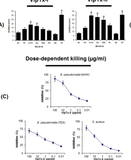
3.4. In vitro antimicrobial activity
[image:5.595.68.509.58.607.2]Purified proteins (VipTx-I and VipTx-II) were tested for their antibacterial properties against Gram-positive and Gram-negative bacteria at a 100
l
g/ml concentration. The enzyme exhibited broad spectrum activity against a wide range of pathogenic organisms. The VipTx-II enzyme showed very strong antimicrobial actionagainstS. aureus,B. pseudomallei(KHW),P. vulgaris,E. aerogenes, and P. mirabilis(Fig. 2A and B). The most promising activity of VipTx-II was compared with standard antibiotics (i.e. Ceftazidime, Chloramphenicol, Penicillin, Streptomycin, and Vancomycin). The inhibitory potential of VipTx-II was equal to that of standard antibiotics like Streptomycin, Chloramphenicol and Ceftazidime. However, VipTx-I exerted a very weak antimicrobial effect against all the tested bacteria. Particularly it’s devoid of activity againstP. aeruginosa. However, this VipTx-II protein displayed the most potent antibacterial activity compared to that of the VipTx-I pro-tein. Similarly, the antimicrobial activity of VipTx-II prompted us to conduct a further testing of MIC determinations by a broth dilu-tion method.
3.4.1. Dose-dependent antimicrobial activity
Antibacterial susceptibility of the most effective protein (VipTx-II) was further assayed against multi-drug resistant (MDR)B. pseudomallei(strain of KHW) andS. aureus. The inhibitory potential of VipTx-II was equal against both types of bacteria; however, this protein was slightly more effective in controlling the growth ofB. pseudomalleiat 10
l
g/ml (Fig. 2C).3.4.2. Minimum inhibitory concentrations (MICs)
The MICs of proteins (VipTx-I and VipTx-II) were determined by broth-dilution assay with initial sample concentrations of 100, 50, 25, 12.5, 6.25, 3.125 and 1.56
l
g/ml tested against Gram-positive and Gram-negative strains, including B. pseudomallei (TES & KHW). The MIC values are expressed as the lowest concentrationthat caused 100% bacterial growth inhibition. The VipTx-II exhib-ited marked activity againstB. pseudomalleiKHW & TES (MICs of 6.25
l
g/ml), S. aureus, (MICs 6.25l
g/ml), P. mirabilis (MICs of 12.5l
g/ml),P. vulgaris(MICs of 25l
g/ml), whereasE. coli,E. aero-genesandP. aeruginosashowed weaker inhibitory MIC effects at all dilutions (100–1.56l
g/ml) shown in Fig. 3A–H. Interestingly, VipTx-II exhibited significant inhibition at the lowest dilution againstB. pseudomalleiandS. aureus(MIC of 6.125l
g/ml). The bac-tericidal (killing) potential of proteins was further quantified by a TS and MH broth dilution method (100–1.56l
g/ml) as shown inTable 2. The MBCs result revealed that VipTx-II exerted most sig-nificant inhibition against KHW strains ofB. pseudomallei andS. aureusat the lowest dilutions (MBC 6.125
l
g/ml) versus bacterial control. The VipTx-I only weakly killed multi-drug resistant strains ofB. pseudomalleiat all tested concentrations.3.4.3. Mechanisms of action for VipTx-II
[image:6.595.58.549.156.439.2]Smooth membrane structure was clearly evident in control bacteria without treatment,B. pseudomallei (KHW) treated with VipTx-II resulted in numerous mushroom-shaped blebs, thickening irregular shapes and retraction of cytoplasm, while bacteria appeared to be losing cell contents particularly at the division septa after 24 h (Fig. 4A and B). Particularly after the treatment of VipTx-II with Gram-positive (S. aureus), while fibrous and pre-sumably cell contents (granular material) appeared to exude from the damaged membranes (Fig. 4C–F) and the inner membrane was difficult to discern. Bacteria treated with VipTx-II also lost of the membrane structure and the formed of blebs in
Table 1
Multiple sequence alignments for the N-terminal sequences of VipTx-I and VipTx-II phospholipase A2(PLA2) from theDaboia russelli russelliwas compared with existing snake venom basic phospholipase A2sp|P59071|PA28_DABRR-VRV-PL-VIIIA ofDaboia russelii(Russel’s viper), tr|D0VX11|D0VX11_DABRP-PLA2VRV-PL-VIIIA (Daboia russelliipulchella), tr|B3RFI8|B3RFI8_9SAUR-PLA2(Daboia russellii limitis), tr|A8CG84|A8CG84_DABRU-basic PLA2Drk-b2 (svPLA2) ofDaboia russelii(Russel’s viper or Vipera russelii), sp|P86368|PA23_DABRR-basic PLA2ofDaboia russelii(Russel’s viper), sp|P84674|PA25_DABRR-basic PLA2VRV-PL-V ofDaboia russelii(Russel’s viper), sp| P14424|PA2B_VIPAA- neutral PLA2agkistrodotoxin ATX) ofAgkistrodon halysorGloydius halys(Chinese water mocassin), sp|P00626|PA2A_VIPAA-PLA2, ammodytoxin A [ATXA]ofVipera ammodytes ammodytes(Western sand viper) PLA2, sp|P11407|PA2C_VIPAA-ammodytoxin C [ATXC] ofVipera ammodytes ammodytes(Western sand viper), sp|P59171|PA25_ECHOC-acidic PLA2ofEchis ocellatus(Ocellated saw-scaled viper), basic PLA2myotoxin III (Bothrops asper), sp|P20474|PA21_BOTAS-basic myotoxic PLA2bothropstoxin-2 (BthTX-II) ofBothrops jararacussu(Jararacussu), sp|P45881|PA2B2_BOTJR-and sp|P86974|PA2D_BOTLC-basic PLA2blD-PLA2) ofBothrops leucurus(White-tailed lancehead).
Accession No N-terminal Ca2+ -loob Active site β-wing
1 10 20 30 40 50 60 70 80
(1) VipTx-I SLLGFGC
MILEETGVMIELEKNCNQHPE---(2) VipTx-II SLLEFGMMILEETGKLAVPFYSKYGLYC
GCGGK-TPDD---sp|P59071|PA28_DABRR SLLEFGKMILEETGKLAIPSYSSYGCYCGWGGKGTPKDATDRC-CFVHDC-CYGNLPDCNPKSDRYKYKRVNGAIVCEKGTSCENR
tr|D0VX11|D0VX11_DABRP SLLEFGKMILEETGKLAIPSYSSYGCYCGWGGKGTPKDATDRC-CFVHDC-CYGNLPDCNPKSDRYKYKRVNGAIVCEKGTSCENR
tr|B3RFI8|B3RFI8_9SAUR SLLEFGKMILEETGKLAIPSYSSYGCYCGWGGKGTPKDATDRC-CFVHDC-CYGNLPDCNPKSDRYKYKRVNGAIVCEKGTSCENR
tr|A8CG84|A8CG84_DABRU SLLEFGKMILEETGKLAIPSYSSYGCYCGWGGKGTPKDATDRC-CFVHDC-CYGNLPDCNPKSDRYKYKRVNGAIVCEKGTSCENR
sp|P86368|PA23_DABRR SLLEFGMMILEETGKLAVPFYSSYGCYCGWGGKATPKDATDRC-CFVHDC-CYGNLPDCNPKSDRYKYKRVNGAIVCEQGTSCENR
sp|P84674|PA25_DABRR SLLEFGMMILEETGKLAVPFYSSYGCYCGWGGKGTPKDATDRC-CFVHDC-CYGNLPDCTPKPDRYKYKRVNGAIVCEQGTSCENR
sp|P14424|PA2B_VIPAA SLLEFGMMILGETGKNPLTSYSFYGCYCGVGGKGTPKDATDRC-CFVHDC-CYGNLPDCSPKTDRYKYHRENGAIVCGKGTSCENR
sp|P00626|PA2A_VIPAA SLLEFGMMILGETGKNPLTSYSFYGCYCGVGGKGTPKDATDRC-CFVHDC-CYGNLPDCSPKTDRYKYHRENGAIVCGKGTSCENR
sp|P11407|PA2C_VIPAA SLLEFGMMILGETGKNPLTSYSFYGCYCGVGGKGTPKDATDRC-CFVHDC-CYGNLPDCSPKTDRYKYHRENGAIVCGKGTSCENR
sp|P59171|PA25_ECHOC SVIEFGTMIIEETGRSPFPFYTSYGCYCGLGGKGKPKDDTDRC-CFVHDC-CYGSMPDCSPKTDIYRYHRENGEIICESGTSCEKR
sp|P20474|PA21_BOTAS SLIEFAKMILEETKRLPFPYYTTYGCYCGWGGQGQPKDATDRC-CFVHDC-CYGKLSNCKPKTDRYSYSRKSGVIICGEGTPCEKQ
sp|P45881|PA2B2_BOTJR DLWQFGQMILKETGKLPFPYYTTYGCYCGWGGQGQPKDATDRC-CFVHDC-CYGKLTNCKPKTDRYSYSRENGVIICGEGTPCEKQ
sp|P86974|PA2D_BOTLC DLWQFGQMILKETGKLPFPYYTTYGCYCGWGGQGQPKDATDRC-CFVHDC-CYGKLTNCKPKTDRYSYSRENGVIICGEGTPCEKQ
.: :*. **: ** : ... *: ****** **:.*** *************.:.:*.**.* * * * .* *:* .**.**::
Accession No α-Helix Mass Homology (%) Residues
90 100 110 121
(1) VipTx-I
---(2) VipTx-II
---sp|P59071|PA28_DABRR ICECDKAAAICFRQNLNTYSKKYMLYPDFLCKGE-LKC
---tr|D0VX11|D0VX11_DABRP ICECDKAAAICFRQNLNTYSKKYMLYPDFLCKGE-LKC
---tr|B3RFI8|B3RFI8_9SAUR ICECDKAAAICFRQNLNTYSKKYMLYPDFLCKGE-LKC
---tr|A8CG84|A8CG84_DABRU ICECDKAAAICFRQNLNTYSKKYMLYPDFLCKGE-LRC
---sp|P86368|PA23_DABRR ICECDKAAAICFRRNLNTYSKIYMLYPDFLCKGE-LKC
---sp|P84674|PA25_DABRR ICECDKAAAICFTKNLNTYSKIYMLYPDFLCKGE-LKC
---sp|P14424|PA2B_VIPAA ICECDRAAAICFRKNLKTYNHIYMYYPDFLCKKESEKC
---sp|P00626|PA2A_VIPAA ICECDRAAAICFRKNLKTYNYIYRNYPDFLCKKESEKC
---sp|P11407|PA2C_VIPAA ICECDRAAAICFRKNLKTYNYIYRNYPDILCKEESEKC
---sp|P59171|PA25_ECHOC ICECDKAAAVCFRENLKTYKNKYMVYPDSLCKEESEKC
---sp|P20474|PA21_BOTAS ICECDKAAAVCFRENLRTYKKRYMAYPDLLCKKPAEKC
---sp|P45881|PA2B2_BOTJR ICECDKAAAVCFRENLRTYKKRYMAYPDVLCKKPAEKC
---sp|P86974|PA2D_BOTLC ICECDKAAAVCFRENLRTYKXXYMAYPDVLCKKPAEKC
---*****:***:** .**.**. * *** *** . 13869 13669 13611 13611 15353 15381 13687 15587 15529 15531 15498 15705 15751 15765 13933 86 85 82 82 82 81 78 76 69 69 68 62 60 60 60 28 35 121 121 137 137 121 121 138 138 138 138 138 138 121
B. pseudomalleistrain KHW, clearly shown in transmission electron microscopy (Fig. 4G and H) than the control (ctrl). The ultra-structural studies have proven the pore-forming action of VipTx-II enzyme on bacteria. Generally, viper proteins induced specific structural and morphological changes versus untreated bacterial controls.
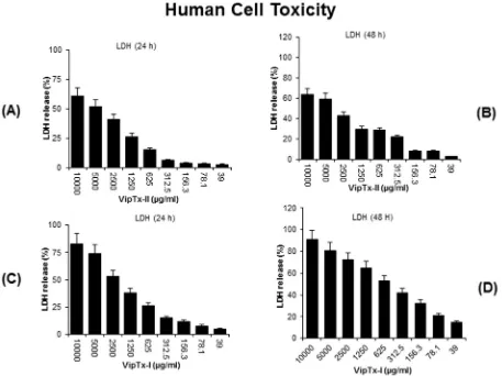
3.5. Morphological and cellular toxicity
[image:7.595.78.499.76.591.2]Cytotoxicity results indicated that VipTx-II did not affect the cell viability up to a concentration of 1000
l
g/ml. Human macro-phages (THP-1) were not affected particularly at 625l
g/ml in a cytotoxicity assay. Exposure to VipTx-II revealed minimalcytotoxicity up to 625
l
g/ml concentrations. The optimum dose of VipTx-II gradually decreased THP-1 cell proliferation after treat-ment with enzymes (Fig. 5A and B). However, cell survival decreased with increasing concentrations of VipTx-I from 39 to 10,000l
g/ml and EC501250l
g/ml (Fig. 5C and D). Themorpholog-ical changes of the cells revealed that membrane disruption, lysis and significant cell death was evident at a 2500
l
g/ml concentra-tion of VipTx-I in a time and dose dependent (24 and 48 h) manner. Sixty percent of the THP-1 cells were inhibited by the exposure of VipTx-I after 48 h than in the control. However, the cytolytic levels were at higher concentrations up to 2500l
g/ml (Fig. 5E–H).3.6. LDH assay
The LDH results revealed that THP-1 cells exposed to VipTx-II were not affected up to 1250
l
g/ml concentrations (Fig. 6A and B). Significant cell death is evident at higher concentration (2500l
g/ml) in a dose and time-dependent manner (24 and 48 h), as a result there is more LDH release into the media. However, the cell proliferation was not affected especially at the optimal dose of VipTx-II (EC501250l
g/m1) than the VipTx-II(Fig. 6C and D). The optimum dose that inhibited bacterial prolifer-ation did not affect the THP-1cells.
4. Discussion
[image:9.595.32.285.95.193.2]Bacterial resistance is a significant problem for treating infec-tions, and thus there is a keen interest in research directed
Table 2
Minimum bactericidal concentrations (MBCs) of viperatoxins isolated from the Indian Russell’s viper snake venom (Daboia russellii russellii).a
Bacteria MBCsa
VipTx-I (lg/ml) VipTx-II (lg/ml)
Escherichia coli 100 100
Enterobacter aerogenes 100 100
Proteus vulgaris 50 12.5
Proteus mirabilis 50 12.5
Pseudomonas aeruginosa 100 100
Staphylococcus aureus 50 6.25
Burkholderia pseudomallei (strainKHW) 100 6.25 Burkholderia pseudomallei (strainTES) 100 6.25
aThe MBC values were determined by a modified micro-broth dilution assay using snake venom proteins (100, 50, 25, 12.5, 6.25, 3.125lg/ml). Antimicrobial activity of viperatoxin (VipTx-I and VipTx-II) was determined by MBCs in a solution killing assay against Gram-negative and Gram-positive bacteria.
[image:9.595.109.474.324.711.2]towards identifying novel agents to treat infections [43]. Most venomous animals contain a variety of venom proteins which participate in both digestion of prey and venom toxicity. Viperi-dae snake venoms represent a source of important bioactive molecules that have led to the development of diverse new drugs in clinical scenario. In this study, novel snake venom proteins were purified and designated as ‘‘Viperatoxin” (VipTx-I and VipTx-II) from the Indian Russell’s viper (D. russelli russelli). The N-terminal amino acid residues of VipTx-I and VipTx-II were sequenced, and compared with existing sequences in the ExPASY proteomics database using BLAST.
The sequence comparison shows that VipTx-II matched 60–86% homology with existing snake venom phospholipase A2s (svPLA2s).
The molecular weight of VipTx-I and VipTX-II slightly differs with the previously reported protein masses from known PLA2s. Our
results corroborate with the N-terminal sequences ofB. neuwiedi pauloensisshowed important homology with Asp49 basic myotoxic PLA2s from other snake venoms [44]. Whereas, Lys49 PLA2
(myotoxin I) elucidated from B. atroxvenom displays very high level of homology with other Lys49 PLA2s, although its primary
and three-dimensional structure show some difference in the C-terminal region [45]. Generally, characterized svPLA2s have a
[image:11.595.63.520.66.408.2]conserved fold with seven disulfide bridges and a histidine at the catalytic site, with calcium (Ca2+) bound at the active site [11,28,46]. The Russell’s viper svPLA2 structure also contains a
Trp-31-containing loop (residues 25-34), b-wing consisting of double-stranded anti-parallel b-strands (residues 74-85) and C-terminal region 9 (residues 119-133). The crystal structures of
complexes with transition analogs provide insights into the poten-tial catalytic mechanisms with the presence of fatty acid in the hydrophobic channel[47].
Several recent studies have shown that antimicrobial proteins and peptides are produced by many living organisms and repre-sent a novel class of antibiotics to treat infectious diseases[8]. Examples include crotamine, a myotoxin from venom of the South American rattlesnake, which is structurally related to beta-defensins. The later are antimicrobial peptides found in vertebrate animals[13], peptides fromNaja najavenom[48], and a venom protein from the inland taipan which all exhibit antimicrobial activity. Previous studies have shown that the naturally occurring proteins display antimicrobial activity[12]. Remarkably, a group of Lys-49 PLA2s homologues present in snake venoms [19], are
bactericidal even though they lack enzymatic activity. Another study shows that myotoxic PLA2s are bactericidal againstE. coli
andS. aureus[20].
In this study, VipTx-II exerted the most potent action against a multi-drug resistant strain (KHW) ofB. pseudomalleias compared to the less resistant TES strains. The inhibitory potential of VipTx-II was quite equivalent to that of standard antibiotics such as streptomycin, chloramphenicol, ceftazidime and vancomycin versus VipTx-I. Further, we tested the antibacterial properties of VipTx-II and found that it killed several strains of Gram-positive and Gram-negative bacteria, with doses ranging from (100–0.01
l
g/ml). The dose-dependant assay revealed bacterial killing by viper protein within 24 h and reached the maximum (75%) activity at a 10l
g/ml concentration. Our earlier studyclearly demonstrated that viper proteins exert the most significant bactericidal effects against B. pseudomallei [49]. Our study, in contrast with the EcTx-I protein from the venoms of Saw-scaled viper species, demonstrated significant bactericidal inhibition of multi-drug resistance (MDR) B. pseudomallei (KHW) and E. aerogenespreviously[38]. VipTx-II exerted the most significant inhibition againstB. pseudomalleiKHW strains even at the lowest dilutions (MICs 6.25
l
g/ml). Interestingly, the VipTx-II protein showed very significant inhibitory effects against S. aureus, P. vulgarisandP. mirabiliseven at 12.25l
g/ml doses (in a dose-dependent manner). Whereas, recently reported studies show that a basic protein of VRV-PL-V fromDaboia russelli pulchella(venom PLA2 fraction V) effectively inhibits Gram-positive bacteria suchas S. aureus and Bacillus subtilis at MICs 13-24
l
g/ml versus Gram-negative E. coli,Vibrio cholerae, Klebsiella pneumoniaeand Salmonella paratyphi [50]. Similarly the PLA2 fraction VIIIA ofD. russelli pulchella(VRV-PL-VIIIa) also controls the growth of bac-teria at 11–19
l
g/ml doses [51], BnpTX-1/II is a basic myotoxic PLA2s obtained fromB. neuwiedi pauloensissnake venom,BnpTX-1 showed the neurotoxic as well as antibacterial activity on E. coliand S. aureus[44]. The Lys49 phospholipase A2(PLA2) of
Bothrops atrox (myotoxin I) displays only a weak antibacterial activity on bacteria[45], whereas synthetic peptide derived from the Lys49 PLA2 of C-terminal segment ofB. asper(myotoxin II)
and tryptophan (Tyr–Trp) substitution enhances antimicrobial potency on Gram-negative (Salmonella typhimurium) and Gram-positive bacteria (S. aureus), with low cytotoxicity on skeletal muscle cells, C2C12 myoblasts[22]. Cardiotoxin 3 (CTX3) isolated from Naja naja atra (Taiwan cobra) exerts the most potent inhibitory effects againstS. aureusthanE. coli[52]. However, the VipTx-II displays the most potent inhibitory effects against B. pseudomallei KHW (MICs 6.25
l
g/ml) as well as S. aureus, P. vulgarisandP. mirabilis(MICs 12.25l
g/ml) at very low concen-trations compared to the existing PLA2s.We found novel bactericidal mechanisms attributed to viper proteins (VipTx-I and VipTx-II) that induced pore formation on clinical isolates such as MDRB. pseudomallei(KHW strain). There were cellular changes that include membrane disintegration. Our study corroborate with membrane damage elicited by the binding of protein to the lipid membrane[53]. Recently, there have been suggestions that transmembrane pore formation is not the only mechanism of microbial killing[54]. Several earlier observations indicate that translocated protein or peptides can alter cytoplasmic membrane septum formation, inhibit cell-wall, nucleic-acid and protein synthesis or inhibit enzymatic activity [54]. Previously study of the mechanisms was clearly evidenced that the antimicro-bial protein as well as peptides induced killing of microorganisms by severely damaging membrane and formation of pores on invading pathogens[38]. Therefore, sPLA2is implicated in the lipid
digestion as a host defence mechanism including the observed antibacterial activities[55].
Enzymatically-independent bactericidal effects of PLA2 has
been demonstrated and mapped to a specific membrane-damaging protein site[20]. A number of Trp substituted peptides derived from svPLA2 can increase microbicidal potency against
both Gram-negative and Gram-positive bacteria [25]. Previous studies also show that the interaction of this peptide with the lipopolysaccharide (LPS) and lipid A or lipoteichoic acid (LA) relies on a membrane-permeabilizing mechanism to exert its bacterici-dal effects[20]. In addition to Trp, Pro and Arg residues are also important in membrane disruption and/or cell entry. Thus these later Pro and Arg residues are an attractive template for designing novel antimicrobial peptides effective against a broad spectrum of microorganisms [56]. In addition, it has been reported that the insertion of positively-charged amino acids can result in big varia-tions and high antibiotic activity. These peptides also significantly
reduce hemolysis and cytotoxicity that correlate with decreased permeabilization of the zwitterionic phosphatidylcholine mem-brane, the major component of outer leaflets from red blood cells
[56].
Several proteins and polypeptides of reptiles have common cytolytic properties, and these cytolysins provide an offensive armament for the animal defense. In this study, cell survival decreased with the increasing concentrations of viper protein (VipTx-I) at 39–10,000
l
g/ml than VipTx-II. However, the enzyme (VipTx-II) has low level of toxicity for eukaryotic cells at higher concentrations. The morphological changes in THP-1 cells show membrane disruption, lysis and significant cell death apparent at 2500l
g/ml with VipTx-I in a time-dependent manner (24 and 48 h). Although, 60% of the cells were killed by the VipTx-I after 48 h, but the observed cytolytic effects were very high at higher protein concentrations (1250l
g/ml). This may be due to the mech-anism for clearing free fatty acids that is toxic to the cell for retain-ing cellular energy reserve. Those fatty acids might be acylated by intracellular located enzymes to membrane-bound proteins. Acyla-tion of these proteins can be potentially one of the mechanisms for disrupting membrane integrity and cellular signaling[57].4.1. Mechanisms of actions
The antimicrobial potency of snake venom is largely unex-plored. Naturally occurring snake venom proteins and peptides possess highly potent antimicrobial activity againstB. pseudomallei
[58]. A family of sPLA2 comprised of low molecular weight
(13 kDa), disulfide-linked proteins[59]depend on Ca2+for enzy-matic activity and play an important role in innate immunity and killing of bacteria[60]. Antimicrobial proteins/peptides generally kill bacteria by permeabilizing and or disrupting their membranes
[61]. The molecular basis for the activity and selectivity of these peptides has been studied in model membranes [61]. Cationic antimicrobial peptides interact preferentially with acidic lipids that are chiefly abundant and found in bacteria [38]. However, the basic proteins manifest strong amphiphilic properties on their molecular surface. For example, apolar tips of loops I-III form a hydrophobic zone flanked by a positively-charged Lys and Arg resi-dues. Another membrane model study demonstrates that the hydrophobic bottom represents a principal membrane-binding motif for protein[62]. In addition, the structural defects in lipid bilayers induced by protein binding to the membrane can lead to the formation of pores[63]. The explanations above provide a basis for the major differences in cell specificity by various antimicrobial peptides/proteins. Interaction of peptides with bilayers alters the organization of the membranes and makes them more permeable to ions [64]. Overall, it is not only the nature of a protein and peptide, but also various characteristics of the cell membrane and metabolic state of the target cell, that ultimately determine the mechanism of antimicrobial activity.
Conflict of interest
The authors confirm that this article content has no conflicts of interest.
Author contributions
Conceived, designed, performed, analyzed data and wrote the manuscript: RPS. Provide facility and materials: AC, MZ, SAA, APK & GS. Corrected and fine-tuned of manuscript: BGS, HKLL, OLF, EGR.
Acknowledgements
This work was supported by Defence Science Technology Agency (DSTA) grant R-181-000-063-422 (DSO), Singapore. The Deanship of Scientific Research, College of Sciences Research Cen-tre, King Saud University, Kingdom of Saudi also supported this work. This work was also supported by grants from the National Medical Research Council of Singapore [R-713-000-177-511], and by the NCIS Yong Siew Yoon Research Grant through donations from the Yong Loo Lin Trust to APK. APK was also supported by the National Research Foundation Singapore and the Singapore Ministry of Education under its Research Centres of Excellence initiative to Cancer Science Institute of Singapore, National University of Singapore. The author OLF is supported by CAPES, CNPq, FAPDF and FUNDECT.
A. Supplementary data
Supplementary data associated with this article can be found, in the online version, athttp://dx.doi.org/10.1016/j.fob.2015.10.004.
References
[1] Moran, G.J., Krishnadasan, A., Gorwitz, R.J., Fosheim, G.E., McDougal, L.K., Carey, R.B., et al. (2006) Methicillin-resistant S. aureusinfections among patients in the emergency department. N. Engl. J. Med. 355, 666–674. [2] Miller, L.G., Perdreau-Remington, F., Rieg, G., Mehdi, S., Perlroth, J., Bayer, A.S.,
et al. (2005) Necrotizing fasciitis caused by community-associated methicillin-resistantStaphylococcus aureusin Los Angeles. N. Engl. J. Med. 352, 1445–1453. [3] White, N.J. (2003) Melioidosis. Lancet 361, 1715–1722.
[4] Subbalaxmi, M.V., Chandra, N., Rao, M.N., Vemu, L. and Raju, Y.S. (2011) Burkholderia pseudomallei: an uncommon cause of bacteraemic pneumoniA in A diabetic. Indian J. Chest Dis. Allied Sci. 53, 185–187.
[5] Sawasdidoln, C., Taweechaisupapong, S., Sermswan, R.W., Tattawasart, U., Tungpradabkul, S. and Wongratanacheewin, S. (2010) GrowingBurholderiA pseudomallei in biofilm stimulating conditions significantly induces antimicrobial resistance. PLoS One 5, e9196.
[6] Mukhopadhyay, C., Chawla, K., Krishna, S., Nagalakshmi, N., Rao, S.P. and Bairy, I. (2008) Emergence ofBurkholderia pseudomalleiand pandrug-resistant non-fermenters from southern Karnataka. India. Trans. R. Soc. Trop. Med. Hyg. 102, S12–S17.
[7] Jenney, A.W., Lum, G., Fisher, D.A. and Currie, B.J. (2001) Antibiotic susceptibility ofBurkholderia pseudomalleifrom tropical northern AustraliA and implications for therapy of melioidosis. Int. J. Antimicrob. Agents 17, 109– 113.
[8] Dawson, R.M., Fox, M.A., Atkins, H.S. and Liu, C.Q. (2011) Potent antimicrobial peptides with selectivity forBacillus anthracisover human erythrocytes. Int. J. Aantimicrob. Agents 38, 237–242.
[9] Koh, D.C., Armugam, A. and Jeyaseelan, K. (2006) Snake venom components and their applications in biomedicine. Cell. Mol. Life Sci. 63, 3030–3041. [10] Sajevic, T., Leonardi, A. and Krizˇaj, I. (2011) Haemostatically active proteins in
snake venoms. Toxicon 57, 627–645.
[11] Ferreira, B.L., Santos, D.O., Andre, Santos L., Rodrigues, C.R., de Freitas, C.C., Cabral, L.M., et al. (2011) Comparative analysis of viperidae venoms antibacterial profile: A short communication for proteomics. Evid. Based Complement. Alternat. Med., 1–4.
[12] de Lima, D., Abreu, P.A., de Freitas, C.C., Santos, D.O., Borges, R.O., dos Santos, T. C., et al. (2005) Snake venom: any clue for antibiotics and CAM? Evid. Based Complement. Alternat. Med. 2, 39–47.
[13] Oguiura, N., Boni-Mitake, M., Affonso, R. and Zhang, G. (2011) In vitro antibacterial and hemolytic activities of crotamine, A small basic myotoxin from rattlesnakeCrotalus durissus. J. Antibiot. (Tokyo) 64, 327–331.
[14]Zhao, H. and Kinnunen, P.K. (2003) Modulation of the activity of secretory phospholipase A2by antimicrobial peptides. Antimicrob. Agents Chemother. 47, 965–971.
[15]Valentin, E. and Lambeau, G. (2000) What can venom phospholipase A2tell us about the functional diversity of mammalian secreted phospholipases A2?. Biochimie 82, 815–831.
[16]Wu, Y., Raymond, B., Goossens, P.L., Njamkepo, E., Guiso, N., Paya, M., et al. (2010) Type-IIA secreted phospholipase A2is an endogenous antibiotic-like protein of the host. Biochimie 92, 583–587.
[17]Vargas, L.J., Londoño, M., Quintana, J.C., Rua, C., Segura, C., Lomonte, B., et al. (2012) An acidic phospholipase A2with antibacterial activity fromPorthidium nasutumsnake venom. Comp. Biochem. Physiol. B. Biochem. Mol. Biol. 161, 341–347.
[18]Gutiérrez, J.M. and Lomonte, B. (1997) Phospholipase A2myotoxins from Bothrops snake venoms in: Venom Phospholipase A2Enzymes: Structure, Function, and Mechanism (Kini, R.M., Ed.), pp. 321–352, John Wiley & Sons, Chichester, England.
[19]Lomonte, B., Angulo, Y. and Calderón, L. (2003) An overview of lysine-49 phospholipase A2myotoxins from crotalid snake venoms and their structural determinants of myotoxic action. Toxicon 42, 885–901.
[20] Páramo, L., Lomonte, B., Pizarro-Cerdá, J., Bengoechea, J.A., Gorvel, J.P. and Moreno, E. (1998) Bactericidal activity of Lys49 and Asp49 myotoxic phospholipase A2 from Bothrops asper snake venom–synthetic Lys49 myotoxin II-(115–129)-peptide identifies its bactericidal region. Eur. J. Biochem. 253, 452–461.
[21]Soares, A.M. and Giglio, J.R. (2003) Chemical modifications of phospholipases A2from snake venoms: effects on catalytic and pharmacological properties. Toxicon 42, 855–868.
[22]Santamaría, C., Larios, S., Angulo, Y., Pizarro-Cerda, J., Gorvel, J.P., Moreno, E., et al. (2005) Antimicrobial activity of myotoxin phospholipases A2 from crotalid snake venoms and synthetic peptide variants derived from their C-terminal region. Toxicon 45, 807–815.
[23]Villarrubia, V.G., Costa, L.A. and Díez, R.A. (2004) Secreted phospholipases A2 (sPLA2): friends or foes? Are they actors in antibacterial and anti HIV resistance? Med. Clin. (Barc) 123, 749–757.
[24]Murillo, L.A., Lan, C.Y., Agabian, N.M., Larios, S. and Lomonte, B. (2007) Fungicidal activity of A phospholipaseA2– derived synthetic peptide variant againstCandidA albicans. Rev. Esp. Quimioter. 20, 330–333.
[25]Araya, C. and Lomonte, B. (2007) Antitumor effects of cationic synthetic peptides derived from Lys49 phospholipase A2homologues of snake venoms. Cell Biol. 3, 263–268.
[26]Costa, T.R., Menaldo, D.L., Oliveira, C.Z., Santos-Filho, N.A., Teixeira, S.S., Nomizo, A., et al. (2008) Myotoxic phospholipases A2isolated fromBothrops brazilisnake venom and synthetic peptides derived from their C-terminal region: cytotoxic effect on microorganism and tumor cells. Peptides 29, 1645– 1656.
[27]Fonseca-Maldonado, R., Ferreira, T.L. and Ward, R.J. (2012) The bactericidal effect of human secreted group IID phospholipase A2 results from both hydrolytic and non-hydrolytic activities. Biochimie 94, 1437–1440. [28]Kang, T.S., Georgieva, D., Genov, N., Murakami, M.T., Sinha, M., Kumar, R.P.,
et al. (2011) Enzymatic toxins from snake venom: structural characterization and mechanism of catalysis. FEBS J. 278, 4544–4576.
[29]Nakayama, D., Ben, Ammar Y., Miyata, T. and Takeda, S. (2011) Structural basis of coagulation factor V recognition for cleavage by RVV-V. FEBS Lett. 585, 3020–3025.
[30] Mandal, S. and Bhattacharyya, D. (2008) TwoL-amino acid oxidase isoenzymes from Russell’s viper (Daboia russelli russelli) venom with different mechanisms of inhibition by substrate analogs. FEBS J. 275, 2078–2095.
[31]Suntravat, M., Yusuksawad, M., Sereemaspun, A., Perez, J.C. and Nuchprayoon, I. (2011) Effect of purified Russell’s viper venom-factor X activator (RVV-X) on renal hemodynamics, renal functions, and coagulopathy in rats. Toxicon 58, 230–238.
[32]Segers, K., Rosing, J. and Nicolaes, G.A. (2006) Structural models of the snake venom factor V activators fromDaboia russelliandDaboia lebetina. Proteins 64, 968–984.
[33]Maity, G., Mandal, S., Bhattacharjee, P. and Bhattacharyya, D. (2011) Thermal detoxification of the venom fromDaboia russelli russelliof Eastern IndiA with restoration of fibrinolytic activity. Toxicon 57, 747–754.
[34]Shevchenko, A., Sunyaev, S., Loboda, A., Shevchenko, A., Bork, P., Ens, W., et al. (2001) Charting the proteomes of organisms with unsequenced genomes by MALDI-quadrupole time-of-flight mass spectrometry and BLAST homology searching. Anal. Chem. 73, 1917–1926.
[35]Bradford, M.M. (1976) A rapid and sensitive method for the quantitation of microgram quantities of protein utilizing the principle of protein-dye binding. Anal. Biochem. 72, 248–254.
[36]Laemmli, U.K. (1970) Cleavage of structural proteins during the assembly of the head of bacteriophage T4. Nature 227, 680–685.
[37]Reynolds, L.J., Hughes, L.L. and Dennis, E.A. (1992) Analysis of human synovial fluid phospholipase A2on short chain phosphatidylcholine-mixed micelles: development of A spectrophotometric assay suitable for A microtiterplate reader. Anal. Biochem. 204, 190–197.
[38]Perumal, Samy R., Gopalakrishnakone, P., Chow, V.T.K. and Ho, B. (2008) Viper metalloproteinase (Agkistrodon halys pallas) with antimicrobial activity against multi-drug resistant human pathogens. J. Cell. Physiol. 216, 54–68. [39]Bauer, A.W., Kirby, W.M. and Sherris, J.C. (1966) Antibiotic susceptibility
[40] Lomonte, B., Moreno, E., Tarkowski, A., Hanson, L.A. and Maccarana, M. (1994) Neutralizing interaction between heparins and myotoxin II, A lysine 49 phospholipase A2 from Bothrops asper snake venom. Identification of A heparin-binding and cytolytic toxin region by the use of synthetic peptides and molecular modeling. J. Biol. Chem. 269, 29867–29873.
[41]Motizuki, M., Itoh, T., Satoh, T., Yokota, S., Yamada, M., Shimamura, S., et al. (1999) Lipid-binding and antimicrobial properties of synthetic peptides of bovine apolipoprotein A-II. Biochem. J. 342, 215–221.
[42]Matsuzaki, K. (1998) Magainins as paradigm for the mode of action of pore forming polypeptides. Biochim. Biophys. Acta 1376, 391–400.
[43]Harris, C.R. and Thorarensen, A. (2004) Advances in the discovery of novel antibacterial agents during the year 2002. Curr. Med. Chem. 11, 2213–2243. [44]Rodrigues, V.M., Marcussi, S., Cambraia, R.S., de Araujo, A.L., Malta-Neto, N.R.,
Hamaguchi, A., et al. (2004) Bactericidal and neurotoxic activities of two myotoxic phospholipases A2fromBothrops neuwiedi pauloensissnake venom. Toxicon 44 (3), 305–314.
[45]Nunez, V., Arce, V., Gutierrez, J.M. and Lomonte, B. (2004) Structural and functional characterization of myotoxin I, A Lys49 phospholipase A2 homologue from the venom of the snakeBothrops atrox. Toxicon 44 (1), 91– 101.
[46]Singh, G., Jasti, J., Saravanan, K., Sharma, S., Kaur, P., Srinivasan, A., et al. (2005) Crystal structure of the complex formed between A group I phospholipase A2 and A naturally occurring fatty acid at 2.7 Å resolution. Protein Sci. 14, 395– 400.
[47]Chandra, V., Jasti, J., Kaur, P., Dey, S., Perbandt, M., Srinivasan, A., et al. (2002) Crystal structure of A complex formed between a snake venom phospholipase A2and A potent peptide inhibitor Phe–Leu–Ser–Tyr–Lys at 1.8 Å resolution. J. Biol. Chem. 277, 41079–41085.
[48]Sachidananda, M.K., Murari, S.K. and Channe, GowdA D. (2007) Characterization of an antibacterial peptide from Indian CobrA (Naja naja). J. Venom Anim. Toxins Trop. Dis. 13, 446–461.
[49]Perumal, Samy R., Pachiappan, A., Gopalakrishnakone, P., Thwin, M.M., Hian, Y. E., Chow, V.T., et al. (2006)In vitroantimicrobial activity of natural toxins and animal venoms tested againstBurkholderia pseudomallei. BMC Infect. Dis. 6, 100.
[50] Sudarshan, S. and Dhananjaya, B.L. (2014) Antibacterial potential of A basic phospholipase A2 (VRV-PL-V) ofDaboia russellii pulchella (Vipera russellii) venom. Biochemistry (Mosc) 79 (11), 1237–1244.
[51]Sudarshan, S. and Dhananjaya, B.L. (2015) Antibacterial potential of A basic phospholipase A2(VRV-PL-VIIIa) fromDaboia russelii pulchella(Russell’s viper) venom. J. Venom Anim. Toxins Incl. Trop. Dis. 21, 17.
[52] Chen, L., Kao, P., Fu, Y., Lin, S. and Chang, L. (2011) Membrane-damaging activity of Taiwan cobrA cardiotoxin 3 is responsible for its bactericidal activity. Toxicon 58 (1), 46–53.
[53] Anderluh, G. and Lakey, J.H. (2008) Disparate proteins use similar architectures to damage membranes. Trends Biochem. Sci. 33, 482–490. [54] Brogden, K.A. (2005) Antimicrobial peptides: pore formers or metabolic
inhibitors in bacteria? Nat. Rev. Microbiol. 238, 239–250.
[55] Yang, S.T., Shin, S.Y., Hahm, K.S. and Kim, J.I. (2006) Design of perfectly symmetric Trp-rich peptides with potent and broad-spectrum antimicrobial activities. Int. J. Antimicrob. Agents 27, 325–330.
[56] Bechinger, B. (1997) Structure and functions of channel-forming peptides: magainins, cecropins, melittin and alamethicin. J. Membr. Biol. 156, 197–211. [57] Suntravat, M., Yusuksawad, M., Sereemaspun, A., Perez, J.C. and Nuchprayoon, I. (2011) Effect of purified Russell’s viper venom-factor X activator (RVV-X) on renal hemodynamics, renal functions, and coagulopathy in rats. Toxicon 58, 230–238.
[58] Oliveira, D.G., Toyama, M.H., Novello, J.C., Beriam, L.O. and Marangoni, S. (2002) Structural and functional characterization of basic PLA2isolated from Crotalus durissus terrificusvenom. J. Protein Chem. 21, 161–168.
[59] Perumal, Samy R., Gopalakrishnakone, P., Thwin, M.M., Chow, T.K., Bow, H., Yap, E.H., et al. (2007) Antibacterial activity of snake, scorpion and bee venoms: A comparison with purified venom phospholipase A2enzymes. J. Appl. Microbiol. 102, 650–659.
[60] Zhao, H., Rinaldi, A.C., Rufo, A., Bozzi, A., Kinnunen, P.K.J. and Di Giulio, A. (2002) Structural and charge requirements for antimicrobial peptide insertion into biological and model membranes in: Pore-forming peptides and protein toxins (Lazarovici, P., Menestrina, G. and DallA Serra, M., Eds.), Harwood Academic Publishers, Reading, UK.
[61] Maget-Dana, R., Lelievre, D. and Brack, A. (1999) Surface active properties of amphiphilic sequential isopeptides: comparison between alpha-helical and beta-sheet conformations. Biopolymers 49, 415–423.
[62] Dubovskii, P.V., Dementieva, D.V., Bocharov, E.V., Utkin, Y.N. and Arseniev, A.S. (2001) Membrane binding motif of the P-type cardiotoxin. J. Mol. Biol. 305, 137–149.
[63] Konshina, A.G., Boldyrev, I.A., Utkin, Y.N., Omelkov, A.V. and Efremov, R.G. (2011) Snake cytotoxins bind to membranes viA interactions with phosphatidylserine head groups of lipids. PLoS One 6, e19064.