Copyright©)1972 AmericanSociety for Microbiology PrintedinU.S.A
Transfection
in
Agrobacterium tumefaciens
VICTOR J. MILANI AND GARY T. HEBERLEIN' Department of Biology, New York University, Bronx,New York 10453
Received forpublication1 March1972
Intact cells of Agrobacteriwn tumefaciens were examined for ability to take up
biologically active LR-4 phage deoxyribonucleic acid (DNA) from the surrounding medium. DNAincorporationasmeasuredbysubsequent plaque formation -(trans-fection) failed to occur when the bacteria were grown in defined minimal salts media, andwasrestrictedtoa4-hr period in theearly log phase of growth in enriched media. In thelatter case, maximal transfection frequencies were obtained after a
25-to30-min incubation with 22.5 ,ug of phage DNA/ml. Higher DNA
concentra-tions or longer incubation times were inhibitory. Transfection was completely inhibited by deoxyribonuclease but not byribonuclease, trypsin, or phage-specific
antisera.
Agrobacterium tumefaciens (Smith and
Town-send) Conn induces autonomous crown gall
tumors in a variety of dicotyledonous plants. There is considerable evidence to suggest that
deoxyribonucleicacid (DNA)fromthebacterium
or one ofits temperate phages plays a passive, if notactive, role in the inductionprocess (2, 11,
12, 19). Nevertheless, the number and nature of
the cistron products responsible for conversion
ofnormal cells to tumor cells has yet to be
re-solved.
In light of the above, it is surprising that so
littleattention has beengiven to thedevelopment
of a well-defined system for genetic analysis in
A. tumefaciens. Conjugation and transduction,
unfortunately, have yet to be demonstrated in
this genus. Early studies by Klein and Klein
(8, 9) and more recently by Kern (6, 7) would
suggest, however, that A. tumefaciensis
capable
of undergoing genetic
transformation,
althoughno reproducible system was defined.
Thepresent study was undertaken inaneffort to confirm "competence" in A. tumefaciens and
to characterizesomeofthe parametersproviding
for maximal DNA uptake. A temperate phage
of A. tumefaciens was used as the source of
bio-logicallyactiveDNA.
MATERIALS AND METHODS
Bacteria and phage. A. tumefaciens (Smith and Townsend) Conn strain B66 and the virulent phage
LR-4 were obtained throughthe courtesy of Robert Manasse of Boyce Thompson Institute for Plant Research, Yonkers, N.Y.B66 was selected forusein this study because ofits sensitivity to LR-4 phage, 1Present address: Biology Department, Universityof Mis-souri-St.Louis,St.Louis,Mo.63121.
and its failure to yield plaque-forming units after treatment with mitomycin C and ultraviolet light.
Mediaandbuffers.Thefollowing eight media were utilized for culturing the bacteria. Basic nutrient broth (BNB) contained 8 g of Difco nutrient broth, 5gof sucrose, and 1 g of Difco yeast extract/liter of distilled water; basic nutrient agar (BNA) was BNB
plus 1.5% Difco agar. Peptone broth (PTB) con-sisted of 1% Difco peptone/liter of distilled water;
peptone agar (PTA) was PTBplus 1.5% Difco agar. Minimal medium broth (MMB) contained 5 g of glucose, 1 g ofKNO3, 1.5 g of NaCl, 2 g of NaH2PO4, 250mgof MgS04 * 7H20, and 25 mg ofCaCl2/liter of distilled water (pH 7.0); minimal medium agar (MMA) was MMB plus
1.5%-7
Difco agar. Sucrose salts broth (SSB) contained 5 g of sucrose, 1 g ofKNO3, 1.5 gofNaCl, 2g ofNaH2PO4, 250 mg of MgSO4- 7H20, and 25 mg ofCaCl2,'liter ofdistilled water (pH 7.0); sucrose salts agar (SSA) was SSB plus
1.5%yo
Difco agar.Twomediawereused to assayforplaque-forming units: Fisherphageoverlay agar (3 g of beef extract, 5 gof peptone, 5 g ofNaCl, 0.2 gofMgSO4-7H20, 0.05g ofMnSO4,0.15gofCaCl2, and 7.0 g of agar/ liter of distilled water) and Fisher phage base agar
(phageoverlay plus 8.0 g of agar/liter).
The following two buffers were employed during the course of this investigation: 0.067 M phosphate buffer, pH 7.0, and0.1 M tris(hydroxymethyl)amino-methane(Tris) buffer (Mann Chemical Co.), pH 7.5. Preparation of phage. A 5-ml amount of LR-4 phagestock wasadded to a 24-hr BNBcultureof B66. Thispreparationwasincubated for12 hr at27C on a gyratoryshaker (ca. 120rev/min) toallowforphage replication and bacterial lysis.Thislysed culture was centrifuged at 12,100 X g for 20 min at 4 C in a Sorvall RC 2-B Superspeedcentrifuge (rotor SS-34). The supernatant fluid was filtered through several membranefilters (0.22blm; Millipore Corp.) toavoid clogging. The sterile filtrate was centrifuged in a Beckman L2-65B ultracentrifuge (rotor 50.1) at 29,473 X gfor 90minat4 C tosedimentthe phage 17
on November 10, 2019 by guest
http://jvi.asm.org/
particles. The phage pellets were resuspended in 0.1
M Tris buffer, pH 7.5, and were maintained at4 C until needed.
Extraction of phage DNA. An equal volume of freshly distilled phenol, neutralized to pH 7.0with NaOH, wasadded to the phage stock. This mixture
wasmildly agitatedfor 30min.Theabovepreparation
was then centrifugedat480 X gfor 20min at4C, in a Sorvall RC 2-B superspeed centrifuge (rotor SS-34). The aqueous supernatant fluid containing the phage DNA was removed and mixed withfresh
phenol. This preparation was again slowly agitated for 30min and centrifuged. Theaqueous layer
con-taining the phage DNA was removed, and dialyzed
at4 C overnight againstasolution of0.067 M
phos-phate buffer, pH7.0. A fewdropsof chloroformwere
added to the dialyzed DNA solution to prevent bacterial contamination.
All DNA preparations were tested for sterility by platingonBNApriortouse.TheDNA concentration
was determined by the diphenylamine method of
Burton (3).
Transfection procedure. Flaskscontaining50 ml of
desired media were inoculated with a loopful of A. tumefaciens B66 that had been cultured ona BNA slant.These cultureswereincubated for 24 hr at 27 C
on a gyratory shaker (ca. 120 rev/min). Side-arm flaskswere then inoculatedwithanadequate sample of eachrespectiveculture togiveanoptical densityof
0.14 withaKlett Summersonphotoelectriccolorimeter
(greenfilterno.54). Sampleswereremoved from these flasksatvarious times andplacedin sterile testtubes. An appropriate concentration of LR-4 phage DNA
wasthenadded to each tube. Sterile preparations of
deoxyribonuclease (10,g/ml, Worthington
Biochemi-cal Corp.), ribonuclease (10 /Ag/ml, Worthington Biochemical Corp.), trypsin(10,gg/ml, Sigma Chemi-calCo.), and LR-4phageantisera (0.1 ml) werealso
addedtocontroltubesatthe time ofDNAaddition.
All tubes were incubated in a tilted position, on a gyratoryshaker (ca. 120 rev/min) at 27 C. Samples
were removed at various times, and were tested for
plaque-forming ability by overlay platingonadefined
medium with 0.5mloflog-phaseB66 cells. Plateswere
incubated overnight at 27 C. A second plating was
accomplished byfirstadding2 ml of0.1 MTrisbuffer
(pH 7.5) toeachplate,andscrapingtheoverlayagar
into a steriletest tube. An additional 10 ml of Tris bufferwasadded, andthe mixturewasvortexedprior
tocentrifugation at12,100 X gat4 Cfor20minin a sterile tube. The supernatant fluid was passed
through a membrane filter (0.22,um) and plated on desired media. Plates were examined for plaques
after overnight incubationat27C. RESULTS
Attempts to characterize DNA uptake (com-petence) in A. tumefaciens B66 by means of
transfection were at first unsuccessful. Plaques failedto develop when cultures of the bacterium wereplated on different media and incubatedat various temperatures after exposure to LR-4 phage DNA (1 to 45 4ug/ml), for periods up to
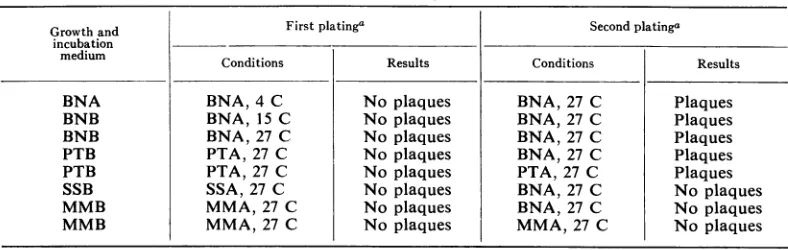
3 hr. This was true regardless of culture age at the time of DNA addition, or the nature of the media used to culture the bacteria (Table 1). Further investigation revealed, however, that some ofthe assay plates contained "intact" in-fectious phage particles after incubation, though plaques werenot apparent. As a consequence, a
second plating was introduced into the transfec-tion assay in all subsequent experiments (see Materials andMethods), so astoallow adequate time for virus expression in the form of plaques. As showninTable 1, only the bacteria originally cultured in SSB and MMB failed to undergo transfection, as indicated by the absence of
plaques afterasecondplating.
Transfectionasobserved abovewascompletely
inhibited by deoxyribonuclease, whereas
ribo-nuclease, trypsin, and LR-4 phage antiserum causedonly minor inhibition (Table 2).
Electronmicroscopy of theparentLR-4phage (from which transfecting DNA was isolated),
and of the transfecting progenyphage, revealed that these phage particles were morphologically
indistinguishable. Furthermore, the progeny
phage andparentphagewerefoundtobe equally susceptibleto neutralization by antisera prepared against LR-4parentphage.
TABLE1. Conditionsleadingtoplaque-formingactivityafterincubation ofA.tumefaciensB66with 22.5 ,ug ofLR-4DNA/ml
Growthand First platinga Second platinga
incubation
medium Conditions Results Conditions Results
BNA BNA, 4 C No plaques BNA, 27 C Plaques
BNB BNA, 15 C No plaques BNA,27 C Plaques
BNB BNA, 27 C No plaques BNA, 27 C Plaques
PTB PTA, 27 C No plaques BNA, 27 C Plaques
PTB PTA, 27 C No plaques PTA, 27 C Plaques
SSB SSA, 27 C No plaques BNA, 27 C No plaques
MMB MMA,27 C No plaques BNA, 27 C No plaques
MMB MMA,27 C No plaques MMA, 27 C No plaques
a SeeMaterials and Methods.
on November 10, 2019 by guest
http://jvi.asm.org/
[image:2.493.58.452.526.651.2]Cultures ofA. tumefaciens B66 were examined forcompetence throughout the completegrowth
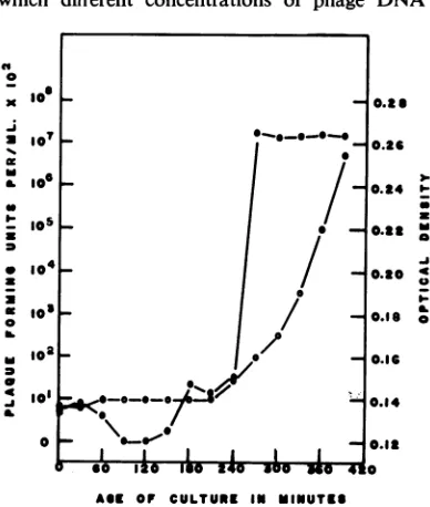
cycle. As shown in Fig. 1 and 2, the ability of
this bacterium to incorporate biologically active phage DNA (22.5
Ag/ml)
and replicate intact infectious phage particles was restricted to aperiod of 2 to 7 hr in the early log phase of
growth in BNB medium. The data indicate that
the bacteria were most competent after 4 hr
of incubationin the abovemedia. Atincubation
times shorterthan 4hr, competence was found to
the maximum. A noticeable degree ofclumping
of cells was observed throughout thecompetent
fluctuate; however, it always remainedless than
period. At maximal competence, optical density
TABLE2. Effectofdeoxyribonuclease, ribonuclease,
trypsin, and LR4 phageantiserum on transfectioninA. tumefaciens B66 by 22.5
lAg
of LR-4 phage DNA permlSubstance added Plaque-formingunits/mi Control... 270
Deoxyribonuclease,
10,ug/ml
0Ribonuclease,
10jug/ml
174Trypsin, 10
jsg/ml
... 164LR-4 phage antiserum, 1:52
di-lution ... 178
a A 1:52dilution neutralized 3.3 X 10⁸
plaque-formingunits of LR-4 phage per ml.
I~
s10
l
°°
3~~~~~~~~
U
U~~~~~~
I-o . . ./. . .
and cell concentration were 0.164 and 4.3 X
105cells/ml, respectively.
Table 3 shows the results of an experiment in
which different concentrations of phage DNA
(I
0 x a a.0
6SI
a-0a
B.
.41
B.
AGE OF CULTURE IN MINUTES
FIG. 2. Transfection competence in A. tumefaciens B66 with LR-4 phage DNA during thelag and early
logphaseof growth. (0) Optical density of the culture at time ofphage addition. (0) Log ofplaque-formning
units permilliliteron secondplating.
0.6
/_ 0.6
/0 0
,o"_ 0.5
z = 0.4 10
I-0.3
0.2 0
0.1
0 I 3 4 5 6 7 * 10 11 12 13 14 IS 1 17 1 1I AGS OF CULTURE IN HOURS
FIG. 1. TransfectioncompetenceinA. tumefaciensB66 with LR-4phageDNAas afunctionofcultureagein
BNBmedium. (0) Optical density ofculture at time ofDNA addition. (0) Logofplaque-formingunitsper milliliteronsecondplating.
3-I..
S A
49 0
I-0
. . . o
19
I I I I I
i
on November 10, 2019 by guest
http://jvi.asm.org/
[image:3.493.256.450.109.338.2] [image:3.493.109.393.397.630.2]were added to bacterial cultures at various
periods ofcompetence. A concentration of 22.5
,ug
ofphage
DNA/ml
gaveoptimal
transfection
frequencies. Thiswas true regardless of the
par-ticularlevel ofcompetence of thebacterial cells.
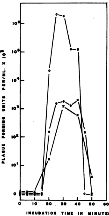
The effect of incubation time on transfection
in A. tumefaciens B66 with LR-4 viral DNA is
shown inFig. 3.Sampleswereremovedfrom the
experimental
culture at 30, 240, and 360 min,and were allowed toincubate with phage DNA
for
various
lengths of time. Transfectionfre-quencies
were found tobe optimal withincuba-bation times between 25 and 30 min,
thereafter
showing a decrease in plaque-forming units.
Inafewcases, however, transfection frequencies
wereobserved to increase again after 90 min of
incubation.
DISCUSSION
Thedatapresented in thispaperclearly
demon-stratethat
extracellular
biologically
active phage DNAcan penetrateviablecells of A. tumefaciens B66.TheextentofDNApenetration varies
with thegrowth
medium,
and isrestrictedto alimitedperiod
in theearly log
phase
ofgrowth.
Theex-tent of DNA uptake is alsoafunction ofDNA
concentration
andincubation time.Competence, as stated
by
Ravin(14),
is anormal
transitory
statein thegrowth
ofaculture.Zelazna (21) reported that in Rhizobium trifolii,
acloserelativeofA.
tumefaciens
(5),
theacquisi-tion of a
specific
celldensity
is essential forattaining
competence for transformation. In the present study, A. tumefaciens competenceTABLE3.Relationbetween LR-4 DNA concentration andtransfection inA. tumefaciensB66
at variousstagesofcompetence
Timesamplewas DNAconcn
Plaque-formiing
removed from
(ug/ml)
units/mi culture(min)30 10 0
180 10 0
240 10 0
300 10 0
360 10 0
30 22.5 8.80 X 10⁵
180 22.5 1.34 X 10⁶
240 22.5 1.28 X 10⁷
300 22.5 1.72 X 10⁹
360 22.5 1.65 X 10⁹
30 45 0
180 45 0
240 45 5.64 X 10³
300 45 322
360 45 242
0 O0 I s o 0
0
I1CU0^T10
10~
0.
FIG. 3. Relationship between incubation time with
LR-4 DNA andtransfectionduringthreestages ofA.
twnefaciens B66 competence. (X) A 0.5-hr culture;
(*)
a4-hrculture; (O) anda6-hrculture.was maximal after cultures reached an
optical
density
of0.164,
from astarting
optical density
of 0.14.
Attempts
tocorrelateaccurately
optical
density
atthetime ofmaximalcompetence
withmean number of cells per milliliterwere
compli-cated
by
extensiveclumping
in the cultures.Clumping,
infact,
was found to be acharac-teristic
property
ofcompetencein thissystem.Possibly
some diffusible substance("transfec-tion
principle")
may be releasedby
cells at aspecific
celldensity,
whichallowsthepopulation
to becomecompetent.
Cell competence factorshave been demonstrated in Bacillus subtilis
(1),
B.
sterothermophilus
(18),
andpneumococci (20),
and appear atleast insome casestobe
proteins.
Wide variations in the concentration of
bac-teriophage
DNArequired
foroptimal
on November 10, 2019 by guest
http://jvi.asm.org/
[image:4.493.263.444.72.430.2] [image:4.493.53.244.456.646.2]tionfrequencies have been reported. Romig (16) observed maximal transfection frequencies when
cells of B. subtilis were infected with DNA
preparations of SP3 in a concentration of 100
,ug/ml.
Meyeretal. (13) found transfection to beoptimal when DNA in a concentration of 35
,ug/ml
was used to infectprotoplasts
ofEsch-erichiacoli.Transfectionfrequenciesinourstudy
were optimal with a concentration of 22.5
Ag
ofphenol-extracted LR-4 phage DNA/ml. Low DNA concentrations (1 and 10 ,ug/ml)
were not sufficient to promote transfection, and
ahigher concentration(45 ,ug/ml)wasinhibitory.
The reasonfor thislatter inhibition isnotknown; however, it is conceivable that high DNA
con-centrations stimulate deoxyribonuclease
pro-ductionorexcretion, orboth.
Transfection was completely inhibited by
deoxyribonuclease, whereas ribonuclease, trypsin, and LR-4phageantiserumresultedinonlyminor inhibition (Table 2). The inhibition observed with ribonuclease andtrypsinmaywellhave been
due to residual deoxyribonuclease in these
pur-chased preparations. Some of the DNA strands in our stock may have had adhering antigenic
proteins, in which case the antisera might have
prevented DNA from penetrating thecompetent
cells. 'The possibility, however, that foreign
pro-teins (antibodies, enzymes, etc.) in themselves
act as inhibitory agents cannot be excluded.
The results from this study indicate thata
30-min incubationperiodyielded maximal
transfec-tion frequencies. The data also suggest (Fig. 3)
that phage DNA may require at least 10 to 15
minofincubation time topenetrateoradhere to
the bacterial cell. In a few
experiments,
lesserpeaks in transfection
frequency
occurred afterprolonged incubation with DNA. This
phenom-enonmightbe dueto
physiological changes
withinthe bacterial cellpopulation, to modifications of
thetransfectingDNAdueto theaccumulation of
extracellular enzymes, or to both.
The necessity for the second, or "indirect,"
plating suggests that during the first
plating
theoriginal transfected viralgenomeeither
replicates
at a slower than normal rate or results in the
productionofonlyafewphage.
Attemptstoencourageplaqueformation in the first plating, by reducing the rate of bacterial growth relativeto therateofviralreplication by plating on suboptimal media and incubating at
suboptimal temperatures, proved unsuccessful
(Table 1). Strauss (17) showed that energy is
requiredfor the entry oftransformingDNAinto
B. subtilis. Transforming DNA may remain in
the extramembrane space beneath the cell
wall,
until energy is made available for it to be
trans-ported across the cell membrane. Epstein (4) suggested that, to incorporate DNA, bacterial cells must make room for the incoming DNA. They accomplish this by halting DNA synthesis for one cell division. The virus DNA that enters the bacterial cell at this time may then have to wait for bacterial DNA synthesis to begin, or for some other specific stage of growth to occur. It is also conceivable that the viral DNA may have to attach to some specific site in the cell in order toreplicate; such seems to be the case with d.x 174 (10). Ifthe number ofreplication sites is assumed tobe limited, it would not be surprising that several bacterialreplication cycles are neces-sary to insure attachment to the appropriate
site. Riggsand Rosenblum(15)reported thatonly
a single viral progeny is obtained from each cell
as a result of transfection in Staphylococcus aureus.If the burst size in this transfection system is also one, this would account for the delay in viralexpression.
TheLR-4 phage stocks have atiter of approxi-mately 4.2 X 10¹¹ infectious
particles,/ml.
Op-timal transfectionfrequenciesareachieved with a concentration of 22.5MAgof LR-4 DNA 'ml. Thisisequivalent to 3.4 X 10¹⁰ viral genomes per ml.
The concentration of bacteria at the period of
optimal competence was 4.3 X 0l
cells/ml.
Thus, 7.9 X 10⁴ viral genomes were required to infect asinglebacterium.
ACKNOWLEDGMENTS
Weare indebted to BarryWilkes forskillful preparation of phage antisera, and to Richard Stephens forassistance in the preparation of electron micrographs.
This work was supported by Public Health Service grants CA 13187-01 from the NationalCancerInstitute andbyagrant from the Jane CoffinChildsMemorial Fund forMedical Research
toG. T.Heberlein.
LITERATURE CITED
1. Akrigg, A.,andS. R.Ayad. 1970.Studiesonthe competence inducingfactorofBacillus subtilis.Biochem. J.117:397-403. 2. Beltra, R.,and J.Rodriguez. 1971.Aseptic induction ofthe
crowngalltumorsbythenucleic acid fractionfrom Agro-bacterium tumefaciens. Phytopathology 70:357-358. 3. Burton, K. 1956. Astudyof theconditionsandmechanism
of thediphenylamine reactionfor thecolorimetric estima-tion of deoxyribonucleic acid. Med. Res. Counc. (Gt. Brit.) Dept. Biochem. 62:315-322.
4. Epstein, H. T. 1968. Factorsaffecting bacterial competence for transfectionand transfection enhancement. Bacteriol. Rev.32:313-319.
5. Heberlein, G. T., J. DeLey, and R. Tijtgat. 1967. Deoxyri-bonucleic acidhomologyand taxonomy ofAgrobacteriuimii, Rhizobium,and Chromobacterium. J.Bacteriol. 94:116-124. 6. Kern,H. 1965.Untersuchungenzurgenetischen Transforma-tion zwischen A. tumefaciens und Rhizobiuin sp. Arch. Mikrobiol.51:140-155.
7. Kern, H. 1969. Interspezifische Transformationen zwischen A. tumefaciens und Rhizobium leguminosarum. Arch. Mikrobiol. 66:63-68.
8. Klein,D. T.. and R. M. Klein. 1953.Transmittanceof tulmiior-21
on November 10, 2019 by guest
http://jvi.asm.org/
MILANI AND HEBERLEIN
inducing abilitytoavirulent crown-gall and related bacteria. J. Bacteriol. 66:220-228.
9. Klein, D.T.,andR. M.Klein. 1956. Quantitativeaspectsof transformation of virulence in Agrobacterium tumefaciens. J.Bacteriol. 72:308-313.
10. Knippers,R., and R. L. Sinsheimer. 1968. Theprocess of infection with bacteriophage <>X 174. Attachment of the parentalDNAofbacteriophagetoafastsedimentingcell component. J. Mol. Biol. 34:17-29.
11. Kovoor, A. 1967. Sur la transformation de tissusnormauxde
Scorsonere provoquee in vitro par l'acide desoxyribonu-cleique d' Agrobacteriumni tuinefaciens. C.R.H. Acad. Sci. 265:1623-1626.
12. Leff, J., and R. Beardsley. 1970. Action tumorigne de l'acide nucleiqued'unbacteriophage presentdans les cultures de
tissu tumoral de tournesol (Helianithus annus). C.R.H.
Acad. Sci. 270:2505-2507.
13. Meyer, F.,R. P. Mackal,M.Tao,and E. A. Evans. 1961.
InfectiousDNAfromlambdabacteriophage.J. Biol. Chem.
236:1141-1143.
14. Ravin, A. W.1961.The genetics of transformation. Advan.
Genet. 10:61-163.
15.Riggs, M. G.,and E. D.Rosenblum. 1969.Transfectionof
lysostaphin treatedcellsofStaphlococcusaureus.J. Virol. 3:33-37.
16. Romig,W. R.1962.Infection of Bacillussubtiliswith phenol
extracted bacteriophages. Virology 16:452-459.
17.Strauss, N. 1970.Early energy-dependentstepin theentryof
transforming DNA. J. Bacteriol. 101:35-37.
18. Streips, U.N.,and N. E.Welker. 1971. Competence-inducing factor of Bacillussterothermophilus. J. Bacteriol 106:955-959.
19. Stroun, M., P. Anker, andG. Auderset.1970.Natural release
of nucleic acids from bacteria into plant cells. Nature (London) 227:607-608.
20. Tomasz, A. 1970. Cellular metabolism in genetic
transforma-tion of pneumococci: requirement for protein synthesis during induction ofcompetence.J. Bacteriol. 101:860- 871. 21. Zelazna, I. 1964. Transformation in Rhizobium trifolii.
Microbiol. Polonica 13:283-289.
J. VIROL.