Int. J. Electrochem. Sci., 14 (2019) 2405 – 2413, doi: 10.20964/2019.03.41
International Journal of
ELECTROCHEMICAL
SCIENCE
www.electrochemsci.org
Electrochemical Myoglobin Biosensor Based on Magnesium
Metal-Organic Frameworks and Gold Nanoparticles Composite
Modified Electrode
Guiling Luo1, Hui Xie1, Yanyan Niu1, Juan Liu2, Yaqi Huang1, Binghang Li1, Guangjiu Li2, Wei Sun1*
1 Key Laboratory of Laser Technology and Optoelectronic Functional Materials of Hainan Province,
Key Laboratory of Functional Materials and Photoelectrochemistry of Haikou, College of Chemistry and Chemical Engineering, Hainan Normal University, Haikou 571158, P R China
2 Key Laboratory of Optic-electric Sensing and Analytical Chemistry for Life Science of Ministry of
Education, College of Chemistry and Molecular Engineering, Qingdao University of Science and Technology, Qingdao 266042, P R China
*E-mail: [email protected]
Received: 10 December 2018 / Accepted: 14 January 2019 / Published: 7 February 2019
A carbon ionic liquid electrode (CILE) made with 1-hexylpyridinium hexafluorophosphate was used as the substrate electrode. The magnesium metal-organic framework (Mg-MOF-74) was casted on CILE surface and gold nanoparticles (AuNPs) was further assembled on electrode through electrodeposition to construct a nanocomposite interface. Myoglobin (Mb) was further modified on AuNPs/Mg-MOF-74/CILE and Nafion film was used to get the working electrode (Nafion/Mb/AuNPs/Mg-MOF-74/CILE). Morphology of the nanoparticles was recorded by SEM. UV-Vis and FT-IR spectroscopy proved that Mb on the composite film retained its original structure. The Mb on AuNPs/Mg-MOF-74/CILE displayed a pair of redox peaks with the formal peak potential of -0.158 V (vs. Ag/AgCl) in 0.1 mol/L pH 2.0 PBS. Mb remained the bioactivity and electrocatalyzed the reduction of trichloroacetic acid and sodium nitrite with relative wide concentration range and low detection limit. The fabricated electrochemical biosensor gave high stability and reproducibility, which extended the application of MOF based composite in electroanalysis.
Keywords: Magnesium metal-organic framework, Gold nanoparticle, Myoglobin, Electroanalysis, Trichloroacetic acid
1. INTRODUCTION
as model molecule for the investigation on direct electron-transfer (DET) of hemoproteins or redox enzymes [2]. In general, electron transfer of Mb with a conventional electrode is difficult, which is due to the macro-biostructures that prevent the electron movement from heme active center to the electrode, or the adsorption and passivation on the electrode. To overcome these questions, nanomaterials modified electrodes are devised for proteins electrochemistry. Gold nanoparticles (AuNPs) have been widely reported due to its unusual optical and electrochemical properties. Because of their size dependent electrochemistry and high chemical stability, AuNPs have been the model in various fields including self-assembly, catalysis and electron-transfer theories [3,4]. Various procedures have been devised for AuNPs synthesis. Among them electrodeposition can not only provide a fast and convenient process for electrode modification with strong adherence on the electrodes surface, but also make the biosensor more stability [5]. As one of the hot topics in material chemistry, metal-organic frameworks (MOFs) exhibit the properties including large specific surface areas, regular pores, high porosity and stable framework [6]. M-MOF-74 contains series iso-structure with different metals (M= Mn, Co, Ni, Mg, Zn) and organic ligand 2,5-dihydroxyterephthalic acid (DHTP) [7]. The metals inside MOF-74 form six-coordinated with five oxygen atoms in DHTP and one solvent molecule [8]. Due to its highest density of metal sites and high stability, MOF-74 has been used in adsorption [9], separation [10], storage [11] and heterogeneous catalysis [12].
In the present work, a carbon ionic liquid electrode (CILE) was home-made with 1-hexylpyridinium hexafluorophosphate (HPPF6). Then Mg-MOF-74 was applied on the CILE surface to
construct the biocompatible interface (MOF-74/CILE). The further formation of AuNPs on Mg-MOF-74/CILE was achieved by electrodeposition to get AuNPs/Mg-Mg-MOF-74/CILE, which was dried in air with Mb and Nafion further casted on the surface step-by-step. After dried in air, Nafion/Mb/AuNPs/Mg-MOF-74/CILE was got, which displayed fast electron transfer and prominent catalytic ability to trichloroacetic acid (TCA) and sodium nitrite (NaNO2). Therefore this paper
proposed a new way to extend the application of Mg-MOF-74 in electrochemical sensors and the fabrication procedure of this Nafion/Mb/AuNPs/Mg-MOF-74/CILE was depicted in the following scheme 1.
Scheme 1. Fabrication procedure of Nafion/Mb/AuNPs/Mg-MOF-74/CILE.
2. EXPERIMENTAL
2.1 Reagents and apparatus
Tech. Co., China), Mg-MOF-74 (2~200 nm, Nanjing XFNANO Materials Tech. Co., China), HPPF6
(Lanzhou Yulu Fine Chem. Co., China), graphite powder (particle size 30 nm, Shanghai Colloid Chem. Co., China), chloroauric acid (Shanghai Chem. Reagent Research Institute Ltd. Co., China) and TCA (Tianjin Kemiou Chem. Co., China) were used as received. 0.1 mol/L phosphate buffer solutions (PBS) were the supporting electrolyte and the reagents used of analytical grade without purification.
Voltammetry was performed on a CHI 604E electrochemical analyzer (Shanghai Chenhua Co., China). A conventional three-electrode model was employed with a modified CILE as working electrode, a platinum wire as auxiliary electrode, and a saturated Ag/AgCl electrode as reference. FT-IR spectra were recorded on a Nicolet 6700 spectrometer (Thermo Fisher Scientific Inc., USA) with UV-Vis spectra on a TU-1901 double-beam spectrometer (Beijing Purkinje General Instrument Co., China). The morphology of the sample was performed with a JSM-7100F scanning electron microscope (Japan Electron Co., Japan).
2.2 Construction of biosensor
CILE was home-manufactured on the basis of a procedure with ionic liquid HPPF6 [13]. 6 µL
of 0.3 mg/mL Mg-MOF-74 suspension was coated on CILE and the electrode was dried under room temperature to acquire Mg-MOF-74/CILE. The modification of Mg-MOF-74/CILE by AuNPs was carried out by electrodeposition using control-potential method at potential of -0.3 V for 200 s with a 2.0 mmol/L HAuCl4 solution. Then 15.0 mg/mL Mb solution was dropped onto
AuNPs/Mg-MOF-74/CILE and stored for 10 h at 4 ℃ to get Mb/AuNPs/Mg-MOF-74/CILE. Finally, 6.0 µL of 0.5% Nafion ethanol solution was casted on electrode to form a stable membrane. For comparison, Nafion/Mb/CILE, Nafion/Mb/AuNPs/CILE and Nafion/Mb/Mg-MOF-74/CILE were prepared with similar procedure without the adding AuNPs or Mg-MOF-74, respectively. All electrodes were washed with deionized water and putted in a 4 ℃ refrigerator when not in use.
3. RESULTS AND DISCUSSION
3.1 SEM characterization
Figure 1. SEM images of (A) Mg-MOF-74 and (B) AuNPs/Mg-MOF-74/CILE.
3.2 UV-Vis and FT-IR characterization
Spectroscopic methods are commonly used to probe the structure change of heme proteins [14]. In Fig. 2A UV-Vis absorption spectra of Mb in solution (curve a) and Mb/Mg-MOF-74 mixture (curve b) showed the typical Soret band at 408.5 nm, indicated that Mb mixture with Mg-MOF-74 retained its biological activity without denaturation. FT-IR spectra of Mb and Mb/Mg-MOF-74 were shown in Fig. 2B. The peaks at 1648.92 and 1529.35 cm−1 could be ascribed to amide I and II infrared
absorbance of Mb (Fig. 2B a), which had similar values with that of Mb/Mg-MOF-74 composite (1650.21 and 1525.49 cm−1, Fig. 2B b). Therefore spectroscopic results proved that Mb kept its
[image:4.596.149.454.410.523.2]original structure after contacted with Mg-MOF-74.
Figure 2. (A) UV-Vis absorption spectra of Mb (curve a) and Mb/Mg-MOF-74 mixture (curve b) in water; (B) FT-IR spectra of Mb (a) and Mb/Mg-MOF-74 (b).
3.3 EIS results
Figure 3. EIS of (a) Nafion/AuNPs/CILE, (b) Nafion/Mb/AuNPs/Mg-MOF-74/CILE, (c) Nafion/Mb/CILE, (d) Nafion/Mg-MOF-74/CILE, (e) CILE in 0.01 mol/L K3[Fe(CN)6]/K4[Fe(CN)6] and 0.1 mol/L KCl mixed solution with the frequencies ranging from
0.1 to 104 Hz.
3.4 Effect of pH
The pH of supporting electrolyte can influence three-dimensional structure of redox protein and the resulting electrochemical response [15]. Cyclic voltammetric (CV) responses were checked in pH range (2.0~8.0) with the changes of curve shape (Fig. 4). In pH 2.0 PBS the symmetrical cyclic voltammograms (CVs) could be observed with largest peak current, which proved that the configuration were related to H2O and the protonation of amino acids around the heme active group,
defined as the Redox Bohr Effect [16]. The dependence of formal peak potential upon pH resulted in a linear regression equations as E0’ (V)=-0.076-0.0407pH (γ=0.996). The slope (40.7 mV/pH) was
smaller than theoretical data (59.2 mV/pH) at 25 ◦C for a single-proton reversible single-electron
transfer process [17]. The reason might be the influence of the protonation states of transligands to the heme iron and amino acids around the heme, or the protonation of H2O molecule coordinated to the
central iron [18].
Figure 4. CVs of Nafion/Mb/AuNPs/Mg-MOF-74/CILE in different pH PBS (from a to g: 2.0, 3.0, 4.0, 5.0, 6.0, 7.0, 8.0) at the scan rate of 100 mV/s.
3.5 Direct electrochemistry
[image:5.596.203.393.73.200.2] [image:5.596.216.367.530.656.2]
currents were present at Nafion/CILE (curve a) and Nafion/AuNPs/Mg-MOF-74/CILE (curve b). On Mb based electrode a pair of redox peaks presented was resulted from DET of Mb with electrode. On Nafion/Mb/AuNPs/Mg-MOF-74/CILE (curve d) the currents were larger than that of Nafion/Mb/CILE (curve c) with good shape, proving positive effects of AuNPs/Mg-MOF-74 on the electron transfer. The presence of AuNPs/Mg-MOF-74 resulted in a rough interface with good conductivity that was benefit for electron communication. At Nafion/Mb/AuNPs/Mg-MOF-74/CILE (curve d) the peak potentials were -0.207 (Epc) and -0.108 V (Epa) with △Ep of 99 mV and E0’ of -0.158 V, the typical
peak of heme Fe(II)/Fe(III) redox couples [19]. Therefore AuNPs/Mg-MOF-74 nanohybrid provided a suitable interface for Mb to transfer electrons with the electrode at faster rate.
Figure 5. (A) CVs of (a)Nafion/CILE, (b)Nafion/AuNPs/Mg-MOF-74/CILE, (c)Nafion/Mb/CILE, (d)Nafion/Mb/AuNPs/Mg-MOF-74/CILE in pH 2.0 PBS at the scan rate of 100 mV/s; (B) CVs of Nafion/Mb/AuNPs/Mg-MOF-74/CILE with different scan rates (from a to j: 100, 200, 300, 400, 500, 600, 700, 800, 900, 1000 mV/s).
Fig. 5B showed CVs of Nafion/Mb/AuNPs/Mg-MOF-74/CILE in pH 2.0 PBS at different scan rates. In scan rate from 100 to 1000 mV/s, the redox peak potentials of Mb shifted with the △Ep increased. The redox peak potential (Ep) increased linearly with ln ν, and the redox peak currents (Ip) increased linearly with scan rates, indicating a surface-controlled process. According to Faraday’s law, Q=nFA* [20], the surface concentration (*) of electroactive Mb was calculated as 5.36×10⁻¹¹
mol/cm2, bigger than that of single-layer coverage (2.0×10⁻¹¹ mol/cm2) [21].
3.6 Electrocatalysis to reduction of TCA
[image:6.596.148.440.235.339.2]
Electrocatalysis of Nafion/Mb/AuNPs/Mg-MOF-74/CILE to reduction of NaNO2 was recorded
with the curves present in Fig. 6B. When NaNO2 was added into pH 2.0 PBS, a new reduction peak
appeared at -0.535 V, indicating a promoting action of immobilized Mb to the electrochemical reduction of NaNO2. The linear relationship of the electrocatalytic reduction current and the NaNO2
[image:7.596.171.412.186.296.2]concentration was from 0.8 to 18.0 mmol/L with regression equation as Ipa (µA) = 5.37 + 115.68C (mmoL/L) (n=11, γ=0.999), the detection limit as 0.267 mmol/L (3σ) and KMapp as 10.25 mmol/L.
Figure 6. (A) CVs of Nafion/Mb/AuNPs/Mg-MOF-74/CILE in pH 2.0 PBS with 4, 10, 20, 30, 40, 50, 60, 80, 100, 120, 150, 170, 200 mmol/L TCA (curves a-m) at the scan rate of 100 mV/s. Inset: Linear relationship of catalytic currents vs. TCA concentration; (B) CVs of Nafion/Mb/AuNPs/Mg-MOF-74/CILE in pH 2.0 PBS with 0.8, 1.5, 2.0, 3.0, 4.0, 5.0, 6.0, 7.0, 8.5, 10.0, 16.0, 18.0 mmol/L NaNO2 (curves a-l) at the scan rate of 100 mV/s. Inset: Linear
relationship of catalytic currents vs. NaNO2 concentration.
3.7 Stability and reproducibility
The reproducibility of this modified electrode was studied by successfully scanning for 200 cycles, which gave no obvious changes in CVs. Under the same conditions, six electrodes were home-made one-by-one, which gave the RSD of 3.1% for the analysis of 80.0 mmol/L TCA. Therefore this modified electrode exhibited excellent stability and reproducibility.
3.8 Applications in sample analysis
The TCA contented in the medical skin lotion (35%, Shanghai EKEAR Bio. Tech. Co., China) was tested by the proposed method. As shown in table 1, the samples were diluted by pH 2.0 PBS and used directly. The recovery was determined by adding the standard TCA solution to the sample with the results between 102.6% and 104.7%, indicating the practical application.
Table 1. Test results of TCA in medical skin lotion. (n=3)
Sample (mmol/L) Labled (mmol/L) Detected (mmol/L) Added (mmol/L) Total Recovery (%)
35% TCA 30.00 30.06
10.00 41.10 102.6
20.00 52.14 104.7
[image:7.596.42.556.660.771.2]
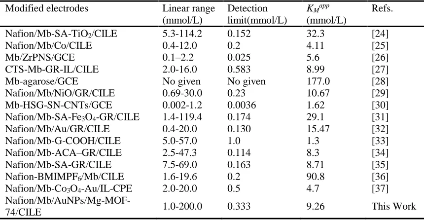
4. CONCLUSIONS
In this paper AuNPs and Mg-MOF-74 were modified on the CILE surface and used as accelerator for Mb electrochemistry. The resulted Nafion/Mb/AuNPs/Mg-MOF-74/CILE exhibited excellent electron-transfer rate for Mb with improved bioelectrochemical catalytic effects to TCA and NaNO2. The Mg-MOF-74 on the electrode gave a rough interface and electrodeposited AuNPs could
modulate the interfacial conductivity. A systematic comparison of the electrochemical and analytical parameters of this biosensor with other Mb modified electrodes was listed in table 2, which showed widely linear range and relatively low detection limit. Therefore AuNPs/Mg-MOF-74 composite exhibited as excellent candidate for the preparation of electrochemical enzyme sensors.
Table 2. Comparison of electrochemical parameters of different Mb modified electrodes to TCA detection. (n=3)
Modified electrodes Linear range
(mmol/L)
Detection limit(mmol/L)
KMapp (mmol/L)
Refs.
Nafion/Mb-SA-TiO2/CILE 5.3-114.2 0.152 32.3 [24]
Nafion/Mb/Co/CILE 0.4-12.0 0.2 4.11 [25]
Mb/ZrPNS/GCE 0.1–2.2 0.025 5.6 [26]
CTS-Mb-GR-IL/CILE 2.0-16.0 0.583 8.99 [27]
Mb-agarose/GCE No given No given 177.0 [28]
Nafion/Mb/NiO/GR/CILE 0.69-30.0 0.23 10.67 [29]
Mb-HSG-SN-CNTs/GCE 0.002-1.2 0.0036 1.62 [30]
Nafion/Mb-SA-Fe3O4-GR/CILE 1.4-119.4 0.174 29.1 [31]
Nafion/Mb/Au/GR/CILE 0.4-20.0 0.130 15.47 [32]
Nafion/Mb-G-COOH/CILE 5.0-57.0 1.0 1.3 [33]
Nafion/Mb-ACA–GR/CILE 2.5-47.3 0.114 8.3 [34]
Nafion/Mb-SA-GR/CILE 7.5-69.0 0.163 8.71 [35]
Nafion-BMIMPF6/Mb/CILE 1.6-19.6 0.2 90.8 [36]
Nafion/Mb-Co3O4-Au/IL-CPE 2.0-20.0 0.5 4.7 [37]
Nafion/Mb/AuNPs/Mg-MOF-74/CILE 1.0-200.0 0.333 9.26 This Work
ACKNOWLEDGEMENT
This project was financially supported by the Hainan Provincial National Natural Science Foundation of China (2017CXTD007), the Key Science and Technology Program of Haikou City (2017042), Graduate Student Innovation Research Project of Hainan Province (Hys2018-212) and Research Fund from Beijing Innovation Center for Future Chips (KYJJ2018006).
References
1. A. K. Yagati, T. Lee, J. Min, J. W. Choi, Biosens. Bioelectron., 47(2013)217. 2. H. Liu, L. Weng, C. Yang, Microchim. Acta, 184(2017)1267.
3. S. Guo, E. Wang, Anal. Chim. Acta, 598(2007)181.
4. M. Grzelczak, J. Pérez-Juste, P. Mulvaney, L. M. Liz-Marzán, Chem. Soc. Rev., 37(2008)1783. 5. C. Liu, X. Guo, H. Cui, R. Yuan, J. Mol. Catal. B-Enzym., 60(2009)151.
6. U. Mueller, M. Schubert, F. Teich, H. Puetter, K. Schierlearndt, J. Pastré, Chem. inform, 16(2006)626.
7. H. Wu, W. Zhou, T. Yildirim, J. Am. Chem. Soc., 131(2009)4995.
[image:8.596.83.513.296.519.2]
8(2016)26817.
9. L. Valenzano, B. Civalleri, S. Chavan, G. T. Palomino, C. Areán, S. Bordiga, J. Phys. Chem. C., 114(2010)11185.
10.E. D. Bloch, W. L. Queen, R. Krishna, J. M. Zadrozny, C. M. Brown, J. R. Long, Science, 335(2012)1606.
11.T. Pham, K. A. Forrest, R. Banerjee, G. Orcajo, J. Eckert, B. Space, J. Phys. Chem. C, 119(2014)1078.
12.J. Kim, S. N. Kim, H. G. Jang, G. Seo, W. S. Ahn, Appl. Catal. A. Gen., 453(2013)175. 13.W. C. Wang, X. Q. Li, X. H. Yu, L. J. Yan, Z. F. Shi, X. Y. Wen, W. Sun, J. Chin. Chem. Soc.,
63(2016)298.
14.P. George, G. A. Hanania, Biochem. J., 55(1953)236.
15.H. H. Liu, Z. Q. Tian, Z. X. Lu, Z. L. Zhang, M. Zhang, D. W. Pang, Biosens. Bioelectron., 20(2004)294.
16.Y. X. Sun, S. F. Wang, Bioelectrochemistry, 71(2007)172.
17.A. M. Bond, Modern polarographic methods in analytical chemistry, CRC Press, 1980. 18.C. I. Yamazaki, T. Araiso, Y. Hayashi, H. Yamada, R. Makino, Adv. Biophys., 11(1978)249. 19.H. Zhang, H. Lu, N. Hu, J. Phys. Chem. B., 110(2006)2171.
20.A. J. Bard, L. R. Faulkner, Electrochemical Methods, Fundamentals and Applications, 2ed edition, Wiley, (2001) New York, the United States of America.
21.S. F. Wang, T. Chen, Z. L. Zhang, X. C. Shen, Z. X. Lu, D. W. Pang, K. Y. Wong, Langmuir., 21(2005) 9260.
22.W. Sun, Y. Q. Guo, X. M. Ju, Y. Y. Zhang, X. Z. Wang, Z. F. Sun, Biosens. Bioelectron., 42(2013)207.
23.R. A. Kamin, G. S. Wilson. Anal. Chem., 52(1980)1198.
24.H. Q. Yan, X. Q. Chen, Z. F. Shi, Y. H. Feng, J. C. Li, Q. Lin, X. H. Wang and W. Sun, J. Solid State Electrochem., 20(2016)1783
25.W. Sun, X. Q. Li, P. Qin and K.Jiao, J. Phys. Chem. C., 113(2009)11294 26.Y. H. Zhang, X. Chen and W. S. Yang, Sens. Actuators, B., 130(2008)682
27.C. X. Ruan, T. T. Li, Q. J. Niu, M. Lu, J. Lou, W. M. Gao and W. Sun, Electrochim. Acta, 64(2012)183
28.S. F. Wang, T. Chen, Z. L. Zhang, X. C. Shen, Z. X. Lu, D. W. Pang and K. Y. Wong, Langmuir, 21(2005)9260
29.W. Sun, S. X. Gong, Y. Deng, T. T. Li, Y. Cheng, W. C. Wang and L. Wang, Thin Solid Films, 562(2014)653
30.C. Y. Liu and J. M. Hu, Biosens. Bioelectron., 24(2009)2149
31.X. Q. Chen, H. Q. Yan, Z. F. Shi, Y. H. Feng, J. C. Li, Q. Lin, X. H. Wang and W. Sun, Polym. Bull., 74(2016)1
32.G. N. Li, T. T. Li, Y. Deng, Y. Cheng, F. Shi, W. Sun and Z. F. Sun, J. Solid State Electrochem., 17(2013)2333
33.W. Zheng, W. S. Zhao, W. Chen, W. J. Weng, Z. W. Liao, R. X. Dong, G. J. Li and W. Sun, Int. J. Electrochem. Sci., 12(2017)4341
34.X. Q. Chen, H. Q. Yan, W. Sun, G. Y. Chen, C. J. Yu, W. Feng and Q. Lin, RSC Adv., 8(2018)38003 35.X. Q. Chen, M. X. Feng, H. Q. Yan, W. Sun, Z. F. Shi and Q. Lin, Int. J. Electrochem. Sci.,
12(2017)11633
36.W. Sun, X. Q. Li and K. Jiao, Electroanalysis, 21(2009)959

![Figure 3. EIS of (a) Nafion/AuNPs/CILE, (b) Nafion/Mb/AuNPs/Mg-MOF-74/CILE, (c) Nafion/Mb/CILE, (d) Nafion/Mg-MOF-74/CILE, (e) CILE in 0.01 mol/L K3[Fe(CN)6]/K4[Fe(CN)6] and 0.1 mol/L KCl mixed solution with the frequencies ranging from 0.1 to 104 Hz](https://thumb-us.123doks.com/thumbv2/123dok_us/1758136.129400/5.596.203.393.73.200/figure-nafion-nafion-nafion-nafion-solution-frequencies-ranging.webp)