Review Article
Role of toll-like receptors in multiple sclerosis
Socorro Miranda-Hernandez, Alan G Baxter
Comparative Genomics Centre, Molecular Sciences Building 21, James Cook University, Townsville, QLD 4811, Australia
Received December 12, 2012; Accepted January 16, 2013; Epub February 27, 2013; Published March 9, 2013
Abstract: Multiple Sclerosis (MS) is an autoimmune disease in which Central Nervous System (CNS) lesions result from perivascular immune cell infiltration associated with damage to myelin, oligodendrocytes and neurons. CNS autoimmunity and its regulation are dominated by the inflammatory cytokines IL17 and IFNγ, and the opposing regulatory cytokines IL10 and the type I IFNs. Toll-like receptors (TLR) play a critical role in modulating cytokine and chemokine secretion in response to exogenous Pathogen Associated to Molecular Patterns and endogenous Danger-Associated to Molecular Patterns. Here, we systematically examine the evidence that TLR play a major role in the initiation disease, the triggering of relapses, and regulation of CNS damage. Data from human studies are supported analyses of a variety of animal models, including Experimental Autoimmune Encephalomyelitis in TLR-deficient mice.
Keywords: Multiple sclerosis, toll-like receptors, hygiene hypothesis, microbial flora, gene/environment interac-tions, autoimmunity, TLR1, TLR2, TLR3, TLR4, TLR5, TLR6, TLR7, TLR8, TLR9, TLR10
Introduction
Multiple Sclerosis (MS) is an autoimmune dis-ease in which Central Nervous System (CNS) lesions result from perivascular immune cell infiltration associated with damage to myelin, oligodendrocytes and neurons. Clinically, symp -toms include numbness, weakness, loss of muscle coordination, and problems with vision, speech, and bladder control. The timing and severity of each attack are unpredictable, and can vary in severity from being detected only by magnetic resonance imaging or neural conduc -tion studies, to being devastating, leaving the patient severely disabled. Three patterns of dis -ease are seen: relapsing-remitting (RRMS), in which episodic exacerbations are separated by periods of recovery, secondary progressive (SPMS), which can develop in patients who ini-tially present with RRMS and is characterized by progressive disability, and primary progres -sive (PPMS), in which disability progresses steadily from disease onset.
The evidence that MS is an autoimmune dis -ease includes the following features it shares with other autoimmune diseases: an HLA asso
-ciation [1]; asso-ciations with other autoimmune diseases and their autoantibodies [2]; the pres-ence of oligoclonal autoreactive T and B cell expansion in the target organ [3, 4]; therapeutic efficacy of corticosteroids [5], plasmapheresis [6] and antilymphocyte globulin [7]; and cure by lymphoid ablation and autologous haematopoi -etic cell transplantation [8].
In MS, leukocytes such as monocytes, dendritic cells (DCs), NK cells, CD4+ and CD8+ T cells and
B cells, migrate to the CNS and mediate myelin destruction, axon damage and neuronal cell
[image:2.612.105.516.74.566.2]77 Am J Clin Exp Immunol 2013;2(1):75-93 TLR expression in the CNS has also been impli
-cated in neuro-protective and restorative func-tions [21-23].
Toll-like receptor one in multiple sclerosis
TLR1 is expressed as a heterodimer with TLR2, and binds bacterial triacyl lipopeptides. It is widely expressed on the APC monocytes, mac-rophages, DC and B cells. It is also expressed on Human NT2-N and CHP-212 neuronal cell lines [19, 20, 24] and has been identified by RT PCR on microglia [25]. TLR1 is down regulated in peripheral blood mononuclear cells (PBMC) of MS patients and up regulated in patients treated with INFβ [26, 27].
Toll-like receptor two in multiple sclerosis
TLR2 is expressed as both a homodimer and as a heterodimer, partnered with either TLR1 or TLR6. It is expressed on monocytes, macro -phages and myeloid DC, and can bind a wide range of ligands, including lipoteichoic acid from Gram-positive bacteria, bacterial lipopep-tides and glycolipids, fungal beta glucan (zymo -san) and the endogenous DAMPs Hyaluronan, HSP70 and HMGB1. TLR2 has been identified on CNS endothelial cells, microglia, astrocytes and oligodendrocytes [25, 28] and on infiltrat -ing cells in MS (Figure 1). It is up regulated on PBMCs, cerebrospinal fluid (CSF) mononuclear cells and in demyelinating lesions of MS patients [19, 20, 24, 25, 29].
MS relapses have been reported during bacte -rial infections [30]. Monocyte-derived dendritic cells from MS patients with bacterial infections express high levels of HLA-DR and costimula -tors than those from uninfected patients and drive higher production of IL12, IL17 and INFγ [31]. Several TLR2 ligands have been identified in the brains and CSF of MS patients. For exam-ple, the TLR2 ligand peptidoglycan, a major component of the Gram-positive bacterial cell walls, has been reported as present in the brains of MS patients within macrophage/ DC-like APC that express co-stimulatory mole -cules (CD80, CD86 and CD40) and proinflam -matory cytokines (IL1α, IL6, IL12, TNF and INFγ; [32]. High numbers of macrophages and microglia expressing the endogenous TLR2 ligand HMGB1 are also found in acute and RRMS [33]. The migration and differentiation of oligodendrocyte precursor cells (OPC) play
important roles in myelin repair after inflamma -tory damage. MS lesions contain hyaluronan deposits that, once fragmented by the hyal -uronidases expressed by OPCs, inhibit the mat -uration of OPC and remyelination via TLR2 liga -tion [10, 29, 34].
The TLR2 ligand zymosan can modulate the severity of MS by inducing peripheral blood DC from MS patients treated with INFβ to secrete IL10, which suppresses IL23 and IL1β produc -tion [35]. Similarly, surface expression of TLR2 on B cells and DC was significantly higher in helminth-infected MS patients, who had better clinical and radiological outcomes than unin -fected patients. Protection was associated with regulatory T cell induction and increased TGFβ and IL10 levels. In contrast, immunization with S. pneumoniae exacerbates MS [36].
Toll-like receptor three in multiple sclerosis
TLR3 is expressed in DC and B cells and binds double-stranded (viral) RNA and the endoge -nous microtubule regulator stathmin. TLR3 liga -tion induces the activa-tion of NF-κB via the adaptor TRIF to increase production of type I interferons. Cerebral endothelial cells [28], neurons, microglia, astrocytes and oligoden -drocytes express TLR3 [19, 25, 37, 38]. Normal adult human astrocytes increase the produc-tion of anti-inflammatory cytokines such as IL10 and downregulate proinflammatory cyto -kines such as IL12 (p40) and IL23 in response to TLR3 ligation [25]. The endogenous TLR3 ligand stathmin was identified in astrocytes, microglia, and neurons of MS-affected human brain, and was shown by cDNA arrays to initiate the same set of neuroprotective factors as the synthetic TLR3 agonist polyinosinic: polycyti -dylic (poly I:C) acid [39].
Association studies of TLR3 sequence variants have failed to identify any significant associa -tion with MS [40, 41].
Toll-like receptor four in multiple sclerosis
TLR4 expression has been identified in cerebral endothelial cells [28] and microglia by RT-PCR [25]. Both TLR4 and its endogenous ligand HMGB1 are increased in expression in the CSF mononuclear cells of MS patients compared to healthy controls [33]. Association studies of functional (missense) mutations in TLR4 (Asp299Gly and Thr399Ile) failed to identify any association with MS [42, 43]. A subsequent study of nine TLR4 single nucleotide polymor -phisms (SNP) tested for association with MS in 362 MS patients and 467 healthy controls also failed to identify any significantly associated loci [44].
Toll-like receptor five in multiple sclerosis
TLR5 binds bacterial flagellin and is expressed on monocytes and macrophages, some DC and intestinal epithelium; its expression has been identified in microglia by RT-PCR [25]. Little has been published on any role it may play in MS.
Toll-like receptor six in multiple sclerosis
TLR6 is expressed on monocytes and macro -phages, B cells and mast cells and it binds to diacyl lipopeptides from Mycoplasma. It has been identified in cerebral endothelial cells and microglia by RT-PCR [25]. The TLR6 SNP rs5743810 was associated with the develop-ment of INFβ -specific neutralizing antibodies in men but not in women after 24 month of treat-ment with INFβ [23].
Toll-like receptor seven in multiple sclerosis
TLR7 is expressed in monocytes and macro -phages, plasmacytoid DC and B cells, and binds to single-stranded (viral) RNA. TLR7 expression has been identified in microglia by RT-PCR [25].
The pro-inflammatory cytokine IL17 plays a crit -ical role in the immunopathogenesis of MS and EAE [45-49] and its production is downregulat -ed by type I IFNs [50, 51]. In vitro treatment of human monocyte-derived DCs with IFNβ1a induced the expression of TLR7 and, in a TLR7-dependent fashion, the members of its down-stream signaling pathway (MyD88, IRAK4, and TRAF6), but inhibited the expression of IL1R. TLR7 expression was also necessary for IFNβ1a-induced secretion of IL27 by DCs and the inhibition of IL1β and IL23. Supernatants
from IFNβ1a-treated DCs inhibited Th17 differ -entiation of CD4 T cells, with down regulation of retinoic acid-related orphan nuclear hormone receptor C (RORC) and IL17A gene expression and IL17A secretion. Again, inhibition of IL17A was TLR7 dependent and could be blocked by TLR7 siRNA silencing [52].
At the onset of MS, a subset of patients (11 of 61) expressed elevated mRNA levels of TLR7, together with RIG-1 and IFIH1 – an IFN expres -sion signature potentially attributable to an overactivity of IFN-stimulated gene factor 3 (ISGF3, a complex formed by STAT1, STAT2 and IFN regulatory factor 9). This phenotype was shared by a subset of healthy control subjects [53]. Patients with a relatively high IFN expres -sion signature at baseline showed no signifi -cant modulation in the expression of the genes involved in IFN -related pathways during IFNβ therapy. In contrast, patients with a low endog -enous IFN gene signature showed strong gene induction after 1 month of treatment [53].
Toll-like receptor eight in multiple sclerosis
TLR8 is expressed on monocytes and macro -phages, a subset of DC and mast cells; it binds to single stranded (viral) RNA. TLR8 expression has also been identified in microglia by RT-PCR [25]. As is the case for TLR7, at the onset of MS, a subset of patients and healthy controls express elevated mRNA levels of TLR8, as part of an endogenous IFN gene signature [53].
Toll-like receptor nine in multiple sclerosis
79 Am J Clin Exp Immunol 2013;2(1):75-93 Following stimulation of TLR9 by CpG-DNA (with
or without stimulation via the B cell receptor and CD40), the B cells of MS patients secrete more lymphotoxin (LT), TNF and IL12, and less IL10, than those of healthy controls [57, 58]. The TLR9-stimulated production of IL10 corre -lates with TLR9 expression levels in CD27+
(memory) B cells, which is significantly reduced in MS patients [58].
Toll-like receptor ten in multiple sclerosis
Little is known about TLR10, its tissue distribu -tion, its specificity or any possible role in MS.
Toll-like receptor eleven in multiple sclerosis
TLR11 is expressed on monocytes and macro -phages, as well as in the liver and in kidney and urinary bladder epithelial cells. It binds profilin from the parasite Toxoplasma gondii and an unidentified ligand from uropathogenic Escherichia coli. Little has been published on any role TLR11 may play in MS.
Animal models of multiple sclerosis
Clinical research of MS is supported by animal models of the pathogenesis and immunoregu -lation of CNS autoimmunity. Commonly used models in the research of MS include:
Cuprizone
Cuprizone is a copper chealator that induces oligodendrocyte cell death, reversible demye -lination, axonal injury and microglial activation [59, 60]. It is used to model aspects of demye-lination, OPG migration and activation, and remyelination.
Theiler’s murine encephalomyelitis virus
Theiler’s murine encephalomyelitis virus (TMEV) is a single stranded RNA murine cardio -virus used to infect genetically susceptible mice (e.g. SJL strain) intra-cerebrally, resulting in persistent infection and chronic demyelinat-ing disease [61]. The TMEV DA strain infects astrocytes, microglia and macrophages, result -ing in chronic encephalomyelitis that resembles the chronic and progressive forms of MS [62, 63]. The disease can also be induced in resis -tant mouse strains (e.g. C57BL/6 strain) by activating innate immunity with two LPS injec -tions after TMEV infection [61].
Experimental autoimmune encephalomyelitis
Experimental autoimmune encephalomyelitis (EAE) is a family of models of autoimmune CNS damage induced by the immunization of experi -mental animals with CNS components (either an extract, purified protein or a peptide), usu -ally in the presence of an adjuvant [64].
Experimental autoimmune encephalomyelitis
The pathogenesis of CNS damage caused by EAE, and the specific CNS components target -ed, are affected by strain and species of ani-mal, choice of antigen (e.g. myelin oligodendro -cyte glycoprotein (MOG), proteolipid protein (PLP) or myelin basic protein (MBP)) and adju -vant (e.g. Complete Freund’s Adju-vant (CFA), CpG, LPS or Pertussis toxin (PTX)), and whether the disease studied is active (i.e. that occurring in the immunised individual) or passive (i.e. that occurring in the recipient of adoptively transferred T cells from the immunised individ -ual). Various combinations can result in mono -phasic, relapsing–remitting or chronic EAE [65, 66]
As a generalization, EAE in mice is associated with an autoimmune CD4 T cell response domi -nated by the production of IFNγ and IL6, IL23 and IL17; features it shares with MS [66-72]. Damage to the blood-brain barrier permits the influx of monocytes and macrophages, DCs, NK cells, CD4+ and CD8+ T cells, NKT cells and
B-lymphocytes. CNS resident cells respond with astrocytic hypertrophy, microglial activa -tion and OPG migra-tion and activa-tion [38, 73-76]. The model is characterized by autoanti -body production and inflammation, demyelin -ation, axon damage and atrophy of the CNS [77-80]. CNS inflammation in the mouse model is predominately restricted to the spinal cord, causing an ascending flaccid paralysis that starts in the tail and progresses to the hind limbs and then the forelimbs [66].
into the CNS [83], increase secretion of IL12 and IL6 [84, 85], decrease production of IL10 and decrease differentiation of CD4+CD25+
regulatory T cells (Tregs; [86]).
This system has significant limitations in mod -eling some aspects of MS, but these limitations are generally well characterized: i) The initiation is unlikely to reflect a natural correlate in MS; ii) Where a single protein or peptide has been introduced, the diversity of antigenic targets seen in MS is not reflected; iii) As disease is induced by introduction of an extrinsic antigen, the roles of MHC class I presentation and CD8 T cells in induction are completely absent, and in pathogenesis are imperfectly modelled; and iv) Not all molecular and cellular components of the immune system have identical functions in non human species.
These limitations have resulted in failure of some therapies identified in preclinical studies to exhibit significant activity in clinical trials. In particular, therapies targeted at specific HLA/ peptide/TCR interactions resulting from EAE studies using defined induction antigens have performed poorly (reviewed in Baxter, [64]). Nevertheless, EAE has been, and remains, a critically important model system for studying many aspects of CNS autoimmunity, such as: immune cell trafficking, CNS entry and apopto -sis; roles of endogenous and infiltrating cells in CNS damage and repair; interactive cellular and cytokine networks; and the immunoregula -tion of remission and relapse. To date, every effective therapy for MS has been successfully trialed in EAE.
The mouse model of EAE has a particular advantage in studying the roles of TLR in CNS autoimmunity, because it provides a well-vali-dated platform for specific gene deletion.
MyD88 in animal models of multiple sclerosis
There is a consensus that EAE is dependent on MyD88 [87-89], an adaptor protein for both TLR and cytokine signaling [90, 91]. The cytoplas -mic portions of TLR receptors include a con -served motif, termed the toll/interleukin-1 receptor (TIR) domain. The TIR domains of TLRs are homologous with the respective domain of the interleukin 1 receptor (IL1R) and the cyto -plasmic adaptor protein family. The TIR domains of the adaptor proteins interact with those of
TLRs or IL1R and trigger the activation of down -stream protein kinases and multiple transcrip -tion factors, including the NFκB family. All TLR except TLR3 signal through the adaptor protein MyD88; TLR3 signals through a MyD88-independent, TRIF-dependent pathway and TLR4 uses both MyD88-dependent and -inde -pendent pathways [92].
Increased expression of MyD88 mRNA was found in the MOG35-55/CFA+PTX induced model of EAE in C57BL/6 mice [87] and targeted gene deletion of Myd88 reduced expression of sev-eral key inflammatory cytokines, including IL6, IL23 and IL17, and prevented EAE [87-89]. Paradoxically, MyD88-signaling in B cells induced IL10, which inhibited secretion of IL-6, IL-12, IL-23, and TNF by CpG-activated DC, sup -pressing inflammatory T cell responses in EAE and aiding recovery from disease [93].
Toll-like receptor one in animal models of multiple sclerosis
Although Tlr1 mRNA expression is increased in the MOG35-55/CFA+PTX induced (active) model of EAE, the disease is unaffected by targeted Tlr1 gene deletion in C57BL6 mice ([87, 89];
Table 1).
Toll-like receptor two in animal models of multiple sclerosis
Increased expression of TLR2 mRNA was found in the MOG35-55/CFA+PTX induced model of EAE in C57BL/6 mice [87], as well as in the EAE model induced in rats immunized with recombi-nant rat MOG in IFA [33] and in mice after TMEV infection [61].
The effects of Tlr2 targeted deletion on active EAE appear to be operator dependent. While Prinz et al [87] and Hermann et al [94] reported mice deficient of TLR2 developing a normal clinical course of active EAE, Shaw et al [95] reported a mild decrease in the clinical scores in female mice, and Reynolds et al [96] described a similar result in adoptive transfer recipients of Tlr2-/- bone marrow. We reported a partial resolution of this discrepancy when we described a mild reduction in EAE clinical scores of female, but not male, Tlr2-/- C57BL/6 mice [89].
81 Am J Clin Exp Immunol 2013;2(1):75-93
Table 1. Toll-like receptors in EAE
Mutation Strain mouse Sex Auto antigen Adjuvant Enhancement Age of induction Result Reference TLR1-/- C57BL/6 Male
Female MOGMOG35–5535–55 CFACFA PTXPTX 7-13 weeks7-13 weeks SusceptibleSusceptible [89][89] TLR2-/- C57BL/6
C57BL/6 C57BL/6 C57BL/6 Male Female Female Female MOG35–55 MOG35–55 MOG35–55 MOG35–55 CFA CFA CFA CFA PTX PTX PTX PTX 7-13 weeks 7-13 weeks 6-10 weeks 7 weeks Susceptible Ameliorated Susceptible Ameliorated [89] [89] [87] [95]
TLR3-/- Unknown Unknown Unknown Unknown Unknown Unknown Unknown Unknown
TLR4-/- C57BL/6
C57BL/6 C57BL/6 C57BL/10ScCr C57BL/10ScCr Male Female Female Not specified Not specified MOG35–55 MOG35–55
Recombinant rat MOGprotein MOG35–55 MOG35–55 CFA CFA CFA CFA CFA PTX PTX PTX PTX PTX 7-13 weeks 7-13 weeks 8-12 weeks 9 weeks 9 weeks Susceptible Susceptible Exacerbated Susceptible Ameliorated [89] [89] [88] [102] [102]
TLR5-/- Unknown Unknown Unknown Unknown Unknown Unknown Unknown Unknown
TLR6-/- C57BL/6
C57BL/6 C57BL/6 Male Female Female MOG35–55 MOG35–55
Recombinant rat MOGprotein CFA CFA CFA PTX PTX PTX 7-13 weeks 7-13 weeks 8-12 weeks Susceptible Susceptible Susceptible [89] [89] [88]
TLR7-/- Unknown Unknown Unknown Unknown Unknown Unknown Unknown Unknown
TLR8-/- Unknown Unknown Unknown Unknown Unknown Unknown Unknown Unknown
TLR9-/- C57BL/6
C57BL/6 C57BL/6 Male Female Female MOG35–55 MOG35–55
Recombinant rat MOGprotein CFA CFA CFA PTX PTX PTX 7-13 weeks 7-13 weeks 8-12 weeks 6-10 weeks Susceptible Ameliorated Exacerbated [89] [89] [88]
C57BL/6 Female MOG35–55 CFA PTX Ameliorated [87]
TLR10
-/-TLR11
-/-TLR12
-/-TLR13
-/-Unknown Unknown Unknown Unknown Unknown Unknown Unknown Unknown
MyD88-/- C57BL/6
C57BL/6 C57BL/6 C57BL/6 C57BL/6 Male Female Female Female Not specified MOG35–55 MOG35–55 MOG35–55
Recombinant rat MOGprotein MOG35–55 CFA CFA CFA CFA CFA PTX PTX PTX PTX PTX 7-13 weeks 7-13 weeks 6-10 weeks 8-12 weeks Not specified Resistant Resistant Resistant Resistant Resistant [89] [89] [87] [88] [93] TLR2/4-/- C57BL/6 Not specified MOG
35–55 CFA PTX Not specified Susceptible [93]
TLR2/9-/- C57BL/6 Male
Female MOGMOG35–5535–55
CFA
CFA PTXPTX 7-13 weeks7-13 weeks AmelioratedAmeliorated [89][89]
T cells from Tlr2-/- C57BL/6 mice into wild type (WT) or Tlr2-/- mice resulted in ameliorated dis-ease [89, 96]. The dependency of passive EAE on TLR2 expression in the recipient suggested the presence of tonic signaling through the receptor.
In this context, TLR2 signaling was associated with detectable levels of circulating IL6, reduced numbers of central (CD62L-expressing) Treg and increased recruitment of activated, IL17-screting CD4 T cells to the brain [89]. Cells differentiated in the presence of the TLR2 ligand Pam3Cys showed a ∼50% increase in the proportion expressing IL17, while Tlr2−/− T cells did not exhibit obvious differences in Th17 polarization in the absence of exogenous TLR2 ligands compared to WT controls; the number of IL-17-producing Tlr2−/− cells was also unaffected when stimulated with Pam3Cys [96].
Exacerbation of MS after active immunization with a pneumococcal vaccine has been report-ed [36], and Herrmann et al [94] confirmreport-ed a similar effect of Streptococcus pneumonie infection on EAE; this effect was TLR2 depen -dent. Similarly, phosphorylated dihydrocerami-des from the common human oral bacterium Porphyromonas gingivalis induced dendritic cell IL6 production, decreased spinal cord Foxp3+ T cells and enhanced EAE in a
TLR2-dependent manner [97, 98].
Visser et al [10, 99] hypothesized that peptido -glycan can contribute to disease development and progression in MS and EAE in the absence of infection or bacterial replication. They found that bacterial peptidoglycan was able to be substituted for heat killed Mycobacteria tuber -culosis in CFA in the induction of active EAE [10]. They subsequently reported persistence of TLR ligands in the CNS in MS patients as well as in two nonhuman primate models of EAE, associated with reduced local expression of two major PGN-degrading enzymes, lysozyme and N-acetylmuramyl-l-alanine amidase [99]. As peptidoglycan can be sensed by cytoplasmic PAMP receptors (NOD1 and NOD2) in addition to TLR2, Shaw et al [95] compared induction of
MOG35-55/CFA+PTX EAE in female Tlr2-/-,
Nod1−/−, Nod2−/−, and Ripk2−/− mice. Tlr2-/- mice developed a severity of disease similar to that reported by ourselves [89], while Nod1−/−, Nod2−/−, and Ripk2−/− mice showed arguably
greater protection. The authors make a good case for RIP2 at least contributing to activation of CNS-infiltrating dendritic cells, and thereby EAE, in WT/Ripk2−/− bone marrow chimeric mice [95].
As for MS, DAMPs that are TLR2 ligands have also been associated with EAE. For example, HMGB1 has been identified in active EAE lesions and its levels correlate with active inflammation [33]. Increased serum concentra -tions of 15-α-hydroxicholestene (15-HC) have also been identified in mice with secondary pro -gressive EAE and 15-HC activated microglia, macrophages and astrocytes, and enhanced expression of TNF, iNOS and CCL2 mRNA in CNS-infiltrating monocytes/macrophages, through a pathway involving TLR2 [100].
Toll-like receptor three in animal models of multiple sclerosis
Although increased expression of TLR3 was not found in the MOG35-55/CFA+PTX induced model of EAE in C57BL/6 mice [87], it was in mice susceptible to demyelinating disease (SJL strain) after TMEV infection, but not in resistant mice [61].
Repeated i.p. injections of the TLR3 ligand poly I:C (a double-stranded RNA analog) induced expression of endogenous IFNβ and the periph -eral induction of the CC chemokine CCL2, and strongly inhibited EAE induced in SJL/J mice by immunization of PLP peptide 139-151 in CFA [101].
Toll-like receptor four in animal models of multiple sclerosis
Increased expression of Tlr4 mRNA was found in the MOG35-55/CFA+PTX induced model of EAE
in C57BL/6 mice [87], as well as in the Dark Agouti rat EAE model with MOG emulsified in incomplete Freud’s adjuvant [33].
83 Am J Clin Exp Immunol 2013;2(1):75-93 was unaffected by targeted deletion of Tlr4 [89,
102], unless a marginal dose of M. tuberculosis was used in the inoculum, in which case the severity of disease was sometimes reduced compared to C57BL/6 in a mechanism that appeared to involve pertussis toxin (PTX) sig -naling through TLR4 [102]. In a model of active EAE induced in C57BL/6. Rag1-/- hosts recon-stituted by CD4 T cell adoptive transfer from either WT or Tlr4-/-mice, the prevalence of EAE induced by MOG35-55/CFA+PTX was halved in the recipients of Tlr4-/-T cells [103]. The protect -ed mice had r-educ-ed numbers of infiltrating cells and consequently reductions in Il17, Ifng, Ccr6 and Ccl2 transcripts in total CNS mRNA analyses.
The TLR4 ligand poly-γ-glutamic acid from Bacillus subtilis signals naive CD4+ T cells via
TLR4 and MyD88 to induce TGFβ and upregu -late FoxP3 expression, suppressing EAE in the C57BL/6 MOG35-55/CFA+PTX model [104]. Complement C5a synergizes with TLR4 ligation by LPS to induce APC to produce serum factors, including IL6, that drive Th17-cell differentia -tion [105]. In the passive (adoptive transfer) model of MOG38-50/CFA, if the T cells to be adoptively transferred were first re-stimulated in vitro in the presence of serum from mice treated with C5a in addition to LPS, a greater severity of EAE resulted [105].
Toll-like receptor five in animal models of
multiple sclerosis
Little is known about the role of TLR5 in animal models of MS. TLR5 was not increased in expression in the MOG35-55/CFA+PTX induced model of EAE in C57BL/6 mice [87].
Toll-like receptor six in animal models of mul-tiple sclerosis
Increased expression of Tlr6 mRNA was found in the MOG35-55/CFA+PTX induced model of EAE in C57BL/6 mice [87], as well as in mice sus -ceptible to demyelinating disease (SJL strain) after TMEV infection, but not in resistant mice [61]. Targeted deletion of Tlr6 did not affect the severity of EAE in C57BL mice [87-89].
Toll-like receptor seven in animal models of
multiple sclerosis
Increased expression of Tlr7 mRNA was found in the MOG35-55/CFA+PTX induced model of EAE
in C57BL/6 mice [87] as well as in mice after TMEV infection [61].
Repeated low dose administration of the syn-thetic TLR7 agonist 9-benzyl-8-hydroxy-2-(2-methoxyethoxy) adenine upregulated expres -sion of the TLR signal inhibitors IRAK-M, and SHIP-1, and induced hyporesponsiveness to TLR2, -7 and -9, resulting in reduced EAE clini -cal scores in the MOG35-55/CFA+PTX induced model [106]. The TLR7 agonist Imiquimod, administered on days one, three and five post administration of MOG35-55/CFA+PTX also delayed disease onset and reduced EAE clini-cal scores; the treatment was associated with the endogenous production of IFNβ [107].
Toll-like receptor eight in animal models of multiple sclerosis
Increased expression of Tlr8 mRNA was found in the MOG35-55/CFA+PTX induced model of EAE in C57BL/6 mice [87] as well as in mice sus -ceptible to demyelinating disease (SJL strain) after TMEV infection, but not in resistant mice [61]. Intra-axonal accumulations of TLR8 pro -tein were confirmed for the MOG35-55/CFA+PTX model [108]. Little else is known about the potential role of TLR8 in animal models of MS.
Toll-like receptor nine in animal models of multiple sclerosis
Increased expression of Tlr9 mRNA was found in the MOG35-55/CFA+PTX induced model of EAE in C57BL/6 mice [87], as well as in mice after TMEV infection [61].
Whereas C57BL/6.Tlr9-/- mice showed a
decreased severity of disease following EAE induction with MOG35-55 [87], disease was exac -erbated in TLR9-deficient mice treated with recombinant rat MOG [88]. In our hands, the EAE clinical scores were reduced in female, but not male, C57BL/6 mice in which disease was induced with MOG35-55/CFA+PTX [89]. Lampropoulou et al [93] examined EAE suscep -tibility of mice bearing TLR9-deficiency on only B cells by using a mixed bone marrow chimeric system with μMT mice (carrying a gene deletion of the µ heavy chain) and Tlr9−/−Cd40−/− mice as donors and WT mice as recipients. The onset and recovery from EAE was indistinguishable from that of control mice [93].
able to induce EAE. Mice that expressed the transgenic TCR 5B6, which is specific for the PLP peptide 139-151, on the EAE-resistant (EAE-resistant) B10.S background rarely devel -oped spontaneous EAE, in contrast to 5B6 transgenic mice on the EAE-susceptible SJL background. The relative resistance to sponta -neous EAE in transgenic B10.S mice appeared to be due to a lower activation state of the APCs. When APCs in 5B6 transgenic B10.S mice were activated by TLR9 ligation with CpG DNA, T cell tolerance was broken, resulting in encephalomyelitis [17]. Similar results were obtained in an analogous, but non-transgenic system: Adult SJL mice injected i.p. with a PLP peptide emulsified in IFA fail to mount prolifera -tive or cytokine responses and are protected from EAE upon subsequent challenge with the PLP/CFA. Again, the tolerized PLP-specific lymph node cells regained the ability to transfer EAE once reactivated in vitro in the presence of CpG oligonucleotides [109]. Finally, a combina -tion of TLR4 and TLR9 agonists (CpG-ODN and LPS) was able to replace mycobacteria in Freunds adjuvant to induce EAE in Lewis rats immunized with MBP peptide 68-86 [110].
Toll-like receptors eleven, twelve and thirteen
in animal models of multiple sclerosis
Nothing has been published on the potential roles of these TLR in animal models of MS.
Importance of toll-like receptors to adjuvants
used in experimental autoimmune encepha-lomyelitis
The induction of EAE commonly involves the use of CFA, which contains Mycobacteria culosis. While the adjuvant activity of M. tuber-culosis is primarily mediated by NOD2 recogni -tion of muramyl dipeptide, TLR do play nonredundant roles in cytokine responses to mycobacteria as cell lines transfected with human TLR2 or TLR4 were responsive to M. tuberculosis [111]. The mycobacterial TLR2 ligand 19 kDa triacyl lipoprotein (LpqH), and the NOD2 ligand muramyl dipeptide synergized in the induction of cytokines, and this synergism was lost in cells defective in either TLR2 or NOD2 [111]. TLR1 contributes to the TLR2 rec -ognition of the 19 kDa lipoprotein as a compo -nent of the TLR1/TLR2 heterodimer [112-114]. Despite the ability of the M. tuberculosis 19 kDa lipoprotein to activate innate immune func
-tions early in infection, it induces TLR2-dependent inhibition of MHC-II expression and Ag processing during later phases of macro -phage infection [113].
Several other mycobacterial lipoproteins stimu-late TNF secretion by macrophages via TLR2 ligation, including PhoS1 (38-kDa lipoprotein; [115], LprA [116] and LprG [117]. Like the 19 kDa lipoprotein, most of these subsequently inhibit MHC-II expression and Ag processing [115-117]. Mycobacterial phosphatidyl-myo-inositol mannosides can activate primary mac-rophages to secrete TNF via TLR2 [118] and TLR4 [119] and mycobacterial DNA contributes to the adjuvant properties of BCG [120] via the recognition of CpG motifs by TLR9 [121].
As there is no evidence of an adjuvanted immu-nization event in the initiation of MS, the depen-dence of EAE on induction by CFA containing M. tuberculosis raises the concern that TLR-dependencies identified in the active model of EAE represent limitations of the model, and not characteristics of the disease. In our own work, we partly addressed this issue by examining the effects of targeted TLR gene deletion on the passive (adoptive transfer) model of EAE. For both TLR2 and TLR9, we confirmed depen -dence [89]. We did not examine the role of TLR4 in passive EAE, because we found no evi -dence of a role in active EAE.
85 Am J Clin Exp Immunol 2013;2(1):75-93 density-gradient column and termed “pertussi
-gen” [126]. The major molecular mechanism appeared to be via vasoactive amine sensitiza-tion, consistent with pertussigen being PTX [83]. Using intravital microscopy of the murine cerebromicrovasculature, Kerfoot et al [102] demonstrated that PTX alone induces the recruitment of leukocytes, including activated T cells, via induction of P-selectin expression; P-selectin blockade prevented the PTX-induced increase in permeability across the blood-brain barrier, demonstrating that permeability is a secondary result of recruitment, rather than the primary mechanism by which PTX induces disease [102]. This effect on the vasculature is enhanced by its effect on peripheral naïve T cells, which undergo proliferation, cytokine polarization, increased expression of CD49d and reduction in CD62L [127].
Kerfoot et al [102] proposed that PTX-induced leukocyte recruitment is dependent on TLR4 signaling and suggested that the disease-inducing mechanisms initiated by PTX are also at least partly dependent on TLR4. They illus -trated independent experiments in which EAE in C57BL/6.Tlr4-/- mice was either ameliorated, prevented, or unaffected. In our own work, we found no significant role for TLR4 in active EAE, and while clinical signs were ameliorated in C57BL/6.Tlr4-/- mice in the passive model, the inhibition of disease was not as great as in the mice that did not receive PTX, indicating that, if PTX does act through TLR4, it is also likely to have activities mediated by other mechanisms in this model [89]. A partial explanation for these discrepancies is provided by the finding of Millward et al [128] that IFNγ-induces expres -sion of the chemokines CXCL10 and CCL5, which synergize with PTX to promote T cell entry to the central nervous system. Neither IFNγ-induced chemokine expression alone, nor PTX alone, led to histologically detectable inflam -mation [128]. These results suggest that the ephemeral TLR4-dependence of the action of PTX may not be due to direct activity, but rather due to TLR4 signaling enhancing the produc -tion of IFNγ.
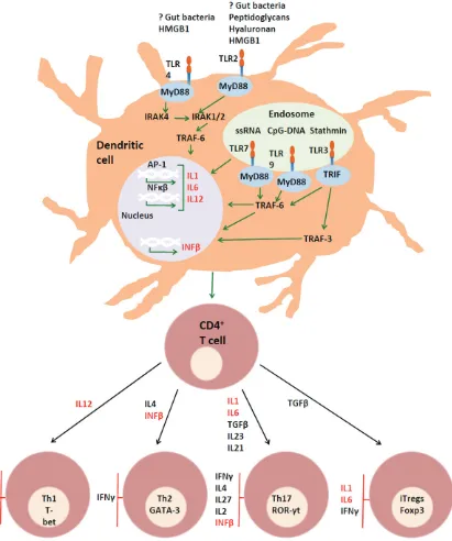
A model of the role of toll-like receptors in multiple sclerosis
Three groups of cytokines appear to play key roles in both MS and EAE. The production of
IL17-secreting CD4 (Th17) T cells appears to play a consistent role in generating CNS auto -immune damage in both diseases [45-49, 129]. The production of this lineage(s) of cells is dependent on IL23, the expression of which is dependent on that of TGFβ and IL6. In con -trast, IL2, IL4, IFNγ, and IL27 inhibit the differ -entiation of Th17 cells [130]. TGFβ is produced within the intestinal immune system in response to the development of a normal microflora [131, 132] and IL6 is generated in response to ligation of TLR2 and TLR4 [133-136].
Members of the second group of cytokines also contribute to disease pathogenesis: IL12 drives the production of IFNγ, which in turn contrib -utes to leukocyte migration to the CNS, vascu -lar adhesion and exocytosis, by upregulating the expression of ICAM-1 and VCAM-1 on CNS vasculature [137-139].
Lymphocytes obtained from the blood of RRMS patients have an increased tendency to express both ROR-γt and T-bet, and secrete both IFNγ and IL17, following expansion in the presence of IL23. IFNγ/IL17 dual expressing T cells showed a selective advantage in migrating across blood-brain barrier endothelial cells and T lymphocytes coexpressing IL17 and IFNγ are found disproportionately in the brain tissue of MS patients [139]. Similarly, during the devel -opment of EAE, IFNγ in the spinal cord was pro -duced almost exclusively by cells that had also produced IL17 [140].
Ligation of TLR2 or TLR4 on myeloid DC can induce the production of IL23, which supports the production of IL17A by CD4 T cells [141] as well as the emergence of IL17/IFNγ-producing cells [139, 140]. Conditioned media from PBMC’s stimulated with a TLR4 agonist were able to elicit IL17 secretion by CD4 T cells – even in the absence of APC [142]. TLR4 ligation also promotes the production of IL12 p70, which in its turn induces IFNγ [143]. These con -sequences of TLR ligation can therefore con -tribute to CNS autoimmunity, and may do so even in the absence of other causes of IL17 and IFNγ polarisation.
from either the donor T cells, the recipient, both or neither. Adoptive transfer of WT lymphocytes into WT recipients resulted in robust signs of EAE as expected. The adoptive transfer of C57BL/6.Tlr2-/- cells into either WT or C57BL/6.
Tlr2-/- recipients resulted in ameliorated
dis-ease, similar to active EAE in female C57BL/6. Tlr2-/- mice. In contrast, the transfer of WT cells
into C57BL/6.Tlr2-/- recipients resulted in
com-plete protection; suggesting that the presence of TLR2 at the induction of disease creates a dependence on TLR2 signaling in the effector phase [89]. Given the role of IL6 in Th17 cell differentiation, via sequential engagement of the IL21 and IL-23 pathways, our finding of IL6 circulating in the plasma of WT, but not C57BL/6.Tlr2-/- mice may provide a potential
mechanism.
Opposing the actions of CNS-damaging IL17/ IFNγ-producing cells, is type I IFN which induc -es IL27, which in turn induc-es the secretion of IL10 [144-148]. IFNβ directly decreases pro -duction of IL17 by T cells in a dose-dependent manner. It also induces the production of IL27 and acts in synergy with IL27 to inhibit the pro -duction of IL17 and promote the secretion of IL10 [148]. IL10 inhibits the production of IFNγ by downregulating IL12 [149]. The production of IL10 is, itself, suppressed by either IL1β
[149] or the combination of IFNγ and TLR2 liga -tion. IFNγ can alter TLR2-induced signal trans -duction by increasing GSK3 activity and sup -pressing MAPK activation, leading to diminished IL-10 production [150].
Plasmacytoid DC are the primary source of type I IFN and predominantly express TLR7 and TLR9. There is some suggestion that the TLR9/ IFN axis is deficient in MS patients [58]. The EAE model does not appear to mimic the activi-ties of TLR9 in this regard [17, 110]. It is possi -ble that this discrepancy results from the man-ner of experimental administation of the TLR9 ligands in these experiments, as it was unlikely to result in targeting of TLR9 ligands to plasma -cytoid DC. TLR3 ligation also upregulates IFNb1 [151], resulting in the production of IL10 and downregulation of IL12 and IL23 [25]. TLR3 is widely expressed in the CNS [19, 25, 37, 38], including on cerebral endothelial cells [28] and the presence of its endogenous ligand stath -min in the CNS suggests the possibility of anti-inflammatory, homeostatic pathways.
Acknowledgements
This work was funded by the Australian National Health and Medical Research Council, the Australian Research Council (NHMRC), The Lions Clubs of Australia and Multiple Sclerosis Research Australia. AGB is supported by an NHMRC Senior Research Fellowship. We thank Dr Margaret Jordan and Mr Benjamin Crowley for corrections and critical comments.
Address correspondence to: Dr. Alan G Baxter, Comparative Genomics Centre, Molecular Sciences Building 21, James Cook University, Townsville, QLD 4811, Australia. Phone: 4781 6265; Fax: 61-7-4781 6078; E-mail: Alan.Baxter@jcu.edu.au
References
[1] Tiwari JL, Hodge SE, Terasaki PI and Spence MA. HLA and the inheritance of multiple sclero-sis: linkage analysis of 72 pedigrees. Am J Hum Genet 1980; 32: 103-111.
[2] De Keyser J. Autoimmunity in multiple sclero-sis. Neurology 1988; 38: 371-374.
[3] Brennan KM, Galban-Horcajo F, Rinaldi S, O’Leary CP, Goodyear CS, Kalna G, Arthur A, Elliot C, Barnett S, Linington C, Bennett JL, Ow-ens GP and Willison HJ. Lipid arrays identify myelin-derived lipids and lipid complexes as prominent targets for oligoclonal band anti-bodies in multiple sclerosis. J Neuroimmunol 2011; 238: 87-95.
[4] Allegretta M, Nicklas JA, Sriram S and Albertini RJ. T cells responsive to myelin basic protein in patients with multiple sclerosis. Science 1990; 247: 718-721.
[5] Miller DM, Weinstock-Guttman B, Bethoux F, Lee JC, Beck G, Block V, Durelli L, LaMantia L, Barnes D, Sellebjerg F and Rudick RA. A meta-analysis of methylprednisolone in recovery from multiple sclerosis exacerbations. Mult Scler 2000; 6: 267-273.
[6] Khatri BO. Experience with use of plasmapher-esis in chronic progressive multiple sclerosis: the pros. Neurology 1988; 38: 50-52.
[7] Ring J, Seifert J, Lob G, Coulin K, Angstwurm H, Frick E, Brass B, Mertin J, Backmund H and Brendel W. Intensive immunosuppression in the treatment of multiple sclerosis. Lancet 1974; 2: 1093-1096.
[8] Reston JT, Uhl S, Treadwell JR, Nash RA and Schoelles K. Autologous hematopoietic cell transplantation for multiple sclerosis: a sys-tematic review. Mult Scler 2011; 17: 204-213. [9] Weber MS, Starck M, Wagenpfeil S, Meinl E,
87 Am J Clin Exp Immunol 2013;2(1):75-93 in vitro and in vivo. Brain 2004; 127:
1370-1378.
[10] Visser L, Jan de Heer H, Boven LA, van Riel D, van Meurs M, Melief MJ, Zahringer U, van Stri-jp J, Lambrecht BN, Nieuwenhuis EE and La-man JD. Proinflammatory bacterial peptidogly-can as a cofactor for the development of central nervous system autoimmune disease. J Immunol 2005; 174: 808-816.
[11] Takeuchi O, Kawai T, Sanjo H, Copeland NG, Gilbert DJ, Jenkins NA, Takeda K and Akira S. TLR6: A novel member of an expanding toll-like receptor family. Gene 1999; 231: 59-65. [12] Hemmi H, Takeuchi O, Kawai T, Kaisho T, Sato
S, Sanjo H, Matsumoto M, Hoshino K, Wagner H, Takeda K and Akira S. A Toll-like receptor recognizes bacterial DNA. Nature 2000; 408: 740-745.
[13] Alexopoulou L, Holt AC, Medzhitov R and Fla-vell RA. Recognition of double-stranded RNA and activation of NF-kappaB by Toll-like recep-tor 3. Nature 2001; 413: 732-738.
[14] Wang T, Town T, Alexopoulou L, Anderson JF, Fikrig E and Flavell RA. Toll-like receptor 3 me-diates West Nile virus entry into the brain caus-ing lethal encephalitis. Nat Med 2004; 10: 1366-1373.
[15] Toneatto S, Finco O, van der Putten H, Abrig-nani S and Annunziata P. Evidence of blood-brain barrier alteration and activation in HIV-1 gp120 transgenic mice. Aids 1999; 13: 2343-2348.
[16] Deng C, Radu C, Diab A, Tsen MF, Hussain R, Cowdery JS, Racke MK and Thomas JA. IL-1 receptor-associated kinase 1 regulates sus-ceptibility to organ-specific autoimmunity. J Im-munol 2003; 170: 2833-2842.
[17] Waldner H, Collins M and Kuchroo VK. Activa-tion of antigen-presenting cells by microbial products breaks self tolerance and induces autoimmune disease. J Clin Invest 2004; 113: 990-997.
[18] Zekki H, Feinstein DL and Rivest S. The clinical course of experimental autoimmune encepha-lomyelitis is associated with a profound and sustained transcriptional activation of the genes encoding toll-like receptor 2 and CD14 in the mouse CNS. Brain Pathol 2002; 12: 308-319.
[19] Lafon M, Megret F, Lafage M and Prehaud C. The innate immune facet of brain: human neu-rons express TLR-3 and sense viral dsRNA. J Mol Neurosci 2006; 29: 185-194.
[20] Zhou Y, Ye L, Wan Q, Zhou L, Wang X, Li J, Hu S, Zhou D and Ho W. Activation of Toll-like recep-tors inhibits herpes simplex virus-1 infection of human neuronal cells. J Neurosci Res 2009; 87: 2916-2925.
[21] Bsibsi M, Persoon-Deen C, Verwer RW, Meeu-wsen S, Ravid R and Van Noort JM. Toll-like re-ceptor 3 on adult human astrocytes triggers production of neuroprotective mediators. Glia 2006; 53: 688-695.
[22] O’Brien K, Fitzgerald DC, Naiken K, Alugupalli KR, Rostami AM and Gran B. Role of the innate immune system in autoimmune inflammatory demyelination. Curr Med Chem 2008; 15: 1105-1115.
[23] Enevold C, Oturai AB, Sorensen PS, Ryder LP, Koch-Henriksen N and Bendtzen K. Polymor-phisms of innate pattern recognition recep-tors, response to interferon-beta and develop-ment of neutralizing antibodies in multiple sclerosis patients. Mult Scler 2010; 16: 942-949.
[24] Prehaud C, Megret F, Lafage M and Lafon M. Virus infection switches TLR-3-positive human neurons to become strong producers of beta interferon. J Virol 2005; 79: 12893-12904. [25] Bsibsi M, Ravid R, Gveric D and van Noort JM.
Broad expression of Toll-like receptors in the human central nervous system. J Neuropathol Exp Neurol 2002; 61: 1013-1021.
[26] Fernald GH, Knott S, Pachner A, Caillier SJ, Na-rayan K, Oksenberg JR, Mousavi P and Baran-zini SE. Genome-wide network analysis reveals the global properties of IFN-beta immediate transcriptional effects in humans. J Immunol 2007; 178: 5076-5085.
[27] Singh MK, Scott TF, LaFramboise WA, Hu FZ, Post JC and Ehrlich GD. Gene expression changes in peripheral blood mononuclear cells from multiple sclerosis patients undergoing beta-interferon therapy. J Neurol Sci 2007; 258: 52-59.
[28] Nagyoszi P, Wilhelm I, Farkas AE, Fazakas C, Dung NTK, Hasko J and Krizbai IA. Expression and regulation of toll-like receptors in cerebral endothelial cells. Neurochem Int 2010; 57: 556-564.
[29] Sloane JA, Batt C, Ma Y, Harris ZM, Trapp B and Vartanian T. Hyaluronan blocks oligodendro-cyte progenitor maturation and remyelination through TLR2. Proc Natl Acad Sci U S A 2010; 107: 11555-11560.
[30] Correale J, Fiol M and Gilmore W. The risk of relapses in multiple sclerosis during systemic infections. Neurology 2006; 67: 652-659. [31] Correale J and Farez M. Monocyte-derived
den-dritic cells in multiple sclerosis: the effect of bacterial infection. J Neuroimmunol 2007; 190: 177-189.
[33] Andersson A, Covacu R, Sunnemark D, Danilov AI, Dal Bianco A, Khademi M, Wallstrom E, Lo-bell A, Brundin L, Lassmann H and Harris RA. Pivotal advance: HMGB1 expression in active lesions of human and experimental multiple sclerosis. J Leukoc Biol 2008; 84: 1248-1255. [34] Back SA, Tuohy TM, Chen H, Wallingford N,
Craig A, Struve J, Luo NL, Banine F, Liu Y, Chang A, Trapp BD, Bebo BF Jr, Rao MS and Sherman LS. Hyaluronan accumulates in demyelinated lesions and inhibits oligodendrocyte progenitor maturation. Nat Med 2005; 11: 966-972. [35] Sweeney CM, Lonergan R, Basdeo SA, Kinsella
K, Dungan LS, Higgins SC, Kelly PJ, Costelloe L, Tubridy N, Mills KHG and Fletcher JM. IL-27 mediates the response to IFN-β therapy in mul-tiple sclerosis patients by inhibiting Th17 cells. Brain Behav Immun 2011; 25: 1170-1181. [36] de la Monte SM, Ropper AH, Dickersin GR,
Har-ris NL, Ferry JA and Richardson EP. Relapsing central and peripheral demyelinating diseas-es. Unusual pathologic featurdiseas-es. Arch Neurol 1986; 43: 626-629.
[37] Farina C, Krumbholz M, Giese T, Hartmann G, Aloisi F and Meinl E. Preferential expression and function of Toll-like receptor 3 in human astrocytes. J Neuroimmunol 2005; 159: 12-19.
[38] Jack CS, Arbour N, Manusow J, Montgrain V, Blain M, McCrea E, Shapiro A and Antel JP. TLR signaling tailors innate immune responses in human microglia and astrocytes. J Immunol 2005; 175: 4320-4330.
[39] Bsibsi M, Bajramovic JJ, Vogt MHJ, van Dui-jvenvoorden E, Baghat A, Persoon-Deen C, Tielen F, Verbeek R, Huitinga I, Ryffel B, Kros A, Gerritsen WH, Amor S and van Noort JM. The microtubule regulator stathmin is an endoge-nous protein agonist for TLR3. J Immunol 2010; 184: 6929-6937.
[40] Szvetko AL, Jones A, Mackenzie J, Tajouri L, Csurhes PA, Greer JM, Pender MP and Griffiths LR. An investigation of the C77G and C772T variations within the human protein tyrosine phosphatase receptor type C gene for associa-tion with multiple sclerosis in an Australian population. Brain Res 2009; 19: 148-152. [41] Szvetko AL, Jones A, Mackenzie J, Tajouri L,
Csurhes PA, Greer JM, Pender MP and Griffiths LR. Investigation of the [-/A]8 and C1236T ge-netic variations within the human Toll-like re-ceptor 3 gene for association with multiple sclerosis. Neurol Res 2010; 32: 438-441. [42] Kroner A, Vogel F, Kolb-Maurer A, Kruse N,
Toy-ka KV, Hemmer B, Rieckmann P and Maurer M. Impact of the Asp299Gly polymorphism in the toll-like receptor 4 (tlr-4) gene on disease course of multiple sclerosis. J Neuroimmunol 2005; 165: 161-165.
[43] Reindl M, Lutterotti A, Ingram J, Schanda K, Gassner C, Deisenhammer F, Berger T and Lo-renz E. Mutations in the gene for toll-like recep-tor 4 and multiple sclerosis. Tissue Antigens 2003; 61: 85-88.
[44] Urcelay E, Blanco-Kelly F, de Las Heras V, de la Concha EG, Arroyo R and Martinez A. TLR4 haplotypes in multiple sclerosis: a case-control study in the Spanish population. J Neuroimmu-nol 2007; 192: 215-218.
[45] Matusevicius D, Kivisakk P, He B, Kostulas N, Ozenci V, Fredrikson S and Link H. Interleu-kin-17 mRNA expression in blood and CSF mononuclear cells is augmented in multiple sclerosis. Mult Scler 1999; 5: 101-104. [46] Lock C, Hermans G, Pedotti R, Brendolan A,
Schadt E, Garren H, Langer-Gould A, Strober S, Cannella B, Allard J, Klonowski P, Austin A, Lad N, Kaminski N, Galli SJ, Oksenberg JR, Raine CS, Heller R and Steinman L. Gene-microarray analysis of multiple sclerosis lesions yields new targets validated in autoimmune enceph-alomyelitis. Nat Med 2002; 8: 500-508. [47] Hofstetter HH, Ibrahim SM, Koczan D, Kruse N,
Weishaupt A, Toyka KV and Gold R. Therapeu-tic efficacy of IL-17 neutralization in murine ex-perimental autoimmune encephalomyelitis. Cell Immunol 2005; 237: 123-130.
[48] Chen Y, Langrish CL, McKenzie B, Joyce-Shaikh B, Stumhofer JS, McClanahan T, Blumenschein W, Churakovsa T, Low J, Presta L, Hunter CA, Kastelein RA and Cua DJ. Anti-IL-23 therapy in-hibits multiple inflammatory pathways and ameliorates autoimmune encephalomyelitis. J Clin Invest 2006; 116: 1317-1326.
[49] Hecker M, Paap BK, Goertsches RH, Kandulski O, Fatum C, Koczan D, Hartung HP, Thiesen HJ and Zettl UK. Reassessment of blood gene ex-pression markers for the prognosis of relaps-ing-remitting multiple sclerosis. PLoS One 2011; 6: 27.
[50] Guo B, Chang EY and Cheng G. The type I IFN induction pathway constrains Th17-mediated autoimmune inflammation in mice. J Clin In-vest 2008; 118: 1680-1690.
[51] Kurtuncu M, Tuzun E, Turkoglu R, Petek-Balci B, Icoz S, Pehlivan M, Birisik O, Ulusoy C, Shugaiv E, Akman-Demir G and Eraksoy M. Ef-fect of short-term interferon-beta treatment on cytokines in multiple sclerosis: significant modulation of IL-17 and IL-23. Cytokine 2012; 59: 400-402.
[52] Zhang X, Jin J, Tang Y, Speer D, Sujkowska D and Markovic-Plese S. IFN-beta1a inhibits the secretion of Th17-polarizing cytokines in hu-man dendritic cells via TLR7 up-regulation. J Immunol 2009; 182: 3928-3936.
89 Am J Clin Exp Immunol 2013;2(1):75-93 Koczan D, Thiesen HJ and Zettl UK. Elevated
type I interferon-like activity in a subset of mul-tiple sclerosis patients: molecular basis and clinical relevance. J Neuroinflammation 2012; 9: 140-140.
[54] Liu YJ. IPC: professional type 1 interferon-pro-ducing cells and plasmacytoid dendritic cell precursors. Annu Rev Immunol 2005; 23: 275-306.
[55] Balashov KE, Aung LL, Vaknin-Dembinsky A, Dhib-Jalbut S and Weiner HL. Interferon-beta inhibits toll-like receptor 9 processing in multi-ple sclerosis. Ann Neurol 2010; 68: 899-906. [56] Aung LL, Fitzgerald-Bocarsly P, Dhib-Jalbut S
and Balashov K. Plasmacytoid dendritic cells in multiple sclerosis: chemokine and chemo-kine receptor modulation by interferon-beta. J Neuroimmunol 2010; 226: 158-164.
[57] Bar-Or A, Fawaz L, Fan B, Darlington PJ, Rieger A, Ghorayeb C, Calabresi PA, Waubant E, Haus-er SL, Zhang J and Smith CH. Abnormal B-cell cytokine responses a trigger of T-cell-mediated disease in MS? Ann Neurol 2010; 67: 452-461.
[58] Hirotani M, Niino M, Fukazawa T, Kikuchi S, Yabe I, Hamada S, Tajima Y and Sasaki H. De-creased IL-10 production mediated by Toll-like receptor 9 in B cells in multiple sclerosis. J Neuroimmunol 2010; 221: 95-100.
[59] Kipp M, Clarner T, Dang J, Copray S and Beyer C. The cuprizone animal model: new insights into an old story. Acta Neuropathol 2009; 118: 723-736.
[60] Werner SR, Saha JK, Broderick CL, Zhen EY, Higgs RE, Duffin KL and Smith RC. Proteomic analysis of demyelinated and remyelinating brain tissue following dietary cuprizone admin-istration. J Mol Neurosci 2010; 42: 210-225. [61] Turrin NP. Central nervous system Toll-like
re-ceptor expression in response to Theiler’s mu-rine encephalomyelitis virus-induced demye-lination disease in resistant and susceptible mouse strains. Virol J 2008; 5: 154-154. [62] Theiler M. Spontaneous Encephalomyelitis of
Mice, A New Virus Disease. J Exp Med 1937; 65: 705-719.
[63] Dal Canto MC and Lipton HL. Multiple sclero-sis. Animal model: Theiler’s virus infection in mice. The American Journal of Pathology 1977; 88: 497-500.
[64] Baxter AG. The origin and application of experi-mental autoimmune encephalomyelitis. Nat Rev Immunol 2007; 7: 904-912.
[65] Bettelli E, Pagany M, Weiner HL, Linington C, Sobel RA and Kuchroo VK. Myelin oligodendro-cyte glycoprotein-specific T cell receptor trans-genic mice develop spontaneous autoimmune optic neuritis. J Exp Med 2003; 197: 1073-1081.
[66] Stromnes IM and Goverman JM. Passive induc-tion of experimental allergic encephalomyeli-tis. Nat Protoc 2006; 1: 1952-1960.
[67] Gijbels K, Van Damme J, Proost P, Put W, Car-ton H and Billiau A. Interleukin 6 production in the central nervous system during experimen-tal autoimmune encephalomyelitis. Eur J Im-munol 1990; 20: 233-235.
[68] Jewtoukoff V, Amzazi S, Lebar R, Bach MA and Marche PN. T-cell receptor identification of an oligodendrocyte-specific autoreactive cytotoxic T-cell clone without self restriction. Scand J Im-munol 1992; 36: 893-898.
[69] Kuchroo VK, Martin CA, Greer JM, Ju ST, Sobel RA and Dorf ME. Cytokines and adhesion mol-ecules contribute to the ability of myelin pro-teolipid protein-specific T cell clones to medi-ate experimental allergic encephalomyelitis. J Immunol 1993; 151: 4371-4382.
[70] Holmoy T. Immunopathogenesis of multiple sclerosis: concepts and controversies. Acta Neurol Scand Suppl 2007; 187: 39-45. [71] Aranami T and Yamamura T. Th17 Cells and
autoimmune encephalomyelitis (EAE/MS). Al-lergol Int 2008; 57: 115-120.
[72] Komiyama Y, Nakae S, Matsuki T, Nambu A, Ishigame H, Kakuta S, Sudo K and Iwakura Y. IL-17 plays an important role in the develop-ment of experidevelop-mental autoimmune encephalo-myelitis. J Immunol 2006; 177: 566-573. [73] Sriram S, Solomon D, Rouse RV and Steinman
L. Identification of T cell subsets and B lympho-cytes in mouse brain experimental allergic en-cephalitis lesions. J Immunol 1982; 129: 1649-1651.
[74] Zamvil SS and Steinman L. The T lymphocyte in experimental allergic encephalomyelitis. Annu Rev Immunol 1990; 8: 579-621.
[75] Jaskiewicz E. Epitopes on myelin proteins rec-ognized by autoantibodies present in multiple sclerosis patients. Postepy Hig Med Dosw 2004; 58: 472-482.
[76] von Budingen HC, Menge T, Hauser SL and Ge-nain CP. Restrictive and diversifying elements of the anti-myelin/oligodendrocyte glycopro-tein antibody response in primate experimen-tal allergic encephalomyelitis. Immunogenet-ics 2006; 58: 122-128.
[77] Karlsson J, Zhao X, Lonskaya I, Neptin M, Hol-mdahl R and Andersson A. Novel quantitative trait loci controlling development of experi-mental autoimmune encephalomyelitis and proportion of lymphocyte subpopulations. J Im-munol 2003; 170: 1019-1026.
[79] Hafler DA, Slavik JM, Anderson DE, O’Connor KC, De Jager P and Baecher-Allan C. Multiple sclerosis. Immunol Rev 2005; 204: 208-231. [80] Sospedra M and Martin R. Immunology of
mul-tiple sclerosis. Annu Rev Immunol 2005; 23: 683-747.
[81] Stefferl A, Brehm U, Storch M, Lambracht-Washington D, Bourquin C, Wonigeit K, Lass-mann H and Linington C. Myelin oligodendro-cyte glycoprotein induces experimental autoimmune encephalomyelitis in the “resis-tant” Brown Norway rat: disease susceptibility is determined by MHC and MHC-linked effects on the B cell response. J Immunol 1999; 163: 40-49.
[82] Billiau A and Matthys P. Modes of action of Freund’s adjuvants in experimental models of autoimmune diseases. J Leukoc Biol 2001; 70: 849-860.
[83] Linthicum DS, Munoz JJ and Blaskett A. Acute experimental autoimmune encephalomyelitis in mice. I. Adjuvant action of Bordetella pertus-sis is due to vasoactive amine sensitization and increased vascular permeability of the central nervous system. Cell Immunol 1982; 73: 299-310.
[84] Hofstetter HH, Shive CL and Forsthuber TG. Pertussis toxin modulates the immune re-sponse to neuroantigens injected in incom-plete Freund’s adjuvant: induction of Th1 cells and experimental autoimmune encephalomy-elitis in the presence of high frequencies of Th2 cells. J Immunol 2002; 169: 117-125. [85] Racke MK, Hu W and Lovett-Racke AE. PTX
cruiser: driving autoimmunity via TLR4. Trends Immunol 2005; 26: 289-291.
[86] Pasare C and Medzhitov R. Toll pathway-de-pendent blockade of CD4+CD25+ T cell-medi-ated suppression by dendritic cells. Science 2003; 299: 1033-1036.
[87] Prinz M, Garbe F, Schmidt H, Mildner A, Gutch-er I, WoltGutch-er K, Piesche M, SchroGutch-ers R, Weiss E, Kirschning CJ, Rochford CDP, Bruck W and Becher B. Innate immunity mediated by TLR9 modulates pathogenicity in an animal model of multiple sclerosis. J Clin Invest 2006; 116: 456-464.
[88] Marta M, Andersson A, Isaksson M, Kampe O and Lobell A. Unexpected regulatory roles of TLR4 and TLR9 in experimental autoimmune encephalomyelitis. Eur J Immunol 2008; 38: 565-575.
[89] Miranda-Hernandez S, Gerlach N, Fletcher JM, Biros E, Mack M, Korner H and Baxter AG. Role for MyD88, TLR2 and TLR9 but not TLR1, TLR4 or TLR6 in experimental autoimmune enceph-alomyelitis. J Immunol 2011; 187: 791-804. [90] Adachi O, Kawai T, Takeda K, Matsumoto M,
Tsutsui H, Sakagami M, Nakanishi K and Akira
S. Targeted disruption of the MyD88 gene re-sults in loss of IL-1- and IL-18-mediated func-tion. Immunity 1998; 9: 143-150.
[91] Zhang FX, Kirschning CJ, Mancinelli R, Xu XP, Jin Y, Faure E, Mantovani A, Rothe M, Muzio M and Arditi M. Bacterial lipopolysaccharide acti-vates nuclear factor-kappaB through interleu-kin-1 signaling mediators in cultured human dermal endothelial cells and mononuclear phagocytes. J Biol Chem 1999; 274: 7611-7614.
[92] Kumar H, Kawai T and Akira S. Toll-like recep-tors and innate immunity. Biochem Biophys Res Commun 2009; 388: 621-625.
[93] Lampropoulou V, Hoehlig K, Roch T, Neves P, Calderon Gomez E, Sweenie CH, Hao Y, Freitas AA, Steinhoff U, Anderton SM and Fillatreau S. TLR-activated B cells suppress T cell-mediated autoimmunity. J Immunol 2008; 180: 4763-4773.
[94] Herrmann I, Kellert M, Schmidt H, Mildner A, Hanisch UK, Bruck W, Prinz M and Nau R. Streptococcus pneumoniae Infection aggra-vates experimental autoimmune encephalo-myelitis via Toll-like receptor 2. Infect Immun 2006; 74: 4841-4848.
[95] Shaw PJ, Barr MJ, Lukens JR, McGargill MA, Chi H, Mak TW and Kanneganti TD. Signaling via the RIP2 adaptor protein in central nervous system-infiltrating dendritic cells promotes in-flammation and autoimmunity. Immunity 2011; 34: 75-84.
[96] Reynolds JM, Pappu BP, Peng J, Martinez GJ, Zhang Y, Chung Y, Ma L, Yang XO, Nurieva RI, Tian Q and Dong C. Toll-like receptor 2 signal-ing in CD4(+) T lymphocytes promotes T helper 17 responses and regulates the pathogenesis of autoimmune disease. Immunity 2010; 32: 692-702.
[97] Nichols FC, Housley WJ, O’Conor CA, Manning T, Wu S and Clark RB. Unique lipids from a common human bacterium represent a new class of Toll-like receptor 2 ligands capable of enhancing autoimmunity. Am J Pathol 2009; 175: 2430-2438.
[98] Nichols FC, Yao X, Bajrami B, Downes J, Fine-gold SM, Knee E, Gallagher JJ, Housley WJ and Clark RB. Phosphorylated dihydroceramides from common human bacteria are recovered in human tissues. PLoS One 2011 Feb 11; 6: e16771.
91 Am J Clin Exp Immunol 2013;2(1):75-93 [100] Farez MF, Quintana FJ, Gandhi R, Izquierdo G,
Lucas M and Weiner HL. Toll-like receptor 2 and poly(ADP-ribose) polymerase 1 promote central nervous system neuroinflammation in progressive EAE. Nat Immunol 2009; 10: 958-964.
[101] Touil T, Fitzgerald D, Zhang GX, Rostami A and Gran B. Cutting Edge: TLR3 stimulation sup-presses experimental autoimmune encephalo-myelitis by inducing endogenous IFN-beta. J Immunol 2006; 177: 7505-7509.
[102] Kerfoot SM, Long EM, Hickey MJ, Andonegui G, Lapointe BM, Zanardo RCO, Bonder C, James WG, Robbins SM and Kubes P. TLR4 contrib-utes to disease-inducing mechanisms result-ing in central nervous system autoimmune disease. J Immunol 2004; 173: 7070-7077. [103] Reynolds JM, Martinez GJ, Chung Y and Dong
C. Toll-like receptor 4 signaling in T cells pro-motes autoimmune inflammation. Proc Natl Acad Sci U S A 2012; 109: 13064-13069. [104] Lee K, Hwang S, Paik DJ, Kim WK, Kim JM and
Youn J. Bacillus-derived poly-γ-glutamic acid reciprocally regulates the differentiation of T helper 17 and regulatory T cells and attenu-ates experimental autoimmune encephalomy-elitis. Clin Exp Immunol 2012; 170: 66-76. [105] Fang C, Zhang X, Miwa T and Song WC.
Com-plement promotes the development of inflam-matory T-helper 17 cells through synergistic interaction with Toll-like receptor signaling and interleukin-6 production. Blood 2009; 114: 1005-1015.
[106] Hayashi T, Gray CS, Chan M, Tawatao RI, Ro-nacher L, McGargill MA, Datta SK, Carson DA and Corr M. Prevention of autoimmune dis-ease by induction of tolerance to Toll-like re-ceptor 7. Proc Natl Acad Sci U S A 2009; 106: 2764-2769.
[107] O’Brien K, Fitzgerald D, Rostami A and Gran B. The TLR7 agonist, imiquimod, increases IFN-beta production and reduces the severity of experimental autoimmune encephalomyelitis. J Neuroimmunol 2010; 221: 107-111.
[108] Soulika AM, Lee E, McCauley E, Miers L, Ban-nerman P and Pleasure D. Initiation and pro-gression of axonopathy in experimental auto-immune encephalomyelitis. J Neurosci 2009; 29: 14965-14979.
[109] Ichikawa HT, Williams LP and Segal BM. Activa-tion of APCs through CD40 or Toll-like receptor 9 overcomes tolerance and precipitates auto-immune disease. J Immunol 2002; 169: 2781-2787.
[110] Wolf NA, Amouzegar TK and Swanborg RH. Synergistic interaction between Toll-like recep-tor agonists is required for induction of experi-mental autoimmune encephalomyelitis in Lew-is rats. J Neuroimmunol 2007; 185: 115-122.
[111] Ferwerda G, Girardin SE, Kullberg BJ, Le Bourhis L, de Jong DJ, Langenberg DML, van Crevel R, Adema GJ, Ottenhoff THM, Van der Meer JWM and Netea MG. NOD2 and toll-like receptors are nonredundant recognition sys-tems of Mycobacterium tuberculosis. PLoS Pathog 2005; 1: 279-285.
[112] Faure E, Equils O, Sieling PA, Thomas L, Zhang FX, Kirschning CJ, Polentarutti N, Muzio M and Arditi M. Bacterial lipopolysaccharide activates NF-kappaB through toll-like receptor 4 (TLR-4) in cultured human dermal endothelial cells. Differential expression of TLR-4 and TLR-2 in endothelial cells. J Biol Chem 2000; 275: 11058-11063.
[113] Noss EH, Pai RK, Sellati TJ, Radolf JD, Belisle J, Golenbock DT, Boom WH and Harding CV. Toll-like receptor 2-dependent inhibition of macro-phage class II MHC expression and antigen processing by 19-kDa lipoprotein of Mycobac-terium tuberculosis. J Immunol 2001; 167: 910-918.
[114] Takeda K, Takeuchi O and Akira S. Recognition of lipopeptides by Toll-like receptors. J Endo-toxin Res 2002; 8: 459-463.
[115] Jung SB, Yang CS, Lee JS, Shin AR, Jung SS, Son JW, Harding CV, Kim HJ, Park JK, Paik TH, Song CH and Jo EK. The mycobacterial 38-kilo-dalton glycolipoprotein antigen activates the mitogen-activated protein kinase pathway and release of proinflammatory cytokines through Toll-like receptors 2 and 4 in human mono-cytes. Infect Immun 2006; 74: 2686-2696. [116] Pecora ND, Gehring AJ, Canaday DH, Boom WH
and Harding CV. Mycobacterium tuberculosis LprA is a lipoprotein agonist of TLR2 that regu-lates innate immunity and APC function. J Im-munol 2006; 177: 422-429.
[117] Gehring AJ, Dobos KM, Belisle JT, Harding CV and Boom WH. Mycobacterium tuberculosis LprG (Rv1411c): a novel TLR-2 ligand that in-hibits human macrophage class II MHC anti-gen processing. J Immunol 2004; 173: 2660-2668.
[118] Gilleron M, Quesniaux VF and Puzo G. Acyla-tion state of the phosphatidylinositol hexaman-nosides from Mycobacterium bovis bacillus Calmette Guerin and mycobacterium tubercu-losis H37Rv and its implication in Toll-like re-ceptor response. J Biol Chem 2003; 278: 29880-29889.
[119] Abel B, Thieblemont N, Quesniaux VJF, Brown N, Mpagi J, Miyake K, Bihl F and Ryffel B. Toll-like receptor 4 expression is required to control chronic Mycobacterium tuberculosis infection in mice. J Immunol 2002; 169: 3155-3162. [120] Ronaghy A, Prakken BJ, Takabayashi K,
DNA sequences influence the course of adju-vant arthritis. J Immunol 2002; 168: 51-56. [121] Krieg AM. CpG motifs in bacterial DNA and
their immune effects. Annu Rev Immunol 2002; 20: 709-760.
[122] Lee JM and Olitsky PK. Simple method for en-hancing development of acute disseminated encephalomyelitis in mice. Proc Soc Exp Biol Med 1955; 89: 263-266.
[123] Wiener SL, Tinker M and Bradford WL. Experi-mental meningoencephalomyelitis produced by hemophilus pertussis. AMA Arch Pathol 1959; 67: 694-699.
[124] Levine S and Wenk EJ. Allergic Encephalomy-elitis: A Hyperacute Form. Science 1964; 146: 1681-1682.
[125] Levine S, Wenk EJ, Devlin HB, Pieroni RE and Levine L. Hyperacute allergic encephalomyeli-tis: adjuvant effect of pertussis vaccines and extracts. J Immunol 1966; 97: 363-368. [126] Bergman RK, Munoz JJ and Portis JL. Vascular
permeability changes in the central nervous system of rats with hyperacute experimental allergic encephalomyelitis induced with the aid of a substance from Bordetella pertussis. In-fect Immun 1978; 21: 627-637.
[127] Fujimoto C, Yu CR, Shi G, Vistica BP, Waw-rousek EF, Klinman DM, Chan CC, Egwuagu CE and Gery I. Pertussis toxin is superior to TLR ligands in enhancing pathogenic autoimmuni-ty, targeted at a neo-self antigen, by triggering robust expansion of Th1 cells and their cyto-kine production. J Immunol 2006; 177: 6896-6903.
[128] Millward JM, Caruso M, Campbell IL, Gauldie J and Owens T. IFN-gamma-induced chemo-kines synergize with pertussis toxin to promote T cell entry to the central nervous system. J Im-munol 2007; 178: 8175-8182.
[129] McGeachy MJ and Cua DJ. The link between IL-23 and Th17 cell-mediated immune patholo-gies. Semin Immunol 2007; 19: 372-376. [130] Maddur MS, Miossec P, Kaveri SV and Bayry J.
Th17 cells: biology, pathogenesis of autoim-mune and inflammatory diseases, and thera-peutic strategies. Am J Pathol 2012; 181: 8-18.
[131] Atarashi K, Nishimura J, Shima T, Umesaki Y, Yamamoto M, Onoue M, Yagita H, Ishii N, Ev-ans R, Honda K and Takeda K. ATP drives lam-ina propria T(H)17 cell differentiation. Nature 2008; 455: 808-812.
[132] Ivanov II, Frutos RdL, Manel N, Yoshinaga K, Rifkin DB, Sartor RB, Finlay BB and Littman DR. Specific microbiota direct the differentia-tion of IL-17-producing T-helper cells in the mu-cosa of the small intestine. Cell Host Microbe 2008; 4: 337-349.
[133] Takeuchi O, Hoshino K and Akira S. Cutting edge: TLR2-deficient and MyD88-deficient mice are highly susceptible to Staphylococcus aureus infection. J Immunol 2000; 165: 5392-5396.
[134] Thoma-Uszynski S, Kiertscher SM, Ochoa MT, Bouis DA, Norgard MV, Miyake K, Godowski PJ, Roth MD and Modlin RL. Activation of toll-like receptor 2 on human dendritic cells triggers induction of IL-12, but not IL-10. J Immunol 2000; 165: 3804-3810.
[135] McCurdy JD, Lin TJ and Marshall JS. Toll-like receptor 4-mediated activation of murine mast cells. J Leukoc Biol 2001; 70: 977-984. [136] Mu HH, Sawitzke AD and Cole BC. Presence of
Lps(d) mutation influences cytokine regulation in vivo by the Mycoplasma arthritidis mitogen superantigen and lethal toxicity in mice infect-ed with M. arthritidis. Infect Immun 2001; 69: 3837-3844.
[137] Frohman EM, Frohman TC, Dustin ML, Vayuve-gula B, Choi B, Gupta A, van den Noort S and Gupta S. The induction of intercellular adhe-sion molecule 1 (ICAM-1) expresadhe-sion on hu-man fetal astrocytes by interferon-gamma, tu-mor necrosis factor alpha, lymphotoxin, and interleukin-1: relevance to intracerebral anti-gen presentation. J Neuroimmunol 1989; 23: 117-124.
[138] Tsukada N, Matsuda M, Miyagi K and Yanagi-sawa N. Adhesion of cerebral endothelial cells to lymphocytes from patients with multiple sclerosis. Autoimmunity 1993; 14: 329-333. [139] Kebir H, Ifergan I, Alvarez JI, Bernard M, Poirier
J, Arbour N, Duquette P and Prat A. Preferential recruitment of interferon-gamma-expressing TH17 cells in multiple sclerosis. Ann Neurol 2009; 66: 390-402.
[140] Hirota K, Duarte JH, Veldhoen M, Hornsby E, Li Y, Cua DJ, Ahlfors H, Wilhelm C, Tolaini M, Men-zel U, Garefalaki A, Potocnik AJ and Stockinger B. Fate mapping of IL-17-producing T cells in inflammatory responses. Nat Immunol 2011; 12: 255-263.
[141] Roses RE, Xu S, Xu M, Koldovsky U, Koski G and Czerniecki BJ. Differential production of IL-23 and IL-12 by myeloid-derived dendritic cells in response to TLR agonists. J Immunol 2008; 181: 5120-5127.
[142] Kattah MG, Wong MT, Yocum MD and Utz PJ. Cytokines secreted in response to Toll-like re-ceptor ligand stimulation modulate differentia-tion of human Th17 cells. Arthritis Rheum 2008; 58: 1619-1629.