STUDIES ON THE ROLE OP HISTONES
IN THE STRUCTURE AND FUNCTION OF CHROMATIN
Thesis by
John Edward Smart
In Partial Fulfillment of the Requirements For the Degree of
Doctor of Philosophy
California Institute of Technology Pasadena, California
1970
•
ACKNOWLEDGMENTS
While studying at the California Institute of Tech-nology, I have had the enjoyable and very rewarding ex-perience of working closely with five undergraduates. In chronological order, they are: Simon Rock Levinson (Caltech undergraduate, 1968) who investigated "The. Selective.Dis-sociation of Histones from Chromatin . by Guanidine Hydro-chloride" (manuscript in preparation); Steven H. Radler
(Caltech undergraduate, 1969) who worked extensively on the construction of "auxin columns" for investigation of auxin receptors in plant tissues, and who contributed signifi-cantly to "Studies on the Mechanism of Selective Dissoci-ation of Histones from Chromatin by Sodium Deoxycholate" (manuscript in preparation); Michael B. Farber (Caltech undergraduate, 1969) who investigated "The Kinetics of the Reaction of Formaldehyde with Chromatin (manuscript in pre-paration); James H. Richards (Caltech undergraduate, 1970) who investigated "The Isolation and Characterization of Histone Deficient Portions of Chromatin" (manuscript in preparation); and Jonathan Weintraub (Reed College under-graduate, 1969) who worked on "Thealaracterization of Chro-matin from Valencia Orange" (manuscript in preparation).
his continued support and advice during the course of this work. The freedom which I was allowed during my four years as a graduate student enabled me to pursue ideas wherever they led and to test myself as a relatively independent research advisor.
Michael E. Dahmus and John W. Sedat deserve a special thanks for the companionship, encouragement (M. E. D.), "torture" (J. W. S.), and critical advice which they pro-vided. Roger G. Chalkley, Sally R. Elgin, Douglas Famtrough, Ronald H. Jensen, Sandra Winicur, and several other members of the Biology department are gratefully acknowledged for their contributions to this thesis. The assistance of Ludia Brown in the bi-weekly preparation of chromatin and in keeping 021 Kerckhoff reasonably neat was indispensible.
Most importantly, my wife, Mary Lou, two sons, Dill and Tom, and (to a small extent) my daughter, Cassie,
de-serve immeasurable thanks for their toleration, under-standing, encouragement, and diversion.
Finally, I thank the National Science Foundation and the Public Health Service for their financial support.
•
ABSTRACT
Studies on the dissociation of histones from chro-matin by increasing concentrations of sodium deoxycholate
(Doc) have shown that histone II is removed at lowest con-centrations of DOC, while slightly higher concon-centrations remove histories III and IV. Still higher concentrations remove histone I.
The complete separation of chromatin and 14C-D00 by sucrose sedimentation indicated that the binding of DOC to chromatin is readily and completely reversible.
The dissociation of histories from chromatin by in-
411
creasing concentrations of related cholanic acids and someof their conjugated derivatives was studied. The results suggested that the driving force for the interaction be-tween the cholanic acid anion and histories is the lowering of the activity coefficient of the cholanic acid anion which occurs when it is partially removed from solution by interaction with hydrophobic regions of the positively charged histories.
•
Vconcentrations, while hiFher concentrations remove histones II, III, and IV). Properties studied included thermal de-naturation, sedimentation velocity, flow dichroism, relaxa-tion times of molecules oriented in a flow field, and the irreversible disruption of a 130 S, cross-linked component of sheared chromatin. The data indicated that none of the structural or chemical parameters with which these proper-ties are correlated show a dependence on the presence of one particular histone fraction.
The template activity (ability to prime a 0.2 M K01 DNA-dependent REA synthesis system catalysed by E. coli RNA polymerase) increases from that of native chromatin (approx-imately 25 per cent of that of pure DNA) to that of pure DNA in a fashion which shows a nearly linear relationship to the amount of histone cov erage of the template. The precipitability of Partially dehistonized chromatin samples in 0.15 M Fa01 shows a large dependence on the presence of histone I.
vi
TABLE OF CONTENTS
CHAPTER PART
1
•
II
2
TITLE PAGE
Acknowledgments Abstract
GENERAL INTRODUCTION
THE SELECTIVE DISSOCIATION OF HIS- TONES FROM CHROMATIN BY SODIUM DE-OXYCHOLATE
ii iv
3
A NEW ORDER OF REMOVAL 4
Introduction 5
Methods and Materials 6
Results 11
Discussion 31
A POSSIBLE MECHANISM OF REMOVAL 36
Introduction 37
Results 38
Discussion 56
References 62
STUDIES ON THE ROLE OF HISTONES 66
IN
THE
STRUCTURE OF CHROMATINIntroduction 67
Methods and Materials 69
Results 73
Discussion 103
vii
TABLE OF CONTENTS
CHAPTER PART
Appendix
411
TITLE
THE SELECTIVE DISSOCIATION OF EISIONES FROM CHROMATIN BY
SO-DIUM CHLORIDE References
STUDIES ON THE ROLE OF HISTONES IN THE BEHAVIOR OF CHROMATIN AT PHYSIOLOGICAL IONIC STRENGTHS
(TEMPLATE ACTIVITY AND PRECIPI-TATION)
Introduction
Methods and Materials Results
Discussion References
PAGE
111
118 121
122 124 128 151 160
•
•
GENERSLL INTRODUOTION2 •
GENERAL INTRODUCTION
In the nuclei of higher organisms, chromosomal DNA exists in close association with specific proteins. The major portion of these proteins are the histones (Bonner Ts'o, 1964). Most of the chromosomal material can be separated from other cell components by differential centrifugation (Bonner et al., 1968). Chemically, the preparation of chromatin is mild and work in this field is based upon the concept that this material may serve as worthwhile models for nuclear chromatin. An impression of the type and degree of interaction between chromosomal DNA
411
and the histones can be obtained by finding various con-ditions for selectively dissociating the chromatin complex and then observing the physical, chemical, and biological effects of doing so.
REFERENCES
1. Bonner, J., Chalkley, G. R., Dahmus, M., Fambrough, D.,
Fujimura, F., Huang, R. C. 0., Huberman, J., Marushige,
K., Ohlenbusch, H., Olivera, B., and Widholm, J.
(1968a), Methods in Enzymoloa, v. 12, Academic Press,
New York.
2. Bonner, J. and _Ts l o,-P. 0 P. ed. '(1964), in ihe
Chapter 1
THE SELECTIVE DISSOCIATION OF HISIONES FROM CHROMATIN BY SODIUM DEOXYCHOLATE
•
•
4
•
Chapter 1 Ta-rt
A NE U ORDER OF REMOVAL
•
5INTRODUCTION
The genetic material in the nucleus of eukaryotic
organisms is present as a nucleoprotein complex, chromatin. Chromatin can be isolated as a chemically defined entity of DNA, RNA, histone and nonhistone proteins. The histones, a family of seven major components, comprise the major portion of the proteins (Bonner & Ts'o, 1964; Murray, 1964; H. Busch,
1965). Although the biological functions of the histones are not completely understood, a large body of data supports the hypotheses that histones are involved in repression of genetic activity (Huang & Bonner, 1962; Allfrey, Littau, &
!II
Mirsky, 1963; Marushige & Bonner, 1966) and that they confersome structural restraints on chromosomal DNA (Doty, Marmur, Eisner, & Schildkraut, 1960; Sameftaa& Yang, 1965; Ohba, 10.‘0;
Pardon, :Wilkins, & Richards, 1967; Tuan & Bonner, 1969). Several workers have studied the contribution of the various histone fractions to the structure and function of
•
6sodium perchlorate, and guanidine hydrochloride. All
present methods, however, yield the same general sequence of histone removal - histones I (lysine-rich) are removed at
the lowest concentration of dissociating agent, while higher concentrations remove histones II (slightly lysine-rich), and histones III and IV (arginine-rich).
A selective dissociation agent which produced a differ-ent order of histone fraction removal would be extremely useful in the interpretation of selective dissociation studies. We have investigated the dissociation of histones from chromatin by increasing concentrations of sodium deoxy-cholate (DOC). We have found that histone II is removed at
!II
the lowest concentrations of DOC, while slightly higher con-centrations remove histones III and IV. Still higher con-centrations of DOC remove histone I.
METHODS AND MATERIALS
Preparation of Chromatin. Sucrose purified chromatin was prepared from pea buds according to the method of Bonner
7
•
enizing in a Potter-Elvehjem homogenizer. The chromatin solution was dialyzed overnight against 0.0025 M Tris, pH 8, sheared in 40 m1 aliquots in the Virtis "45" homogenizer at:50 volts for 90 seconds, and then centrifuged at 10,000 x for
30 minutes. The resulting supernatant was further fraction-ated by sedimenting the sheared chromatin into a cushion of 1.2 M sucrose - 0.0025 M Tris, pH 8, for the equivalent of 10 hours at 50,000 rpm (Spine° Ti-50 rotor). Th2 super-natant was removed and discarded. The pellet was resus-pended as above, and dialyzed extensively against 0.0025 M Tris, pH 8. This solution, referred to as chromatin, con-stituted the starting material. The chromatin has a final
41O
concentration of 20 to 40 A260 111 , a ratio A 230mp/A26041 ofapproximately 0.75, and a ratio A320/ 11-260mp of less than 0.054. All steps were carried out at 0 to 40 C.
Histone Dissociation Studies. To assure that the chromatin was not subjected to a sodium deoxycholate (BOO) concentration higher than the desired final one, the fol-lowing procedure was adopted. The chromatin was diluted with 0.0025 M Tris, pH 8, so that a final volume of 10 ml
and a final concentration 5 to 7 2, -
m
8 •
pH 8, were gently layered at the bottom of the tube. The partially dehistonized DNA was then separated from the dis-sociated protein by sedimentin; the DNA into the sucrose cushion at the equivalent of 50,000 rpm for 10 hours (Spinco Ti-50 rotor). The top 11 ml of the resulting supernatant were removed and discarded. The remaining loosely packed pellet (contained in 1 ml of sucrose solution) was then re-moved and resuspended by homogenization in a Teflon
homog-enizer as above. The samples were exhaustively dialized against 0.0025 II Tris, pH 8, and then centrifuged at 10,000 x liEfor 10 minutes. The resulting supernatants were then used for further characterization of the partially dehis-
411
tonized chromatin.Thermal Denaturation and Ultraviolet Absorption. The
partially dehistonized samples were dialyzed exhaustively against 2.5 x 10 EDTA, pH 8, diluted to approximately
1 A260mp with dialysate, and melted in a Gilford Model 2000
multiple sample absorbance recording apparatus adapted for the recording of melting profiles. The rate of temperature increase was 0.5 to 1.0 degree/minute. Ultraviolet absorP-tion spectra were determined with a Cary recording spec-trophotometer, Model 11.
Free-Zone ElectroPhoresis. Free-zone electrophoresis
was performed as described by Olivera, Bathe, and Davidson
9 •
apparatus. Data were fitted to a straight line usinc a least sauares computer program.
Sedimentation AnaLysis. Sedimentation velocity was studied using band-sedimentation techniques on preformed sucrose gradients in a Spinco Model L2-65 ultracentrifu7e. All steps were performed at 0 to 40 C.
Chromato.c4raphz_._ Chromatographic separation of chroma-tin from DOC was accomplished as follows. The appropriate concentration of sodium deoxycholate was added to the chro-matin solution. The mixture was then applied to a jaciceted,
2 cm x 30 cm Biogel P-150 column, which had been
equili-brated with 0.0025 M Tris, p]i
8.
The column was eluted with • 0.0025 h Tris, pH 8, and the fractions analyzed for A 2 6 0mu and 0 14 cts./Min. All steps were performed at 0 to 4 0 C.
Chemical Analysis. REA was separated from DNA by the modified Schmidt-Tannhauser procedure of Ts'o and Sato
(1959). DNA concentration was determined by the diphenyla-mine assay of Burton (1956) and by ultraviolet absorption. R1TA concentration was determined by the orcinol reaction of Dische and Schwartz (1937). Purified calf thymus DNA
(Worthinoton Biochemical Corp.) and yeast RYA (Sigma) were used as standards.
•
10components. The histone samples were prepared by adding 0.25 ml of 2 N H 2 SO4 per ml of chromatin solution. The solution was vigorously mixed, allowed to stand at 0 0 C. for 30 minutes with occasional mixing, and then centrifuged at 24,000 x for 20 minutes. The supernatant was removed by pipetting and analyzed for protein content by ultraviolet absorption. Three volumes of
95
per cent ethanol were then added and the proteins precipitated at -20° C. for 24 hours. They were pelleted by centrifugation at 24,000 x for 20 minutes, and washed twice with cold95
per cent ethanol.The pellet was air-dried and dissolved in the proper amount of
8
VI urea to make the final solution approximately 1 mg protein/ml solution. Acid-insoluble material was washed once with95
per cent ethanol, air-dried, and then dissolved in 1 IST NaOH. Calf thymus histories and bovine serum albumin(Sigma) were used as standards.
The absorptivity of total histone at 230 mu is 4.15 1/cm g (Jensen, 1966); for DNA contained in chromatin it is 22 1/cm g at 260 mu (Tuan, 1966); for RNA contained in chro-matin it is assumed to be 25 1/cm
E
at 260 mu.Disc Gel ElectrorhoreSis of Histories. Acrylamide disc gel electrophoresis of isolated histories was performed by the method of Bonner et al. (1968a). The quantity of each electrophoretic component was determined by densitometry •
Chemicals. Deoxycholic acid-24-C 1 (Nuclear Equipment Chemical Corp.), 3.6 mC/MM, was dissolved in 0.5 N NaOH and then adjusted to pH 8.
All cholanic acids and their conjugated derivatives were obtained from Mann Research Laboratories, Inc.
Note: All assays were performed within one week of the preparation of chromatin.
The weight fractions of total histone protein, non-his -tone protein, and RNA remaining bound to the DNA of pea bud chromatin as a function of the molarity of sodium deoxy-cholate (DOC) used for dissociation are shown in Figure 1. As is the case with other agents that have been used to
selectively dissociate the nucleoprotein complex, increasing concentrations of DOC principally dissociate histone protein. Relatively little nonhistone Protein or RNA, amounting to no more than 20 per cent of initial amounts, is extracted by DOC over the concentration range studied.
12
Figure 1. The weight fraction of components of chromatin remaining bound to DNA after extraction of chromatin with increasing concentrations of DOC. Each point for histone protein is the avera,7e value from five experiments. Each point for RYA is the average value from three experiments. Each point for both types of nonhistone protein is the av-erage value from two experiments.
•
•
.1 3
•
I I I I I I • _I:Djn
D (-5 (7) I — (1) U) — C I 0 I a) 0 Z D — a.) - cLi2
— T5 (75 L} cn u) 0 Cc z - —
— •
_
cr
cr -
0
0
a)
U
)co _o
<
(" \J (" ■ 1 D Z I = 0 ---- U) I0
a) c
_
a.)
I — c — C • 0 -1 (1) C_)4
U ) E0 C 00
•
0
Z _c
–
Z
–
I
•
/
_
–
•
4
a)
C
o I
<
•
Z
4 ,
_
1
_
•
4
1J°
1---
"."---In
4
_. •
1
I I 1 • ,- 0 — 0 0 0 0IRON0d1A103 AO NO113VLIA _LHOGM
14
nucleoprotein complex. Nonhistone protein has previously been defined as that fraction of chromatin-bound protein which remains insoluble upon subjectimT the chromatin to
extraction with 0.4 N H 2 SO4, followed by the modified
Schmidt-Tannhauser procedure of Ts t o and Sato (1959) (0.3 M KOH, 37 0 C. for 18 hours; 0.5 M HC104, 100 0 C. for 10
minutes), and which is subsequently soluble in 1 N NaOH (Bonner et al., 1968). As is shown in Figure 1, this type of nonhistone protein amounts to approximately 0.17 weight fraction of the DNA in chromatin. The discrepancy in the yields obtained by the two methods can be explained by as-suming that a step(s) in the modified Schmidt-Tannhauser • procedure causes a loss of some nonhistone protein, and/or
a change in some nonhistone protein which renders it insol- uble in 1 N NaOH. That the second explanation is apparently correct is supported by the repeated observation that after hydrolysis of the 0.4 N H2 50 4 insoluble material of pea bud chromatin in hot 0.5 M HC10 4 a significant amount of 1 N
NaOH insoluble material remains. Fambrough (1967) found that this material was almost totally soluble in 1 per cent sodium dodecylsulfate - 8 M urea, and could be purified by precipitation with ammonium sulfate, a method developed for the study of the nonhistone protein of rat liver chromatin
(Marushige, Brutlag, & Bonner, 1969). This material was
15
DNA from which it was isolated. Studies on the dissociation of chromatin with increasing concentrations of Na01 indicate that a large portion of this nonhistone protein is not dis-sociated from DNA by extraction of chromatin with 2 M NaC1
(Fambrough, 1967).
An increase in the weight fraction of nonhistone pro-tein above control levels can be seen in the chromatin samples previously extracted with 0.025 and 0.0375 M DOC. This increase appears to be due to histone and nonhistone protein aggregation (Levinson, Smart, & Bonner, 1969) and/Or to histone-nonhistone-DOC micelle formation.*
• *Extraction of chromatin with concentrations of sodium de-oxycholate between 0.0625 and 0.1 molar form a small amount of white precipitate which remains insoluble after exten-sive dialysis against 0.0025 M Tris, pH 8, or 2.5 x 10 -4 M EDT, pH 8. The precipitate is also insoluble in 0.4 IT
F SO
-
but is soluble in 1 N NaOH or 1 per cent sodium do- 2 4'decylsulfate (SDS). Examination of the proteins contained in the precipitate by SDS disc gel electrophoresis shows a
simnificant amount of proteins which have the same mobili-ties as the histone fractions (Elgin, 1969). Although this precipitate pellets upon centrifugation at 10,000 x g for
16
•
Data on the fraction of each histone component dis-sociated from chromatin by increasing concentrations of sodium deoxycholate are presented in Figure 2. Histone II
(slightly lysine-rich) is most readily dissociated by BOO. Histones III and IV (arginine-rich) are extracted by
slightly higher concentrations of DOC, while histone I
(lysine-rich) is least readily dissociated by BOO. The same general pattern of histone removal has been found with calf thymus chromatin. Consequently, this method for the selec-tive dissociation of histones produces a pattern of histone removal which is different from that produced by increasing concentrations of Ka01, HaC104, and guanidine hydrochloride
411
(G1101). Sodium deoxycholate extraction of histones is par-ticularly useful because it selectively removes histones II, III, and 'Tat low concentrations of dissociating agent,
•
17Figure 2. The fraction of individual histone components remaining bound to the DNA after extraction of chromatin with increasing concentrations of DOC. Histone I =
lysine-rich = fl; histone II = slightly lysine-lysine-rich = f2b and f2a2; histone III = arginine-rich = f3; histone IV = ar-ginine-rich = f2al.
•
1=1
0 d (1) a 0 ir) 0 0E
o
0
0cn
ro
o
a c\J 0I-
w
I
I
I
I
-1-10
o
. 00 co •1 -. c\J Q0
c5
o
0o
•
19
while leaving histone I bound to the DNA; whereas, all other reported dissociation agents selectively remove histone I at low concentrations, while leaving histone II, III, and IV bound to the "D*IU.. It should also be noted that the concentration of dissociating agent required for the extraction of a given weight fraction of total histone is approximately ar order of magnitude less for DOC than for other dissociating agents.
Free-zone electrophoresis of partially dehistonized samples has been considered as a method which provides an independent indication of the histone coverage of the
samples (Ohlenbusch, Olivera, Tuan, & Davidson, 1967; Levin-
411
son, Smart, & Bonner, 1969). The relationship of electro- Phoretic mobility of the partially dehistonized samples to molarity of DOS used for dissociation of chromatin is pre-sented in Figure 3. These data show that extraction of chromatin with 0.037 5 1,1 DOS removes approximately 67 per cent of total histone. Such partially dehistonized chro-matin exhibits an electrophoretic mobility of 1.75 x 10 4cm2sec-1volt-1 , which is intermediate (51 per cent) be-tween that of fully covered chromatin (1.31 x 10 4 cm 2 5ec -1 -
volt -1 ) a:d pure DNA (2.17 x 10 4 cm2 sec -l volt -/ ). These data
may be compared with those of Ohlenbusch, Olivera, Tuan, and Davidson (1967), who studied Na01 extraction of calf
•
20
Figure 3. The free-zone electrophoretic mobility of chro-matin samples partially dehistonized by increasing, concen-trations of DOC. Each point represents the avera.e value from four experiments. Pree-zone electrophoresis was done in 0.01 I NaC1, 0.001 M Vris, PH 7.5
•
21
•
0
•
N)
.
0")
.
N-.
LQ
re)
(v01
X
i_OeS
i_A
z
WO
tri)
Ainieow
•
22411
III
aminowho studied guanidine hydrochloride extraction of pea bud chromatin. Both found an increase in electrophoretic mobility of approximately 50 per cent after removal of approximately 30 per cent of total histone by extraction with 0.6 M NaC1 or 0.5 M GuCl. In both cases, the large change in electrophoretic mobility was consistent with the fact that histone I, the most positively charged histone, was selectively removed. Our data are consistent with the fact that histone II, the least positively charged histone, as well as a small amount of histones III and IV are re- moved by 0.0375 M BOO. In fact, consideration of the net positive charge density of the histones (calculated from
acid composition data of Fambrough, 1967) removed by this concentration of BOO shows that 52 per cent of the net positive charges of the total histones have been removed. This number is in very good agreement with the 51 per cent increase in electrophoretic mobility of this sample. These data also indicate that the negatively charged BOO molecule does not remain bound to the partially dehiston-ized samples to any significant extent.
Figure 4 shows the electrophoretic dispersion (defined as the band width of the migrating 11
-260 mp peak at one half peak height; native chromatin is assigned the value of 1.0) of chromatin samples which have been extracted with
•
23Figure 4. The free-zone electrophoretic dispersion (the band width of the 11260 91 Peak at one half peak height) of chromatin samples partially dehistonized by increasinf con-centrations of DOO. The electrophoretic dispersion of native chromatin is assi ,;ned the value of 1.0. Free-zone electrophoretic dispersion of native chromatin is assi:ned the value of 1.0. Free-zone electrophoresis was done in 0.01 PaC1, 0.001 N rris, ph 7.5.
•
•
2
2
'
0
0
0
LC)
0
LC)
0
o
c\-1.
c\i
—
—
—
NOI Se:JdS KJ
•
25
•
hlstonized samples all migrat e as single A260
mp
peaks, it is clear that partial, removal of the histones from chroma-tin, creates a set of molecules of much higher diversity of charge distribution than that of either unextracted chroma-tin or pure DNA.The thermal denaturation behavior of chromatin from which discrete histone fractions have been removed provides yet another independent indication of the amount of histone coverage of each sample. It is also of interest because it may provide some insight into the distribution of histones along the DYA chain. Figure
5
shows the melting profiles of chromatin samples which have been partially dehistonized41,
by various concentrations of DOC. Under the conditions em- ployed in this study, the T m (midpoint of thermal transi-tion) of native chromatin is in the temperature range,73
to 76 0 C., while that of DNA is 43 to 45 0 C. The values of Tm, hyperchromicity, and dispersion (defined as that temper-ature span required to raise the A260 mia from 0.333 to 0.667 of the final hyperchromicity) for partially dehistonized•
26Figure 5. Melting profiles of chromatin samples partially dehistonized by increasing concentrations of DOG. Meltirv; was done in 2.5 x 10 -4 M EDTA, pH 8. No correction for thermal expansion.
•
•()
riUj
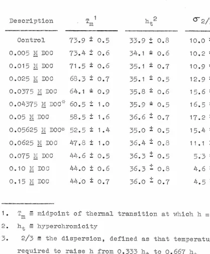
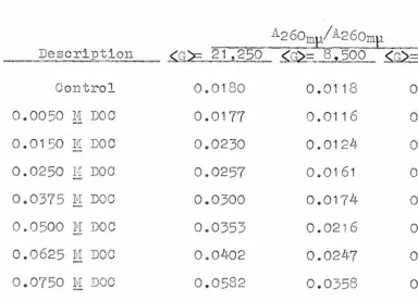
28 Table 1
Thermal Denaturation Measurements
Description . T m1 -t
Control 73.9 ± 0.5 33.9 t 0.8 10.0 ± 0.2
0.005 7.4 D00 73.4 ± 0.6 54.1 i: 0.6 10.2 * 0.2
0.015 li. DOC 71.5 i 0.6 p5.1 0.7 10.9 * 0.3 0.025 H LOU 68.3 ± 0.7 35.1 = 0.5 .1_ 12.9 = .4. 0.3 0.0375 111 Doc 64.1 * 0.9 35.8 ± 0.6 15.6 * 0.5 0.04575 k'.1 JOC * 60.5 ± 1.0 35.9 * 0.5 16.5 * 0.9
•
0.05 M DOC 58.5 ± 1.6 36.6 ± 0.7 17.2 i 0.5 0.05625 M DOC* 52.5 i 1.4 35.0 i 0.5. 15.4 * 1.50.0625 m L100 47.8 ± 1.0 36.4 ± 0.8 11.1 ± 1.5
0.075 m DO0 44.6 ± 0.5 36.3 ± 0.5 5.'3 ± 0.3
0.10 IT too 44.0 ± 0.6 36.3 - 0.8 -1- _ 4.6 ± 0.3
-1-
0.15 M DOO 440 ± 0.7 36.0 - 0.7 4.5 ± 0.4
1. T midpoint of thermal transition at which h = 0.5 h t 2. h., 1." hyperchromicity
3. 2/3 = the dispersion, defined as that temperature span required to raise h from 0.333 h t to 0.667 h t
* Data from three independent experiments; all other points from F;reater than 7 independent experiments
29
•
nucleoprotein complex from
73.9 to 64.1 0
O. This corre-sponds to a33
Per cent decrease in T m (where 73.9 = 0% and 44.0 = 100%). It therefore appears that decreases in the Tm -f o partially dehistonized samples lag considerably - behind decreases in the weight fraction of histones com-plexed with the DNA, and also behind decreases in the net positive charges contributed by the histones.A general observation from Figure
5
is that the melting: profiles of partially extracted chromatin samples are con-siderably broadened, but not cleanly divided into a DNA-like region and a native chromatin region. The general broadening of the melting profiles can be more clearly seen by inspection of the values for30
samples show considerable broadening, but are not cleanly divided into DUA-like and native chromatin regions, has been observed with chromatin dissociation by increasing concen-trations of sodium chloride (Ohlenbusch, Olivera, Tuan, Davidson, 1967; Smart C.: Bonner, 1969) and with increasin,g concentrations of guanidinium hydrochloride (Levinson, Smart,
&
Bonner, 1969).DNA in chromatin is hyperchromic with respect to pure DNA. (Doty, Marmur, Eigner, & Schildkraut, 1960; Tuan
Bonner, 1969). Data for the values of the hyperchromicity of chromatin samples which have had increasing amounts of total histone removed by extraction with DOC are presented in Table 1. The anticipated increase in hyperchromicity upon melting of the partially debistonized samples is ob-served. The increase in the hyperchromicity, however, is rather gradual over the intermediate ranges of histone re-moval. This same gradual increase in hyperchromicity upon melting is observed in chromatin samples that have been
partially dehistonized with increasing concentrations of sodium chloride (Tuan & Bonner, 1969; Smart
&
Bonner, 1969) and rmanidinium hydrochloride (Levinson, Smart, & Bonner, 1969). Tuan and Donner (1969), who have studied calf thymils, found a similar rather gradual decrease in the molarex-tinction coefficients of the DNA contained in samples of
31
•
extraction with increasin concentrations of BaCl.
DISCUSSION
ill
III
agent.We have found that increasing concentrations of sodium deoxycholate (DOC) dissociate the various histone fractions in an order which is different from the order observed with other previously reported dissociating agents. DOC most readily dissociates histone II (slightly lysine-rich), while histones III and IV (arginine-rich) are dissociated by
slightly higher concentrations of DOC. Histone I (lysine- rich) is the last histone fraction to be extracted by this
All other reported methods of histone dissociation . (Ohlenbusch, Olivera, Tuan, Davidson, 1967; Murray, 1966;
Levinson, Smart, (1 Bonner, 1969) result in the selective removal of histone I at the lowest concentrations of dis- sociating agent. Extraction of chromatin with 0.05 M DOC yields a nucleoprotein sample which is almost completely devoid of histones II, III, and IV, but which retains Prac- tically all of histone I. This agent thus provides a method for the interpretation of the roles of the various histone fractions in the structure and function of chromatin.
Our studies show that sodium deoxycholate is about ten times as effective as sodium perchlorate in the dissociation of total histone from chromatin. Sodium perchlorate, in
-Th
•
32turn, is about twice as effective as sodium chloride or guanidinium hydrochloride, and requires about one half the molar concentrations of the latter to cause an equivalent amount of histone dissociation. These facts., together with the evidence presented in the second section of this paper, suggest that the effectiveness of a given salt in dissoci-ating histones is more dependent upon differences in the binding of the anion to specific sites of the protein, than upon differences in the binding of the cation to the phos-phate residues of DNA.
Ilyin and Georgiev (1969) have found that when 0.6 M Na01 extracted chromatin of calf thymus or of Ehrlich as-
411
cites carcinoma cells is treated with formaldehyde to pre- vent dissociation of the complex in high salt concentra-tions, and then centrifuged to equilibrium in a Cs01 density gradient containing 2 M urea, it yields a wide plateau with two partially resolved peaks of 1=260/all material. The total protein to DNA ratio in the denser peak is approximately 0.95, while that in the less dense peak is about 1.3. Since 0.6 M Na01 removes histone I, they explain the presence of two peaks by suggesting that the remaining histone fractions(II, III, IV) are unequally distributed in the partially dehistonized complex. Ohlenbusch, Olivera, Tuan, and
33
trophoresis. They reported that although the electro-phoretic band was broader than that of pure DNA or native chromatin, it was not bimodal. As has been mentioned earlier, extraction of chromatin with 0.05 11 DOC yields a nucleoprotein sample which is almost completely devoid of histones II, III, and IV, but which still has practically all of histone I still attached. Free-zone electrophoresis of this sample also yields a broader electrophoretic band; however, again there is no indication of any bimodal char-acteristics. It, therefore, appears that the 25 to 30 per cent difference in the distribution of total protein in the 0.6 M NaC1 extracted chromatins observed by Ilyin and Goer-
411
giev (1969) is not completely due to differences indis-tribution of histones II, III, and IV. Moreover, since free-zone electrophoresis, which separates mainly on the basis of charge differences in chromatin molecules, shows no bimodal nature, we must look to other sources for the
cause of the bimodal distribution of formaldehyde-fixed, 0.6 M NaCl extracted samples in a OsC1 density gradients.
Thermal denaturation and electrophoretic mobility
data both eliminate the possibility that a large number of molecules of a single histone fraction sit side by side over long stretches of DNA. They, however, do little more to precisely define the situation. One possible arrangement
34
be the situation in which individual molecules of the seven major species of histones succeed one another down the chain
in a regular, repeating fashion, producing a repeating and completely invariant sequence of histone molecules. Since partially dehistonized samples yield more broadly migrating 11260my peaks upon free-zone electrophoresis than does pure DNA or native chromatin, the DNA segments (molecular weight approximately
3 x 10 6 )
of chromatin must be complexed with varying amounts of the different histone fractions.Con-sequently, partial removal of histones generates a set of nucleoprotein molecules of higher diversity in charge dis -tribution than would be expected if the histones were dis-tributed along the DNA chain in an invariant, repeating sequence. We conclude that the various histone fractions are somewhat heterogeneously distributed along the DNA.
DNA contained in native chromatin has undergone a con-formational change which is expressed as an increased
absorptivity at 260 and a reduced hyperchromicity upon melting (Doty, 1,1arMur, Eigner, SchildkraUti 1960; Tuan & Bonner, 1969). Although the dissociation of a small amount of RNA of unknown secondary structure in pea bud chromatin makes the interpretation of melting profiles more difficult, we observe a gradual increase in hynerchromicity upon
melting as increasing amounts of total histone are removed
35
of DOC. The same gradual increase in hyperchromicity is seen with samples which have had increasing amounts of total histone removed by NaC1 (Tuan & Bonner, 1969) and GuCl
(Levinson, Smart, & Bonner, 1969). If this increase in hyperchromicity upon melting represents a loss of the con-formational change characteristic of DNA contained in native chromatin, then the change would not appear to be dependent upon the presence of any one particular histone fraction.
•
Chapter 1
Part II
37
•
IETRODUCTIOY
Deoxycholic acid and cholic acid have been used ex-tensively in the isolation of many subcellular components. Practically all of the procedures employed have taken ad-vantage of their detergent-like properties to solubilize various components of the cellular homogenate. Some ex-amples include gentle lysis of bacterial membranes (Razin
e
Arsaman, 1963; .,:-odsen c?,! Sinsheimer, 1967), isolation of specific membrane fractions from several organisms (Burk- hard L. :cropf, 1964; Lenaz etal,, 1968), isolation of poly-some and other ribopoly-some fractions (Monroy, Mag7io, & Rinal-111
di, 1965), andCasper, & Ellem,
purification of nucleic acids (Colter, 1962). Despite the widespread use of de- oxycholic acid and cholic acid, relatively little has been done towards furthering the understanding of the mechanism by which these agents act.
38
•
positive charge per amino acid, mole per cent hydrophobic amino acid content, andcK-helical content (Fambrough, 1968; Tuan & Bonner, 1969). In an attempt to gain some insight into the mechanism by which sodium deoxycholate dissociates the various histone fractions from chromatin, we have
studied the reversibility of the binding of DOC to chroma-tin and the effects of increasing concentrations of a few cholanic acids and their conjugated derivatives on histone
dissociation patterns.
RESULTS
111
Because irreversible binding of the detergent-like DUOanion to partially dehistonized nucleoprotein would signi-ficantly alter biophysical and biological properties of the nucleoprotein complex, we have investigated binding of c 14_ labelled DOC to chromatin.
The reversibility of binding of DOC to native chromatin was measured by incubating chromatin with varying
concen-trations of 0 14 -DOC - for 30 minutes at 00 C., and then subjecting the chromatin-DOC mixture to centrifugation
•
39
Figure 6. Separation of chromatin and 140-DO0 by sedimen- tation through sucrose. 0.29 ml of native chromatin (34.3 11260mia) was incubated at 0 0 C. for 30 minutes with 0.01 ml
14
of 0-24-D00 (approximately 65,000 cts/min under the con- ditions employed). The 140-D0C-chromatin solution was then layered over a linear 5 to 20 per cent sucrose gradient
(4.83 ml) and centrifuged at 65,000 rpm. for 3 hours at 40 in a Spinco aFir65 rotor. Fractions were collected dropwise, analyzed for A 2 6 0 , and then plated on stainless steel
rap.
0.4
0.2
1.4
10,000
5000
ft
200
Cts/min
-150
40
1.2
LO
100
50
•
•
0
A 0
0
5
10
15
20
25
Fraction Number
411
41
micelles. The results obtained from such an experiment are shown in Figure 6. It is clear that no 0 14 -DOC molecules sediment with theA26 0m11 peak. If each histone molecule in this sample had irreversible bound one DOD molecule, then
A260 of chromatin would have approximately 3000 cts/Min
of -D00 bound to it. The same result, indicating no ir-reversible binding of DOD to native or partially dehiston-ized chromatin, has been obtained for samples treated with increasing, concentrations of DOD (up to 0.1 molar) and then separated from DOD by sucrose gradient centrifu gation or by chromatography on Diogel I,'-150. Therefore, the binding of
sodium deoxycholate to fully covered or partially dehiston-
III
ized chromatin appears to be readily and completelyrever-sible.
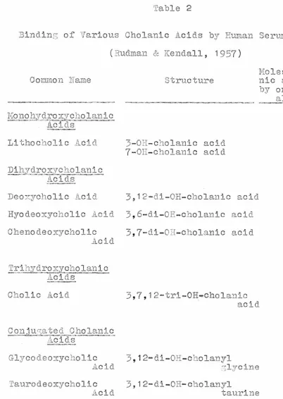
Rudman and Kendall (1957) studied the effect of pH upon the binding of deoxycholic acid by human serum albu-min. They found a suppression of binding above PH 9. They
suggested that this result was compatible with the existence of an electrostatic bond between the positively charged
•
42 Table 2Binding of Various Cholanic Acids by Human Serum Albumin l (Rudman & Kendall, 1957)
Moles of chola- Common Name Structure nic acid bound
by one mole Of albumin
•
Monohydyoxvcholanic Acids Lithocholic Acid Dihydroycholanic Acids Deo-Tcholic Acid Hyodeomycholic Acid Chenodeoxycholic Ac 1(1 3-0H-cholanic acid 7-0H-cholanic acid 3,12-di-OH-cholanic acid 3,6-di-OH-cholanic acid 3,7-di-OH-cholanic acid 6,5 6.7 2.3 3.2 3.0 Trihydroaycholanic AcidsCholic Acid 3,7,12-tri-OH-cholanic
acid 0.94
•
43
Table 2 (continued)
Bindin of Various Oholanic Acids by Human Serum Albumin
1. Ten ml. of a 1 per cent human serum albumin solution -
(1.4 x 10 -) mM of albumin) was equilibrated by dialysis with 50 ml of buffer containin 12.7 x 10 -3 of cholanic acid. After equilibration with buffer or serum protein, the cholanic acid concentration in the outer solution was determined spectrophotometrically.
•
44
•
concentrations of DOG, we would expect to find that histone I (25.5 mole per cent lysine) would be removed at the low-est concentrations of dissociating agent. Histone II (16.1 mole per cent lysine) would follow at slightly higher
con-centrations of DOG, while histones III (8.6 mole per cent lysine) and IV (8.5 mole per cent lysine) would be the last histone fractions to be dissociated. Since the
experimen-tally observed order of dissociation is histone II, fol-lowed closely by histones III and IV, which are in turn followed lastly by histone I, we feel that the formation of such an electrostatic bond is not the primary force respon-sible for the histone dissociation patterns observed when
II!
chromatin is extracted with increasing concentrations ofDOG.
Rudman and Kendall (1957) also reported that the affin-ity of various cholanic acids for albumin is reduced by the introduction of polar groups into the steriod nucleus.
Table 2 shows their results on the binding of various cho-lanic acids by human serum albumin. It is readily apparent
that the extent of binding decreases in the order monohy-droxy > dihymonohy-droxy >trihymonohy-droxy cholanic acid. This vari-ation in binding: among closely related cholanic acids
indicates the existence of forces, other than electrostatic, between albumin and cholanic acids. The data suggested
45
that information of interest might be obtained from studying how changes in the structure of cholanic acids affect
his-tone dissociation patterns obtained when chromatin is ex-tracted with increasing concentrations of various cholanic acids.
The fraction of total histone which remains bound to DNA of chromatin after extraction with the sodium salts of several cholanic acids is shown in Figure 7. Sodium cholate is 3 to 5 times less effective as a histone dissociating agent than is sodium deoxycholate. Movement of one of the hydroxyl groups of sodium deoxycholate from the 12-position to the 6-position (sodium hyodeoxycholate) has relatively
111
little effect on the amount of histone dissociated by a given concentration of cholanic acid. The monohydroxycho-lanic acid series was not sufficiently soluble in our buffer system to allow a histone dissociation study. Conjugation of the carboxyl group of deoxycholic acid or cholic acid with either glycine or taurine causes approximately a 1.5 fold reduction in the histone dissociating capacity of the cholanic acid. Conjugation of cholic acid with glutamic acid, however, restores the ability to dissociate histones to its original level (same as unsubstituted cholic acid).46
•
Figure 7. The weight fraction of total histone remaining -bound to DYA after extraction of chromatin with increasin g
concentrations of the sodium salts of various cholanic acids and some of their conjugated derivatives.
•
0 0
•
/ / / / -.
/ •.
41 a) 4-- o o _c 0 0 C.) 2-. . ° E=
'6 • o / 4 a) -.- o -5 _c 0 E 0 4.S 0 m 'Es 0 U).
... ...• I a) o -E) _c (....) E n .-i5 0 U .) a) 4- o a) oT5
o o , >, -c x 0 0 ›.-, 0 Q.) X ( 7, "0 0 0 a) _c o ID o >, o >, x 0 = o E mo a) E0 E 2 '5 'fi D 0 • .-E3 00 (r) 0 (1)1— / (I)•
p
illg•
a)0
75 _c 0 > n X 0 cv so-0 E o U) _ — _ - 0 Cq CO. Cr. ( 'J 0 0 0 0 dVW] 01
aNnoe
ONINIVINA8
J1\101SIH 1\7101 NO110V8A
•
0
48
ability to dissociate histone from chromatin, its ability to selectively dissociate the various histone fractions was investigated. Figure 8 presents data on the fraction of each histone fraction remaining bound to the DNA of chro-matin after extraction with increasing concentrations of
sodium cholate. At a given amount of total histone removal, more histone I is removed by sodium cholate than by sodium
deoxycholate. The ability to selectively leave histone I attached to the DNA while removing histone II, III, and IV is apparently diminished by the higher concentrations of dissociating agent required for a given amount of total histone removal when a third hydroxyl group is introduced
411
into the steroid nucleus.Thermal denaturation of partially dehistonized samples was studied in order to provide an independent indication
of the degree and type of histone fraction removal. Figure
9 shows the T m of samples which have been partially dehis-tonized by extraction with increasing concentrations of de-oxycholic acid, cholic acid, and a few of their conjugated derivatives. In order to provide a clearer feeling of the type of histone fraction being removed at a given per cent total histone removal, the data have been replotted as frac-tion decrease in T (where 74 0 C. = 0 and 44 0 C. 1.00)
m
versus fraction of total histone remaining bound to the
49
_Azure 3. The fraction of individual histone components remaining bound to MA after extraction of chromatin with increasinL, concentrations of sodium cholate. -Aston° I = lysine-rich = fl; histone II = slightly lysine-rich = f2b and f2a2; histone III = ar-injne-rich = f3; histone IV = arTs'inine-rich = f2al.
•
50
•
oco.
•1-
o
—
o
o
o
o
VNC1 01
aNnoe
ON 1 NIVEN3ed
1NANOcilAJO0 ANO1S1H NO110V8A
•
51Fi ,Jure 9. The Tm of chromatin samples partially dehiston-ized by increasing concentrations of the sodium salts of various cholanic acids and some of their conjugated deri-
-4
vatives. Melting was done in 2.5 x 10
m
,DTA, pH 8. No correction for thermal expansion.•
o
a
ro
O()
(73
C.) — c\J Z<
0
o
u_
0
Ln >-
51—
E
_J 00o 2
•
52 _ 1 I I I I I a) _ _ cs o _c 0 o 0 - >, _ 0 a) E0
_
—,_,
C
E -;-- n ..., - _c -- - - o E n '-o- a) o o-
(f)
_
(7)
(.)
I
/111"
_c
>, x
I
o a) o cl • Elf/
a) (..) E -5 i o >, x n :15_;....-n 9_ >" /
c 2 E mo 0 .,-; P o - cr) -0 0 <I) t..i
la
>, _
0 I--0/
0
(f) i
53 DNA (Figure 10).
The stabilization of the DNA double helix against thermal denaturation by increases in the ionic strength of the melting medium has been well documented, and for a given DNA increases by approximately 18 ° C./log
DTA
(Dove & [image:60.612.68.498.123.636.2]Davidson, 1962). Therefore, a decrease in the ?ni t s of par-tially dehistonized samples of chromatin would be expected to show some dependence on the number of histone positive charges still attached to the DNA, and, consequently, some dependence on the arsinine plus lysine content of the his-tone fractions still attached to the DNA. Comparison of sodium chloride data (Smart
&
Bonner, 1969) and sodium de oxycholate data yields the expected differences in Tm changes. Thus, removal of 20 per cent of total histone by NaC1 (only histone I removed) produces a decrease in the T m of the partially dehistonized sample which is approximately 2 to3
times as great as that observed when 20 per cent of total histone is removed by DOC (only histone II removed). These decreases are in good agreement with the differences in the net positive charge per amino acid of the two his-tone fractions (from amino acid composition data of Fam-browdi, 1967); histone I has 0.183 net positive charges per amino acid, while histone II has only 0.084.•
54Figure 10. The fraction decrease in the T m (where
740
C. 0 and 44 0 C. = 1.00) versus the fraction of total histone removed from DNA after extraction of chromatin withmr)
• Sodium Deoxycholate
o
Sodium Hyodeoxycholate
• Sodium Glycodeoxycholate
A
Sodium Taurodeoxycholate
0
Sodium Cholate
1.0
0.8
•
v
x
Sodium Glycocholate
Sodium Glutamylcholate
Sodium Chloride
/1//
// /
/
/
///
/
o
o
- a
, 0.6
a
w
c)
0.4
a2
0
// / ‹// /
/
/ /
/
/
,
/
/
/
/
'0
/ --x/
/>*---Na Cl
0
• A
77 A • 0
/
/
/
/ /
/
/
/
i
o
,)V
A • //0
DOC
56
duced by extraction of chromatin with increasing concentra-tions of various deoxycholic acid derivatives as a function of totallAstone remaining bound to the DFA closely parallel those for deoxycholic acid. On the other hand, decreases in Tm produced by extraction of chromatin with increasing concentrations of cholic acid and its derivatives are
slightly greater per 'weight fraction histone removed. This observation is in accord with the relatively early removal of histone I by sodium cholate (see Figure 2 and 8). It also appears that the sodium salts of the various deriva-tives of deoxycholic acid and cholic acid selectively dis-sociate histone fractions in a pattern similar to that of the parent compounds.
DISCUSSION
The first conclusion of this section is that the binding of sodium deoxycholate to chromatin components
appears to be readily and completely reversible. Secondly, we conclude that the formation of an electrostatic bond between the positively charged e-amino group of lysine and the negatively charged carboxylate group of deoxycholic acid
57
force responsible for the histone dissociation pattern ob-served when chromatin is extracted with increasing concen-trations of BOO.
The lowering of the chemical potential of the aqueous chromatin-cholanic acid solution, which occurs when the cho-lanic acid complexes with histones and other chromatin com-ponents, certainly involves changes in the activity co-efficients of histones and DNA. These changes, however, are very complex and the actual effect on the activity co-efficients of the DNA and histones is difficult to predict. We feel, on the other hand, that the complexing of the
relatively hydrophobic DOC anion with hydrophobic regions
41!
of the positively charged histones, certainly lowers the activity coefficient of the cholanic acid.Consideration of the amino acid composition data of Fambrough (1967) shows that the various histone fractions contain the hydrophobic amino acid residues of valine, iso-leucine, iso-leucine, tyrosine, and phenylalanine in the
following amounts: histone I, 12.1 mole per cent; histone II, 25.4 mole per cent; histones III and IV, 26.0 mole per cent. Although these data do not consider sequential
arrangement of these residues in the histones, they are consistent with the hypothesis that DOC should form more stable hydrophobic complexes with histones II, III, and IV
58
than with histone I. ith no correction for amide groups, the (lysine + arginine) - (glutamic acid + aspartic acid) content for the various histone fractions is: histone I,
18.3 mole per cent; histone II,
8.4
mole per cent; histones III and IV, 9.1 mole per cent. The mole per cent of net positive charges per histone fraction, therefore, is an inverse function of the order of removal with increasing concentrations of DOC.We suggest that the driving force for the interaction between the cholanic acid anion and histones is the lowering of the activity coefficient of the cholanic acid anion which occurs when it is partially removed from aqueous solution
111
by interaction with hydrophobic regions of the histones.When a cholanic acid anion interacts with a histone mole-cule, it effectively lowers the net positive charge of the histone molecule, thereby lowering the ionic strength of dissociating agent required to remove the histone molecule. This binding also probably cancels some of the histone-his-tone and hishistone-his-tone-DNA hydrophobic interactions. It is pos-sible that these interactions may account for the fact that ionic dissociating agents, such as liaC1, ilaC104, and CuCl selectively remove histone I, the histone fraction with the highest net positive char57e density per amino acid, at lower concentrations of dissociatirv , agent than those required to
59
remove histones II, III, and IV.
We propose that cholanic acid anions preferentially interact with those histone fractions which contain the highest mole per cent hydrophobic amino acids, namely his-tones II, III, and IV. This interaction will lower the net positive charge of these histone fractions and reduce his-tone-histone and histone-DNA hydrophobic interactions. with these assumptions we expect sodium deoxycholate to
remove histones at much lower concentrations of dissociating agent than do ionic dissociating agents - such is the ex-perimental observation. We also expect DOC to selectively remove histones II, III, and IV at lower concentrations of dissociating. agent than those required for removal of his-tone I - this is the experimentally observed pattern of
histone dissociation. Since the introduction of a hydroxyl group into the steroid nucleus effectively lowers the ac-tivity coefficient of the cholanic acid in aqueous solution, we expect sodium cholate to be less effective in. dissocia-tion of histones from chromatin, as is experimentally ob-served. Conjugation of the cholanic acid with a compound which is relatively less hydrophobic will result in a
6o
should, and does, make the product less effective in the dissociation of histories than the original cholanic acid. The importance of the negatively charged carboxylate group of the cholanic acid in the original binding of the cho-lanic acid anion to histories, and/or the subsequent lowering of the net positive charge of the histone to which it has bound, and/or its contribution to the ionic strength of the dissociating medium, is demonstrated by the effectiveness of sodium glutam7lcholate as a histone dissociating agent.
It appears that the presence of the additional negatively charged carboxyl group compensates for the expected lowering of the activity coefficient (due to the presence of the hy-
III
drophilic carboxyl group) of sodium glutamylcholate in aqueous solution.Since addition of a third hydroxyl group to the steroid nucleus lowers the activity coefficient of the cholanic • acid in aqueous solution, less cholate anion is expected to be bound to the histories than deoxycholate anion at a given molarity of cholanic acid. Consequently, a given molarity
of cholic acid would not lower the net positive charge of
the various historic fractions as much as the same
concen-tration of deoxycholic acid. Dissociation of histories by
61
dium, and consequently, would be expected to yield a dis-sociation pattern of the various histone fractions which would be intermediate between sodium deoxycholate and ionic
dissociating agents, such as sodium chloride. The data show that although sodium cholate is about 7 to 10 times as effective as sodium chloride in the dissociation of total histone, it is only 2 to 3 times as effective in the dissociation of histone I.
62
REFERENCES
1. Allfrey, V. C., Littau, V. C., and hirsky, A. E. (1963),
Proc. Nat. Acad. Sci. (hash.) 50, 1026.
2. Bonner, J., Chalkley, G. R., Dahmus, M., Fambrou-h, D.,
Tlujimura, Y., Evan - , E. C. C., Fuberman, J., 2arushige, K., Chlerbusch, H., Olivera, B., and Tridholm, J.
(1968a), Methods in 'Snzymoloqy, v. 12, Academic Press, New fork.
3. Bonner, J., Dahmus, M., Bambrou ,-;h 1 D., Huan-;, R. C. C.,
Marushi7,e, K., and Tuan, D., (1968b), Science, 159, 47.
4. Bonner, J. and 2s 1 0, F. 0. P., ed. (1364), in The
!II
Yucleohistones, Holden-Day, San Francisco, Calif.5, 1-Ai.rkhard, R. K. and Mrovf, C. (1964), Siochim. Pio-
phys. Acta, 90, 393.
6. Burton, K. (1956), Biochem. J. 62, 315.
7. Busch, H. (1965), ISistones and ether Nuclear Proteins,
Academic Press, Rev- York.
8. Colter, J. S., Casper, H. 11., and Ellen, K. A. (1962),
Nature, 195, 585.
9. Dische, Z. and Schwarz, K. (1937), mikrochim. Acta, 2,
13.
10. Doty, P., Marmur, J., Eilner, J., and Schildkraut, C.
63
11. Dove, W. F., Jr. and Davidson, N. (1962), J. Mol. Biol. 5, 467.
12. Elgin, S. R. (1969), personal communication.
13 FambrouEh, D. (1967), Ph.D. Thesis, California Insti- tute of Technology.
14. Fambrough, D. and Bonner, J. (1966), Biochemistry, 5
2563.
15. Fambroush, D. and Bonner, J. (1963), Biochim. - io-phys. Acta, 154, 601.
16. Godson, G. N. and Sinsheimer, 2. (1967), J. Mol. Biol. 23, 495.
17. Huang, R. C. C. and Bonner, J. (1962), Proc. Pat. Acad.
`4110 Sci. (Wash.) 48, 1216.
18. Ilyin, Y. V. and Geor , iev, G. P. (1969), J. Zol, Diol. 41, 299.
19. Jensen, R. (1966), personal communication.
20. Kedes, L. H. and Gross, P. H. (1969), J. Hal. Biol. 42 559.
21. Lenaz, G., aard, I. F., Lauuers, A., Allmenn, D. %., and Green, D. E. (1968), Arch. Ir3ioc'ler. Bio -ohys, 126, 746.
22. Levinson, S. R., Smart, J. E., and Bonner, J. (1969), in preparation.
•
65240, 2094.
37. Smart, J. E. and Bonner, J. (1969), in preparation.
38. Ts T o, P. 0. P. and Sato, C. (1939), Exptl. Cell Res.
17, 227.
39. Tuan, D. (1966), Ph.D. Thesis, California Institute
of Technology.
40. Than, D. and Bonner, J. (1969), in -preparation.
66
STUDIES ON THE ROLE OF HISTONES IN THE STRUCTURE OF CHROMATIN
67
INTRODUCTION
The DNA contained in the nucleus of higher organisms is complexed with proteins and usually a small amount of T. This nucleoprotein complex, as found in interphase cells, is referred to as chromatin. The major portion of the proteins contained in chromatin are histones, a family of seven major species all of which, possess a 20 to 30 mole per cent arginine plus lysine content. The complexing of histones to DNA results in neutralization of a large portion of the negative phosphate groups of the DNA., and a few
con-comitant structural changes in the DNA. Although the
!II
structure of chromatin is complex, and probably concernsseveral levels or organization, re :lent studies indicate that its primary organization is at the level of supercoiling of the individual chromatin fibers (Griffith. & Bonner,
1969; Pardon, Wilkins, .& Richards, 1967; Moundrianakis, 1969). The model which appears to best account for experi-mental observations is that of a DNA double helix (width
68
•
molecular axis of the chromatin molecule. It is also observed experimentally that the DNA contained in chroma-tin has undergone a conformational change which is expressed as an increased absorptivity and, also, a decreased hyper-chromicity upon melting (Tuan & Bonner, 1969).
The dissociation of chromatin with increasino, con-centrations of YlaC1 selectively removes histone I (lysine-rich) at lowest concentrations of dissociating agent. Higher concentrations of NaO1 remove histone II (slightly lysine-rich) and histones III and IV (arginine-lysine-rich) (Ohlenbusch, Olivera, Tuan, & Davidson, 1967; Fambrough & Bonner, 1968; Georgiev, Ananieva, & Kozlov, 1966; Tuan & Bonner, 1969;
411
Smart & Bonner, 1969). Smart, Hadler, and Bonner (1969)have reported that increasing concentrations of sodium de-oxycholate (D00) dissociate histone II at lowest concen-trations of dissociating agent, while slightly higher
•
69cross-linked component of sheared chromatin. The data indicate that as evidenced by selective removal of hi-stone studies, none of the structural or chemical parameters with which these properties are correlated show a dependence on
the presence of one particular histone fraction.
METHODS AND MATERIALS
Preparation of Chromatin. Pea bud and calf thymus chromatins were prepared by the methods previously des-cribed (Smart, Hadler, & Bonner, 1969). The chromatin had a final concentration of 20 to 40 A 2 60141 , a ratio of A230 mp/
of approximately 0.75, and a ratio A, 20 /A 2 6Om o A260
less than 0.034. The pea bud DNA contained in chromatin is complexed with hi-stone protein, nonhistone protein, and RNA in the mass ratios DNA, 1.0; histone, 1.05; nonhistone, 0.50; ANA, 0.12. Calf thymus DNA is complexed in the mass ratios DNA, 1.00; hi-stone, 1.00; nonhistone, 0.30.
Isolation of Chromatins of Differing; Sedimentation Co-efficients. Chalkley and Jensen (1968) reported that prep- __
•
70as previously described (Chalkley & Jensen, 1968). Histone Dissociation Studies. To assure that the chromatin was not subjected to a sodium deoxycholate or so-dium chloride concentration higher than the desired final one, partially dehistonized samples were prepared as des-cribed previously (Smart, Hadler, & Bonner, 1969).
Thermal Denaturation and Ultraviolet Absorption. The partially dehistonized samples were dialyzed exhaustively against 2.5 x 10 -4 M EDTA, pH 8, diluted to approximately
1 k with dialysate, and melted in a Gilford Model 2000 260
multiple sample absorbance recording apparatus adapted for the recording of melting profiles. The rate of temperature increase was 0.5 to 1.0 de?ree/minute.. Ultraviolet absorp-tion spectra were determined with a Cary recording spectro-photometer, Model 11.
Sedimentation
Analy21.a.:_
Sedimentation velocity was studied using band-sedimentation techniques either on pre-formed sucrose gradients in a Spinco Model L ultracentri-fuge or on self-generating density gradients in a Spine° Model E analytical ultracentrifuge (Vinograd, 1963). In the analytical centrifuge D 20 was used to form the gradients.•
71& Bonner, 1969).
Flow Dischroism Measurements. Flow dichreism was per-formed as described by Callis and Davidson (1969a) in 0.0025 M Tris, pH 8, using their apparatus. In their apparatus, the flow is down a long, narrow channel with an unpolarized light beam alonp: the direction of flow. The apparatus has the following advantages: dilute macromolecule solutions can be used (0.2 An gn ,,vmp ), high shear gradients are easily obtained (up to 21,000 sec -1 ), and only small volumes of solution are needed.
Relaxation Times of Molecules Oriented in a Flow Field. The relaxation of the flow dichroism signal of native and
411
dehistonized chromatin was performed as described by Callisand Davidson (1969b) in 0.0025 M Tris, PH 8, using their apparatus. Under the conditions employed in this study, the actual time required for stoppage of flow is 3 to 5 x 10 -3 sec.
Precipitation of Chromatin. In order to obtain a stan-dard and reproducible measure of precipitation, we have de-fined precipitated chromatin as that material sedimented from solution (0.15 H NaC1 - 0.0025 M Tris, pH 8) in 10 minutes at 10,000 x L. The assay was performed by slowly
adding, 1/3 volume of 0.6 M NaC1 - 0.0025 M Tris, pH 8, to 1 volume of chromatin solution (approximately 10 A260rop) c o
•
72Vortex mixer. After 10 minutes on ice, the solution was centrifuged in a Servall 55-34 rotor at 10,000 rpm for 10 minutes. The supernatant was removed by Pipetting,
ana-lyzed for A 2 60 and then dialyzed extensively against 0.0025 M Tris, pH 8. The pellet was removed, re-suspended by Teflon homogenization, and dialyzed extensive-ly against 0.0025 M Tris, pH 8. Diaextensive-lysis of supernatants and pellets was carried out in a glass stoppered, gradu-ated cylinder mounted on a slowly rotating carrier. A small air bubble in the dialysis tubes resulted in con-stant stirring_ All operations were carried out at 0 to 4° C.
Chemical Analysis. DNA, histone protein, nonhistone protein, and RNA analysis were performed as previously des-cribed (Smart, Hadler, & Bonner, 1969).
Disc Gel Electrophoresis of Histones. Acrylamide disc gel electrophoresis of isolated histories was performed by the method of Bonner et al. (1968a). The quantity of each electrophoretic component was determined by densitometry after Fambrough (1967).
Note: All assays were performed within one week of the preparation of chromatin.
73
•
RESULTS
Samples of chromatin which have been partially de-histonized by increasing concentrations of sodium chloride
(NaC1) or sodium deoxycholate (DOC) are more heterogeneous in protein distribution than native chromatin or pure DNA. This is shown by an increase in the band width of formal-dehyde-fixed samples in a cesium chloride density gradient
(Ilyin & Georgiev, 1969; Farber, Smart, & Bonner, 1 969). These samples also have an increased heterogeneity in charge distribution over that of native chromatin or pure DNA, as shown by the increase in electrophoretic dispersion
111
(band width) (Ohlenbusch,of the samples upon free-zone electrophoresis Olivera, Tuan, & Davidson, 1967; Smart &
74
•
precipitability of chromatin to decrease as increasing amounts of histone are removed. If partially dehistonized chromatin samples are in fact composed of molecules which contain varying proportions of DNA to histone, we would anticipate that partially dehistonized samples should be precipitable to an extent intermediate between the observed 90 to 98 per cent characteristic of native chromatin and the
0 per cent characteristic of pure DNA. The pellet (preci-pitated portion) of samples with intermediate precipitation values should consist of a class of nucleoprotein molecules of a higher histone to DNA ratio than that found in the supernatant (soluble portion). Figure la presents the
111
melting profiles of a chromatin sample which has been ex- tracted with 0.04375 M DOC, and of the supernatant and pel-lets obtained from this sample by fractionation in 0.15 NaC1 (approximately 66 per cent of the A260 mater