Study of the Anatomical Variations of
the Liver in Human
Dissertation submitted for
M.D Anatomy Branch V Degree Examination,
The Tamil Nadu Dr. M.G.R. Medical University
TABLE OF CONTENTS
1. INTRODUCTION ... 10
2. AIM AND OBJECTIVES... 15
3. LITERATURE REVIEW ... 17
3.1 Fissures of the liver: ... 17
3.2 Segments of the liver: ... 20
3.3 Variation in liver morphology ... 22
3.4 Vascular system of Liver ... 29
4. MATERIALS AND METHODS ... 49
4.1 Gross anatomical variation ... 49
4.2 Branching pattern of the hepatic artery and portal vein ... 49
4.2.1 Luminal casting ... 50
4.2.2 Radiological study ... 52
5. RESULTS ... 54
5.1. Morphological variations of the liver ... 54
5.2. Hepatic artery branching pattern ... 63
5.3 Branching pattern of the portal vein ... 79
6. DISCUSSION ... Error! Bookmark not defined. 6.1. Morphological variations of the Liver ... 96
6.2 Hepatic artery ... 103
6.3 Portal vein ... 109
7. HIGHLIGHTS OF THE STUDY ... 114
7.1 Hepatic artery branching pattern ... 115
7.2 Portal Vein branching pattern... 116
8. CONCLUSION ... 119
9. LIMITATIONS ... 121
10. FUTURE SCOPE ... 123
ACKNOWLEDGEMENT
I thank God Almighty for helping me finish this thesis with His
presence in each and every step.
I am grateful to the Institutional Review Board (IRB) of Christian
Medical College, Vellore for giving me the permission and funding
for this project.
I sincerely thank Dr. Suganthy J, my guide for entrusting and
encouraging me to start this study and for being an inspiration and
supporting and helping me all the time.
Dr. Reetu Amrita John, Assistant professor and Dr. Anu Eapen,
Professor, Department of Radiology for helping me in getting the images
and explaining the images
Dr. Manbha Rymbai, Assistant surgeon, Department of Hepatopancreato
biliary surgery for accepting the concept
Dr. Devakumar Devadhas, Associate professor, Department of Nuclear
Medicine for his guidance
Dr. Pauline Shanthi, Assistant professor, Department of Anatomy for her
guidance and help
Mr. Prakash R, Senior demonstrator, Department of Biostatistics for
helping me in calculation of sample size and Ms. Hepsy.Y.S for
helping me in analysis of the thesis data
All Associate Professors and Assistant professors for their help in
times of need
All co - postgraduates for their valuable suggestions, help and
encouragement
To all non- teaching staff in the Department of Anatomy –
Mr. S. Thirumurugan (late), Mr. Antonyswamy, Mr. R. Gopi, Mr.
Sanmughan, Mr. Dhandapani, Mr. Babu, Mr. Collin, Mr. N. Narayanan
for their help with arrangement of the liver specimens and support
all the time
Mr. V. Gopinath (Secretary) and Mr. G. Rajkumar (Artist) in the
Department of Anatomy for their timely help
To all generous volunteers who donated bodies
1. INTRODUCTION
The liver is the largest abdominal organ. It occupies the right hypochondrium,
epigastrium and a small part of the left hypochondrium. It has been divided into the
right, left, caudate and quadrate lobes based on the surface peritoneal and ligamentous
attachments.1 The right and left lobes are partitioned by falciform ligament anteriorly
and by the fissure for ligamentum venosum and fissure for ligamentum teres
inferiorly. Functionally, Cantlie's line which runs between the fossa for gall bladder
and inferior vena cava on the diaphragmatic surface and divides the liver into right
and left lobes.2 This corresponds to middle hepatic vein. The structures present at the
hilum of the liver include the hepatic artery, portal vein and the bile duct. The
branching patterns of blood supply and biliary drainage in the liver form the lobes and
further subdivision form the segments or sectors of the liver.1 According to Couinaud‟s division of the liver, there are eight functional segments (I to VIII).
Distribution of the portal venous system and the hepatic veins forms the basis of this
division.3
In addition to the right, left, caudate and quadrate lobes, accessory lobes and
fissures have been described. It has been found out in Indian population, that
accessory lobes are present in 10% of the population.3 Various other anomalies like
quadrate lobe with complete transverse fissure dividing the lobe into a superior and an
inferior lobe, pons hepatis connecting the left lobe with the quadrate lobe,3 hypoplasia
of right lobe of the liver4 have been reported. A defect in the development or
lobe might be mistaken for a lymph node due to its small size and removed during the
surgeries. Torsion of the vascular pedicle may require urgent manipulation.3 Presence
of accessory fissures are the potential sources of diagnostic errors during imaging. It
may be mistaken for a liver cyst, hematoma or abscess when there is a collection of
fluid in these fissures. Metastatic tumour cells getting lodged into these spaces may
mimic intrahepatic focal lesions. Knowledge of such possible variations is thereby
important during radiological investigation and surgery.3
The liver receives upto about 25% of the cardiac output. It is supplied by
hepatic artery which is a branch of coeliac trunk and contributes to about 25% to 30 %
of the blood supply to the liver. It also gets blood supply from the portal vein which
contributes to the remaining 70%-75%. The arterial and the venous blood ultimately
gets mixed up in the sinusoids of the liver and through the right, middle and left
hepatic veins, the venous blood drain into the inferior vena cava.5
Variations in the extrahepatic branching pattern of the hepatic artery and portal
vein have been reported earlier. The common hepatic artery arises from celiac trunk
and gives off the right gastric, the right gastroduodenal and the proper hepatic artery.
The proper hepatic artery gives off the right hepatic, left hepatic and middle hepatic
arteries supplying the right, left and quadrate lobe of the liver respectively.6
Extrahepatic arterial variations have been classified into 10 types by Michael.7
Kamath, reported that in 35% of Indian population, the middle hepatic artery
supplying the quadrate lobe of the liver was given extrahepatically. In 17.5% the
common hepatic artery trifurcates into gastroduodenal, right and left hepatic arteries
present.6 But there is a paucity of literature in the intrahepatic branching pattern of the
hepatic artery.
The portal vein begins at the level of the second lumbar vertebra and is formed
by the union of the superior mesenteric and splenic veins.1 It is approximately 8cm
long and lies anterior to the inferior vena cava and posterior to the neck of the
pancreas. It ascends behind the first part of the duodenum, the common bile duct and
gastroduodenal artery. It enters the right border of the lesser omentum, ascends
anterior to the epiploic foramen to reach the portahepatis. It then divides into right and
left main branches which accompany the corresponding branches of the hepatic artery
into the liver.1 Guler et al. (2013) reported that 12.6% donors have portal vein
variations.8 Preoperative assessment of the portal vein system is essential for safe
hepatectomy.9,10 With the growing popularity of complex hepatobiliary surgical and
percutaneous procedures, including trisegmentectomy, portal vein embolization, and
transjugular intrahepatic portosystemic shunts (TIPS), the detection and recognition of
portal vein variants are increasingly relevant.
A reduction in iatrogenic complications during hepatobiliary surgeries can be
achieved with the knowledge of the variation of hepatobilary system. Though there
are a few studies available regarding the intrahepatic branching pattern of the
hepatobilary system in other population, there is no data available for Indian
2. AIM AND OBJECTIVES
Aim:
To describe the anatomical variations of the hepatic artery and portal vein
within the liver in terms of branching pattern and determine the frequency of each
pattern.
Objectives:
- To determine the gross anatomical variations of the liver
- To determine the variation in the branching pattern of hepatic artery and
portal vein by modified corrosion casting technique
- To determine the variation in the branching pattern of the hepatic vasculature
3.
LITERATURE REVIEW
The liver is located in the right hypochondrium, epigastrium and extends to the
left hypochondrium. Based on the surface peritoneal and ligamentous attachments, the
liver can be anatomically divided into the right, the left; the caudate and the quadrate
lobes.1 The part of the caudate lobe adjacent to the inferior vena cava is known as the
paracaval portion and the part which is adjacent to the fissure for ligamentum venosus
is known as Spiegel's lobe.11 The caudate lobe has caudate process and papillary
process.11
3.1 Fissures of the liver:
Knowledge of the fissures of the liver is essential for understanding liver
surgery. The liver has 3 major fissures and 3 minor fissures.The three major fissures
are the main, left and right portal fissure. They are not visible on the surface. They run
through the liver parenchyma and harbour the three main hepatic veins (Figure 3.1).
Three minor fissures are the umbilical fissure, the venous fissure and the fissure of
Gans. They are visible on the liver surface.1
Major fissures:
Main portal fissure:
The main fissure extends from the tip of the gallbladder back to the midpoint of
the inferior vena cava and contains the middle (main) hepatic vein. It separates the
liver into right and left hemi-livers. Segments V and VIII lie to the right and segment
Left portal fissure:
The left fissure divides the left hemi-liver into medial (anterior) and lateral
(posterior) sectors. It extends from the midpoint of the anterior edge of the liver
between the falciform ligament and the left triangular ligament to the point which
marks the confluence of the left and middle hepatic veins. It contains the left hepatic
vein and separates the left anterior and left posterior sectors: segment III lies
[image:18.595.89.474.356.622.2]anteriorly and segment II lies posteriorly.1
Right Portal fissure:
The right portal fissure divides the right hemi-liver into lateral (posterior) and
medial (anterior) sectors. This fissure contain the right hepatic vein. It divides the
right anterior sector (segments V and VIII) from the right posterior sector (segments
VI and VII). The right portal fissure marks the thickest point of liver parenchyma
which is commonly transected during liver resection.1
Minor fissures:
Umbilical fissure:
The umbilical fissure separates segment III from segment IV and contains a
main branch of the left hepatic vein (the umbilical fissure vein). It is marked by the
attachment of the falciform ligament. It is often avascular and can be divided safely
with diathermy during a surgical approach. In addition to the umbilical portion of the
left portal vein it also contains the final divisions of the left hepatic duct and the left
hepatic artery branches. The umbilical portion of the left portal vein is an important
landmark. Access to this vein and mobilization of the left portal vein are essential
steps in surgery for hilar cholangiocarcinoma. A knowledge of the arrangement of the
portal vein, hepatic artery and bile duct within the umbilical fissure is essential for live
Venous fissure:
The venous fissure is a continuation of the umbilical fissure on the under
surface of the liver and contains the ligamentum venosum. It lies between the caudate
lobe and the left lobe.5
Fissure of Gans:
The fissure of Gans lies on the undersurface of the right lobe of the liver behind
the gallbladder fossa. It often contains the portal pedicle to the right posterior sector
and is thought to correspond to the right fissure as it relates to the separation of the
sectors of the liver.1
3.2 Segments of the liver:
(Figure 3.1)According to Couinaud‟s division of the liver, there are eight functional
segments (I to VIII). Distribution of the portal venous system and the location of
hepatic veins forms the basis of this division.1
Segment I
Segment I corresponds to the anatomical caudate lobe and lies posterior to segment
IV. It receives vessels independently from the left and right portal veins and hepatic
arteries, and it drains independently into the inferior vena cava by multiple small
branches. The bile ducts draining the segment are closely related to the confluence of
the right and left hepatic ducts.
Segments II
Segment III
Segment III lies between the umbilical fissure and the left fissure, it drains into the left
hepatic vein.
Segment IV
Segment IV lies between the umbilical fissure and the main fissure, The main venous
drainage segment is into the middle hepatic vein; the segment can also drain into the
left hepatic vein through the vein of the falciform ligament.
Segment V
Segment V lies between the middle and the right hepatic veins. Venous drainage is
into the right and middle hepatic veins.
Segment VI
Segment VI lies posterior to the right portal fissure. Venous drainage is normally into
the right hepatic vein.
Segment VII
Segment VII lies behind the right hepatic vein. The venous drainage is into the right
hepatic vein.
Segment VIII
3.3 Variation in liver morphology
The segmental anatomy of the liver has been extensively researched, but there
are very few studies which have dealt with surface variations of the liver.12 Variations
in the liver morphology can be either congenital or acquired.
The liver starts its organogenesis early during 3rd week of intrauterine life and
develops more rapidly. In spite of its complex development, developmental anomalies
are rare.13 The congenital anomalies of liver can be divided into anomalies due to
defective development and anomalies due to excessive development.13 It was
described that the hepatic malformations are common in perinatal age group and liver
undergoes reformation postnatal. Accordingly all fissures and lobes of liver should
disappear during postnatal.14 There are many kinds of described congenital
abnormalities of the liver. It could be:15,16
a) agenesis of lobes
b) absence of its segments c) deformed lobes
d) accessory lobes e) lobar atrophy
f) presence of only one lobe g) presence of multiple lobes h) hypoplastic lobes
i) peduncular lobes j) lobes without division k) Riedel‟s lobe
l) Transposition of the gall bladder
Acquired variations in liver could be due to the pressure given by diaphragm,
peritoneal ligaments and other organs in relation with liver so developed during
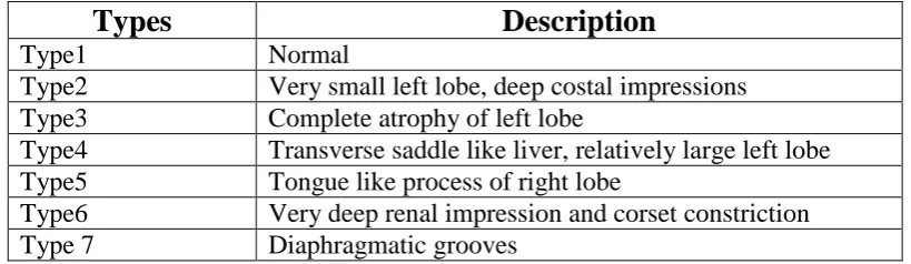
Netter classified the morphological variations of the liver into 7 types as
[image:23.595.63.473.221.340.2]follows:
Table 3.1. Netter's classification of morphological variations of liver
Types
Description
Type1 Normal
Type2 Very small left lobe, deep costal impressions Type3 Complete atrophy of left lobe
Type4 Transverse saddle like liver, relatively large left lobe Type5 Tongue like process of right lobe
Type6 Very deep renal impression and corset constriction Type 7 Diaphragmatic grooves
Agenesis of liver lobes:
Congenital agenesis of liver lobe affects the left lobe more than the right.17–19
Aktan et al. reported absence of the left liver lobe in 11 cases out of 383 CT images.15
Agenesis of the right lobe of the liver is a rare anomaly and only 42 cases have been
reported earlier.20 It occurs slightly more often in men. In patients with agenesis of
the right lobe of the liver, the right hapatic vein will be absent.19,21 Agenesis of the
right lobe might be associated with billiary tract disease, portal hypertension, and
other congenital anomalies.18
Morphological variations of Right lobe:
Agenesis and atrophy of the right hepatic lobe has been reported ealier by
various authors.17–20,22,23,24 Hypoplasia of the right lobe of the liver was reported by
umbilical vein.4 Fissures and accessory lobes including Riedel's lobe are the other
variations associated with the right lobe.
Morphological variations of Left lobe:
Morphological variations of left lobe include hypertrophy of left lobe,
hypoplasia of left lobe and presence of fissures.11 Enlargement of left lobe associated
with agenesis of right lobe was reported by Nikam and Kitture on radiological
study.25 Shankar and Rabi reported enlargement of left lobe with hypoplasia of right
lobe in a cadaver.4 Abnormal „L‟ shaped large left lobe, with the shift of quadrate lobe
and fissure for ligamentum teres to right was noticed by Saritha et al.14 Hypoplastic
left lobes were reported in 3.44% specimens by Vinnakota and Jeyasree.26 Liver with
elongated left lobe is otherwise called as Beaver's lobe or lingual process or Netter's
type 5 variation. Such extremely long left lobe was reported by various authors
including Nayak et al. who reported in one liver out of 5527 and 2 out of 50.14
Elongated left lobe was reported in 4% by Mamatha et al.28 and in 1.72% by
Vinnakota and Jayasree.26 Chaudhari et al. reported elongated left lobe in 12.5% of the
80 livers studied.12 Shivarama and Kumar reported a lingular prolongation of the left
lobe capping the spleen.29
Morphological variations of Caudate lobe:
Phad et al.(2014) reported morphological variations of caudate lobe in 30% of
the 80 livers studied. They found enlargement of caudate process and paracaval
lobe or Couinaud‟s segment) was seen in 5%. Notch or fissure separating papillary
process from rest of the caudate lobe was seen in 2.5%.11
Abraham et al. observed vertical fissure extending upwards from lower border
of caudate lobe in 5.1% and also oblique fissures in caudate lobe.30 In addition, he also
reported prominent papillary process in 1.81% cases and absence of papillary process
in 33%.
Various shapes of the caudate lobe were observed. Phad et al. observed
variations in shape of the caudate lobe in 12.5% of specimens including streak shaped
hypoplastic caudate lobe in one specimen.11 Sarala et al.(2015) observed rectangular
shaped caudate lobe in 58%, pear shaped in 10%, irregular in 20%, triangular in 8%,
others (square, heart shape, inverted pear) in 4% of the livers studied.31 Chavan and
Wabale observed rectangular shaped caudate lobe in 48%, pear shaped in 26%, oval in
14%, square 6%, triangular in 4% and inverted flask shaped in 2% out of 50 livers
studied.32 Abraham et al reported rectangular shaped caudate lobe in 57.6%,
bicornuate in 12%, pyriform in 18.6%, inverted pyriform in 3.4%, globular or heart
shaped in 10.2% out of 59 livers studied.30
Accessory fissures and accessory lobes in the caudate lobe has been described
earlier. Vinnakota and Jayasree reported accessory fissures and accessory lobes in the
caudate lobe in 8 specimens out of 31 liver specimens studied.26 Singh reported an
accessory caudate lobe associated with hypoplastic left lobe of liver.33
Morphological variations of Quadrate Lobe:
Phad et al. reported morphological variations in quadrate lobe in 12.5% of
specimens. These include presence of pons hepatis, i.e segment of hepatic tissue
connecting quadrate lobe to left lobe over the fissure for ligamentum teres hepatis,
presence of horizontal fissures and variation in shape.11 Presence of accessory lobes
in the quadrate lobe, accessory fissures, various shapes ranging from triangular to
irregular, and also various size from very narrow to ill-defined and pons hepatis was
noted by Vinnakota and Jayasree.26 Abnormal quadrate lobe in the form of absence of
quadrate lobe, deeply buried, transverse fissure and fusion with right and left lobe was
also observed.28
Pons hepatis was reported in 4% of the livers by Saritha et al. and 14% by
Khedekar and Hattangdi.34 Vinnakota and Jayasree observed accessory fissures in
quadrate lobe. They have reported a complete transverse fissure dividing the quadrate
lobe into a superior and an inferior lobe.26 Saritha et al. also reported such a complete
transverse fissure in the quadrate lobe. In addition, they reported a mini accessory
quadrate lobe in 2%.14 Absence of quadrate lobe has also been reported in one liver.35
Morphological variations of surface of liver
Costal impressions of the liver were noted in previous studies. Liver with
diaphragmatic impression (Netter‟s Type 7) has been reported by Vinnakota and
Jeyasree.26 Deep renal impressions with corset constrictions were reported by Saritha
In addition to the surface variations, notches along the inferior border were
reported.36
Accessory fissures of liver:
Accessory fissures were noted in the right, left, caudate and quadrate lobes of
the liver by various authors. Saritha et al. reported accessory fissures in 30% and
Khedekar and Hattangdi in 42% of the livers studied.14,35 Khedekar and Hattangdi also
have reported the number of fissures ranged from 1-5 in each liver. Accessory
fissures in the right lobe26,28, in the left lobe26, fissure extending over
postereo-superior surface28 and between caudate process and duodenal impression26 have been
reported.
Accessory lobes of the liver
Accessory lobe of the liver was a rare congenital anomaly found incidentally at
autopsy or laparotomy. They are commonly found on the undersurface of the liver and
seen on the gallbladder surface, hepatogastric ligament near umbilicus, adrenal gland,
pancreas and the thoracic cavity.37 Sato et al. in a series of 1800 laproscopices
described that the accessory lobe was a congenital anomaly found in approximately
0.7%.38 Maharana reported in 4.76% of cases, Saritha et al., in 16% and Muktyaz et al.
Reidel’s lobe:
Riedel‟s lobe is described as an inferior, tongue-like projection of the anterior
border of the right lobe of the liver to the right of the gallbladder.40 It was first described by Corbin in 1830 and it was defined by Riedel in 1888, as a „„round tumor
on the anterior side of the liver, near the gallbladder, to its right‟‟.41 The incidence of
Riedel lobe is approximately 25% in 20-45 year-olds and 60% in 45-65 year-olds,
with female: male ratio being 3:1.42 It may be clinically confused with an abdominal
mass or pathological hepatomegaly.43 The complications of Riedel's lobe include lobar
torsion, mass effect induced obstruction, interference with laparoscopic surgical
procedures and unnecessary imaging.42
Types of accessory lobes:
The accessory lobes can also be classified into three types, based on the biliary
drainage pattern.44
In type 1, the duct of the accessory lobe drains into an intra–hepatic bile duct of the
normal liver;
in type 2, the duct of the accessory lobe drains into an extra–hepatic bile duct of the
normal liver, and
in type3, the accessory lobe and the normal liver have a common capsule and the bile
3.4 Vascular system of Liver
The liver receives its blood supply from the hepatic artery which is a branch of
coeliac axis and through the portal vein which is formed by the joining of splenic and
superior mesenteric vein.5
Preoperative assessment of the hepatic artery, portal vein and bile duct is
essential for safe hepatectomy.9,10 In living donor liver transplantation, careful
manipulation of the vasculobiliary system is critical to avoid causing injury to the
biliary duct, portal vein and hepatic vein in the residual liver and/or the graft.10,45,46,47
Prompt identification of anatomical anomalies can help the surgeon to avoid
postoperative complications.
Hepatic Artery
The hepatic artery is a branch of the coeliac trunk. In adults the hepatic artery is
intermediate in size between the left gastric and splenic arteries. In foetal and early
postnatal life, it is the largest branch of the coeliac axis.1 From the coeliac trunk to the
origin of the gastroduodenal artery, it is called as common hepatic artery, and from
that point to its bifurcation it is called as proper hepatic artery.1 Within the free border
of the lesser omentum the hepatic artery is medial to the common bile duct and
anterior to the portal vein. The branches of the hepatic artery are right gastric,
gastroduodenal and cystic branches. At the porta hepatis it divides into right and left
branches.1
The right hepatic artery divides into an anterior division supplying segments V
anterior division often gives a branch to segment I and the gallbladder. The segmental
arteries are end-arteries although some collateral circulation may occur.1 The right
hepatic artery usually crosses posterior to the common hepatic duct and therefore the
right hepatic artery is involved in bile duct cancer earlier than the left hepatic artery.
Occasionally the right hepatic artery crosses anterior to the common bile duct and
therefore, may be injured in surgery of the common bile duct.1
The cystic artery is a branch of the right hepatic artery which is given off inside
the Calot‟s triangle. Inside the triangle it bifurcates into superficial and deep branches
which enter the neck or body of the gallbladder.48 Various studies have reported that
the cystic artery may arise from other sources like common hepatic artery or proper
hepatic artery. Aristotle reported that in 40 livers which he dissected, cystic artery
arose from right hepatic artery in 92.5%, proper hepatic artery in 5% and from
common hepatic artery in 2.5%.48 In addition to this, Williams et al. reported that
cystic artery may arise from left hepatic artery, gastroduodenal trunk, superior
pancreaticoduodenal artery and superior mesenteric artery.49 Balija et al. reported that
the prevalence of cystic artery arising from the left hepatic artery is 1%.50 Nowak et al
reported that the cystic artery may also arise from the right gastric artery and celiac
trunk.51 Double cystic artery has been reported in 15% - 25% of patients by Hugh et
al. and in 12.2% during laparascopic cholecystectomy by Ding et al.52,53
The left hepatic artery divides into two sub-branches; medial and lateral
segmental arteries. The medial segmental artery supplies the quadrate lobe and
anterior region of the left lobe. The lateral segmental artery further divides into
According to Kamath, a typical „normal‟ hepatic artery divides into three main
branches - the right hepatic, the left hepatic and the middle hepatic supplying the right,
left and quadrate lobe of the liver respectively.6 The middle hepatic artery supplying
the quadrate lobe is otherwise called as artery to segment IV.
[image:31.595.88.454.230.469.2]
Figure 3.2 showing the branches of hepatic artery. The picture is reproduced from Gray’s Anatomy 40th
Aberrant hepatic artery
If the hepatic artery arises from other than the coeliac trunk, it is known as
'aberrant hepatic artery'. It is of two types - accessory and replaced. The term
„accessory‟ hepatic artery is used if one hepatic artery arises from the coeliac trunk
and there is an additional artery from other sources. When the normal right or left
hepatic artery arising from coeliac trunk is missing and the replaced vessel coming
from another source supplying the right or left lobe, then it is termed as a „replaced‟
right or left hepatic artery 55
Variations in the origin and branching patterns of hepatic artery are quite
common. Congenital anomalies of the hepatic arterial supply are thought to occur
because of the persistence of vitelline arteries during embryologic development.56
Anatomical variations of the hepatic arteries are of considerable importance in
liver transplants, laparoscopic surgery, radiological abdominal interventions and
penetrating injuries to the abdomen.57 Rela et al. observed that the frequency of
iatrogenic injury to hepatic artery rises in presence of aberrant hepatic arteries.58
Michel's described the hepatic arterial anatomy and its variations after 200
cadaveric dissections and identified 10 types of hepatic arterial anatomy (Table 3.2).55
Hiatt et al. in 1994 modified Michel's classification and described 6 subtypes as it is
difficult to distinguish between accessory and replaced hepatic arteries by
angiography. Further, other authors including Adachi in 1928 and Abdullah in 2006
Table 3.2 Hepatic artery variations according to Michel's and Hiatt's classification
Description Michel's
classification
Hiatt's classificatio
n
RHA,MHA and LHA arise from CHA Type I Type I Replaced LHA from the LGA Type II Type II Replaced RHA from the SMA Type III Type III Replaced RHA and LHA Type IV Type IV RHA,MHA; and LHA arise from CHA; accessory
LHA from the LGA
Type V Type II
RHA,MHA and LHA arise from CHA; accessory RHA from SMA
Type VI Type III
Accessory LHA from LGA and accessory RHA from SMA
Type VII Type IV
Replaced RHA and Accessory LHA Type VIII Type IV Entire hepatic trunk arise from SMA Type IX Type V Entire hepatic trunk arise from LGA Type X NOD CHA directly originating from the aorta NOD Type VI RHA - Right hepatic artery, MHA - Middle hepatic artery, LHA - Left hepatic artery, CHA - Common hepatic artery, LGA - Left gastric artery, SMA - Superior mesenteric artery; NOD - Not otherwise described.
The most common variations are a replaced right hepatic artery arising from
the superior mesenteric artery in 10–15% of cases and a left hepatic artery originating
from the left gastric artery in 3–10% of patients.55,59
Kamath mentioned that out of the 40 cadavers studied, text book description of
common hepatic artery originating from celiac trunk and dividing into right and left
hepatic artery, and sometimes middle hepatic artery was seen in 30 (75%) cases only.
He reported aberrant hepatic arteries in 25% of cases.6
Ugurel et al. reported that either coeliac trunk or hepatic artery variation was
normal hepatic arterial system has been reported in 51–80% of cases in studies
conducted using digital substraction angiography.61,62
Ramanadham et al. demonstrated a retroportal replaced right hepatic artery
originated from the superior mesenteric artery and a replaced left hepatic artery
originated from the left gastric artery.63
In a prospective investigation done in 1,081 donor cadaveric livers,
Lopez-Andujar et al. reported the classical pattern in 30% of the livers. The most common
variant was a replaced left hepatic artery arising from the left gastric artery (9.7%)
followed by a replaced right hepatic artery arising from the superior mesenteric artery
(7.8%).64
Sureka et al. did a retrospective review of multidetector CT abdominal
angiography scans performed in patients between January 2012 and February 2013. A
total of 600 patients were evaluated. In their study, normal origin of right hepatic
artery from hepatic artery proper was seen in 79.6% patients. Replaced origin of right
hepatic artery was seen in 15.16% cases and accessory origin of right hepatic was seen
in 5.16% cases. Left hepatic artery originated from hepatic artery proper in 81.5%
patients. Replaced left hepatic artery origin was seen in 10.8% cases and accessory
left hepatic artery origin in 7.6% cases.65
In cadaveric dissections also, aberrant hepatic arteries have been reported.
Suganthy et al reported in a case of multiple vascular variations of splanchnic
branches of abdominal aorta, both accessory and replaced right hepatic artery and
replaced left hepatic artery.66 Yan et al. reported both replaced right and left hepatic
Covey et al. (2002) used digital subtraction angiography among 600 patients.
In their study, in two patients, gastroduodenal artery arose from the celiac axis and the
proper hepatic artery stemmed as the first branch of the superior mesenteric artery.68
In a study done in a total of 19,013 patients, 81% of cases displayed normal
anatomy. A replaced right hepatic artery arose from the superior mesenteric artery in
3.7% of cases, while a replaced left hepatic artery stemmed from the left gastric artery
in 3% of cases. Both replaced right and left hepatic arteries were found in 0.8% of
cases, while an accessory left hepatic artery and an accessory right hepatic artery
were present in 3.2% and 1.6% of cases respectively.69
Shukla et al. reported replaced right hepatic artery in 11-21% of cases and
replaced left hepatic artery in 3.8-10% of cases, while accessory right hepatic artery
and left hepatic artery have a frequency of 0.8-8%.70
Rare anomalies that are not consistent with any type described under Michel‟s
and Hiatt‟s classifications can also be seen. Trifurcation of common hepatic artery
into gastroduodenal, right and left hepatic arteries was seen in 17.5% cases. Hepatic
artery proper was absent in these cases.6 Ugurel et al. reported a right hepatic artery
originated from the middle colic artery and another from the abdominal aorta and left
hepatic artery arising from the common hepatic artery.60 A replaced right hepatic
artery that originated from the aorta was also reported by Koops et al.71 Common
hepatic artery along with the left gastric artery forming a separate trunk called as the
hepatogastric trunk has also been reported.72 Takeishi et al. reported a middle hepatic
artery originating either from the gastroduodenal artery or common hepatic artery; a
posterior branch originating from the common hepatic artery. They also reported
artery to segment III directly originating from the common hepatic artery.73
Artery to Segment IV
Normally, the artery supplying segment IV arises from left hepatic artery.1,54
On contrary, various studies have mentioned that the incidence of artery to segment
IV arising from right hepatic artery was more than the left. Table 3.3 shows the
reports by various authors regarding the origin of artery to segment IV. According to
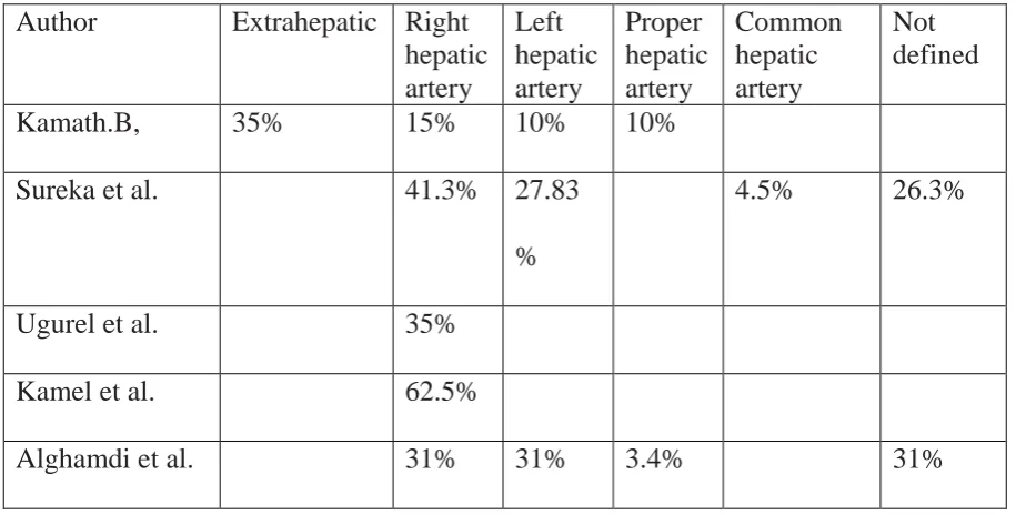
Kamath, it was given off extrahepatically in 35% cases and from the right hepatic
artery in 15%, left hepatic artery in 10% and hepatic artery proper in 10% cases.6
Sureka et al. reported middle hepatic artery originated from right hepatic artery in
41.3%, left hepatic artery in 27.83% and from common hepatic artery in 4.5%
patients. Origin of middle hepatic artery could not be defined in 26.3% of patients.74
Ugurel et al. reported that arteries supplying liver segment IV originating from the
right hepatic artery in 35 of the 100 cases (Ugurel et al. 2010). In an multi-detector CT
study conducted by Kamel et al, the segment IV artery was reported to originate from
the right hepatic artery in 62.5% of cases.62
Alghamdi et al. dissected a total of 29 livers and injected water and ink into the
various arterial branches supplying segment IV and studied the arterial pattern
supplying segment IV. In their study, the middle hepatic artery arose from the right
hepatic artery in nine livers and from the left hepatic artery in a further nine instances,
giving a frequency of 31% for each occurrence. The middle hepatic artery originated
(31%). The middle hepatic artery was doubled in one instance: one arm arising from
[image:37.595.65.523.190.422.2]the right hepatic artery and the other from the left hepatic artery.75
Table 3.3 Origin of artery to segment IV reported by various authors
Author Extrahepatic Right
hepatic artery Left hepatic artery Proper hepatic artery Common hepatic artery Not defined
Kamath.B, 35% 15% 10% 10%
Sureka et al. 41.3% 27.83
%
4.5% 26.3%
Ugurel et al. 35%
Kamel et al. 62.5%
Alghamdi et al. 31% 31% 3.4% 31%
Portal vein
The portal vein begins at the level of the second lumbar vertebra and is formed
by the union of the superior mesenteric and splenic veins posterior to the neck of the
pancreas. The main portal vein, which carries as much as 80% of the blood supply to
the liver, typically divides at the hilus into the left and right portal branches. The left
portal vein is often of smaller caliber and it has horizontal and vertical portions. It
courses medially to the umbilical fissure and supplies segments II, III, and IV and
sector trunk, which in turn divides into segment V and segment VIII branches, and the
right posterior sector trunk, which supplies segments VI and VII.76 (Figure 3.2) The
normal portal vein anatomy occurs in 90% of cases.77
Embryologically, the portal vein develops from the right and left vitelline
veins. At 5 weeks gestational age, the right and left vitelline veins form a venous
plexus around the duodenum, comprising two components ventral to the duodenum
and one component dorsal to the duodenum, before terminating in the sinus venosus.
By 10 weeks, selective involution of portions of this venous plexus gives rise to the
adult portal vein. The main portal vein arises from the left vitelline vein and dorsal
anastomoses.The right portal vein arises from the right vitelline vein, and the left
portal vein from the left vitelline vein and ventral anastomoses.78 Deviations in the
development and selective involution of this venous plexus lead to portal vein
variation.
The right portal vein is only 2–3 cm in length and usually divides into a right
medial (anterior) sectoral division supplying segments V and VIII, and a right lateral
(posterior) sectoral division supplying segments VI and VII. The medial division may
Figure 3.3. The main portal vein and its intra-hepatic branches. (Right lateral = right posterior; right medial = right anterior.). Picture is reproduced from Gray’s Anatomy, 40th Edition Pg. No. 1171
Variations of portal vein branching have been reported by various authors. Nakamura
classified the portal vein variations into 5 types as shown in table 3.4
Table 3.4. Variations of portal vein according to Nakamura et al. 2002
Type Description
Type A The usual bifurcation of main portal vein into right and left portal vein
Type B The trifurcation pattern without the trunk of a right branch of the portal vein
Type C A right paramedian sector branch or a right lateral sector branch bifurcates separately from the left portal
vein,originated from the proximal, or extraparenchymal site Type D A right paramedian sector branch or a right lateral sector
branch bifurcates separately from the left portal vein originated from a distal, or intraparenchymal
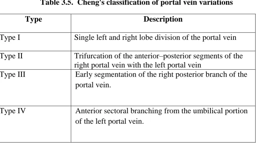
[image:39.595.64.533.474.710.2]Cheng classified portal vein variations into four types.79 (Table 3.5)
Table 3.5. Cheng's classification of portal vein variations
Type Description
Type I Single left and right lobe division of the portal vein
Type II Trifurcation of the anterior–posterior segments of the right portal vein with the left portal vein
Type III Early segmentation of the right posterior branch of the portal vein.
Type IV Anterior sectoral branching from the umbilical portion of the left portal vein.
The classification of branching pattern of the portal vein in current literature is
as follows80
Type 1 - The main portal vein bifurcates into right and left portal vein trunks. The
right portal vein further bifurcates into an anterior branch supplying liver segments V
and VIII and a posterior branch supplying segments VI and VII.
Type 2 - The main portal vein trifurcates into a left portal vein, right anterior portal
vein supplying liver segments V and VIII, and right posterior portal vein supplying
liver segments VI and VII at the same craniocaudal level.
Type 3 - Early branching of the right posterior portal vein. The first branch arising
segments VI and VII. Beyond this early branch, the left portal vein supplying the left
hepatic lobe and the right anterior portal vein supplying liver segments V and VIII
bifurcate at the same level
Type 4 - The right portal vein bifurcates into two vessels – one larger vessel
supplying liver segments V, VI, and VIII and a smaller vessel supplying liver segment
VII only – beyond the left portal vein origin from the main portal vein.
Type 5 - The right portal vein bifurcates into two vessels – one supplying liver
segment VI only and another branch supplying liver segments V, VII, and VIII –
beyond the left portal vein origin from the main portal vein.
Macdonald et al. (2005) reported in a study done using preoperative hepatic
computed tomography scans and intraoperative cholangiograms from 39 consecutive
living liver donors, portal venous anomalies were seen in 18%.45
Kishi et al. studied 287 living donor livers. They reported 91% of the portal
vein branching belonged to the normal pattern Type A. 6% showed trifurcation (Type
B). In their study, they did not observe Type E variation.77
Takeishi et al. 2015, reported that in the cohort of 407 donors, three types of
portal vein were identified, 89% had a normal bifurcation pattern (type 1), 6.1% had
trifurcation (type 2), 4.7% had a type 3 portal vein. In two donors, they noted a rare
variation, where a posterior and a segment V portal vein branch originating from the
Koç et al.2007, did a retrospective study done in 1384 patients (721 males, 663
females) using routine abdominal multidetector CT. Normal portal vein branching
pattern was observed in 72.6% of the patients. Portal vein variants and anomalies were
identified in 27.4%. The most frequent types of these variations were trifurcation in
11.1% and right posterior portal vein as the first branch of the main portal vein in
9.7%. Variation in the origin of the segmental portal vein that traversed the interlobar
boundary was identified in 4%.81
Sureka et al. (2015) studied 967 patients using a triphasic multidetector CT
abdomen scans. Normal anatomy (Type 1) was seen in 79.94%, Trifurcation (Type 2
anomaly) was seen in 6.83% of cases. Right posterior vein as first branch of main
portal vein (Type 3 anomaly) was seen in 4.96% of cases. Type 4 anomaly and Type 5
anomaly was seen in 2.69% and 1.34% cases respectively. In 1.96% other types of
variations were seen.
In addition to the types described, nonstandard miscellaneous variations have also
been described by various authors. Covey et al. have described the following
miscellaneous variations:68
Trifurcation of the right portal vein into the right anterior sectoral portal trunk
and into segment VI and segment VII branches.
Division of the main portal vein into segment VI, segment VII, right anterior
Trifurcation of the right portal vein into branches supplying segment V,
segment VIII and the right posterior sectoral trunk
Segments IV and VII branches originating from the right anterior portal vein
An accessory segment VI branch from the right portal vein in a patient with
type 5 portal vein branching
Trifurcation of the main portal vein into segment VI branch, left and right
anterior sectoral branches, and segment VII branch.
Gunasekaran et al. reported a right portal vein bifurcation into separate superior
and inferior branches supplying liver segments VI and VII and V and VI,
respectively80
Koc et al reported the following miscellaneous variation:81
Separate origin of segment IV portal vein branch from the main portal vein
Quadrification
Separate origin of the liver segments VI and VII branch from the main portal
vein
Sureka et al. described the following variations:74
Segment V branch arising from right posterior portal vein
Separate origin of branch to segment VI and segment VII branch from right
portal vein
Accessory segment VI branch arising from the right anterior portal vein
Branch to segment IV being supplied by right portal vein and left portal vein
Segment II branch directly from main portal vein bifurcation
Segment VIII branch arising from left portal vein
Separate origin of segment V from right portal vein
Absence of portal vein bifurcation
Interestingly, while the right portal vein showed substantial anatomic variation, left
portal vein anomalies were less frequently encountered.80
Radiological study
Hepatic artery and portal vein anatomy can be determined noninvasively by
various radiological techniques like three-dimensional (3D) CT, multidetector CT.
Schroeder et al. noted that 3D integrated images contributed to surgical simulation in a
better way. Sakai et al. defined the variations of the segmental hepatic structures
accurately by multi-detector CT images of the portal vein, the hepatic artery and the
bile duct.82 He reported that, comparing with conventional angiography, 3D CT
angiography depicted the extra hepatic arteries and the aberrant arteries successfully
and also depicted the intrahepatic segmental arteries with a high detection rate.82
Excellent delineation of the portal vein and its branches was achieved on CT
conventional arterial portograms in patients in whom categorization of the portal vein
anatomy using CT alone was difficult.68
Luminal casting
The technique of luminal / corrosion cast has advanced since its first
application by Leonardo da Vinci using wax in 16th century where the cast of the
cerebral ventricles were made. Various materials such as metal alloys, celloidin, latex
gum, synthetic resins/epoxy resins, polyester resins, and silicone have been used to
prepare cast of vasculature and ductal architecture.83
An ideal casting media/resin should be of a low viscosity and nontoxic liquid,
should have the property to polymerize within a short period following injection with
minimal shrinking and it should resist corrosion, cleaning, and dissection and having
these properties, makes it easy to inject the resin into the blood vessels or ductal
system of an organ.83
The modern corrosion casting methods are based upon the idea of Jan
Schwamnerdan of the 17th century following the injection of wax into the blood
vessels the surrounding parenchyma was dissolved by a corroding agent. Hinmann in
1923 formulated the basic principle of injecting low viscosity resins which could enter
vessels of small caliber. Cellulose acetyl butyrate and epoxy resin with the addition of
Murakami in 1971 initiated revolutionary advancement of corrosion casting
techniques. A semipolymerized methyl methacrylate was introduced for the purpose
of cast making and later the casts was observed under the Scanning electron
microscope.83
Routinely the silicon resin/gel has also been used to produce casts of various
structures. These cast are of execellent quality in terms of flexibility, clarity of details
and anatomical accuracy and also provide a visual representation of the internal
structures.84
Recently Polyurethane (diphenylmethane-4,4-diisocyanate; Souda foam) foam
is found to be a suitable material to make accurate casts of vessels and viscera, and to
develop a method based on its use for anatomical studies. Injection of Soudafoam is a
new technique of luminal casting and the method is inexpensive, simple and requires
no special equipment. The polyurethane foam does not need a catalyst. It is simply
diluted with acetone, which does not cause shrinkage of the cast due to evaporation
during hardening. The foam naturally expands into the cavities without high pressure
of the inoculum. The polyurethane foam was tested primarily in the lungs of various
animal species, but also in renal, intestinal and equine digital vessels.
The drawbacks following the used of saudafoam are, multiple injections cannot
be made in the same cavity since the foam solidifies quickly; the second is the slight
brittleness of the cast, due to the low elasticity of polyurethane foam.85
The three-dimensional structure of blood vessels, ductal system, and cavities of
serves an excellent tool to have a detailed knowledge of the structures studied; as the
replica of the vasculature and ductal system anatomy is retrived.83
More recently, expensive specific products such as dental polymers (Gordon et al.
2007) or methylmethacrylate (Casteleyn et al. 2010; Debbaut et al. 2011; Madrahimov
4. MATERIALS AND METHODS
The study was conducted in the Department of Anatomy and Department of
Radiology, Christian Medical College, Vellore after obtaining the ethical clearance
from the Institutional Review Board. It was an observational study. In this study, the
gross anatomical variations of the liver and the branching pattern of hepatic artery and
portal vein were studied.
4.1 Gross anatomical variation
Seventy liver specimens available in our Department were used for the study.
The liver specimens were removed during routine dissection for medical
undergraduate teaching and were preserved in 10% formalin. All the livers were
apparently normal and without major gross deformity. The morphological variations of
the liver such as changes in size and shape, presence of pons hepatis, accessory lobes
and fissures, were noted. Photographs were taken to document the variations. The
results obtained were then tabulated.
4.2 Branching pattern of the hepatic artery and portal vein
Sample size determination
The sample size was calculated as 113 based on the incidence of hepatic artery
The formula used,
N = __________
where, N is the number of samples
p is the prevalence
d is the precision.
The branching pattern of hepatic artery and portal vein was studied using two
techniques:
1. Luminal casting (modified)
2. Radiological study
For luminal casting 30 liver specimens were used, 15 for hepatic artery
branching pattern and 15 for portal vein branching pattern. For radiological study 100
CECT images were used for each. Thus a total sample of 115 was used for each.
4.2.1 Luminal casting
Materials used:
Sauda Foam (polymethylenepolyphenylisocyanate)
BOSS FLEXSIL GP (Silicone Sealant)
Acetone 100%
I/V set tube / butterfly tube / micropipette tip
Syringe (50/20/10cc)
Artery clamp
Forceps
Scissors
Thread
Silicon gun
4pq
Procedure:
The liver specimens without major gross deformity was chosen. The formalin
fixed liver was then washed in running water overnight. Then the liver was kept in a
bath which contained the anticoagulant, sodium tri-citrate for 5-6 hrs. Again the liver
was washed in running water, and also syringing water through the lumen of blood
vessels in the porta hepatis.
SoudaFoam (polymethylenepolyphenylisocyanate) was used to study the
branching pattern of hepatic artery. The chemical was mixed with acetone in a bowl.
The hepatic artery was identified in the porta hepatis and either a canula, I/V set
tube/butterfly tube or micropipette tip was inserted and then tied with thread. Then
using a 50cc syringe the mixture was injected and then clamped using artery clamp.
After the injection of the chemical was completed, the specimen was kept in a freezer
overnight (2-5 degree celcius). After keeping the casted specimen in freezer overnight,
the following day the specimen was shifted to a bowl filled with water and diluted HCl
and kept for another day. On the 3rd day the specimen was taken out from the water
mixed with diluted HCl.
For the portal vein, glass sealant (silicon) was used for casting the specimens.
The same steps were followed which was adopted for casting structures using Souda
Foam while injecting with silicon material. While doing the procedure necessary
Dissection was carried out using the finger fracture technique for tracing the
vascular structures. The forceps was used to tease away the liver parenchyma to
expose the vessels. The trunks of the hepatic artery and portal vein were identified and
their pattern of division was traced and noticed. The branching pattern was recorded
by drawing the line diagram.
4.2.2 Radiological study
One hundred 3D reconstructed contrast enhanced CT (CECT) images were
used for this study. These images were from patients who underwent CECT for
various ailments like bladder with ureteric tumour, periampullary carcinoma,
suspected carcinoma of gall bladder, carcinoma of breast for metastatic evaluation,
recurrent acute pancreatitis, mass arising from the tail of pancreas, carcinoma of
ovary, but devoid of any pathology in the liver. These images were from 52 males
and 48 females, age ranged from 8yrs to 86yrs. CECT images were obtained at
2.5-mm-thick slices using (GE- Discovery. USA). Enhancement was achieved by
intravenous bolus administration of 120 ml of a nonionic contrast medium
(Iopalmidol/Iohexol;) at a speed of 3 mL/s. 3D reconstruction of the hepatic artery and
portal vein was performed using AW-SERVER. The branching pattern of hepatic
artery and portal vein was studied. .
The data obtained was statistically analysed with software STATA V.13.1.
Fisher's exact test was done to find out whether there was any gender differences in
5. RESULTS
5.1. Morphological variations of the liver
Seventy livers have been studied for the morphological variations. Various
gross variations were observed in the study like presence of accessory fissures,
grooves on the surface of the livers, lobulations, conical shaped right lobe, notched
border, underdeveloped caudate process, abnormal papillary process with fissure,
fissure in the caudate lobe, bilobed caudate lobe, fissure in the quadrate lobe, quadrate
lobe with tongue like projection, pons hepatis and presence of accessory lobe. The
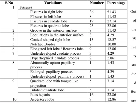
[image:54.595.66.526.382.727.2]frequencies of variations in morphology of liver has been tabulated in Table 5.1.
Table 5.1. Morphological variations of the liver (n = 70)
Out of the 70 live rs stu die d fiss ure
s of various size and orientations were encountered in 57 specimens (81.4 %). In 36
S.No Variations Number Percentage
1 Fissures
Fissures in right lobe 36 51.43 FIssures in left lobe 8 11.43 Fissures in caudate lobe 19 27.14 Fissures in quadrate lobe 23 32.86 2 Groove in the anterior surface 8 11.43 3 Lobulations in the anterior surface 3 4.29 4 Conical shaped right lobe 13 18.57
5 Notched Border 7 10.00
6 Elongated left lobe / Beaver's lobe 9 12.86 7 Underdeveloped caudate process 3 4.29
Hypertrophied caudate process 2 2.86 8 Abnormally upturn papillary
process
1 1.43
Enlarged papillary process 3 4.29 Underdeveloped papillary process 1 1.43 9 Quadrate lobe with tongue like
projection
5 7.14
Bilobed quadrate lobe 5 7.14
Pons hepatis 16 22.86
specimens (51.43%), accessory fissure was present on the right lobe (Figure 5.1.1). In
8 specimens (11.43%) accessory fissure was present on the left lobe also (Figure
5.1.2 ). Accessory fissure were present in the caudate lobe in 27.1% and in quadrate
lobe in 32.86%. There were 4 instances where the fissure was present on the right lobe
along with the fissure on caudate and quadrate lobe (Figure 5.1.3). Most often the
fissures were present on the visceral surface and in a few cases, deep fissures were
seen on the anterosuperior surface. These fissures on the anterosuperior surface were
either single (10%) (Figure 5.1.4) or multiple (4.3%) (Figure 5.1.5). In 2 livers, the
superior surface was irregular due to visceral impression (Figure 5.1.6).
Other than the fissures, 13 specimens (18.57%) showed conical shaped right
lobe (Figure 5.1.4). Elongated left lobe or Beaver's lobe (Netter's type 4) was observed
in 9 specimens (12.86 %) (Figure 5.1.7). Netter type 2 liver was seen in one specimen
(Figure 5.1.4), which is characterized by a small left lobe with deep costal
impressions.
Various morphological variations were observed in caudate lobe in addition to
the presence of fissure. They included underdeveloped caudate process in 4.29%,
hypertrophied caudate process in 2.86% (Figure 5.1.8), l in enlarged papillary process
in 4.29% (Figure 5.1.8) or underdeveloped papillary process in 1.43%. Caudate lobe
Twenty three liver specimens (32.86%) showed the presence of fissure in
quadrate lobe. Tongue like projection was seen in 5 (7.14%) specimens (Figure
5.1.10) and bilobed quadrate lobe 5 specimens (7.14%) (Figure 5.1.11). Pons hepatis
was seen in 16 specimens (22.9 %) (Figure 5.1.12)
Notched border was encountered in 7 specimens (10 %) (Figure 5.1.13).
Accessory lobe was seen in 9 specimen (12.86%) either in caudate lobe or in
quadrate lobe or adjacent to these lobes (Figure 5.1.14).
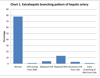
5.2. Hepatic artery branching pattern
Extrahepatic branching pattern
The extra-hepatic branching pattern of the hepatic artery was studied
radiologically using CECT in 100 subjects. In the current study normal anatomy was
observed in 78% subjects while the remaining 22% subjects showed variations. Figure
5.2.1 shows the normal branching pattern of the hepatic artery. The common hepatic
artery arose from the coeliac trunk, from which the proper hepatic artery was given
off. The proper hepatic artery divided into the right and left hepatic arteries. The
variations noted in this study were, common hepatic artery arising from the superior
mesenteric artery in 1% (Figure 5.2.2), a replaced right hepatic artery in 13% (Figure
5.2.3) and, accessory left hepatic artery from left gastric artery in 3 cases (Figure
5.2.4) and early branching of right hepatic artery from common hepatic artery just
beyond common hepatic artery origin was seen in one case. Of the variations noted,
was no gender difference regarding the variation in the branching pattern (Table 5.2)
Table 5.2 Extrahepatic branching pattern of hepatic artery (n=100)
S.No Branching pattern Female
(n=44) N (%) Male (n=54) N % Total in % P value
1. Normal 33 (75) 45(81.5%0 78
0.568 2. CHA arising from SMA 0 (0) 1 (1.9) 1
3. Replaced LHA 3 (6.8) 1 (1.9) 4 4. Replaced RHA 6(11.4) 7 (13.0) 13 5 Accessory LHA from LGA 2 (4.6) 1 (1.9) 3 6 Early branching of RHA from
CHA just beyond origin of CHA
1 (2.3) 0 (0) 1
p value <0.05 is significant
CHA - Common hepatic artery; SMA - Superior mesenteric artery; LHA - Left hepatic artery; RHA - Right hepatic artery; LGA - Left gastric artery
0 10 20 30 40 50 60 70 80 90
Normal CHA arising from SMA
Replaced LHA Replaced RHA Accessory LHA from LGA
Early branching of RHA from CHA
Per
ce
n
tage
Intrahepatic branching pattern
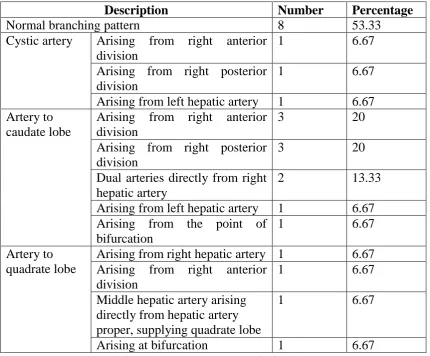
The intrahepatic branching pattern of the hepatic artery was studied in 15
formalin embalmed liver specimens by modified luminal cast technique using Sauda
Foam. Figure 5.2.5 shows the branching pattern of the hepatic artery in 15 livers. In
the current study 8 specimens (53.33%) displayed normal anatomy. The proper
hepatic artery divided into right and left hepatic arteries. The right hepatic artery
divided into right anterior and right posterior divisions. The left hepatic artery
supplied segments II, III and IV. The cystic artery arose from the right hepatic artery
and the artery to the caudate lobe arose from right hepatic artery (Figure
5.2.5.a,b,c,d,e,g,h,j, Figure 5.2.6). Table 5.3 shows the variation in the origin of cystic
artery, artery to the caudate lobe and artery to the quadrate lobe.
In 3 specimens (20%), the origin of cystic artery was varied; in one specimen,
it was given off from right anterior division (Figure 5.2.5.b), in another from the right
posterior division (Figure 5.2.5.f) and in one specimen, from the left hepatic artery
(Figure 5.2.5.l, Figure 5.2.7).
The caudate lobe received its blood supply from right hepatic artery in 14
specimens. Of which, in 3 (20%) specimens artery to caudate lobe was given off from
right posterior division (Figure 5.2.5.k,n,o) and in 3 (20%) specimens from the right
anterior division (Figure 5.2.5. c,f,m), and in two specimens, the right hepatic artery
gave dual branches to the caudate lobe before it divided into right anterior and right
posterior divisions (Figure 5.2.5.e,l, Figure 5.2.8). In one specimen the artery to
off from the point of bifurcation of the \proper hepatic artery (Figure 5.2.9). In one
specimen.(6.6 %), the artery to caudate lobe arose from the left hepatic artery (Figure
5.2.5.i, Figure 5.2.10).
Quadrate lobe received its blood supply from left hepatic artery in most of the
cases. Variations in the arterial supply to the quadrate lobe was observed in 3
specimens. In one specimen (6.6 %), the right hepatic artery gave a branch to quadrate
lobe (Figure 5.2.5.k, Figure 5.2.11) and in another right anterior division of right
hepatic artery gave a branch to quadrate lobe (Figure 5.2.5.n). in another specimen the
hepatic artery proper trifurcated into right hepatic artery, left hepatic artery and middle
hepatic artery and in this case the middle hepatic artery gave a branch to the quadrate
lobe (Figure 5.2.5.m, 5.2.12).
Segmental branching pattern
Segment V in addition being supplied by right anterior division, in two
specimens was supplied by right posterior division (Figure 5.2.5.n,o). Segment VIII
was supplied by right posterior division in three specimens (Figure 5.2.5l,n,o). In one
specimen, the right hepatic artery divided into superior and infe