0022-538X/07/$08.00⫹0 doi:10.1128/JVI.01343-07
Copyright © 2007, American Society for Microbiology. All Rights Reserved.
Complexes between Herpes Simplex Virus Glycoproteins gD, gB, and
gH Detected in Cells by Complementation of Split Enhanced
Green Fluorescent Protein
䌤
Elisa Avitabile, Cristina Forghieri, and Gabriella Campadelli-Fiume*
Department of Experimental Pathology, Section on Microbiology and Virology, University of Bologna, Via San Giacomo, 12, 40126 Bologna, Italy
Received 20 June 2007/Accepted 25 July 2007
The interactions between herpes simplex virus gD and its nectin1 receptor or between gD, gB, and gH were analyzed by complementation of the N and C portions of split enhanced green fluorescent protein (EGFP) fused to the glycoproteins. The gDN-NectCcomplex was readily detected; the gDN-gCCcomplex was
undetect-able, highlighting the specificity of the assay. Split EGFP complementation was detected between proteins designated gDNⴙgHC, gDNⴙgBC, and gHNⴙgBCⴙwtgD (gB was deleted of endocytosis motifs), both in cells
transfected with two-tree glycoproteins and in syncytia. The in situ assay provides evidence that gD interacts with gH and gB independently of each other and supports a model whereby gH and gB in complex exert their activities to gD.
The entry of herpes simples virus (HSV) into cells requires a multipartite fusion system made of a quartet of glycoproteins (3, 24, 28). The receptor-binding glycoprotein gD interacts with three alternative receptors, nectin1, herpesvirus entry me-diator, and modified heparan sulfate (5, 11, 22, 27). gD also encodes a profusion domain at the ectodomain C terminus, which is required to trigger fusion (4). In the unliganded gD, the ectodomain C terminus folds around the N terminus. At receptor binding, the C terminus is displaced, gD adopts an open conformation, and fusion is triggered (8, 20). Three gly-coproteins conserved across theHerpesviridaefamily, gB and gH䡠gL, execute fusion (2, 7, 25). The identity of the execu-tor—whether it is gB, gH䡠gL, or the three glycoproteins to-gether—remains unclear. Thus, gB exhibits a trimeric struc-ture, properties typical of class I and II viral fusion proteins, and a candidate fusion loop (16, 17). On the other hand, gH exhibits elements typical of class I fusion glycoproteins, includ-ing two heptad repeats able to form a coiled coil and a candi-date fusion peptide, besides having additional hydrophobic regions (9, 10, 12–15). Hemifusion (the fusion of the outer layers of the virion and cell membranes) requires gD and gH䡠gL; complete fusion (the mixing of both outer and inner lipid layers) additionally requires gB (29). Fusion between perinuclear virions and the outer nuclear membranes, which culminates in capsid release into the cytoplasm, requires gD plus either gB or gH䡠gL, implying that, under particular con-ditions, either gB or gH䡠gL suffice for fusion execution (6).
A key question in HSV entry/fusion centers on how gD signals the encounter with its receptor to the downstream gly-coproteins and thus triggers fusion. The working model inves-tigated in this laboratory envisions that the receptor-bound gD
forms complexes with the downstream glycoproteins or with a subset of them (3). Indeed, by coimmunoprecipitation, gD was shown to be in a complex with gH (23).
The aim of this work was to investigate, by means of a protein complementation assay (CA) (19, 21), in intact cells, the molecular interactions that take place between gD and nectin1 and between the four glycoproteins. In the CA, pro-teins like enhanced green fluorescent protein (EGFP) are split into two portions that, if brought to an 8- to 10-Å proximity of each other, refold together and emit fluorescence (19, 21). In the current adaptation to membrane proteins, the EGFP N terminus (N; amino acids [aa] 1 to 157) was fused to the endodomain of HSV type 1 gD or gH, and the C terminus (C) was fused to the endodomain of gB, gH, gC, or nectin1. The glycoproteins under examination interact, or are likely to in-teract through their ectodomains; the EGFP portions were located in the endodomains. The assumption was made that any specific interaction occurring between the ectodomains would result in refolding and fluorescence emission of the endodomain-located EGFP fragments. Fluorescence intensity from the complemented EGFP was reported to be about 10% of that produced by the unsplit protein, essentially because only a subset of the fragments have a likelihood of associating with each other (see reference 19). This drawback is balanced by the fact that complementation is driven by specific interac-tions that occur between the proteins under investigation, to which the EGFP portions are fused. Moreover, the refolded EGFP adopts an irreversible conformation that contributes to stabilizing the complex and thus enables the detection of tran-sient and weak complexes (18, 19).
Plasmid construction.The mammalian expression plasmids for gH in the MTS vector, and gD and nectin1 in pcDNA3.1 (5, 31) were site-directed mutagenized 0 to 10 aa upstream of the stop codon, in order to generate restriction sites for the inser-tion of N or C amplimers. The sites were SphI for gH and BglII for gD or nectin1. Where necessary, the BglII site of pcDNA3.1 was preliminarily eliminated by digestion, filling in
* Corresponding author. Mailing address: Department of Experi-mental Pathology, Section on Microbiology and Virology, University of Bologna, Via San Giacomo, 12, 40126 Bologna, Italy. Phone: 39 051 2094733. Fax: 39 051 2094735. E-mail: [email protected].
䌤Published ahead of print on 1 August 2007.
11532
on November 8, 2019 by guest
http://jvi.asm.org/
by T4-DNA polymerase and religation. N and C sequences were PCR amplified from pCMS-EGFP (Clontech) with the primer pairs (or variations thereof) 5⬘CCCAGATCTC CATG GTGAGC AAGGGCGAGG AGCTGT plus 5⬘GGGAAG CTTC TACTTGTCGG CCATGATATA GACGTTG or 5⬘C CCGCTAGCT CAGAAGAACG GCATCAAGGT GAACT plus 5⬘GGGAGATCTT ACTTGTACAG CTCGTCCATG CCGAGA, respectively. N amplimer was ligated with BglII-HindIII-digested gD plasmid or SphI-BglII-digested gH plas-mid, generating gDN and gHN. The C-EGFP amplimer was
ligated with digested nectin1 (BglII-XhoI) and gH (SphI-BglII) plasmids, generating NectCand gHC. The gC gene
se-quence was PCR amplified from DNA of HSV type 1 (F) with primers 5⬘AGATCTAGGC CTATGGCCCC GGGGCG GGTG GGCCTTGCCG TGGTCCTGTG GAGCCTG and 5⬘GAAGATGCGG CCGCTTAGCT AGCCGCCGAT GAC GCTGCCG CGACTGTGAT GTGCG. The StuI-NheI-di-gested gC amplimer and the NheI-BglII-diStuI-NheI-di-gested C amplimer were ligated with StuI-BglII-digested MTS vector. The gB-encoding plasmid in pcDNA3.1 was deleted of the endodo-main sequences that carry endocytosis motifs, from aa 867 to the stop codon (gB⌬867), in order to maximize gB expression
(1). The gBC chimera was generated by mixing gB⌬867and C
amplimers, generated with primer pairs 5⬘GGCTGGATCC TCCCCGTAGT CCCGCCATGC plus CCTTGATGCC GTT CTTCTGA GATCTCTTCT TCTTGGCCTT GTGTTC and 5⬘GAACACAAGG CCAAGAAGAA GAGATCTCAG AAG AACGGCA TCAAGG plus 5⬘GGGAAGCTTT TACTTG TACA GCTCGTCCAT GCCGAGA, followed by ligation with BamHI-HindIII-digested pcDNA3.1. Arrestin-transfected 293T or COS cells, mounted without fixation with Fluoromount, were observed with a Leica TCS-SL confocal microscope, set at 100% excitation at 488 nm with emission between 490 to 540 nm. Im-ages were collected with a 63⫻1.62 Leica oil immersion objec-tive; confocal slices were 1.7 to 2.3m thick. For each experi-mental series—see Figure 2A to T and U to Z and Fig. 3A to F, G to I, J to L, and M to P—images were collected on the same day, under the same settings, applying 1,024- by 1,024-pixel res-olution and an 8-bit intensity scale. Specifically, the first sample to be analyzed was the negative one, containing gCc; for subsequent observations of the samples belonging to the same series, the settings were then kept unmodified. Figure 1 shows the electro-phoretic mobility of the N-EGFP or C-EGFP fusion proteins generated in this study, made in transfected COS or 293T cells, detected by Western blotting with monoclonal antibodies H170, H1817, H633, and CK6 to gD, gB, gC, and nectin1, respectively, and polyclonal antibodies to gH. As expected, all the fusion pro-teins exhibited a slower electrophoretic mobility relative to their respective wild-type versions.
gD-nectin1 complex detection.Inasmuch as EGFP-CA has been applied mainly to analysis of soluble mammalian or bacterial proteins (19), we first validated its application to membrane proteins—in particular, to HSV glycoproteins—by analysis of gD and its nectin1 receptor. Figure 2C and M documents complex formation between gDN and NectC as
fluorescence emission from EGFP-CA in transfected 293T or COS cells. Cells were observed 36 h after arrestin-mediated transfection. Results with the two cell lines were essentially similar, although the level of expression and number of fluo-rescent cells was higher with 293T cells. In agreement with
previous reports, the overall fluorescence emitted by comple-mentation of split EFGP fragments was lower than that from the unsplit protein (19).
The specificity controls that validated the assay were as fol-lows. First, neither gDNnor NectCemitted fluorescence when
transfected singly (Fig, 2A, B, K, and L) or in combination with the wild-type alleles of nectin1 or gD (not shown), ruling out autofluorescence. Second, fluorescence was reconstituted only when the EGFP chimeric proteins were in a specific complex and not simply present in the same subcellular compartment. For this control, we selected gC, which is involved in virus attachment but not virus entry and is present in the same subcellular compartments as gD. The coexpression of gDNand
gCCresulted in no or background fluorescence (Fig. 2D and
N), ruling out the possibility that proteins that exhibit no spe-cific interaction, but that are abundantly present in the same cellular compartment, give rise to EGFP complementation. We took advantage of the lack of EGFP complementation by gCC-containing samples and, in all experiments, used the gCC
[image:2.585.338.503.69.393.2]-containing sample to adjust the confocal microscope settings. The settings were then kept constant throughout the observa-tion period of a same series of samples. Third, we ascertained by immunofluorescence assay (IFA) that all proteins were ex-pressed, even those expressed singly (gDN, NectC) (Fig. 2E, F, FIG. 1. Electrophoretic mobility of proteins fused to N- or C-EGFP portions, or their wild-type alleles, expressed in COS or 293T cells. Numbers on the left indicate the migration positions of molecular mass markers (in kilodaltons).
VOL. 81, 2007 NOTES 11533
on November 8, 2019 by guest
http://jvi.asm.org/
11534
on November 8, 2019 by guest
O, and P) or in the gDN-gCC combination that did not yield
EGFP fluorescence (Fig. 2I, J, S, and T). Importantly, the EGFP-glycoprotein chimeras were not hampered in plasma membrane localization (Fig. 2). We conclude that EGFP-CA fulfills the criteria for detection of specific interactions be-tween membrane-bound proteins, particularly HSV-1 gD and its receptor.
Complexes between HSV glycoproteins.The second series of experiments was performed with 293T and COS cells trans-fected with three membrane proteins in combinations that included gDN⫹gBC, gDN⫹gB⌬867, gDN⫹gHC⫹wtgL, gDN⫹
wtgH⫹wtgL, and gDN⫹gCC. We used a form of gB deleted for
endocytosis motifs, to maximize its expression and localization in exocytic and plasma membranes (1). Transfection mixtures were made equal in DNA amounts (900 ng/well, 300 ng/plas-mid) by the addition of a plasmid encoding epidermal growth factor receptor 1 deleted of signaling sequences (26). This control ensured that exocytic membranes were loaded with comparable amounts of proteins. In both cell types observed 36 h after transfection, the gDN⫹gHC⫹wtgL combination
re-sulted in a readily detectable fluorescence (Fig. 3B and E). The gDN⫹gBCcombination gave rise to a somewhat weaker
fluo-rescence (Fig. 3A and D) that nonetheless was much higher than the background fluorescence emitted by the gDN⫹gCC
combination (Fig. 3C and F). Even though the subcellular localization cannot be clearly defined, EGFP appeared to lo-calize to a perinuclear position, consistent with a Golgi com-partment localization, to a cytoplasmic reticular comcom-partment, consistent with endoplasmic reticulum, and to nuclear mem-branes. By IFA, all proteins resulted to be expressed, even those that did not yield EGFP fluorescence (not shown). We infer that gD can recruit gH to a complex. gD can also recruit gB to a complex. The gD-gH combination results in a stronger EGFP fluorescence than the gD-gB combination, possibly re-flecting a stronger interaction, a more stable or longer half-life complex, a higher number of complexes at steady state, or peculiar behaviors of the fusion proteins.
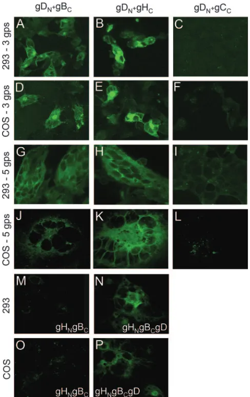
Cells transfected with the quartet of gD, gB, gH, and gL form syncytia (30). A series of experiments was designed to verify whether the glycoprotein-EGFP chimeras were still functional in cell-cell fusion, and whether complexes were de-tectable under conditions that lead to cell-cell fusion. 293T or COS cells were cotransfected with combinations of five plas-mids (1.25g/well, 250 ng/plasmid) encoding gD, gB⌬867, gH,
gL, and gC or their EGFP chimeras. The transfected combi-nations included gDN⫹gBC⫹wtgH⫹wtgL⫹wtgC, gDN⫹
gHC⫹wtgL⫹gB⌬867⫹wtgC, and gDN⫹gB⌬867⫹wtgH⫹wtgL⫹
gCC. Cells were observed 24 h (293T) or 40 h (COS) after
transfection. The results in Fig. 3G to L show that syncytia were formed for any combination, indicating that the EGPF-glycoprotein chimeras were not hampered in fusion activity. The strongest fluorescence was observed with the combination that included gDN⫹gHC (panels H and K). A somewhat
weaker fluorescence was observed with the combination that included gDN⫹gBC(panels G and J), particularly in COS cells.
No fluorescence above background level was observed with the combinations that included gDN⫹gCC(panels I and L). The
[image:4.585.299.543.68.455.2]stronger fluorescence in panels B relative to H and in panels E relative to K reflects (i) higher amounts of transfected DNA for each plasmid, (ii) a longer time interval after transfection (panels B versus H), (iii) the lack of dilution of complemented
FIG. 2. EGFP CA between gDNand NectCor between gHN⫹gHc, and lack of complementation between gDNand gCC. COS or 239T cells were
transfected with the indicated plasmids, gDN(A, E, K, and O), NectC(B, F, L, and P), gDN⫹NectC(C, G, H, M, Q, and R), gDN⫹gCc(D, I, J,
N, S, and T), and gHN⫹gHc(U to Z). (A to D, K to N, U, and X) CA. (E to J, O to T, V, W, Y, and Z) IFA. Green, gD; red, all others. Antibodies
used were R8 to gD, R1.302 to nectin1, H633 to gC, and 53S to gH (reacts to a gL-dependent epitope).
FIG. 3. EGFP CA between HSV glycoproteins. COS or 239T cells were transfected with the indicated plasmids, gDN⫹gBC(A, D, G, and
J), gDN⫹gHC(B, E, H, and K), gDN⫹gCC(C, F, I, and L), gHN⫹gBc
(M and O), and gHN⫹gBc⫹gD (N and P). Cells transfected with three
glycoproteins (gps) received, in addition, plasmids encoding wtgL or epidermal growth factor receptor, as appropriate. Cells transfected with five glycoproteins received gD, gB⌬867, gH, gL, and gC as the wild
type or EGFP chimeras, as indicated.
VOL. 81, 2007 NOTES 11535
on November 8, 2019 by guest
http://jvi.asm.org/
EGFP molecules consequent to fusion of transfected fluores-cent cells with adjafluores-cent untransfected nonfluoresfluores-cent cells, or (iv) possibly a longer half-life of the complexes when cell-cell fusion does not ensue.
We next tested whether gH and gB interact with each other and whether the interaction was dependent on the presence of gD. Cells were transfected with gHN⫹wtgL⫹gBC in the
ab-sence or preab-sence of wtgD. Interaction between gHN-gBCwas
readily documented in the presence (Fig. 3N and P) but not in the absence (Fig. 3M and O) of gD.
While gB is known to be a trimer (17), the oligomeric state of gH is unknown. Here, we addressed the question of whether the split EGFP-CA was suitable to define the oligomeric state of gH. Cells were transfected with gHN⫹gHC⫹wtgL. As shown
in Fig. 2U to Z, the CA readily documented that the gH䡠gL heterodimer (detected by its reactivity to MAb 53S) formed oligomeric structures in transfected cells.
Concluding remarks. We validated the adaptation of the EGFP-CA to membrane proteins by first applying it to the gD-nectin1 interaction. The fluorescence emitted from the gDN
-NectCcombination was readily detectable, whereas that from the
gDN-gCC combination was detected at only background levels,
testifying to the assay specificity. For every series of observations, the gCC-containing sample was therefore used to adjust the
con-focal microscope settings. Samples exhibiting readily detectable fluorescence under these conditions were considered positive.
We detected a complex made of gD and gH, in agreement with coimmunoprecipitation data (23). The complex formed even in the absence of gB. In addition, we detected a complex made of gD and gB that formed even in the absence of gH䡠gL. We further documented the interaction between gH and gB; its gD dependence suggests that the interaction is triggered by gD. A notable property of the EGFP-CA as ap-plied here was that complex formation between HSV glyco-proteins was detected in intact cells, i.e., in the intracellular compartment and microenvironment and under the very con-ditions in which the interactions do occur. Importantly, the EGFP chimeric glycoproteins were not hampered in cell-cell fusion activity. Hence, the detected interactions were a faithful mirror of the interactions that take place under conditions that lead to cell-cell fusion. Inasmuch as EGFP reconstitution from split portions is an irreversible reaction, the assay does not allow us to infer whether the complexes between the HSV glycoproteins were stable or transient.
By taking advantage of the fact that CHO but not Vero cells lack the lipid ganglioside GM1, hemifusion, i.e., the mixing of the outer lipid leaflets of the virion envelope and cell mem-brane or cell-cell memmem-branes, was differentiated from fusion, i.e., complete lipid mixing and content mixing (29). In that assay, gD and gH䡠gL are sufficient to induce hemifusion. Complete fusion additionally requires gB (29). Those findings support the view that HSV fusion occurs through steps, i.e., the juxtaposition of membranes and the triggering of fusion, hemi-fusion, and complete fusion. They do not shed light on the sequential order and mechanism of gB recruitment. Current data agree with those from the hemifusion study and, more-over, argue that hemifusion is carried out by the gD-gH䡠gL complex. Of note, the fact that three independent assays— coimmunoprecipitation, hemifusion and split EGFP
CA—con-cordantly showed the interaction between gD and gH strongly substantiates the current approach.
Cumulatively, the current assay provides in situ evidence for the following. (i) gD recruits gH䡠gL and gB to complexes. (ii) gH and gB can be recruited to gD independently of one an-other. Thus, gD carries binding sites for both gH䡠gL and gB. The independent recruitment of these glycoproteins to gD is consistent with and substantiated by the observation that, at the outer nuclear membrane, virions deleted for gB but carry-ing gD⫹gH, or deleted for gH but carrying gD⫹gB are capable of fusion (6). (iii) Once gH䡠gL and gB are recruited to gD, they possibly interact with each other. (iv) gH䡠gL and gB are not necessarily recruited in a sequential order or one to the other. Current data support a model of HSV entry-fusion whereby gH and gB exert their activity through complex for-mation with gD, or following activation mediated by complex formation with gD.
We thank our colleagues G. Cohen and R. Eisenberg (University of Pennsylvania), T. Minson (Cambridge University), ande M. Lopez (INSERM, Marseille, France) for gifts of antibodies; C. Taddei and L. Dipietrangelo for invaluable assistance with confocal microscopy; the members of our laboratory for providing constructs; and Elisabetta Romagnoli for helpful assistance.
The work was supported by EU contract TargetHerpes-VI FP LSHG-CT-2006-037517, MIUR-PRIN-2005, University of Bologna RFO, and Fondi Roberto e Cornelia Pallotti from our Department.
REFERENCES
1.Avitabile, E., G. Lombardi, T. Gianni, M. Capri, and G. Campadelli-Fiume.
2004. Coexpression of UL20p and gK inhibits cell-cell fusion mediated by herpes simplex virus glycoproteins gD, gH-gL, and wild-type gB or an en-docytosis-defective gB mutant and downmodulates their cell surface expres-sion. J. Virol.78:8015–8025.
2.Cai, W. H., B. Gu, and S. Person.1988. Role of glycoprotein B of herpes simplex virus type 1 in viral entry and cell fusion. J. Virol.62:2596–2604. 3.Campadelli-Fiume, G., M. Amasio, E. Avitabile, A. Cerretani, C. Forghieri,
T. Gianni, and L. Menotti.2007. The multipartite system that mediates entry of herpes simplex virus into the cell. Rev. Med. Virol.17:313–326. 4.Cocchi, F., L. Menotti, V. Di Ninni, M. Lopez, and G. Campadelli-Fiume.
2004. The herpes simplex virus JMP mutant enters receptor-negative J cells through a novel pathway independent of the known receptors nectin1, HveA, and nectin2. J. Virol.78:4720–4729.
5.Cocchi, F., L. Menotti, P. Mirandola, M. Lopez, and G. Campadelli-Fiume.
1998. The ectodomain of a novel member of the immunoglobulin superfam-ily related to the poliovirus receptor has the attributes of a bona fide receptor for herpes simplex virus types 1 and 2 in human cells. J. Virol.72:9992– 10002.
6.Farnsworth, A., T. W. Wisner, M. Webb, R. Roller, G. Cohen, R. Eisenberg, and D. C. Johnson.2007. Herpes simplex virus glycoproteins gB and gH function in fusion between the virion envelope and the outer nuclear mem-brane. Proc. Natl. Acad. Sci. USA104:10187–10192.
7.Forrester, A., H. Farrell, G. Wilkinson, J. Kaye, N. Davis Poynter, and T. Minson.1992. Construction and properties of a mutant of herpes simplex virus type 1 with glycoprotein H coding sequences deleted. J. Virol.66:341– 348.
8.Fusco, D., C. Forghieri, and G. Campadelli-Fiume.2005. The pro-fusion domain of herpes simplex virus glycoprotein D (gD) interacts with the gD N terminus and is displaced by soluble forms of viral receptors. Proc. Natl. Acad. Sci. USA102:9323–9328.
9.Galdiero, S., A. Falanga, M. Vitiello, H. Browne, C. Pedone, and M. Galdiero.2005. Fusogenic domains in herpes simplex virus type 1 glycopro-tein H. J. Biol. Chem.280:28632–28643.
10.Galdiero, S., M. Vitiello, M. D’Isanto, A. Falanga, C. Collins, K. Raieta, C. Pedone, H. Browne, and M. Galdiero.2006. Analysis of synthetic peptides from heptad-repeat domains of herpes simplex virus type 1 glycoproteins H and B. J. Gen. Virol.87:1085–1097.
11.Geraghty, R. J., C. Krummenacher, G. H. Cohen, R. J. Eisenberg, and P. G. Spear.1998. Entry of alphaherpesviruses mediated by poliovirus receptor-related protein 1 and poliovirus receptor. Science280:1618–1620. 12.Gianni, T., R. Fato, C. Bergamini, G. Lenaz, and G. Campadelli-Fiume.
2006. Hydrophobic␣-helices 1 and 2 of herpes simplex virus gH interact with lipids, and their mimetic peptides enhance virus infection and fusion. J. Vi-rol.80:8190–8198.
on November 8, 2019 by guest
http://jvi.asm.org/
13.Gianni, T., P. L. Martelli, R. Casadio, and G. Campadelli-Fiume.2005. The ectodomain of herpes simplex virus glycoprotein H contains a membrane
␣-helix with attributes of an internal fusion peptide, positionally conserved in theHerpesviridaefamily. J. Virol.79:2931–2940.
14.Gianni, T., L. Menotti, and G. Campadelli-Fiume.2005. A heptad repeat in herpes simplex virus gH, located downstream of the ␣-helix with attributes of a fusion peptide, is critical for virus entry and fusion. J. Vi-rol.79:7042–7049.
15.Gianni, T., A. Piccoli, C. Bertucci, and G. Campadelli-Fiume.2006. Heptad repeat 2 in herpes simplex virus-1 gH interacts with heptad repeat 1 and is critical for virus entry and fusion. J. Virol.80:2216–2224.
16.Hannah, B. P., E. E. Heldwein, F. C. Bender, G. H. Cohen, and R. J. Eisenberg.2007. Mutational evidence of internal fusion loops in herpes simplex virus glycoprotein B. J. Virol.81:4858–4865.
17.Heldwein, E. E., H. Lou, F. C. Bender, G. H. Cohen, R. J. Eisenberg, and S. C. Harrison.2006. Crystal structure of glycoprotein B from herpes simplex virus 1. Science313:217–220.
18.Hu, C. D., Y. Chinenov, and T. K. Kerppola.2002. Visualization of interac-tions among bZIP and Rel family proteins in living cells using bimolecular fluorescence complementation. Mol. Cell9:789–798.
19.Kerppola, T. K.2006. Visualization of molecular interactions by fluorescence complementation. Nat. Rev. Mol. Cell Biol.7:449–456.
20.Krummenacher, C., V. M. Supekar, J. C. Whitbeck, E. Lazear, S. A. Connolly, R. J. Eisenberg, G. H. Cohen, D. C. Wiley, and A. Carfi.2005. Structure of unliganded HSV gD reveals a mechanism for receptor-mediated activation of virus entry. EMBO J.24:4144–4153.
21.Michnick, S. W.2001. Exploring protein interactions by interaction-induced folding of proteins from complementary peptide fragments. Curr. Opin. Struct. Biol.11:472–477.
22.Montgomery, R. I., M. S. Warner, B. J. Lum, and P. G. Spear.1996. Herpes simplex virus-1 entry into cells mediated by a novel member of the TNF/ NGF receptor family. Cell87:427–436.
23.Perez-Romero, P., A. Perez, A. Capul, R. Montgomery, and A. O. Fuller.2005. Herpes simplex virus entry mediator associates in infected cells in a complex with viral proteins gD and at least gH. J. Virol.
79:4540–4544.
24.Rey, F. A.2006. Molecular gymnastics at the herpesvirus surface. EMBO Rep.7:1000–1005.
25.Roop, C., L. Hutchinson, and D. C. Johnson.1993. A mutant herpes simplex virus type 1 unable to express glycoprotein L cannot enter cells, and its particles lack glycoprotein H. J. Virol.67:2285–2297.
26.Rovero, S., A. Amici, E. D. Carlo, R. Bei, P. Nanni, E. Quaglino, P. Porcedda, K. Boggio, A. Smorlesi, P. L. Lollini, L. Landuzzi, M. P. Colombo, M. Giovarelli, P. Musiani, and G. Forni.2000. DNA vaccina-tion against rat her-2/Neu p185 more effectively inhibits carcinogenesis than transplantable carcinomas in transgenic BALB/c mice. J. Immunol.
165:5133–5142.
27.Shukla, D., J. Liu, P. Blaiklock, N. W. Shworak, X. Bai, J. D. Esko, G. H. Cohen, R. J. Eisenberg, R. D. Rosenberg, and P. G. Spear.1999. A novel role for 3-O-sulfated heparan sulfate in herpes simplex virus 1 entry. Cell
99:13–22.
28.Spear, P. G., and R. Longnecker.2003. Herpesvirus entry: an update. J. Vi-rol.77:10179–10185.
29.Subramanian, R. P., and R. J. Geraghty.2007. Herpes simplex virus type 1 mediates fusion through a hemifusion intermediate by sequential activity of glycoproteins D, H, L, and B. Proc. Natl. Acad. Sci. USA104:2903–2908. 30.Turner, A., B. Bruun, T. Minson, and H. Browne.1998. Glycoproteins gB, gD,
and gHgL of herpes simplex virus type 1 are necessary and sufficient to mediate membrane fusion in a Cos cell transfection system. J. Virol.72:873–875. 31.Zhou, G., E. Avitabile, G. Campadelli-Fiume, and B. Roizman.2003. The
domains of glycoprotein D required to block apoptosis induced by herpes simplex virus 1 are largely distinct from those involved in cell-cell fusion and binding to nectin1. J. Virol.77:3759–3767.
VOL. 81, 2007 NOTES 11537