JOURNAL OFVIROLOGY, Dec. 2010, p. 12733–12753 Vol. 84, No. 24 0022-538X/10/$12.00 doi:10.1128/JVI.01065-10
Copyright © 2010, American Society for Microbiology. All Rights Reserved.
NFAT and CREB Regulate Kaposi’s Sarcoma-Associated
Herpesvirus-Induced Cyclooxygenase 2 (COX-2)
䌤
†
Neelam Sharma-Walia,* Arun George Paul,‡ Kinjan Patel,‡ Karthic Chandran,
Waseem Ahmad, and Bala Chandran
H. M. Bligh Cancer Research Laboratories, Chicago Medical School, Department of Microbiology and Immunology, Rosalind Franklin University of Medicine and Science, North Chicago, Illinois 60064
Received 18 May 2010/Accepted 1 October 2010
COX-2 has been implicated in Kaposi’s sarcoma-associated herpesvirus (KSHV) latency and pathogenesis (A. George Paul, N. Sharma-Walia, N. Kerur, C. White, and B. Chandran, Cancer Res. 70:3697-3708, 2010; P. P. Naranatt, H. H. Krishnan, S. R. Svojanovsky, C. Bloomer, S. Mathur, and B. Chandran, Cancer Res. 64:72-84, 2004; N. Sharma-Walia, A. G. Paul, V. Bottero, S. Sadagopan, M. V. Veettil, N. Kerur, and B. Chandran, PLoS Pathog. 6:e1000777, 2010; N. Sharma-Walia, H. Raghu, S. Sadagopan, R. Sivakumar, M. V. Veettil, P. P. Naranatt, M. M. Smith, and B. Chandran, J. Virol. 80:6534-6552, 2006). However, the precise regulatory mechanisms involved in COX-2 induction during KSHV infection have never been explored. Here,
we identifiedcis-acting elements involved in the transcriptional regulation of COX-2 upon KSHVde novo
infection. Promoter analysis using human COX-2 promoter deletion and mutation reporter constructs revealed that nuclear factor of activated T cells (NFAT) and the cyclic AMP (cAMP) response element (CRE) modulate KSHV-mediated transcriptional regulation of COX-2. Along with multiple KSHV-induced signaling pathways,
infection-induced prostaglandin E2(PGE2) also augmented COX-2 transcription. Infection of endothelial cells
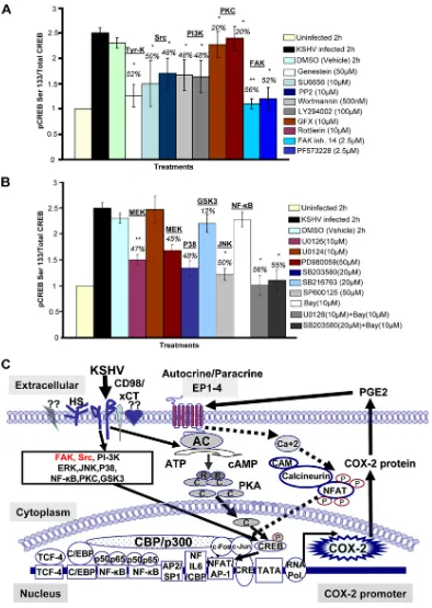
markedly induced COX-2 expression via a cyclosporine A-sensitive, calcineurin/NFAT-dependent pathway. KSHV infection increased intracellular cAMP levels and activated protein kinase A (PKA), which phosphor-ylated the CRE-binding protein (CREB) at serine 133, which probably led to interaction with CRE in the COX-2 promoter, thereby enhancing COX-2 transcription. PKA selective inhibitor H-89 pretreatment strongly inhibited CREB serine 133, indicating the involvement of a cAMP-PKA-CREB-CRE loop in COX-2 transcrip-tional regulation. In contrast to phosphatidylinositol 3-kinase and protein kinase C, inhibition of FAK and Src effectively reduced KSHV infection-induced COX-2 transcription and protein levels. Collectively, our study indicates that mediation of COX-2 transcription upon KSHV infection is a paradigm of a complex regulatory milieu involving the interplay of multiple signal cascades and transcription factors. Intervention at each step
of COX-2/PGE2induction can be used as a potential therapeutic target to treat KSHV-associated neoplasm
and control inflammatory sequels of KSHV infection.
Kaposi’s sarcoma (KS)-associated herpesvirus (KSHV) is associated with a neoplastic angioproliferative endothelial malignancy and two other AIDS-related lymphoid cell malignan-cies called primary effusion lymphoma (PEL) and multicentric Castleman disease (64, 78). The KSHV life cycle displays dis-tinct latent and lytic replication events (64, 78). Viral latency contributes to infected-cell survival and proliferation and la-tency maintenance, whereas the lytic cycle participates in the spread of infection and KS progression (64, 78). KSHV has been shown to utilize a variety of strategies not only to alter host cell metabolism via its signaling proteins but also to hijack cellular signaling pathways, transcription factors, and secreted metabolites to its own advantage, especially to remain latent in the host cell (19, 42, 43, 51, 55, 56).
In our previous studies (19, 55, 56), we reported that COX-2
functions as an important host factor maintaining KSHV la-tency and pathogenesis. The cyclooxygenases catalyzing the rate-limiting step in the synthesis of prostaglandins (PGs) are also referred to as PG endoperoxide synthases and are known to perform two enzymatic functions. As cyclooxygenases, they convert arachidonic acid to prostaglandin G2 (PGG2), and as peroxidases, they convert PGG2 to PGH2. Two forms of the enzyme, named COX-1 and COX-2, have been shown to be expressed in mammalian tissues. COX-1 is present in most tissues as a housekeeper enzyme, whereas COX-2, the product of an 8.2-kb gene containing 11 exons and 10 introns mapping to 1q25.2-q25.3, is the central enzyme in the PG biosynthetic pathway. The gene for COX-2 is considered an immediate-early gene and is stimulated and transcriptionally active during inflammation or pathophysiological process like carcinogenesis and thus plays an important role in the development of human tumors (34, 63). The COX-2/prostaglandin E2(PGE2) connec-tion with KSHV pathogenesis makes it an attractive chemo-therapeutic target.
Levels of COX-2 are tightly controlled in most tissues, and its gene regulation is exclusively dependent on gene transcrip-tion and posttranscriptranscrip-tional events (20). The promoter regions of the human (22), mouse (15), rat (62), and chicken (76) COX-2 genes have been cloned, and their expression is tightly
* Corresponding author. Mailing address: H. M. Bligh Cancer Re-search Laboratories, Chicago Medical School, Department of Micro-biology and Immunology, Rosalind Franklin University of Medicine and Science, North Chicago, IL 60064. Phone: (847) 578-8838. Fax: (847) 578-3349. E-mail: neelam.sharma-walia@rosalindfranklin.edu.
† Supplemental material for this article may be found at http://jvi .asm.org/.
‡ Both authors contributed equally.
䌤Published ahead of print on 13 October 2010.
12733
on November 8, 2019 by guest
http://jvi.asm.org/
regulated at both the transcriptional and posttranscriptional levels. The COX-2 promoter contains a classical TATA box, an E box, and binding sites for transcription factors such as nu-clear factor B, nuclear factor interleukin-6 (IL-6)/CCAAT enhancer-binding protein, two nuclear factor of activated T cells (NFAT) binding sites (NFAT distal site [dNFAT] and NFAT proximal site [pNFAT]) (25, 26) and cyclic AMP (cAMP) response element (CRE)-binding proteins (25, 26). The dNFAT COX-2 site appears to be a pure NFAT site, as evidenced by the absence of any surrounding predicted AP-1 binding sequences and the lack of competition of an AP-1 consensus oligonucleotide for protein binding to this sequence. In contrast, pNFAT contains a highly homologous AP-1 site adjacent to the NFAT core GGAAA motif. Host cell signaling cascade induction has been shown to mediate the recruitment of specific transcription factors to these elements and thus trigger COX-2 activation in other systems (25, 26). However, the underlying mechanism of COX-2 induction upon KSHV infection of endothelial cells has never been reported to date. Therefore, in the present study, we investigated the role of KSHV-induced transcription factors and signaling pathways leading to COX-2 promoter activation, gene transcription, and PGE2secretion.
MATERIALS AND METHODS
Cells.Human microvascular dermal endothelial (HMVEC-d) cells from pas-sages 5 to 7 (CC-2543; Lonza, Walkersville, MD), primary human foreskin fibroblast (HFF) cells (Lonza), and 293 cells were cultured as described before (55). Recombinant green fluorescent protein-KSHV (GFP-KSHV-␥ KSHV.152)-carrying BCBL-1 cells (72) were cultured, and GFP-KSHV was prepared and assessed for infectivity and mycoplasma and endotoxin contamination as de-scribed previously (55). Replication-defective virus (UV-inactivated KSHV) was inactivated with UV light (365 nm) for 20 min at a 10-cm distance (53, 54). KSHV DNA was extracted from live KSHV and UV-inactivated KSHV, and viral copy numbers were quantitated by real-time DNA PCR using primers amplifying the KSHV open reading frame 73 (ORF73) gene as described pre-viously (53–56).
Reagents.LY294002 [20(4-morphodinyl)-8-phenyl-1(4H)-benzopyran-4-one], heparin, sodium orthovanadate, benzamidine, leupeptin, aprotinin, SB216763 (potent and selective cell-permeating, ATP-competitive inhibitor of glycogen synthase kinase 3 [GSK3], a serine/threonine protein kinase), phorbol 12-myris-tate 13-ace12-myris-tate, and mouse anti--actin monoclonal antibody were from Sigma-Aldrich, St. Louis, MO. U0126 (1,4-diamino-2,3-dicyano-1, 4-bis [2-aminophe-nylthio] butadiene, MEK-1,2 inhibitor), U0124, rottlerin (protein kinase C-␦ [PKC-␦]-specific inhibitor), GFX (GF 109203X, a cell-permeating protein kinase inhibitor inhibiting all isoforms of PKC by acting as a competitive inhibitor of the ATP binding site of PKC), COX-2-specific inhibitor NS-398 [N-(2-cyclohexyloxy-4-nitrophenyl)-methanesulfonamide], actinomycin D (RNA polymerase II inhib-itor), ionomycin (100 nM; calcium ionophore), the NFAT antagonist cyclospo-rine A (CsA), PD980059 (2⬘-amino-3⬘-methoxyflavone; MEK-1 inhibitor), SB203580 (10M; specific inhibitor of the p38 mitogen-activated protein kinase [MAPK] pathway), JNK inhibitor II (SP600125; selective inhibitor of c-jun N-terminal kinase), and H-89 (selective inhibitor of protein kinase A [PKA]) were from Calbiochem, La Jolla, CA. Goat polyclonal anti-lamin B, anti-cAMP re-sponse element (CRE)-binding protein (CREB), anti-rabbit PKC, anti-rabbit extracellular signal-regulated kinase 2 (ERK2), and anti-NFAT antibodies were from Santa Cruz Biotechnology, Inc., Santa Cruz, CA. PGE2, PKA selective
inhibitor H-89, and forskolin (adenylate cyclase agonist) were from Cayman Chemical, Ann Arbor, MI. Selective focal adhesion kinase (FAK) inhibitors (FAK inhibitor 14; 1,2,4,5-benzenetetramine tetrahydrochloride) and PF 573228 {3,4-dihydro-6-[[4-[[[3-(methylsulfonyl)phenyl]methyl]amino]-5-(trifluoromethyl)-2-pyrimidinyl]amino]-2(1H)-quinolinone} were obtained from Tocris Bioscience, Ellisville, MO. Anti-mouse COX-1 and COX-2 antibodies were from Cayman Chemicals. Anti-mouse phosphatidylinositol 3-kinase (PI3K) and p-Src antibod-ies were from BD Biosciences and Calbiochem. Anti-mouse p-ERK 1/2 and anti-rabbit Src, p-PKC, and p-PI3K antibodies were from Cell Signaling Tech-nology, Inc. Anti-rabbit PGE2antibody was from Abcam, Cambridge, MA.
Plasmids.Plasmids containing the 5⬘flanking region of human COX-1 and COX-2 promoter deletion and mutation constructs linked to luciferase reporter plasmids were obtained from M. Fresno (Madrid, Spain) and have been de-scribed previously (25, 26). Plasmids for the human COX-2 promoter with wild-type (WT) CRE (CRE-WT;⫺59TTCGTCA⫺53) and mutated CRE (CRE-Mut; TTGAGCT) were obtained from Karsten Schroer (Du¨sseldorf, Germany). These plasmids were constructed into a promoterless luciferase expression plas-mid, pGL3-Basic (Promega). VIVIT, a plasmid containing the NFAT regulatory domain, was obtained from Addgene (2). Plasmids CREB, p-CMV-CREB 133, p-CMV-Kp-CMV-CREB, p-CMV-IKM, and p65 (p65 subunit of NF-B) were purchased from BD Clontech, Mountain View, CA. p-CMV-CREB con-stitutively expresses human WT CREB. The p-CMV-CREB 133 vector expresses a mutant variant of human CREB that contains a serine-to-alanine mutation corresponding to amino acid 133 (S133A). This mutation blocks phosphorylation of CREB that blocks transcription. p-CMV-KCREB vector expresses a mutant variant of human CREB that contains mutations in its DNA-binding domain. KCREB acts as a dominant repressor by forming an inactive dimer with CREB, blocking its ability to bind the cAMP-regulated enhancer element CRE.
Cytotoxicity assay.293, HFF, or HMVEC-d cells were incubated with Dul-becco modified Eagle medium containing different concentrations of various inhibitors for 4 h or 4 days. At different time points, supernatants were collected and assessed for cellular toxicity using a cytotoxicity assay kit (Promega, Madi-son, WI) as described previously (53, 54, 56).
Transient transfection.For reporter gene assays, HFF and 293 cells were transfected using Lipofectamine 2000 (Invitrogen, Carlsbad, CA) as described before (53). Low-passage (passages 5 to 7) HMVEC-d cells were transfected using Effectene reagent (Qiagen, Valencia, CA) in accordance with the manu-facturer’s instructions.
Signaling inhibitor treatment.For luciferase assays, gene expression, PGE2
secretion, and COX-2/COX-1/-actin protein measurements, cells were pre-treated with either solvent controls or signaling inhibitors. Briefly, target cells grown to confluence in 25-cm2flasks were serum starved for 10 h and then
exposed to signaling inhibitors. MEK inhibitors PD980059, U0126, and U0124 (structural analogue of U0126), the general tyrosine kinase (Tyr-K) inhibitor genistein, FAK inhibitors (FAK inhibitor 14 and PF573228), p38 MAPK inhib-itor SB203580, JNK inhibinhib-itor II (SP600125), Src kinase inhibinhib-itors PP2 and SU6656, phosphatidylinositol 3-kinase (PI-3K) inhibitors LY294002 and wort-mannin, and GSK3 inhibitor SB216763 were used for 1 h at 37°C prior to KSHV infection (30 DNA copies/cell). Cells pretreated with the vehicle control (the dimethyl sulfoxide concentration was the same as that used for each specific inhibitor) for 1 h at 37°C prior to KSHV infection were used as a control.
Real-time RT-PCR.Host (COX-1 and COX-2) and viral (ORF73 and ORF50) gene transcripts were detected by real-time reverse transcription (RT)-PCR using gene-specific real-time primers in accordance with the procedures de-scribed previously (56).
Immunofluorescence assay (IFA).Confluent HMVEC-d cells in eight-well chamber slides (Nalge Nunc International, Naperville, IL) were either left un-infected or un-infected with 30 DNA copies of KSHV/cell at 37°C for 30 min, 1 h, or 2 h. For NFAT immunostaining, cells were fixed with 4% paraformaldehyde and permeabilized for 10 min with 0.1% Triton X-100. The cells were incubated for 2 h with anti-NFAT antibody (G1-D10; Santa Cruz Biotechnologies), washed extensively with phosphate-buffered saline, and incubated with a secondary an-tibody (Alexa; Molecular Probes, Eugene, OR) for 1 h at room temperature in the dark. Nuclei were visualized by using 4⬘,6-diamidino-2-phenylindole (DAPI; Ex358/Em461; Molecular Probes) as a counterstain. Stained cells were viewed with appropriate filters under a fluorescence microscope with the Nikon Meta-morph digital imaging system. For immunofluorescence staining to demonstrate translocation of NFAT, HMVEC-d cells were grown and left untreated or pre-treated with CsA (1.0M) before KSHV infection. ORF59 staining in KSHV-infected HMVEC-d cells was performed using ORF59 antibody generated in Bala Chandran’s lab as described before (55).
ELISA for PGE2detection.Levels of PGE2in the supernatants of cell signaling
inhibitor-treated HMVEC-d cells either left uninfected or KSHV infected for 2 h were measured with a commercially available enzyme-linked immunoassay (ELISA) as described previously (55).
Luciferase reporter assays. The effect of KSHV infection on COX-2 full-length promoter (P2-1900), deletion, or mutant constructs (25, 26) and the COX-1 promoter (P2-1009) was measured using a Dual-Luciferase kit according to the manufacturer’s protocol (Promega). 293 cells were transfected using methods described before (56). The relative COX-2 promoter activity or number of relative luciferase units (RLU) was normalized toRenillaluciferase protein levels.
on November 8, 2019 by guest
http://jvi.asm.org/
Preparation of nuclear extracts.HMVEC-d or 293 cells were serum starved for 10 and 24 h, respectively, and then infected with 30 DNA copies of KSHV/cell for 10 min, 15 min, 30 min, 1 h, 2 h, or 4 h at 37°C, and nuclear extracts were prepared using a Nuclear Extract kit (Active Motif Corp., Carlsbad, CA) in accordance with the manufacturer’s instructions. After measurement of protein concentrations with bicinchoninic acid protein assay reagent (Pierce Biotechnol-ogy, Rockford, IL), extracts were aliquoted and stored at⫺80°C until used. The purity of the nuclear extracts was assessed by immunoblotting using anti-lamin B antibodies, and cytoskeletal contamination was checked for by using an anti- -actin antibody.
Transcription factor activation assay.A total of 2g of each nuclear extract was used to measure NFAT, CREB, and phospho-CREB activities using ELISA-based TransAM-NFAT and TransAM-CREB kits (Active Motif Corp.) in ac-cordance with the manufacturer’s instructions. In this assay, transcription factors bind to the immobilized oligonucleotide containing the consensus sequences specific for the particular transcription factor, which is detected by a sandwich ELISA. A competition assay was done by premixing nuclear extracts for 30 min at 4°C with the WT and mutated (mut) consensus oligonucleotides provided in the kit before addition to the probe immobilized on the plate.
Electrophoretic mobility shift assay (EMSA).Nuclear protein extracted from uninfected and KSHV-infected cells (HMVEC-d and 293) by the methods de-scribed before were stored at⫺80°C (53). Double-stranded DNA probes (CRE or dNFAT) were synthesized and end labeled with 50 mCi of [␥-32
P]ATP using T4 polynucleotide kinase, and EMSA was performed as described previously (53). A competition EMSA was performed by adding a 100-fold molar excess of an unlabeled double-stranded oligonucleotide probe. In the case of a supershift, 1l of a 1-g/l concentration of the respective specific antibody (NFAT or CREB) was added to the binding reaction mixture before addition of the labeled probe.
Phospho-MAPK antibody arrays.Lysates from HMVEC-d cells either left uninfected or infected with 30 DNA copies of KSHV/cell for different times or treated with exogenous PGE2(1M) for 2 h were used for analysis of the
phosphorylation state of all MAPKs using a human phospho-MAPK array kit, equivalent to immunoprecipitation and Western blot analysis (R&D Systems, Minneapolis, MN). Arrays were incubated overnight at 4°C with 100g of lysates. The arrays were washed three times and incubated for 2 h with the provided detection antibody cocktail containing phospho site-specific MAPK biotinylated antibodies. The wash steps were repeated, after which the arrays were exposed to chemiluminescent reagents and film. The data on the developed X-ray film were scanned and quantitated using image analysis software.
PKA activity assay.PKA activity measurement in uninfected, KSHV-infected, inhibitor-pretreated, and then KSHV-infected HMVEC-d cells was performed using a PKA activity assay by Assay Designs in accordance with the manufac-turer’s instructions. This kinase assay is based on a solid-phase ELISA that utilizes a specific synthetic peptide as a substrate for PKA and a polyclonal antibody that recognizes the phosphorylated form of the substrate. Phospho-specific substrate antibody is added to the well and binds Phospho-specifically to the phosphorylated peptide substrate. The phospho-specific antibody is subsequently bound by a peroxidase-conjugated secondary antibody, the assay is developed with tetramethylbenzidine (TMB) substrate, a color develops in proportion to the PKA phosphotransferase activity, and the intensity of the color is measured in a microplate reader at 450 nm.
Measurement of GSK3activity by FACE GSK3ELISA.Phospho-GSK3 (p-GSK3) and total GSK3were measured with a FACE (fast activated cell-based) ELISA kit (Active Motif Corp.) according to the manufacturer’s instruc-tions. Briefly, HMVEC-d cells (90% confluence) serum starved for 6 to 8 h were left uninfected or infected with KSHV (30 DNA copies/cell) for various times, fixed in 4% formaldehyde, and assayed for GSK3.
cAMP assay.HMVEC-d cells left uninfected, KSHV infected, or exogenous PGE2treated were lysed and incubated for 30 min at room temperature. Lysed
cells were then scraped into microcentrifuge tubes and centrifuged at 1,000⫻g
for 10 min. The supernatants were collected and subjected to an enzyme immu-noassay for cAMP according to Cayman Chemical recommendations.
RESULTS
KSHVde novoinfection regulates COX-2 transcription and
subsequently PGE2secretion.We previously showed by
con-ventional real-time RT-PCR and ELISA that COX-2 mRNA levels and PGE2secretion are upregulated upon KSHV infec-tion (56). To address whether the levels of COX-2 and PGE2
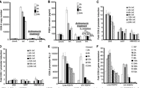
early upon KSHVde novoinfection are regulated at the scriptional level, we pretreated HMVEC-d cells with the tran-scriptional inhibitor actinomycin D (1g/ml) for 4 h prior to KSHV infection. A noncytotoxic dose of actinomycin D was used for the experiments (see Fig. S1A, a, in the supplemental material). As depicted in Fig. 1A and B, an increase in COX-2 mRNA levels and PGE2secretion was detected as early as 2 h postinfection (p.i.) and was efficiently maintained until 24 h p.i. Pretreatment of HMVEC-d cells with actinomycin D drasti-cally inhibited the effect of KSHV infection on both COX-2 mRNA levels and PGE2secretion (Fig. 1A and B). Total RNA polymerase II-dependent transcriptional arrest completely ab-rogating COX-2 levels clearly indicated that COX-2 gene ex-pression during early times of infection is regulated at the transcriptional level. COX-2 protein levels were also checked by Western blotting in serum-starved, uninfected, and KSHV-infected cells (see Fig. S1C, a and b). KSHV infection of HMVEC-d and 293 cells was confirmed by real-time-PCR for ORF50, ORF73 (see Fig. S1B, a and b), and ORF59 staining by IFA (see Fig. S1B, c). Serum starvation was not cytotoxic (see Fig. S1A, b) and did not induce COX-2 (see Fig. S1C, b). In contrast, COX-2 protein levels were induced upon KSHV infection (see Fig. S1C, a).
COX-2 promoter activity is induced in all of the cell types
tested.We have previously shown that COX-2 gene expression
is induced in HFF and HMVEC-d cells upon KSHV infection. Therefore, to further explore the transcriptional regulation of the COX-2 gene, we studied COX-2 promoter activation in transiently transfected 293, HFF, and HMVEC-d cells. Com-pared to the promoterless vector (pXP-2), the COX-2 full-length promoter construct containing the⫺1796 to⫹104 re-gion of the human COX-2 gene (P2-1900) was significantly activated during KSHV infection in all of the cell types tested at the indicated time points compared to the level in the re-spective uninfected cells (Fig. 1C), which was taken as 1-fold. We did not observe induction of control pXP-2 vector lucifer-ase activity upon KSHV infection (data not shown). Compared with that in 293 cells, COX-2 promoter activity induction was lower in HFF and HMVEC-d cells, which might be due to low transfection efficiency in HFF (50%) and HMVEC-d (20 to 30%) cells than in 293 cells (90%). No induction in the lucif-erase activity of the COX-1 promoter construct (P1-1009) was observed upon KSHV infection (Fig. 1D) in the cell types tested.
KSHV gene expression is required for sustained levels of
COX-2.To determine whether COX-2 promoter induction
re-quires a KSHV-induced signal cascade and/or viral gene expres-sion, we examined COX-2 promoter activity in HMVEC-d cells transfected with the COX-2 promoter and then infected with 30 DNA copies of KSHV/cell (either live or UV inactivated and replication incompetent) for the indicated times (Fig. 1E). UV-inactivated KSHV binds and enters HMVEC-d and HFF cells as efficiently as live KSHV (56). As reported before (56), UV-inactivated KSHV did not induce any KSHV latent (ORF73) or lytic (ORF50, K8, and K5) gene expression and was replication incompetent (data not shown). Compared to live KSHV, although UV-inactivated KSHV could also induce COX-2 promoter activity (Fig. 1E) and gene expression (Fig. 1F), this induction was low and was not sustainable for longer times (8 to 24 h) p.i. Our results clearly indicate that viral gene
VOL. 84, 2010 COX-2 PROMOTER REGULATION BY KSHV 12735
on November 8, 2019 by guest
http://jvi.asm.org/
expression is indeed necessary for sustained COX-2 levels in infected cells, but KSHV binding and entry events are good enough to upregulate appreciable levels of COX-2 promoter activation and gene expression. Seventeen-fold COX-2 gene expression was observed even at 30 min p.i. Our previous studies have shown (53) that UV-inactivated KSHV induces the p42/p44 ERK1/2 MAPK pathway, which in turn regulates the activation of multiple transcription factors (Jun, c-Fos, ATF-2, STAT-1␣, and c-Myc) that could potentially regulate COX-2 transcription.
These results suggested that during early times of KSHV infection, virus binding- and entry-initiated signal transduction cascades might be contributing to COX-2 promoter induction via the activation of various transcription factors. COX-2 ex-pression at the later time points might be due to the cumulative effect of viral gene expression, host cell gene products, auto-crine/paracrine signaling by COX-2 metabolite PGE2, and the
presence of various growth factors and cytokines in the infected-cell microenvironment.
PGE2 autoregulates COX-2 promoter activation and gene
expression.Since KSHV infection-induced PGE2can initiate
multiple signaling cascades (JNK-1, ERK1/2, PKC, PI3K-AKT, HPK1, and Src), second messengers, including cAMP, calcium (Ca2⫹), and reactive oxygen species, in an autocrine/ paracrine manner by binding to its specific G-protein-coupled receptors (EP receptors) (3, 4, 6, 16, 18, 27, 32, 33, 35, 37, 38, 46, 52, 73, 77), we tested the role of exogenous PGE2in COX-2
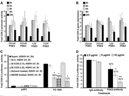
promoter and COX-2 gene expression (Fig. 2A and B). Use of biologically relevant concentrations (250 nM, 500 nM, and 1
[image:4.585.44.541.67.375.2]M) of exogenous PGE2could induce COX-2 promoter acti-vation, as well as COX-2 gene expression (Fig. 2A and B). PGE2concentrations as low as 250 nM could effectively induce COX-2 promoter induction, suggesting that low levels of PGE2 (biologically relevant) in the infected-cell microenvironment
FIG. 1. Effects of KSHV gene expression and PGE2supplementation on COX-2 promoter activity and gene expression. (A) HMVEC-d cells
pretreated with 1g/ml actinomycin D for 4 h were infected with KSHV for the indicated times. These cells were washed and lysed, and total RNA was prepared. COX-2 copy numbers were quantitated by real-time RT-PCR as described before (55, 56). (B) PGE2levels were measured (55, 56)
in the supernatants collected from cells treated as described for panel A. Percent inhibition of PGE2secretion was calculated by taking the PGE2
secretion of cells not pretreated with actinomycin D at the respective points as 100%. Each reaction was done in triplicate, and each point represents the average⫾the standard deviation (SD) of three independent experiments.**, statistically significant atP⬍0.005. (C and D) Effect of KSHV gene expression on COX-2 and COX-1 promoter activities. The indicated cell types were transfected with control pXP-2, COX-2 promoter (P2-1900), and COX-1 promoter (P1-1009)-luciferase constructs. After 24 h, cells were serum starved, infected (Inf.) for different times, lysed, and assayed for luciferase activity as described previously. Data are expressed as mean numbers of RLU after normalization with the cotransfectedRenillaluciferase activity. Each reaction was done in triplicate, and each point represents the average⫾SD of three independent experiments. The activation in uninfected (Uninf.) cells at the respective time points was taken as 1-fold for comparison. (E and F) COX-2 promoter activity (E) and gene expression (F) upon UV-inactivated KSHV (30 DNA copies/cell) or live KSHV (30 DNA copies/cell) infection. HMVEC-d cells grown to 80% confluence were serum starved for 10 h and infected with 30 DNA copies of either live or UV-inactivated KSHV per cell for different times. COX-2 promoter activity (as described for panel C) and gene expression (56) were measured in these cells.
on November 8, 2019 by guest
http://jvi.asm.org/
are sufficient to stimulate the COX-2-PGE2-COX-2 loop to
sustain COX-2. At any given time, PGE2-induced COX-2 pro-moter activity and COX-2 gene expression levels (Fig. 2A and B) were lower than the levels observed during KSHVde novo
infection (Fig. 1E and F). PGE2supplementation for a similar
duration could not change COX-1 promoter activity (data not shown). These results suggest that COX-2 induction upon KSHV infection is the collective outcome of multiple events initiated and maintained by a variety of secreted factors like vascular endothelial growth factor (VEGF), HIF-1␣, and IL-1 which are known to induce COX-2 (29, 74). In other words, PGE2 is not the only secreted factor responsible for COX-2 levels in KSHV-infected cells.
Our previous studies demonstrated that PGE2 production could be diminished upon the treatment of HMVEC-d cells
with COX-2 chemical inhibitors prior to KSHV infection or cells silenced for COX-2 and then KSHV infected (55). To evaluate the role of PGE2present in the KSHV-infected
[image:5.585.79.498.64.375.2]cul-ture supernatant in COX-2 transcriptional regulation, we used supernatants from HMVEC-d cells KSHV infected (solvent control pretreated, COX-2 inhibitor pretreated, and COX-2 silenced) for 2 h (Fig. 2C). Effective reduction (Fig. 2C) was observed in the presence of supernatants obtained from cells pretreated with COX-2 inhibitors (72%) or silenced for COX-2 (68%). Though inhibition of COX-2 primarily affects PGE2levels, the possibility that it affects the secretion of other factors like VEGF and IL-1cannot be ruled out. As VEGF and IL-1have also been shown to modulate COX-2 transcrip-tion in other systems, we extended these studies to evaluate the exclusive contribution of PGE2to COX-2 promoter activation.
FIG. 2. Effect of exogenous PGE2supplementation on COX-2 promoter activity and gene expression. (A) 293 cells transfected with a pXP2
(promoterless construct) or a COX-2 promoter (P2-1900) construct were supplemented with various concentrations of PGE2in their basal growth
medium (no serum) for different times as indicated. Promoter activity was measured using luciferase activity in the lysates prepared from unstimulated and PGE2-supplemented cells as described in Materials and Methods. Each reaction was done in duplicate, and each bar represents
the average⫾SD of three independent experiments. COX-2 promoter activity at 0 h after PGE2supplementation was taken as 1-fold for
comparison. (B) Serum-starved HMVEC-d cells were treated with various doses of exogenous PGE2for the indicated times, RNA was prepared,
and a real-time RT-PCR for COX-2 was performed as described before (55). Each reaction was done in duplicate, and each bar represents the average⫾SD of three independent experiments. COX-2 gene expression at 0 h after PGE2supplementation was taken as 1-fold for comparison.
(C and D) Effect of PGE2in KSHV-infected culture supernatant (Supnt,) on COX-2 promoter activity. Supernatants obtained from HMVEC-d
cells treated with COX-2 inhibitor or silenced for COX-2 prior to KSHV infection were used to assess COX-2 promoter activation. 293 cells were transfected with control PXP-2 and COX-2 (P2-1900)-luciferase constructs. After 24 h, cells were serum starved (10 h) and incubated with various supernatants obtained from uninfected or infected cells (as described above). After 2 h of incubation, cells were lysed and assayed for luciferase activity as described previously. The activation in supernatants from uninfected cell was taken as 1-fold. Percent inhibition was calculated by taking as 100% the levels of COX-2 promoter activity in the presence of supernatant obtained from either Si control (Si-C)-treated and KSHV-infected or solvent-treated and KSHV-infected cells. (D) Supernatant (1 ml) from serum-starved (10 h), KSHV-infected HMVEC-d cells (2 h) was incubated at 37°C for 1 h with the indicated concentrations of PGE2neutralizing antibody or rabbit IgG antibody and used for the COX-2 promoter
assay as described for panel C. Percent inhibition was calculated by taking levels of COX-2 promoter activity in the presence of supernatant pretreated with IgG antibody as 100%.
VOL. 84, 2010 COX-2 PROMOTER REGULATION BY KSHV 12737
on November 8, 2019 by guest
http://jvi.asm.org/
To understand the role of PGE2in regulating KSHV infec-tion-mediated COX-2 promoter transcriptional regulation, we performed neutralization experiments using various concentra-tions of anti-PGE2antibody (Fig. 2D). Incubation of
KSHV-infected culture supernatant with increasing concentrations (2.5, 5, and 10g/ml) of anti-PGE2neutralizing antibody could
effectively reduce COX-2 promoter induction in a dose-depen-dent fashion (42 to 82%) (Fig. 2D). Therefore, data in Fig. 2 suggest that PGE2is one of the key components of COX-2 promoter autoregulation and contributes to the continuous loop of COX-2 maintenance in KSHV-infected cells.
KSHV entry-associated signaling molecules regulate COX-2
promoter activity, gene expression, protein levels, and PGE2
secretion.To define the role of signaling molecules induced
early during KSHV infection (virus binding and entry) in COX-2 regulation, cells transfected with COX-2 full-length promoter and control Renilla luciferase plasmids were pre-treated with noncytotoxic doses of signal cascade inhibitors (see Fig. S2 in the supplemental material) and then infected with KSHV and measured for their luciferase activity (Fig. 3A). Similarly treated cells were used for COX-2 gene expres-sion (Fig. 3B), and their supernatants were analyzed for PGE2 secretion (Fig. 3D). The general Tyr-K inhibitor genistein strongly inhibited COX-2 promoter activity (91%) (Fig. 3A), COX-2 gene expression (84%) (Fig. 3B), and PGE2secretion (74%) (Fig. 3D). The Src kinase inhibitors PP2 and SU6656 also reduced COX-2 promoter activity (75%) (Fig. 3A), COX-2 gene expression (96 and 93%) (Fig. 3B), and PGE2
secretion (75 and 73%) (Fig. 3D), while the PI3K inhibitors wortmannin and LY294002 inhibited COX-2 promoter activity (65 and 82%) (Fig. 3A), COX-2 gene expression (52 and 54%) (Fig. 3B), and PGE2secretion (24 and 23%) (Fig. 3D).
Pre-treatment with the PKC inhibitors GFX and rottlerin could only reduce COX-2 promoter activity by 42 and 34%, respec-tively (Fig. 3A), COX-2 gene expression by 18 and 28% (Fig. 3B), and PGE2secretion by only 9 and 10% (Fig. 3D). FAK
inhibitors like FAK inhibitor 14 and PF573228 effectively re-duced COX-2 promoter activity (83%) (Fig. 3A), COX-2 gene expression (77 and 78%) (Fig. 3B), and PGE2 secretion (66 and 68%) (Fig. 3D). All of these results suggest that the signal molecules, especially FAK and Src, induced upon virus binding and entry are critical for COX-2 promoter activity, gene ex-pression, and PGE2secretion. Similar to COX-2 gene expres-sion data, we found changes in the levels of COX-2 protein (Fig. 3C, a and b). In contrast to PI3K (wortmannin and LY294002) and PKC (GFX and rottlerin) inhibitors, pretreat-ment with general Tyr-K (genistein), FAK (FAK inhibitor 14 and PF573228), and Src (SU6656 and PP2) inhibitors effec-tively inhibited KSHV infection-induced COX-2 protein levels (Fig. 3C).
KSHV infection-induced multiple MAPKs regulate COX-2
promoter activity, gene expression, protein levels, and PGE2
secretion. Many receptor Tyr-Ks (RTKs) and MAPKs have
been shown to regulate COX-2 promoter activity in other systems (9, 66). In our previous studies, we have shown that virus binding and entry are associated with activation of ki-nases like FAK, Src, P13K, and MAPKs (42, 51, 53, 54, 70, 71). Therefore, we assessed the majority of MAPKs (Fig. 4A). Phospho-MAPK arrays were used to assess the role of these MAPK phosphorylations at various serine/threonine positions
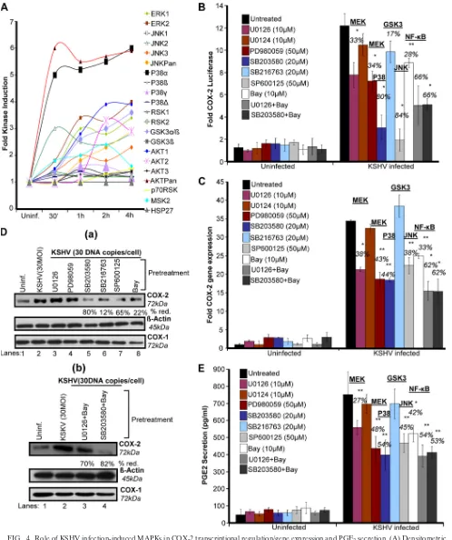
in the context of COX-2 promoter activity in uninfected and KSHV-infected (30 DNA copies/cell) HMVEC-d cell lysates. Of the MAPKs tested (Fig. 4A), KSHV infection induced phosphorylation of ERK1 and -2; JNK2, -3, and -Pan; GSK3␣/and -3␣; AKT1 and -2; and MSK2 by⬃3- to 4-fold at 2 h and this induction was time dependent (Fig. 4A). Five-to 6-fold induction of the levels of AKTPan and p38␣ was observed (Fig. 4A), while very low (1.5- to 2-fold) induction of JNK1; p38, -␥, and -␦; and RSK1, -2 and -p70 levels was observed (Fig. 4A). These results suggest that MAPKs are induced beginning early in KSHV infection. Similarly, stimu-lation of HMVEC-d cells by supplementation with 1⌴PGE2 also induced phosphorylation of MAPKs (data not shown). We hypothesized that these MAPKs induced early during KSHV infection or PGE2stimulation might be playing a critical role in the activation of downstream transcription factors and thus controlling COX-2 promoter activity, gene expression, and eventually PGE2secretion.
KSHV infection-induced MAPK pathways regulate COX-2
gene transcription, protein levels, and PGE2 secretion. To
define the role of MAPKs induced early during infection in COX-2 regulation, cells transfected with the COX-2 full-length promoter and control Renilla luciferase plasmids were pre-treated with signal cascade inhibitors and then infected with KSHV and their luciferase activity was determined (Fig. 4B). Similarly treated cells were used for COX-2 gene expression (Fig. 4C), and their supernatants were assayed for PGE2(Fig. 4E). MEK inhibitors U0126 and PD980059 inhibited COX-2 promoter activity (33 and 34%) (Fig. 4B), COX-2 gene expres-sion (38 and 43%) (Fig. 4C), and PGE2secretion (27 and 48%)
(Fig. 4E). p38 inhibitor SB203580 repressed COX-2 promoter activity (80%) (Fig. 4B), COX-2 gene expression (44%) (Fig. 4C), and PGE2secretion (54%) (Fig. 4E), while JNK inhibitor SP-600125 diminished COX-2 promoter activity (84%) (Fig. 4B), COX-2 gene expression (38%) (Fig. 4C), and PGE2 se-cretion (45%) (Fig. 4E). NF-B inhibitor Bay11-7082 de-creased COX-2 promoter activity (28%) (Fig. 4B), COX-2 gene expression (33%) (Fig. 4C), and PGE2secretion (42%)
(Fig. 4E). The ERK and NF-B or p38 and NF-B combina-tion of inhibitors inhibited COX-2 promoter activity (66%) (Fig. 4B), COX-2 gene expression (62%) (Fig. 4C), and PGE2 secretion (55%) (Fig. 4E).
Of the MAPKs tested, JNK and p38 appeared to control COX-2 promoter activity. The ERK or NF-B inhibitor alone did not noticeably inhibit COX-2 promoter activity, gene ex-pression, protein levels, or PGE2 secretion (Fig. 4B to E)
compared to the NF-B or p38 and NF-B combination of inhibitors. Since we observed a 3- to 4-fold induction of the phosphorylation of GSK3 isoforms (␣, 51 kDa; , 47 kDa) (Fig. 4A), we used a GSK3 inhibitor. GSK3 inhibitor SB216763 pretreatment could not inhibit COX-2 promoter activity (Fig. 4B) like the other MAPK inhibitors; rather, we observed slightly increased COX-2 gene expression (Fig. 4C). GSK3 inhibitor SB216763 pretreatment downregulated COX-2 pro-tein levels by 12% (Fig. 4D, a).
Since early binding and entry events of KSHV infection effectively modulated COX-2 transcription and PGE2 secre-tion, we hypothesized that these signal cascades are critical for the activation of various transcription factors involved in COX-2 promoter regulation. Hence, we next studied the
on November 8, 2019 by guest
http://jvi.asm.org/
FIG. 3. Role of signaling molecules induced early during KSHV infection in COX-2 transcriptional regulation/gene expression, protein level, and PGE2secretion. (A) 293 cells were transfected with COX-2 full-length promoter and controlRenillaluciferase plasmids. Transfected cells were
serum starved and then treated with nontoxic concentrations of various signaling inhibitors (inh.), as indicated, for 1 h at 37°C and then infected with 30 DNA copies/cell KSHV for 2 h. Cells were lysed, and luciferase activity was measured. Data represent mean numbers of RLU after normalization with the cotransfectedRenillaluciferase activity. Each reaction was done in triplicate, and each point represents the average⫾SD of five experiments. The activation in uninfected cells was taken as 1-fold for comparison. Percent inhibition was calculated by taking the COX-2 promoter activity in non-inhibitor-treated, KSHV-infected (2 h) cells as 100%. (B) HMVEC-d cells were treated with nontoxic concentrations of various signaling inhibitors for 1 h at 37°C and then infected with KSHV (30 DNA copies/cell) for 2 h. Total RNA was prepared from these cells, and COX-2 gene expression was quantitated as described before. The COX-2 level in uninfected cells was taken as 1-fold for comparison. Percent inhibition was calculated by taking the COX-2 gene expression in non-inhibitor-treated, KSHV-infected (2 h) cells as 100%. (C, a and b). HMVEC-d cells were treated as described for panel B. Total protein was prepared from these cells, and COX-2/COX-1 protein levels were quantitated by Western blotting. Percent reduction (red.) was calculated by taking the COX-2 protein level in non-inhibitor-treated, KSHV-infected (2 h) cells as 100%.-Actin levels were checked as a loading control. (D) Supernatants collected from the HMVEC-d cells treated as described for panel B were used for PGE2measurement by ELISA as described before. Data are expressed as mean pg/ml. Each reaction was done
in triplicate, and each point represents the average⫾SD of five experiments. Percent inhibition was calculated by taking PGE2secretion from
non-inhibitor-treated, KSHV-infected (2 h) cells as 100%.*and**, statistically significant atP⬍0.01 andP⬍0.005, respectively.
12739
on November 8, 2019 by guest
http://jvi.asm.org/
FIG. 4. Role of KSHV infection-induced MAPKs in COX-2 transcriptional regulation/gene expression and PGE2secretion. (A) Densitometric
analysis of MAPK array blots measuring the activation of MAPKs. The values were normalized to identical background levels using the R&D human MAPK antibody array analysis tool. The fold induction of MAPK was calculated by dividing the respective values obtained from KSHV-infected HMVEC-d cell lysates by the values obtained from uninfected-cell lysates. (B) 293 cells were transfected with COX-2 full-length promoter and controlRenillaluciferase plasmids. Transfected cells were serum starved and then treated with nontoxic concentrations of various signaling inhibitors as indicated for 1 h at 37°C and then infected with 30 DNA copies/cell KSHV for 2 h. Cells were lysed, and luciferase activity
on November 8, 2019 by guest
http://jvi.asm.org/
mechanism of KSHV infection-induced COX-2 transcription utilizing various COX-2 promoter mutations and deletion con-structs.
cis-acting element dNFAT regulates KSHV-induced human
COX-2 promoter activity.It is well known that COX-2
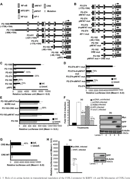
pro-moter activity is regulated in other systems by several tran-scription factors, including NF-B, nuclear factor IL-6, AP-1, CRE, and NFAT (25, 26). To understand the roles of various transcription factors and their involvement in COX-2 upregu-lation upon KSHV infection, we studied the role of KSHVde novo infection in cells transiently transfected with deletion (Fig. 5A) and mutation (Fig. 5B) constructs of the human COX-2 promoter. Details of different COX-2 promoter lucif-erase constructs have been described previously (25, 26), and their maps are given in Fig. 5A and B. The COX-2 promoter has two NFAT cis-acting elements, named COX-2-dNFAT and -pNFAT, which are highly conserved among different an-imal species (25, 26).
KSHV infection induced the activity of the P2-1900 and P2-1102 promoter luciferase constructs similarly (Fig. 5C). Promoter analysis results indicated that promoter elements between ⫺88 and ⫺170 are essential for KSHV-induced COX-2 promoter activity, as an 88% reduction in promoter activity was observed upon the deletion of this region (Fig. 5C). These results suggest that dNFAT/NF-IL-6/AP-2-SP1 is likely to be responsible for promoter activation following KSHV infection. The possibility of NF-B involvement cannot be ruled out either, as we observed a 63% reduction in promoter activity of the construct with oneB site removed (P2-431), whereas the loss of both of the sites binding NF-B (P2-274) relieved this inhibition (25%), suggesting the involvement of NF-B in the regulation of COX-2 promoter activity upon KSHV infection (Fig. 5C). The P2-150 construct containing CRE and the TATA box was appreciably activated by KSHV infection compared to P2-192, suggesting a potential role for CRE in COX-2 promoter activity (Fig. 5C). These results also suggested that pNFAT and AP-1 in P2-192 might be negatively regulating COX-2 promoter activity (Fig. 5C).
To investigate the role of both NFAT sites in detail, we used P2-274-AP-1 mut, P2-274 dNFAT mut, P2-274 pNFAT mut, and P2-274 d⫹pNFAT mut constructs to transfect 293 cells. The transfected cells were serum starved and then infected with KSHV for 2 h. Mutation of dNFAT resulted in a 75% reduction of COX-2 promoter activity with respect to P2-274, suggesting the involvement of dNFAT in the regulation of
COX-2 (Fig. 5D). Mutation of pNFAT did not result in the downregulation of COX-2, ruling out the possibility of pNFAT involvement in COX-2 transcriptional regulation and verifying the results depicted in Fig. 5C. Mutation of both the proximal and distal NFAT sites resulted in a 78% reduction (Fig. 5D). Since the proximal NFAT site is not purely NFAT but is always bound to AP-1, we used P2-274-AP-1 mut to study COX-2 promoter activity. AP-1 mut did not affect COX-2 promoter activation further, verifying the results shown in Fig. 5C. Col-lectively, the data shown in Fig. 5D strongly suggest the in-volvement of dNFAT in KSHV-induced COX-2 promoter ac-tivity. P2-274-AP-1 mut did not show any reduction in luciferase activity, ruling out the possible involvement of AP-1 in COX-2 transcriptional regulation (Fig. 5D).
CRE recognition sequences in the human COX-2 promoter are important for KSHV infection-induced transcriptional
ac-tivity.To investigate the role of CRE, we used P2-192 pNFAT
mut, P2-192 CRE mut, and P2-192 pNFAT mut⫹CRE mut constructs to transfect 293 cells. The transfected cells were serum starved and then infected with KSHV for 2 h. Mutated pNFAT’s failure to inhibit COX-2 promoter activity further ruled out the involvement of pNFAT in KSHV-induced COX-2 (Fig. 5E). Mutation of CRE resulted in an 88% reduc-tion in COX-2 promoter activity compared to that obtained with the P2-192 construct (Fig. 5E). The promoter luciferase activity of the P2-192 pNFAT mut⫹CRE mut construct was similar to that of P2-192 CRE mut, suggesting a role for CRE but not pNFAT in COX-2 promoter induction (Fig. 5E). Col-lectively, COX-2 promoter deletion and mutation analyses showed that dNFAT and CRE recognition sequences present in the human COX-2 promoter are important for transcrip-tional activation upon KSHV infection.
Calcineurin (Cn)/NFAT pathway regulates KSHV-induced
COX-2 promoter activity.To further investigate the
involve-ment of NF-B and NFAT in COX-2 promoter induction, we measured COX-2 full-length promoter activity in the presence of p65, IBM, and VIVIT expression plasmids. As NFAT activity is controlled by Cn phosphatase, we cotransfected 293 cells with COX-2 promoter construct P2-1900 and a plasmid expressing the peptide VIVIT, which competes with NFAT for Cn binding and selectively inhibits NFAT activation without disrupting other Cn-dependent pathways (Fig. 5F). COX-2 luciferase reporter activity was dramatically lowered (81%) in the presence of a plasmid expressing the peptide VIVIT, sug-gesting a role for Cn binding in KSHV-induced NFAT activity
was measured. Data represent mean numbers of RLU after normalization with the cotransfectedRenillaluciferase activity. Each reaction was done in triplicate, and each point represents the average⫾SD of five experiments. The activation in untreated infected cells was taken as 1-fold for comparison. Percent inhibition was calculated by taking the COX-2 promoter activity in non-inhibitor-treated, KSHV-infected (2 h) cells as 100%. (C) HMVEC-d cells were treated with nontoxic concentrations of various MAPK inhibitors for 1 h at 37°C and then infected with KSHV (30 DNA copies/cell) for 2 h. Total RNA was prepared from these cells, and COX-2 gene expression was quantitated as described before. The COX-2 level in uninfected cells was taken as 1-fold for comparison. Percent inhibition was calculated by taking the COX-2 promoter activity in non-inhibitor-treated, KSHV-infected (2 h) cells as 100%. (D, a and b). HMVEC-d cells were treated as described for panel C. Total protein was prepared from these cells, and COX-2/COX-1 protein levels were quantitated by Western blotting. Percent inhibition was calculated by taking the COX-2 protein level in non-inhibitor-treated, KSHV-infected (2 h) cells as 100%.-Actin levels were checked as a loading control. (E) Supernatants collected from HMVEC-d cells treated as described for panel C were used for PGE2measurement by ELISA as described before. Data are expressed as
mean pg/ml. Each reaction was done in triplicate, and each point represents the average⫾SD of five experiments. Percent inhibition was calculated by taking PGE2secretion from non-inhibitor-treated, KSHV-infected (2 h) cells as 100%.*and**, statistically significant atP⬍0.01
andP⬍0.005, respectively.
VOL. 84, 2010 COX-2 PROMOTER REGULATION BY KSHV 12741
on November 8, 2019 by guest
http://jvi.asm.org/
FIG. 5. Role ofcis-acting factors in transcriptional regulation of the COX-2 promoter by KSHV. (A and B) Schematics of COX-2 promoter deletion (A) and mutation (B) constructs.cis-acting consensus sequences are denoted by boxes. The extents of 5⬘deletions are shown, with the numbers indicating their lengths relative to the transcription start site. Transcription factor binding sites are represented on the promoter, while the promoter regions relative to the transcription initiation start site are shown in parentheses. (C) 293 cells were transfected with control pXP2
on November 8, 2019 by guest
http://jvi.asm.org/
(Fig. 5F). We also assessed the effect of NF-B by using the plasmid for IBM (dominant negative mutant IB), which blocks NF-B activation (Fig. 5F). Cotransfection with IBM inhibited COX-2 promoter activation by 56%, whereas the p65 plasmid did not change COX-2 promoter activity appreciably (Fig. 5F, a). Expression of IBM, p65, and VIVIT plasmids was checked by Western blotting (Fig. 5F, b) using antibodies to the respective tags (Myc and GFP). p65 and VIVIT expres-sion in the transfected cells was also checked by observing GFP expression under a microscope. These results supported the data in Fig. 5C suggesting that, along with NF-B, there are other factors regulating KSHV infection-induced COX-2 tran-scription, of which Cn/NFAT appeared to be especially critical.
CRE and NFAT play obligatory roles in KSHV
infection-induced COX-2 promoter activity. To further verify the
in-volvement of CRE and NFAT in COX-2 promoter induction, we measured COX-2 promoter activity using COX-2 CRE-WT and COX-2 CRE-Mut constructs in uninfected and KSHV-infected (2 h) cells. KSHV infection induced COX-2 CRE-WT activation compared to the CRE-Mut construct (Fig. 5G). A 48% reduction was observed in COX-2 CRE-Mut promoter activity upon KSHV infection compared to COX-2-CRE-WT (Fig. 5G).
To evaluate the combined effect of CRE mutation and NFAT blocking on COX-2 promoter activity, a cotransfection experiment was performed using the pCDNA or VIVIT ex-pression plasmid along with the CRE-Mut construct. Cotrans-fection with VIVIT further reduced COX-2 CRE-Mut activity by 74%, suggesting critical roles for CRE and NFAT in KSHV infection-induced COX-2 transcriptional regulation (Fig. 5H, a). Expression of VIVIT was confirmed by Western blotting (Fig. 5H, b).
KSHV infection induces NFAT.NFAT belongs to the family
of transcription factors which comprises four classical bers: NFAT1, NFAT2, NFAT3, and NFAT4. All of the mem-bers of the NFAT family are Ca2⫹responsive and are
regu-lated by the Ca2⫹/Cn signaling pathway (7, 13, 23, 28, 36, 45, 47). NFAT activation is controlled via nuclear import and export involving dephosphorylation by Cn phosphatase,
opti-mal DNA binding, and regulation of the transactivating activ-ity. In resting cells, NFATs are phosphorylated at a cluster of serine residues located in the regulatory domain which effec-tively mask the nuclear localization signal and thereby retain NFAT in an inactive conformation in the cytoplasm (44, 47). Upon stimulation with agonists which increase intracellular Ca2⫹, NFATs are dephosphorylated by the phosphatase Cn and translocate to the nucleus. In the nucleus, NFATs interact with fos and Jun family members to cooperatively bind to and transactivate NFAT sites on the promoter regions of target genes (36, 58, 61). When the cells return to their unstimulated state, NFAT becomes rephosphorylated and is exported out of the nucleus (47).
As COX-2 promoter analysis showed NFAT involvement in COX-2 promoter regulation, we examined whether
KSHVde novoinfection induces NFAT transcription factor
activation in 293 and HMVEC-d cells. Using a transcription factor ELISA, nuclear extracts from uninfected and infected 293 or HMVEC-d cells were assessed for the ability of host transcription factors to bind to their respective WT DNA se-quences. Infection of 293 or HMVEC-d cells with KSHV in-creased NFAT activation in a time-dependent manner (Fig. 6A), although NFAT activation was lower in 293 cells than in HMVEC-d cells (Fig. 6A). Results in Fig. 6B show a repre-sentative example of HMVEC-d cells infected with KSHV for different times. NFAT activity was barely detected in the se-rum-starved uninfected cells (Fig. 6B). To determine the spec-ificity of the above transcription factor detection, uninfected and infected nuclear extracts were preincubated for 30 min with WT or mutated consensus sequences for the respective factors before application to the plate-bound WT DNA se-quences. In this experiment, transcription factors prebound to the WT oligonucleotides will be unable to bind to the plate-bound DNA and hence will be removed in the washing steps. The ability of transcription factors to bind their respective target sequences was significantly inhibited by preincubation with WT oligonucleotides, while no effect was seen with mu-tated oligonucleotides (Fig. 6B), demonstrating the specificity of the assay. Nuclear extracts prepared from the ionomycin
or various COX-2 promoter deletion constructs (P2-1900-luc, P2-1102-luc, P2-431-luc, P2-274-luc, P2-192-luc, and P2-150-luc). At 24 h after transfection, cells were serum starved and then left uninfected or infected with 30 DNA copies of KSHV/cell for 2 h. These cells were harvested, lysed, and assayed for RLU as described before. (D) 293 cells were transfected with P2-274 or P2-274 promoter mutant constructs (P2-274 dNFAT mut, P2-274 pNFAT mut, P2-274 d⫹pNFAT mut, and P2-274-AP1 mut). After 24 h, cells were serum starved for 10 h and infected for 2 h and a promoter assay was performed as described for panel C. (E) 293 cells were transfected with P2-192 promoter mutant constructs (P2-192 pNFAT mut, P2-192 CRE mut, and P2-192 pNFAT mut⫹CRE mut). After 24 h, cells were serum starved for 10 h and infected for 2 h and a promoter assay was performed as described for panel C. Data in panels C, D, and E represent the mean number of RLU after normalization with the cotransfectedRenillaluciferase activity. Each reaction was done in triplicate, and each point represents the average⫾SD of three experiments. (F, a) 293 cells were cotransfected with a COX-2 promoter full-length construct (P2-1900) along with 1g of pcDNA, IM, p65, or VIVIT expression plasmid and left uninfected or KSHV infected for 2 h. Promoter induction in uninfected cells was taken as 1-fold. Percent inhibition of COX-2 induction was calculated with reference to fold induction in pcDNA-transfected and KSHV-infected (2 h) cells as 100%.*and**, statistically significant atP⬍0.01 andP⬍0.005, respectively. (F, b) Expression of IM, p65, or VIVIT at protein level. Lysates prepared from IM-, p65-, or VIVIT-transfected cells were prepared and Western blotted using Myc or GFP antibodies. (G) 293 cells were transfected with CRE-WT and CRE-Mut promoter constructs. After 24 h, cells were serum starved for 10 h and left uninfected or infected for 2 h and a promoter assay was performed as described for panel C. Percent inhibition of COX-2 induction was calculated with reference to induction in CRE-WT plasmid-transfected and then infected cells, which was taken as 100%. (H, a) 293 cells were cotransfected with a COX-2 promoter construct with a CRE-Mut construct along with 1g of a pcDNA or VIVIT expression plasmid. After 24 h, cells were serum starved for 10 h and then infected for 2 h and a promoter assay was performed as described for panel C. Percent inhibition of COX-2 promoter induction was calculated with reference to fold induction in pcDNA-transfected and KSHV-infected (2 h) cells, which was taken as 100%.*and**, statistically significant at
P⬍0.01 andP⬍0.005, respectively. (H, b) Expression of VIVIT at protein level. Lysates prepared from pcDNA- or VIVIT-transfected cells were prepared and Western blotted using GFP antibody.
VOL. 84, 2010 COX-2 PROMOTER REGULATION BY KSHV 12743
on November 8, 2019 by guest
http://jvi.asm.org/
on November 8, 2019 by guest
http://jvi.asm.org/
(NFAT agonist)-stimulated cells showed dramatic (28-fold) induction in NFAT activity and was used as a positive control (Fig. 6C). Altogether, these data suggested a link between NFAT activity and COX-2 induction upon KSHV infection.
Multiple scenarios can be hypothesized for KSHV- or PGE2-induced NFAT activation (Fig. 6D). In other systems,
RTKs have been shown to induce IP3, which can lead to Ca2⫹ release from the endoplasmic reticulum, and this Ca2⫹ can
induce Cn/NFAT signaling, which can dephosphorylate NFAT and augment nuclear translocation of NFAT.
The remarkable (81%) reduction in COX-2 promoter activ-ity upon cotransfection with NFAT peptide inhibitor plasmid VIVIT (Fig. 5F) suggested the importance of Cn signaling in COX-2 transcription regulation. Cn signaling has been impli-cated in a broad spectrum of infections and pathological con-ditions, and there is evidence that NFAT activation is medi-ated by a Ca2⫹/Cn-dependent pathway. To evaluate the contribution of Cn in KSHV infection-induced COX-2 expres-sion, CsA, a specific inhibitor of Cn/NFAT, was employed. First, we checked the role of CsA in KSHV infection-induced NFAT activity (Fig. 6E). Pretreatment of HMVEC-d or 293 cells with various concentrations of CsA (0.25M, 0.5 M, and 1M) was able to effectively inhibit NFAT induction in a dose-dependent manner, as indicated in Fig. 6E. All of the doses of CsA used were noncytotoxic, while the 1M concen-tration was most effective in NFAT inhibition in both of the cell types tested (Fig. 6E). CsA pretreatment also drastically inhibited ionomycin-induced NFAT activity in both of the cell types tested (293 and HMVEC-d), indicating the specificity of NFAT inhibition (Fig. 6E).
KSHV-induced COX-2 promoter activity is preferentially regulated via dNFAT binding to the human COX-2 promoter.
To further confirm a functional activation of NFAT by KSHV, dNFAT DNA binding was assessed by EMSAs with an oligo-nucleotide containing a consensus dNFAT binding site as a
probe (Fig. 6F). Nuclear extracts prepared from HMVEC-d cells left uninfected (Fig. 6F, lane 5) or KSHV infected for 1 h (Fig. 6F, lane 1) or 2 h (Fig. 6F, lane 2) were incubated with consensus oligonucleotides for the dNFAT binding site used as labeled probes. Compared with uninfected cells (Fig. 6F, lane 5), KSHV infection markedly induced the formation of a com-plex with the consensus dNFAT probe (Fig. 6F, lanes 1 and 2). Compared to nuclear extracts prepared from infected cells (Fig. 6F, lanes 1 and 2), CsA-treated and then KSHV-infected cell nuclear lysates (Fig. 6F, lane 4) showed a marked reduc-tion in complex formareduc-tion with the labeled dNFAT probe. A competition assay with an excess of unlabeled probe (COX-2 dNFAT) completely abolished complex formation with the labeled probe, suggesting that the complex is specific to COX-2 dNFAT (Fig. 6F, lane 7). The specificity of in vitro DNA binding was also confirmed by supershift EMSA using nuclear extracts prepared from HMVEC-d cells infected for 1 h (Fig. 6F, lane 3) or 2 h (Fig. 6F, lane 6).
Since NFAT activity is intimately correlated with its nuclear localization, we investigated the subcellular localization of this transcription factor after KSHV infection by microscopy using a specific anti-NFAT antibody. Interestingly, NFAT translo-cates to the nucleus early after KSHV infection (Fig. 6G) and this nuclear translocation was strongly inhibited in cells treated with CsA prior to KSHV infection (data not shown).
The significance of the Cn/NFAT pathway was further as-sessed at the COX-2 gene expression and PGE2 secretion levels. KSHV infection-induced COX-2 gene expression ap-peared to be Cn signaling dependent, as it was suppressed by CsA treatment prior to infection (Fig. 6H) at all of the time points tested. Similar to COX-2 gene expression, PGE2 secre-tion was also reduced in the cells treated with CsA prior to KSHV infection (Fig. 6I). Data presented in Fig. 6 strongly indicate that NFAT activation increases COX-2 expression and PGE2synthesis, and inactivation of NFAT by CsA
signif-FIG. 6. Role of KSHVde novoinfection-induced activation of Cn/NFAT-dependent COX-2 gene expression and PGE2secretion. (A) KSHV de novoinfection of HMVEC-d and 293 cells induces NFAT activation. Relative activation was calculated by taking the activation in uninfected cells as 1-fold at all of the indicated times. (B) To monitor the specificity of NFAT activation, a competition assay using soluble oligonucleotides was performed. Briefly, nuclear extracts prepared from HMVEC-d cells left uninfected or infected with KSHV for different times were incubated with either the WT consensus oligonucleotides (competitor for NFAT binding which will prevent NFAT binding to the probe immobilized on the plate) or mutant oligonucleotides for 30 min prior to their addition to the plate with immobilized oligonucleotides, and an ELISA was performed with an antibody specific to the NFAT transcription factor in accordance with the manufacturer’s instructions. Optical density (OD) values are presented. (C) Nuclear lysates prepared from ionomycin-treated cells were used as a positive control, and the specificity of the assay was verified as shown in Fig. 5B. Data represent the average⫾SD of three experiments. (D) Schematic of the possible pathways of NFAT induction by KSHV infection and PGE2secreted in the infected-cell microenvironment. (E) Histogram depicting NFAT fold induction in nuclear extracts prepared
from HMVEC-d cells left untreated or pretreated with various concentrations of CsA and then infected with KSHV for 2 h. Percent inhibition of NFAT activation upon CsA pretreatment was calculated with respect to the DNA-binding activities in KSHV-infected HMVEC-d cells without CsA pretreatment, which were taken as 100%. Ionomycin-treated nuclear lysates were used as a positive control. Data represent the average⫾ SD of three experiments. (F) EMSA for nuclear extracts prepared from HMVEC-d cells using the dNFAT probe. This EMSA experiment shows the binding of nuclear extracts prepared from virus-infected and uninfected HMVEC-d cells left untreated or pretreated with CsA to the dNFAT probe. The specificity of the DNA-protein interaction was assessed by competition (using a 100-fold molar excess of unlabeled double-stranded oligonucleotide NFAT probe as a competitor) and a supershift EMSA using anti-NFAT antibody. Each EMSA is representative of at least three independent experiments. (G) NFAT’s subcellular localization upon KSHV infection. HMVEC-d cells left uninfected or KSHV infected at the different times indicated were labeled with anti-NFAT antibody and then examined under a microscope. Each image shown corresponds to one of three independent experiments. (H) Effect of CsA on COX-2 gene expression in HMVEC-d cells. cDNA prepared from serum-starved (10 h) uninfected, KSHV-infected (2 h), solvent-pretreated, and then KSHV-infected (2 h) or CsA-treated and then KSHV-infected (2 h) HMVEC-d cells was used for COX-2 gene expression analysis. COX-2 gene expression in uninfected cells was taken as 1-fold. Data represent the average⫾ SD of three experiments. (I) Effect of CsA on PGE2secretion in HMVEC-d cells. Supernatants collected from serum-starved (10 h), uninfected,
KSHV-infected (2 h), solvent-pretreated, and then KSHV-infected (2 h) or CsA-treated and then KSHV-infected (2 h) cells were used for PGE2
quantitation by ELISA, and levels in pg/ml are presented. Data represent the average⫾SD of three experiments.*and**, statistically significant atP⬍0.01 andP⬍0.005, respectively.
VOL. 84, 2010 COX-2 PROMOTER REGULATION BY KSHV 12745
on November 8, 2019 by guest
http://jvi.asm.org/
on November 8, 2019 by guest
http://jvi.asm.org/
icantly diminished them, suggesting that KSHV-induced COX-2 transcription may involve Ca2⫹signaling and
Cn-de-pendent NFAT activation. Although in the present study we have not assessed the role of Ca2⫹using any pharmacological
inhibitors of Ca2⫹ (BAPTA-AM and TMB-8), in a separate study (19), we documented the role of PGE2in Ca2⫹signaling,
which might also be one of the factors inducing NFAT activity in KSHV-infected cells.
Data presented in Fig. 6E to I clearly show that KSHV infection or PGE2per seupon interaction with target cells can
induce NFAT activation (Fig. 6E and F), COX-2 gene expres-sion (Fig. 6H), and PGE2 secretion (Fig. 6I) via Ca2⫹
/Cn-mediated NFAT dephosphorylation and nuclear translocation (Fig. 6G).
CREB serine 133 phosphorylation is involved in
KSHV-induced COX-2 gene expression.Since COX-2 promoter
anal-ysis indicated a role for CRE (Fig. 5C, E, and G), next we assessed whether KSHV infection induces the binding of tran-scription factor CREB to CRE. CREB is a member of a large family of structurally related transcription factors (AFT1 to -4, c-Fos, c-Jun, c-Myc, and C/EBP) (59) which specifically recog-nize the CRE promoter site (5⬘-TGACGTCA-3⬘) (39). CREB transcriptional activity is controlled by homo- and het-erodimers (CREB/ATF-1) of CREB family members binding to the CRE promoter site (30). CREB activity is also con-trolled by its cofactor, CREB-binding protein (5), and a variety of protein kinases like protein kinase A (PKA), PKC, MAPKs, and Ca2⫹/calmodulin-dependent protein kinases that
phos-phorylate CREB at Ser-133 (59), which increases CREB’s af-finity for its promoter site (59) (Fig. 7D).
KSHV infection-induced CREB activity in 293 (⬃2.5-fold) and HMVEC-d (⬃3-fold) cells (Fig. 7A) was measured by a transcription factor ELISA with a CREB-specific probe. Using an ELISA-based technique as described in Fig. 6B, nuclear extracts from uninfected and KSHV-infected HMVEC-d and 293 cells were assessed for the ability of host transcription factors to bind to their respective WT DNA sequences. Figure 7B shows a representative example of the CREB activity in
HMVEC-d cells infected with KSHV for different times. The specificity of activation was assessed by a reduction of binding in the nuclear extracts incubated with WT CREB consensus sequences (Fig. 7B). Levels of these transcription factors were barely detectable in serum-starved uninfected cells (Fig. 7B). Since cAMP induction is required upstream of CREB serine 133 phosphorylation and CRE binding, we prepared nuclear lysates from forskolin-treated HMVEC-d cells (Fig. 7C). These nuclear lysates showed very high activation of CREB-serine phosphorylation and exhibited reduced binding when preincubated with WT consensus sequences (Fig. 7C).
Multiple scenarios can be hypothesized for KSHV- or PGE2-induced CREB serine 133 phosphorylation (Fig. 7D). KSHV binding initiates various MAPKs (p38, JNK), and FAK/ Src kinases can induce production of cAMP, which can activate PKA and mediate phosphorylation of CREB serine 133. PGE2
secreted from infected cells can induce adenylate cyclase acti-vation via binding to its prostanoid receptors (EP receptors) present on the infected cell or the neighboring infected/unin-fected cells. These interactions can upregulate cAMP produc-tion, PKA activaproduc-tion, and CREB serine phosphorylation. Apart from PGE2/EP receptor signaling, Ca2⫹signaling could
also regulate CREB activation, as reported in other systems. To further confirm a functional activation of CREB by KSHV, CREB DNA binding was assessed by EMSAs with an oligonu-cleotide containing a consensus CRE binding site as a probe (Fig. 7E). Nuclear extracts prepared from HMVEC-d (Fig. 7E, lanes 6 to 9) or 293 (Fig. 7E, lanes 2 to 5) cells left uninfected or KSHV infected for different times were incubated with consensus oligo-nucleotides for the CRE binding site as labeled probes. Com-pared with leaving HMVEC-d cells uninfected (Fig. 7E, lane 8), KSHV infection for 1 h (Fig. 7E, lane 7) or 2 h (Fig. 7E, lane 6) markedly induced the formation of a complex with the consensus CRE probe. Compared with leaving 293 cells uninfected (Fig. 7E, lane 4), KSHV infection for 1 h (Fig. 7E, lane 3) or 2 h (Fig. 7E, lane 2) markedly induced the formation of a complex with the consensus CRE probe. Competition assay with an excess of un-labeled probe (COX-2-CRE) completely abolished complex
for-FIG. 7. Effect of KSHVde novoinfection-activated, cAMP-PKA-mediated CREB serine 133 phosphorylation and COX-2 promoter induction. (A) KSHVde novoinfection of HMVEC-d and 293 cells induces CREB activation. Relative activation was calculated by taking activation in uninfected cells as 1-fold. (B) To monitor specificity of CREB activation, nuclear extracts prepared from HMVEC-d cells left uninfected or infected with KSHV for different times were tested for CREB activity by incubating the nuclear extracts with WT consensus oligonucleotides (competitor for CREB binding) or mutant oligonucleotides for 30 min prior to performing ELISA as described for Fig. 6B. (C) Nuclear lysates prepared from forskolin-treated cells were used as a positive control, and the specificity of the assay was verified as shown in Fig. 6B. Data represent the average⫾SD of three experiments. (D) Diagrammatic representation of the possible pathways involved in CREB serine 133 phosphorylation and CRE activation. (E) EMSA for nuclear extracts prepared from HMVEC-d or 293 cells using CRE probe. This EMSA experiment shows the binding of nuclear extracts prepared from virus-infected and uninfected HMVEC-d cells with a CRE probe. The specificity of the DNA-protein interaction was assessed by supershift EMSA using anti-CREB-1 antibody. Each EMSA is representative of at least three independent experi-ments. (F, a) 293 cells cotransfected with a P2-1900 COX-2 promoter full-length construct along with 1g of p-CMV-CREB, p-CMV-CREB-133, or p-CMV-KCREB expression plasmid were left uninfected or KSHV infected for 2 h. The promoter induction in uninfected cells was taken as 1-fold. Percent inhibition of COX-2 induction was calculated with reference to then-fold induction in p-CMV-CREB-transfected and KSHV-infected (2 h) cells, which was taken as 100%. Expression of various CREB constructs was checked by Western blotting (F, b). (G, a and b). Lysates prepared from serum-starved HMVEC-d cells left uninfected or KSHV infected or treated with 1⌴PGE2or forskolin for the indicated times
were used to measure intracellular cAMP levels. Data are expressed as mean pmol/ml. (H, a and b). Lysates prepared as described for panel G (a and b) were used to quantitate PKA levels by the methods described previously. Data are expressed as mean ng/3⫻105HMVEC-d cells. Each
reaction was done in triplicate, and each point represents the average⫾SD of five experiments. (I, a and b). Serum-starved HMVEC-d cells were left untreated or pretreated with PKA inhibitor H-89 and then KSHV infected for the indicated times, treated with 1⌴PGE2for 2 h, or treated
with forskolin for 30 min. Nuclear lysates prepared from these cells were used to quantitate CREB serine 133 phosphorylation levels as described above. Each reaction was done in triplicate, and each point represents the average⫾SD of five experiments. Percent inhibition was calculated by taking the CREB serine 133 phosphorylation in non-inhibitor-treated, KSHV-infected (2 h) cells as 100%.
VOL. 84, 2010 COX-2 PROMOTER REGULATION BY KSHV 12747
on November 8, 2019 by guest
http://jvi.asm.org/
FIG. 8. Roles of multiple signaling pathways involved in regulating transcription factor (CREB) activation involved in KSHV-induced COX-2. (A and B) Nuclear lysates prepared from HMVEC-d cells were used to measure CREB serine 133 phosphorylation by transcription factor ELISA. Activation in uninfected cells was taken as 1-fold for comparison. Percent inhibition was calculated by taking the CREB activation in KSHV-infected cells as 100%.*and**, statistically significant atP⬍0.01 andP⬍0.005, respectively. (C) Schematic diagram depicting the multiple signaling pathways and transcription factors involved in the transcriptional regulation of KSHV-induced COX-2. A variety of transcription factors can stimulate COX-2 expression, as there are several binding sites for a number of transcription factors, including NF-B, NFAT, NF-IL-6/cEBP, AP-1, and CRE, in the 5⬘region of the COX-2 gene. Intervention at each step of COX-2 induction can be used as a potential therapeutic target. The role of cell signaling (NF-B, ERK, p38, JNK, PI3K/AKT, PKC, and Rho-GTPases) in the regulation of COX-2 expression in other systems has been well defined (65, 67, 75). Since KSHV has been shown to induce a variety of overlapping cell signaling cascades (ERK, PI3K, Rho-GTPases, FAK, Src, NF-B, and PKC-) and transcription factors (c-Fos, c-Jun, c-Myc, and STAT1-␣) early during infection (42, 51, 53, 54,