Madrid, Universidad Auto´noma de Madrid, Cantoblanco, 28049 Madrid, Spain
Received 18 March 2010/Accepted 17 May 2010
The first morphological evidence of African swine fever virus (ASFV) assembly is the appearance of precursor viral membranes, thought to derive from the endoplasmic reticulum, within the assembly sites. We have shown previously that protein p54, a viral structural integral membrane protein, is essential for the generation of the viral precursor membranes. In this report, we study the role of protein p17, an abundant transmembrane protein localized at the viral internal envelope, in these processes. Using an inducible virus for this protein, we show that p17 is essential for virus viability and that its repression blocks the proteolytic processing of polyproteins pp220 and pp62. Electron microscopy analyses demonstrate that when the infection occurs under restrictive conditions, viral morphogenesis is blocked at an early stage, immediately posterior to the formation of the viral precursor membranes, indicating that protein p17 is required to allow their progression toward icosahedral particles. Thus, the absence of this protein leads to an accumulation of these precursors and to the delocalization of the major components of the capsid and core shell domains. The study of ultrathin serial sections from cells infected with BA71V or the inducible virus under permissive conditions revealed the presence of large helicoidal structures from which immature particles are produced, suggesting that these helicoidal structures represent a previously undetected viral intermediate.
African swine fever virus (ASFV) (61, 72) is the only known DNA-containing arbovirus and the sole member of the
Asfar-viridaefamily (24). Infection by this virus of its natural hosts,
the wild swine warthogs and bushpigs and the argasid ticks of the genusOrnithodoros, results in a mild disease, often asymp-tomatic, with low viremia titers, that in many cases develops into a persistent infection (3, 43, 71). In contrast, infection of domestic pigs leads to a lethal hemorrhagic fever for which the only available methods of disease control are the quarantine of the affected area and the elimination of the infected animals (51).
The ASFV genome is a lineal molecule of double-stranded DNA of 170 to 190 kbp in length with convalently closed ends and terminal inverted repeats. The genome encodes more than 150 open reading frames, half of which lack any known or predictable function (16, 75).
The virus particle, with an overall icosahedral shape and an average diameter of 200 nm (11), is organized in several con-centric layers (6, 11, 15) containing more than 50 structural proteins (29). Intracellular particles are formed by an inner viral core, which contains the central nucleoid surrounded by a
thick protein coat, referred to as core shell. This core is en-wrapped by an inner lipid envelope (7, 34) on top of which the icosahedral capsid is assembled (26, 27, 31). Extracellular viri-ons possess an additional membrane acquired during the bud-ding from the plasma membrane (11). Both forms of the virus, intracellular and extracellular, are infective (8).
The assembly of ASFV particles occurs in the cytoplasm of the infected cell, in viral factories located close to the cell nucleus (6, 13, 49). ASFV factories possess several character-istics similar to those of the cellular aggresomes (35), which are accumulations of aggregates of cellular proteins that form perinuclear inclusions (44).
Current models propose that ASFV assembly begins with the modification of endoplasmic reticulum (ER) membranes, which are subsequently recruited to the viral factories and transformed into viral precursor membranes. These ER-de-rived viral membranes represent the precursors of the inner viral envelope and are the first morphological evidence of viral assembly (7, 60). ASFV viral membrane precursors evolve into icosahedral intermediates and icosahedral particles by the pro-gressive assembly of the outer capsid layer at the convex face of the precursor membranes (5, 26, 27, 31) through an ATP- and calcium-dependent process (19). At the same time, the core shell is formed underneath the concave face of the viral enve-lope, and the viral DNA and nucleoproteins are packaged and condensed to form the innermost electron-dense nucleoid (6, 9, 12, 69). However, the assembly of the capsid and the internal envelope appears to be largely independent of the components of the core of the particle, since the absence of the viral polyprotein pp220 during assembly produces empty virus-like particles that do not contain the core (9).
* Corresponding author. Mailing address: Centro de Biología Molec-ular Severo Ochoa, CSIC-UAM, c/ Nicolas Cabrera N°1, Universidad Auto´noma de Madrid, Cantoblanco, 28049 Madrid, Spain. Phone: 34 911964467. Fax: 34 911964420. E-mail: mlsalas@cbm.uam.es.
† Present address: Centro Nacional de Microbiología, Instituto Na-cional de Salud Carlos III, Ctra. Majadahonda-Pozuelo Km 2.2, 28220-Majadahonda, Madrid, Spain.
‡ Present address: CENBIMO S. L., c/Dr. Iglesias Otero s/n, Lugo, Spain.
䌤Published ahead of print on 26 May 2010.
7484
on November 8, 2019 by guest
http://jvi.asm.org/
Comparative genome analysis suggests that ASFV shares a common origin with the members of the proposed nucleocy-toplasmic large DNA viruses (NCLDVs) (40, 41). The recon-structed phylogeny of NCLDVs as well as the similitude in the structures and organizations of the genomes indicates that ASFV is more closely related to poxviruses than to other mem-bers of the NCLDVs. A consensus about the origin and nature of the envelope of the immature form of vaccinia virus (VV), the prototypical poxvirus, seems to be emerging (10, 17, 20, 54). VV assembly starts with the appearance of crescent-shaped structures within specialized regions of the cytoplasm also known as viral factories (21, 23). The crescent membranes originate from preexisting membranes derived from some spe-cialized compartment of the ER (32, 37, 52, 53, 67), and an operative pathway from the ER to the crescent membrane has recently been described (38, 39). VV crescents apparently grow in length while maintaining the same curvature until they be-come closed circles, spheres in three dimensions, called imma-ture virions (IV) (22). The uniform curvaimma-ture is produced by a honeycomb lattice of protein D13L (36, 70), which attaches rapidly to the membranes so that nascent viral membranes always appear to be coated over their entirety. The D13L protein is evolutionarily related to the capsid proteins of the other members of the NCLDV group, including ASFV, but lacks the C-terminal jelly roll motif (40). This structural dif-ference is probably related to the fact that poxviruses are the only member of this group without an icosahedral capsid; in-stead, the spherical D13L coat acts as a scaffold during the IV stage but is discarded in subsequent steps of morphogenesis (10, 28, 46, 66). Thus, although crescents in VV and precursors of the inner envelope in ASFV are the first morphogenetic stages discernible in the viral factories of these viruses, they seem to be different in nature. Crescents are covered by the D13L protein and are more akin to the icosahedral interme-diates of ASFV assembly, whereas ASFV viral membrane pre-cursors are more similar to the naked membranes seen when VV morphogenesis is arrested by rifampin treatment (33, 47, 48, 50) or when the expression of the D13L and A17L proteins are repressed during infection with lethal conditional VV vi-ruses (45, 55, 56, 68, 74, 76).
Although available evidence strongly supports the reticular origin of the ASFV inner envelope (7, 60), the mechanism of acquisition remains unknown, and the number of membranes present in the inner envelope is controversial. The traditional view of the inner envelope as formed by two tightly opposed membranes derived from ER collapsed cisternae (7, 59, 60) has recently been challenged by the careful examination of the width of the internal membrane of viral particles and the single outer mitochondrial membrane, carried out using chemical fixation, cryosectioning, and high-pressure freezing (34). The results suggest that the inner envelope of ASFV is a single lipid bilayer, which raises the question of how such a structure can be generated and stabilized in the precursors of the ASFV internal envelope. In the case of VV, the coat of the D13L protein has been suggested to play a key role in the stabiliza-tion of the single membrane structure of the crescent (10, 17, 36), but the ASFV capsid protein p72 is not a component of the viral membrane precursors. The identification and func-tional characterization of the proteins involved in the genera-tion of these structures are essential for the understanding of
the mechanisms involved in these early stages of viral assembly. For this reason, we are focusing our interest on the study of abundant structural membrane proteins that reside at the in-ner envelope of the viral particle. We have shown previously that one of these proteins, p54, is essential for the recruitment of ER membranes to the viral factory (59). Repression of protein p54 expression has a profound impact on virus produc-tion and leads to an early arrest in virion morphogenesis, resulting in the virtual absence of membranes in the viral factory.
Protein p17, encoded by the late gene D117L in the BA71V strain, is an abundant structural protein (60, 65). Its sequence, which is highly conserved among ASFV isolates (16), does not show any significant similarity with the sequences present in the databases. Protein p17 is an integral membrane protein (18) that is predicted to insert in membranes with a Singer type I topology and has been localized in the envelope precursors as well as in both intracellular and extracellular mature particles (60), suggesting that it resides at the internal envelope, the only membranous structure of the intracellular particles.
In this work, we analyze the role of protein p17 in viral assembly by means of an IPTG (isopropyl--D -thiogalacto-pyranoside)-dependent lethal conditional virus. The data pre-sented indicate that protein p17 is essential for viral morpho-genesis. The repression of this protein appears to block assembly at the level of viral precursor membranes, resulting in their accumulation at the viral factory.
From the electron microscopy analysis of serial sections of viral factories at very early times during morphogenesis, we present experimental evidence that suggests that, during as-sembly, viral precursor membranes and core material organize into large helicoidal intermediates from which icosahedral par-ticles emerge. The possible role of these structures during ASFV morphogenesis is discussed.
MATERIALS AND METHODS
Cells and viruses.Vero, Vero C1008, and COS-1 cells were obtained from the American Type Culture Collection and grown in Dulbecco’s modified Eagle’s medium (DMEM) containing 10% fetal calf serum (FCS). The ASFV strain BA71V and the recombinant vGUSREP have already been described (25, 31). The recombinant VV vTF7-3, which expresses bacteriophage T7 RNA polymer-ase (30), was kindly provided by Bernard Moss.
Antibodies.To prepare antibodies against protein p17, the C-terminal region of the ORF D117L, amino acids 59 to 117, was cloned in plasmid pThioHis.C (Invitrogen), expressed inEscherichia coli, and purified using a His-Patch Thio-Fusion expression system following the manufacturer’s procedure (Invitrogen). Antibodies against the purified soluble recombinant protein were raised in rab-bits. The monospecific rabbit polyclonal sera against the polyprotein pp220-derived products p150 and p34, the polyprotein pp62-pp220-derived products p35 and p15, the major capsid protein p72, and the capsid protein pE120R, the mouse monoclonal antibody (MAb) 18HH7 against the polyprotein pp220-derived product p150, the MAbs 17LD3 and 19BA2 against protein p72, and the rat polyclonal antibody against protein p54 have been described previously (4, 8, 59, 62, 63, 64). The MAb specific for protein disulfide isomerase (PDI) was pur-chased from Stressgen.
Plasmid construction.The plasmids used for the generation of the inducible viruses by homologous recombination with virus vGUSREP derive from pIND3 and pIND4, which have been previously reported (27), and were constructed as follows. The flanking region from⫺516 to ⫹10 (relative to the translation initiation codon of the D177L ORF) was amplified by PCR using as primers the oligonucleotides 5⬘-GGAGCCCGGGCAGTGTCCTTTTATTACAATTGA ACAG and 5⬘-GCTGCAGGCGGCCGCGACCTAAAAACTCCTGCGC CTCC and using genomic DNA as the template. The promoter and initiation codon of ORF D117L are included in the flanking region amplified by this PCR.
VOL. 84, 2010 ROLE OF ASFV PROTEIN p17 7485
on November 8, 2019 by guest
http://jvi.asm.org/
were generated by inserting the XbaI/HindIII-digested PCR fragment into the XbaI/HindIII-linearized plasmids pIND3.p17.FL and pIND4.p17.FL, respec-tively. These vectors differ in direction of transcription of thelacZgene with respect to the inducible promoter.
The plasmid pcDNA.D117L, in which the D117L ORF is under the control of a promoter for the T7 RNA polymerase, was constructed by digesting the plasmid pIND3.p17 with XbaI and HindIII and inserting the fragment containing the complete D117L ORF into XbaI/HindIII-digested pcDNA 3.1(⫺).
Generation of recombinant virus v17i.For the generation of the inducible viruses, we followed the previously described procedure (58) with minor modi-fications. Recombinant viruses were generated in vGUSREP-infected COS cells by the transfection of plasmid pIND3.p17 or pIND4.p17 in the presence of different concentrations of IPTG. At 48 h postinfection (hpi), intracellular and extracellular viruses were harvested, and the recombinant viruses were isolated after several rounds of plaque purification in the presence of IPTG. We deter-mined the inducer concentration at which the virus production was maximal, 2.5 mM, which was used for virus growth and all the experiments performed under permissive conditions. A preliminary characterization of the inducible viruses obtained from the two plasmids showed similar results. We selected one virus clone, v17i, derived from the plasmid pIND3.p17, for further characterization.
Metabolic labeling and immunoprecipitation.COS cells were mock infected or infected with 5 PFU of BA71V or v17i virus per cell in the absence or presence of 2.5 mM IPTG. Cells were pulse-labeled from 18 to 20 hpi with 500Ci of [35S]methionine-[35S]cysteine (EXPRE35S35S protein labeling mix; PerkinElmer,
Inc.) per ml. For the immunoprecipitation, the cells were lysed and immunopre-cipitated with the anti-p17 antisera and the 17LD3 monoclonal antibody immo-bilized on protein A-Sepharose as previously described (8). Total extracts and immunoprecipitated proteins were resolved by 12% SDS-polyacrylamide gel electrophoresis and detected by autoradiography.
One-step virus growth curves.COS cell monolayers in 24-well plates were infected with 5 PFU of recombinant v17i or parental BA71V per cell. After a 1-h adsorption, the cells were incubated in DMEM supplemented with 2% FCS. IPTG (2.5 mM) was added immediately after the adsorption period or at 12 or 18 hpi. Infected cells with their culture supernatants were harvested at different times postinfection, sonicated, and titrated by plaque assay in Vero C1008 cell monolayers in the presence of 2.5 mM IPTG as previously described (25).
Immunoblotting. Protein samples were electrophoresed in 12% SDS-poly-acrylamide gels, transferred to nitrocellulose, and probed with antibodies against ASFV structural proteins. Protein detection was performed using peroxidase-conjugated antibodies and an ECL system (GE Healthcare Life Sciences).
Indirect immunofluorescence. For the immunofluorescence of transfected cells, Vero cells were transfected for 1 h at 37°C with 500 ng of DNA per 105cells
of the plasmid pcDNA 3.1 or pcDNA.D117L using Lipofectamine transfection reagent (Invitrogen) according to the manufacturer’s indications. The trans-fected cells were then intrans-fected with 5 PFU of the VV recombinant vTF7-3. Infected cells were incubated in the presence of 40g/ml of cytosine arabinoside, an inhibitor of VV DNA replication and late protein synthesis. Cells were fixed at 7 hpi and processed as described below.
For the immunofluorescence of ASFV-infected cells, Vero cells were grown in coverslips and infected with the parental BA71V or the recombinant v17i with a multiplicity of infection (MOI) of 1 PFU per cell. At 12 or 24 h after infection, the infected cells were fixed for 45 min with 4% paraformaldehyde in 1⫻ phosphate-buffered saline (PBS). Fixed cells were permeabilized for 15 min with 0.1% Triton X-100 in 1⫻PBS and incubated twice for 10 min in 1⫻PBS containing 50 mM NH4Cl. Samples were then sequentially blocked for 30 min
with blocking buffer (1% cold fish skin gelatin, 0.1% Triton X-100, 10% normal goat serum, 1⫻PBS) and incubated for 1 h with primary and the corresponding secondary antibodies diluted with blocking buffer. The cells were finally mounted with Mowiol/Dabco on glass slides. Preparations were examined using a Zeiss
according to standard procedures.
For cryosectioning, cells infected as described above were fixed at 24 hpi for 1 h with 2% paraformaldehyde and 0.2% glutaraldehyde in 100 mM phosphate buffer, pH 7.2, on ice. After fixation, the cells were processed for cryosectioning as detailed by Andre´s et al. (7). Ultrathin thawed cryosections were incubated for 45 min at room temperature with a dilution of the primary antibody, followed by an incubation of 30 min at room temperature with a 1:60 dilution of protein A-gold (diameter, 10 nm; Cell Microscopy Center, Utrecht, Netherlands), except for p54 protein, which was detected with a 1:40 dilution of a gold (10 nm)-conjugated anti-rat antibody (Biocell Research Laboratories). The primary an-tibodies were used at the following dilutions: mouse MAb 19B.A21 against protein p72 at 1/100, rabbit serum against polyprotein pp200/p150 at 1/10, rabbit serum against polyprotein pp62/p35 at 1/200, rabbit serum against protein p17 at 1/200, and rat polyclonal antibody against protein p54 at 1/100. Specimens were examined with a Jeol 1010 microscope.
RESULTS
Protein p17 is targeted to ER membranes in transfected
cells.The ASFV protein p17 is a small (17-kDa) integral
mem-brane protein (18) that has been localized at the viral precursor membranes and intracellular viral particles (60), indicating that it is a component of the inner viral envelope. ASFV structural proteins that reside at the inner viral envelope, like p54 (59), or nonstructural proteins that localize at the precur-sor membranes, like the viral prenyltransferase (1), have an intrinsic affinity for the ER (1, 59). To test whether protein p17 can associate with ER membranes when expressed outside the context of ASFV infection, we analyzed its subcellular local-ization in Vero cells transfected with a plasmid containing the D117L ORF under the control of a T7 RNA polymerase pro-moter and infected with a recombinant VV that expresses the T7 RNA polymerase. Transient expression of p17 was per-formed in the presence of cytosine arabinoside, an inhibitor of VV DNA replication and late gene expression, to minimize any interference from the late gene expression and morpho-genesis of the VV vector. Transfected cells were analyzed at 7 hpi by confocal immunofluorescence after double immunola-beling with the anti-p17 antibody and a serum raised against the ER marker protein disulfide isomerase (PDI). As shown in Fig. 1, the fluorescence signal associated with the anti-p17 antibody essentially colocalizes with the ER-specific labeling, which indicates that p17 is targeted to the ER, the cell com-partment from which the viral envelope precursors are origi-nated (7, 60).
Construction of an ASFV recombinant with an inducible
D117L gene.The intrinsic affinity of the ASFV inner envelope
proteins for the ER is thought to be related to the processes of recruitment and modification of the ER-derived membranes that serve as starting material for the viral precursor mem-branes (7, 60). To study the role of the membrane protein p17 in these morphogenetic processes, we constructed an ASFV
on November 8, 2019 by guest
http://jvi.asm.org/
recombinant, v17i, in which the expression of the p17-encoding gene, D117L, is controlled by theE. coli lacoperator/repressor system (Fig. 2A). To drive the expression of protein p17 in the recombinant virus, we used a synthetic promoter derived from the promoter of the late protein p72 that displays similar ki-netics of expression characteristic of the late ASFV genes (data not shown). To construct the v17i virus, we started with the virus vGUSREP, a BA7IV-derived recombinant virus that expresses constitutively the E. coli lac repressor (31). In vGUSREP, we replaced by homologous recombination the promoter of gene D117L with the synthetic ASFV promoter p72.I*. This is an inducible promoter in which the operator sequence O1is separated by only 2 bp from the strong late viral promoter p72.4 to obtain a potent repression of gene expres-sion (31).
Inducible expression of protein p17 in cells infected with the
recombinant virus v17i. We next characterized the level of
control over the expression of protein p17 that can be obtained in cells infected with v17i. For this, COS cells were infected with the recombinant virus in the presence or absence of IPTG (2.5 mM) and labeled for 2 h at 18 hpi, a period of late gene expression, with [35S]methionine-[35S]cysteine. As negative
and positive controls, we labeled under similar conditions mock- and BA71V-infected cells, respectively. Figure 2B shows the protein profiles of parental BA71V and recombinant v17i infections. We can observe that, with the exception of the extra bands that correspond to the reporters-glucuronidase and
-galactosidase present in the v17i-infected cells, the overall protein profiles are very similar. We analyzed the expression of protein p17 by immunoprecipitation because this protein is not detectable in total cell extracts. As shown in Fig. 1C, the
amount of protein p17 synthesized under permissive condi-tions is lower than the amount expressed in cells infected with parental BA71V during the same period, suggesting that the p72-derived inducible promoter is weaker than the natural promoter of the D117L gene. Without the inducer, however, there is a strong repression of the expression of protein p17.
Effect of the repression of protein p17 expression on the
replication of v17i. To study the effect of the repression of
protein p17 expression on the production of infectious parti-cles in cells infected with the recombinant virus, one-step growth curves were performed by infecting cells at an MOI of 5 PFU/cell in the presence or absence of 2.5 mM IPTG and analyzing the total virus production. Figure 2D shows that under permissive conditions, from 12 hpi onward, the total virus yield of v17i in the presence of IPTG is about 1 log unit lower than that of the parental virus, probably because of the lower level of expression of protein p17 from the inducible promoter. Without the inducer, however, there is a strong reduction in the total virus yield obtained in v17i infections at all postinfection times examined. Compared with the produc-tion under permissive condiproduc-tions, the total virus producproduc-tion of v17i is approximately 3 log units lower than that of the parental virus from 18 hpi onward. This result indicates that protein p17 is essential for viral replication and that the recombinant v17i is an IPTG-dependent lethal conditional mutant.
Effect of the repression of p17 expression on ASFV
polypro-tein processing. The proteolytic processing of the ASFV
polyproteins pp220 and pp62 is a key maturational step during the assembly of the viral core (2, 5). Previous results obtained with other inducible ASFV recombinants indicated that poly-protein processing is very sensitive to alterations in the mor-FIG. 1. Protein p17 is targeted to the ER in transfected cells. Transfected Vero cells expressing protein p17 were fixed and double labeled with a rabbit anti-p17 antibody (␣-p17) and a monoclonal antibody against the ER marker protein disulfide isomerase (␣-PDI). Labeling was detected with an Alexa 555-linked anti-rabbit antibody (␣-p17) and an Alexa 488-linked anti-mouse antibody (␣-PDI). Confocal laser scanning images recorded in green and red channels are presented separately and as a merged image. As a control of the antibody specificity, mock-transfected Vero cells, infected with VV, are also shown.
VOL. 84, 2010 ROLE OF ASFV PROTEIN p17 7487
on November 8, 2019 by guest
http://jvi.asm.org/
phogenetic process (5, 26, 27, 59). We therefore investigated the state of the proteolytic processing of polyproteins pp220 and pp62 in cells infected with the recombinant virus v17i under restrictive conditions. Polyprotein processing was mon-itored by Western immunoblotting of extracts of v17i-infected cells maintained under permissive or restrictive conditions for 12 h using antibodies specific for polyprotein pp220 and its mature product p150 and for polyprotein pp62 and its mature product p35 (Fig. 3). Under permissive conditions, polyprotein processing occurred to an extent similar to that for the control BA71V infections. On the contrary, under restrictive condi-tions, the proteolytic cleavage of both core precursors was severely impaired, which indicates that the expression of pro-tein p17 is required for the correct processing of both
polypro-FIG. 2. Construction and characterization of v17i virus. (A) Genomic structure of the ASFV recombinant v17i. The inducible virus v17i is derived from the intermediate virus vGUSREP, which constitutively expresses thelacIgene encoding thelacrepressor. In v17i, the ORF D117L is under the control of the inducible promoter p72.I*, consisting of the synthetic late promoter p72.4 followed by the
[image:5.585.360.482.68.197.2]lacoperator sequence (●). The reporter geneslacZandgusA, used for the selection of the recombinants, are also shown. The numbers below the scale indicate the position in the genome of BA71V where the repressor cassette has been inserted, as well as the start and end positions of the genomic fragments shown below. (B) Protein profile of cells infected with the recombinant v17i. COS cells were mock infected (lane M) or infected with the parental BA71V virus or the recombi-nant v17i in the presence (⫹) or absence (⫺) of 2.5 mM IPTG. The cells were pulse-labeled with [35S]methionine-[35S]cysteine from 18 to 20 hpi, lysed, resolved by SDS-PAGE, and detected by fluorography. The positions of the molecular mass markers, the polyprotein pp220, the major capsid protein p72, and the reporter proteins ß-galactosidase (ß-gal) and ß-glucuronidase (ß-gus) are indicated. (C) IPTG-depen-dent expression of protein p17. COS cells were mock infected (lane M) or infected with the parental BA71V virus or the recombinant v17i in the presence (⫹) or absence (⫺) of 2.5 mM IPTG. At 12 hpi, the cells were harvested and analyzed by immunoprecipitation with antibodies against proteins p17 and p72, used as a loading control. Arrows indi-cate the positions of proteins p72 and p17. (D) One-step growth curves of v17i. COS cells were infected with 5 PFU of BA71V or v17i per cell. At the indicated times after infection, the total virus titer of each sample was determined by plaque assay. The recombinant virus was grown in the presence (⫹IPTG) or absence (⫺IPTG) of 2.5 mM IPTG.
FIG. 3. Polyprotein processing in v17i-infected cells. COS cells were mock infected (lane M) or infected with the parental BA71V virus or the recombinant v17i in the presence (⫹) or absence (⫺) of 2.5 mM IPTG. At 12 hpi, the cells were lysed, resolved by SDS-PAGE, and analyzed by Western blotting using antibodies against the polyprotein pp220-derived products p150 and p34, the polyprotein pp62-derived product p35, and protein p54, used as a loading control. Polyprotein pp46, an intermediate product in the processing of polyprotein pp62, is also detected by the anti-p35 antibody. Arrows indicate the positions of the different proteins and polyproteins.
on November 8, 2019 by guest
http://jvi.asm.org/
[image:5.585.41.281.74.670.2]teins and that at least some of the processes for which this protein is essential occur before or during core assembly, when polyprotein processing is thought to occur.
Repression of protein p17 alters the subcellular localization
of capsid and core shell proteins.The blockage in the
proteo-lytic processing of the ASFV polyproteins observed under re-strictive conditions indicates that the repression of protein p17 produces an important alteration in viral morphogenesis. To study these effects, we began analyzing the subcellular distri-bution of viral DNA and key proteins belonging to the major structural domains of the viral particle. Vero cells infected with BA71V or with the recombinant virus v17i and maintained for 12 hpi under permissive or restrictive conditions were analyzed by confocal immunofluorescence using antibodies specific for the inner envelope protein p54, the capsid proteins p72 and pE120R, and the core shell polyprotein pp220. To-Pro3 was used to visualize the distribution of cytoplasmic DNA. Figure 4 shows that neither the size nor the position of the viral factory, indicated by the cytoplasmic DNA labeled by To-Pro3,
appears to be affected by the strong repression of protein p17 expression observed under restrictive conditions. Likewise, the inner envelope protein p54, which labels mainly viral mem-brane precursors (14, 57, 59; our unpublished observations), appears to be localized correctly inside the viral factory under restrictive conditions.
We next analyzed the subcellular localization of the cap-sid proteins p72 and pE120R by using protein p54 as a marker for the viral factory localization. In cells infected with BA71V or with v17i under permissive conditions, an-tibodies against these two proteins produce similar immu-nofluorescence patterns characteristic of the ASFV struc-tural proteins, i.e., the viral factory appears as a strong perinuclear fluorescence label, and the intracellular virus particles are detected as a punctate pattern scattered throughout the cytoplasm of the infected cell (Fig. 5). How-ever, when the distribution of these proteins was analyzed under restrictive conditions, we found that the repression of protein p17 expression has a profound effect on the subcel-FIG. 4. Immunofluorescence microscopy analysis of the localization of inner envelope proteins p17 and p54 in v17i-infected cells. Vero cells were fixed 12 h after infection with parental BA71V or with v17i in the presence or absence of 2.5 mM IPTG. Samples were incubated with antibodies against the inner envelope proteins p17 and p54. The labeling was detected by using secondary antibodies coupled to Alexa 488 and 555, respectively. DNA was stained with To-Pro3. Arrows indicate the positions of the viral factories.
VOL. 84, 2010 ROLE OF ASFV PROTEIN p17 7489
on November 8, 2019 by guest
http://jvi.asm.org/
[image:6.585.43.543.67.447.2]lular localization (Fig. 5). Thus, although both antibodies labeled the viral factory, we could not identify the charac-teristic punctate pattern of the viral particles in the cyto-plasm; instead, a diffuse fluorescence signal dispersed throughout the cytoplasm of the infected cell, excluding the nucleus, was detected. The similitude of these diffuse signals suggests that both proteins, which have been shown to in-teract in the infected cells (8), also colocalize in the absence of protein p17.
Next, we analyzed the distribution within the infected cell of polyprotein pp220. Figure 6 shows that the pattern of fluores-cence produced by the anti-pp220 antibody in cells infected with the parental virus or with the mutant v17i virus under permissive conditions is similar to that previously described for ASFV structural proteins. In contrast, under restrictive condi-tions, we can distinguish two cell populations according to the patterns of fluorescence observed for this polyprotein. In the majority (66%) of the infected cells (Fig. 6, v17i-IPTG-66%), the anti-pp220 antibody labels the viral factory, indicated by
the fluorescence label of protein p54, but also produces a diffuse fluorescence signal throughout the cytoplasm of the infected cell, similar to that seen previously for the capsid proteins in cells infected with the recombinant v17i under restrictive conditions. However, in approximately one-third of the infected cells (Fig. 6, v17i-IPTG-34%), the anti-pp220 an-tibody labels the viral factory (purple arrow) where the polyprotein colocalizes with protein p54 and, also, large irreg-ular areas located close to the viral factory (red arrows), which are not labeled by the anti-p54 antibody. Additionally, a weak cytoplasmic fluorescence can be detected in this cell popula-tion. When the distribution of polyprotein pp220 is analyzed after 24 hpi under restrictive conditions, the majority of the cells display the latter pattern of large aggregates (not shown), suggesting a dynamic relationship between both situations where the dispersed polyproteins accumulate in discrete re-gions outside the viral factory.
[image:7.585.41.542.65.445.2]In summary, the localization of the DNA and protein p54 under restrictive conditions suggests that DNA replication and FIG. 5. Immunofluorescence microscopy analysis of the localization of the capsid proteins p72 and pE120R in v17i-infected cells. Vero cells were fixed 12 h after infection with parental BA71V or with v17i in the presence or absence of 2.5 mM IPTG. Samples were incubated with antibodies against the capsid proteins p72 and pE120R and against the inner envelope protein p54, used as a marker for the localization of the viral factory. Labeling was detected by using secondary antibodies coupled to Alexa 488, 555, and 647, respectively. Arrows and arrowheads indicate the positions of the viral factories and viral particles, respectively.
on November 8, 2019 by guest
http://jvi.asm.org/
the processes involved in the recruitment of membranes to the viral factory are unaffected by the repression of protein p17. However, the anomalous localization of key structural proteins belonging to the capsid and the core shell indicates a profound alteration of the assembly of viral particles under restrictive conditions.
Electron microscopy of v17i-infected cells.To investigate in
[image:8.585.107.474.63.547.2]more detail the effects of the repression of protein p17 expres-sion on viral morphogenesis, COS cells infected with BA71V or v17i in the presence or absence of IPTG were analyzed by transmission electron microscopy. As shown in Fig. 7, at 12 hpi the cytoplasmic factories of BA71V contained the expected FIG. 6. Immunofluorescence microscopy analysis of the localization of core shell polyprotein pp220 in v17i-infected cells. Vero cells were fixed 12 h after infection with parental BA71V or with v17i in the presence or absence of 2.5 mM IPTG. Samples were incubated with antibodies against the core shell polyprotein pp220 and the inner envelope protein p54, used as a marker for the localization of the viral factory. Labeling was detected by using secondary antibodies coupled to Alexa 555 and 647, respectively. White arrows and arrowheads indicate the positions of the viral factories and viral particles, respectively. Cells infected with v17i and maintained for 12 h without IPTG produced two different patterns of fluorescence for the polyprotein pp220. Both patterns are shown, and their relative levels of abundance are indicated as percentages with respect to the total cell population. In the v17i-IPTG-34% panels, the fluorescence signal associated with polyprotein pp220 that colocalizes with the label of the anti-p54 antibody is indicated by a purple arrow, and the large irregular structures located close to the viral factory that are not labeled by the anti-p54 antibody are indicated by red arrows.
VOL. 84, 2010 ROLE OF ASFV PROTEIN p17 7491
on November 8, 2019 by guest
http://jvi.asm.org/
types of viral structures: viral precursor membranes (mp), ico-sahedral intermediates (arrowheads), and immature (imv) and mature (mv) icosahedral particles. In contrast, the viral facto-ries of cells infected with v17i under restrictive conditions contained only small membranous structures, with no evidence of icosahedral intermediates or immature particles. When an-alyzed at 24 hpi under restrictive conditions, the viral factories (VF) had grown considerably in size due to the accumulation of these membranous structures. Occasionally we could detect minimal signs of the development of icosahedral intermediates (arrowhead), probably due to the leakage of protein p17 ex-pression from the reex-pression system. Outside the viral facto-ries, large numbers of zipper-like structures (Z) were observed in tight association with ER cisternae, whose luminal spaces could be clearly identified (L, inset).
These zipper-like structures, which are elongated laminar domains composed of the unprocessed core shell precursors pp220 and pp62 (5), probably account for the large irregular areas labeled by the anti-pp220 antibody in the immunofluo-rescence experiments. These aberrant structures have previ-ously been described in cells infected with ASFV recombinants
where the expression of protein p72 (5, 31), pB602L (26), or p54 (59) is repressed.
Immunoelectron microscopy of cells infected with v17i.
[image:9.585.80.502.66.372.2]Dur-ing morphogenesis, viral precursors progress toward icosahe-dral intermediates by the progressive edification of the capsid layer at the convex face while interacting with the core com-ponents at the concave surface (6, 12, 31, 69). However, im-munofluorescence results indicate that capsid proteins p72 and pE120R and core polyprotein pp220 are delocalized to differ-ent degrees in cells infected with v17i under restrictive condi-tions. This suggests that, in the absence of p17, these key structural proteins fail to interact properly with the viral mem-brane precursors and that parts of them drift to the cytoplasm of the infected cell. To verify this hypothesis, we analyzed the levels of these proteins in the viral factory by immunoelectron microscopy. For this, cells infected with BA71V or with the recombinant v17i under restrictive conditions were processed at 12 hpi for cryosectioning. Ultrathin thawed cryosections from the samples were then incubated with antibodies against structural proteins localized at the inner envelope of the viral particle (p54 and p17), the core shell domain (pp62/p35), and FIG. 7. Electron microscopy of v17i-infected cells. COS cells infected with BA71V or with v17i in the presence or absence of IPTG were fixed at 12 or 24 hpi and processed for conventional Epon embedding. At 12 hpi, the viral factories (VF) of the parental BA71V virus contain large numbers of precursor viral membranes (mp), icosahedral precursors (arrowheads in top panel), and immature (imv) and mature (mv) virus particles. In contrast, in the viral factory of cells infected with v17i under restrictive conditions, at the same time after infection the only evidence of viral assembly is an accumulation of small membranous structures similar to the viral precursor membranes. When the infection with v17i under restrictive conditions is allowed to continue up to 24 hpi, the viral factories grow in size due to the accumulation of these membranous structures, and whereas very minor signs of virus assembly (arrowhead in bottom panel) are detected within the factories, large numbers of aberrant zipper-like structures (Z) bound to ER cisternae are found at the periphery of the assembly sites. As shown in the inset on the bottom panel, the zipper-like structures show the characteristic trilaminar structure of the core shell of the viral particle flanked by cisternal (L) membrane profiles (arrows). Bar, 500 nm.
on November 8, 2019 by guest
http://jvi.asm.org/
the capsid (p72), followed by protein A-gold. Table 1 shows a quantification of the labeling density obtained for each anti-body. Examples of the labeling patterns for these proteins are illustrated in Fig. 8. As expected, there is a strong reduction in the density of the label corresponding to protein p17 in viral factories of cells infected with the recombinant virus grown under restrictive conditions compared with that in factories from cells infected with the parental virus (a 94% reduction in the mean density of gold grains per square micrometer). In-terestingly, the density of the label of membrane protein p54 is somewhat higher under restrictive conditions (131% with
re-spect to that of BA71V-infected cells), which probably reflects the higher density of membranous structures found under these conditions and indicates that this component of the viral membrane precursors is not affected by the absence of p17. The density of the label corresponding to proteins from the capsid and the core shell is reduced to 42% in the case of protein p72 and to 53% for polyprotein pp62. These results agree with the previous immunofluorescence results and indi-cate that the interaction of some structural proteins with the precursor membranes produced in the absence of p17 is ab-normal. Proteins p72 and pp220 are believed to interact di-rectly with the internal envelope (9, 31), and the absence of p17 appears to directly affect these interactions. The interaction with membranes appears to occur indirectly through p72 in the case of pE120R (8) and through pp220 in the case of pp62 (5, 69). We have been unable to detect, by immunoprecipitation using antibodies specific for protein p17, an interaction of p17 with any other viral protein or, by using antibodies against the major capsid protein p72 or the polyprotein pp220, an inter-action of these proteins with protein p17 (data not shown).
The repression of protein p17 blocks the maturation of viral
precursor membranes.It is difficult to make a direct
compar-ison between the structure of the precursors found at 12 hpi in the factories of cells infected with parental BA71V and the membranous structures observed in the factories of cells in-fected with v17i and maintained under restrictive conditions. In the first case, one type of viral factory is actively engaged in assembly, displaying a highly heterogeneous appearance, whereas at the factories of cells infected with v17i under re-TABLE 1. Quantification of the amounts of proteins p17, p54,
pp62/p35, and p72 in virus factories, as detected by immunoelectron microscopya
Antibody
Labeling density, mean⫾SE (%)
BA71V v17i-IPTG
Anti-p17 17.47⫾1.10 1.07⫾0.17 (6)
Anti-p54 30.06⫾1.62 39.44⫾3.37 (131) Anti-pp62/p35 21.10⫾2.03 11.18⫾0.94 (53) Anti-p72 43.08⫾3.22 18.02⫾2.34 (42)
a
[image:10.585.43.283.98.170.2]Immunogold labeling was performed as described in the legend for Fig. 8 and in Materials and Methods. To calculate the labeling density, we counted the gold grains inside rectangular areas of known arbitrary size contained inside viral factories and calculated the number of gold grains per square micrometer. For each antibody, we counted at least 10 different virus factories of cells infected with BA71V or v17i and maintained under restrictive conditions. To facilitate the comparison of the results, in the v17i-IPTG column, the mean labeling density found in v17i-infected cells is also expressed as the percentage with respect to the mean density in BA71V-infected cells.
FIG. 8. Immunoelectron microscopy of v17i-infected cells. COS cells were infected with BA71V or v17i in the absence of IPTG. At 12 hpi, the cells were fixed and processed for cryosectioning. Ultrathin sections were incubated with antibodies against structural proteins localized at the inner envelope of the viral particle (p54 and p17), the core shell domain (pp62/p35), and the capsid (p72). Antibodies were detected by protein A-gold (10-nm diameter), except for the p54 protein, which was detected with a gold (10 nm)-conjugated anti-rat antibody. Arrowheads indicate labeling on membrane precursors, and arrows indicate labeling on viral particles. Bar, 250 nm.
VOL. 84, 2010 ROLE OF ASFV PROTEIN p17 7493
on November 8, 2019 by guest
http://jvi.asm.org/
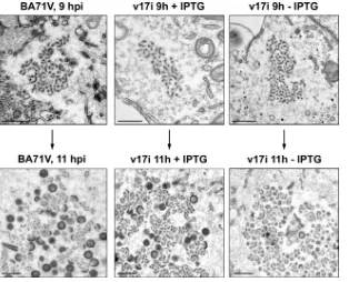
[image:10.585.79.504.415.677.2]strictive conditions, morphogenesis appears to be blocked at the precursor membrane stage by the repression of protein p17 expression. To overcome this problem, we looked for the time, early during ASFV morphogenesis, when the viral assembly had not yet progressed past the viral precursor membrane stage and compared the structures of the viral precursor mem-branes found in the factories of cells infected with BA71V and v17i under permissive or restrictive conditions. As we can see in Fig. 9, at 9 hpi the viral factories in cells infected with BA71V or v17i under restrictive or permissive conditions con-tain only viral precursor membranes that are virtually indistin-guishable by this technique. However, 2 h later the majority of the factories in cells infected with BA71V or with the recom-binant virus under permissive conditions have progressed past this morphogenetic stage, and icosahedral intermediates and immature particles are being actively produced, whereas in cells infected with v17i and maintained for 11 h under restric-tive conditions, morphogenesis appears to be stopped at the level of viral precursor membranes. This result strongly sug-gests that the membranes found at later times after infection in cells infected with the virus v17i under restrictive conditions correspond to bona fide viral membrane precursors.
In summary, electron microscopy of v17i-infected cells indi-cates that the repression of the expression of protein p17 prevents the progression of viral morphogenesis from the pre-cursor membrane stage at the virus factories. Immunoelectron microscopy and immunofluorescence results indicate that the absence of p17 affects the interaction of these precursor mem-branes with capsid and core shell proteins, which delocalize
partially outside the viral factory, where the precursor polypro-teins assemble into zipper-like structures. It is interesting to note that these zipper-like structures show clear luminal spaces, as occurs with those produced when the expression of protein p54 is repressed, which are also found outside the viral factory (59). The repression of the expression of protein p54 blocks the appearance of viral precursor membranes in the viral factory and, similarly, results in the delocalization of the capsid and core shell proteins (59). Zipper-like structures with uncollapsed cisternae are also produced when the core shell polyproteins are cotransfected outside the context of the viral infection (5). This suggests that the formation outside the viral factory of zipper-like structures with uncollapsed cisternae could be induced by the delocalization of core shell polypro-teins. In contrast, the repression of the expression of proteins p72 and pB602L (5, 26, 31), which are involved in the con-struction of the viral capsid, provokes the appearance of the zipper-like structures without luminal spaces inside the viral factory.
Serial section analyses show the presence of large helicoidal
structures in the viral factory.The fact that the absence of
[image:11.585.136.450.67.321.2]protein p17 blocks morphogenesis at the stage of viral precur-sor membranes gives us the opportunity of studying in more detail the structure of these precursors. For this, we analyzed ultrathin serial sections, approximately 80 nm thick, from cells infected with BA71V or v17i and maintained for 12 hpi under permissive or restrictive conditions. We took advantage of the fact that the viral factories which originated from individual incoming particles take a longer time in COS cells than in Vero FIG. 9. Electron microscopy of viral factories at early times of infection. COS cells infected with BA71V or with v17i in the presence or absence of 2.5 mM IPTG were fixed at 9 or 11 hpi and processed for conventional Epon embedding. At 9 hpi, the majority of the viral factories detected are very small and contain exclusively viral membrane precursors surrounded by an electrolucent area. Note that under the three conditions the viral factories are almost indistinguishable. At 11 hpi, the majority of the viral factories are considerably larger and, in the cells infected with BA71V or with the recombinant virus under permissive conditions, show clear evidence of normal viral assembly, like immature particles and icosahedral intermediates. In contrast, under restrictive conditions, no evidence of assembly can be detected. Bar, 500 nm.
on November 8, 2019 by guest
http://jvi.asm.org/
cells to coalesce into a single perinuclear factory (our unpub-lished observations). Thus, in COS cells at 12 hpi it is possible to analyze small factories that contain a reduced number of structures, which facilitates the interpretation of the micro-graphs. Figure 10 shows three correlative sections from small factories of cells infected with BA71V (Fig. 10A) or with v17i under permissive (Fig. 10B) or restrictive (Fig. 10C) condi-tions. The sections have been positioned using as markers cellular structures and viral particles that expanded most of the serial sections. As expected, in factories from cells infected with the parental BA71V virus or with the inducible virus under permissive conditions, viral particles were contained in at least two consecutive sections (Fig. 10A, arrowheads). How-ever, when we analyzed similar serial sections of factories from cells infected with v17i and maintained for 12 hpi under re-strictive conditions (Fig. 10C), we were unable to detect any evident continuity between the viral membrane precursors in the serial sections, which suggests that these precursors are smaller than 80 nm in length or, more likely, that they are so convoluted that it is not possible using this technique to iden-tify any continuity among them.
Unexpectedly, during the analysis of the serial sections of the factories of cells infected with BA71V or with v17i and maintained for 12 h under permissive conditions, we frequently detected the presence of several patterns of electron density that displayed a constant length and width between sections. These patterns were formed by the repetition of elements separated by a constant distance and appeared in almost iden-tical relative positions in contiguous serial sections, which in-dicates that they correspond to correlative sections of the same structure. Several of these patterns can be identified in Fig. 10 (arrows in panels A and B). When these structures are sec-tioned longitudinally, they are completely contained within three serial sections and produce three different and very char-acteristic patterns (Fig. 10A and B, dashed blue boxes). The electron-dense outlines for the patterns boxed in panels A and B have been enlarged and colored in panels D and E, respec-tively. When the colored patterns are superposed (Fig. 10D1 and E1), we can observe that they share identical periodicities. From these superposed images, we can infer that these con-tours correspond to two-dimensional projections of three sec-tions of a two-strand helicoidal structure. This helicoidal na-ture becomes even more evident when the components of each strand in the different sections are identically colored (Fig. 10D2 and E2).
Interestingly, we observed several cases in which a single immature viral particle seemed to emerge from the tip of a helicoidal structure. Close inspection revealed the presence of clear continuities between the emerging immature viral parti-cles and the helicoidal structures (Fig. 10F), suggesting a role for these peculiar structures as precursors of immature viral particles during ASFV assembly in COS cells. Thus, in several sections we can observe that the central rod of the helicoidal precursor is continuous with the core of the viral particle (Fig. 10F, images B to F), whereas the two helicoidal strands appear to be continuous with the inner envelope (Fig. 10F, images F and G), although this is more difficult to observe due to the twisted nature of the membrane strands in the helicoidal pre-cursors. These continuities suggest that the helicoidal precur-sors are formed by a long ribbon of viral membrane precursor,
wrapped around a rod of core material following an overall helicoidal shape, that does not completely enclose the core material (Fig. 10D and E, images 2). Importantly, the mem-brane profiles that outline these structures appear to be con-tinuous, or at least there is no evidence of discontinuities at this resolution. The ends of the viral membrane precursor ribbon are located at the side opposite that from where viral particles emerge. A possible sequence of the events occurring during this process is shown in Fig. 10F.
We have proposed that the interaction of core material with membrane precursors and its subsequent encapsidation begin with the insertion of the myristoyl moiety of polyprotein pp220 into the membrane precursors, since the blockage of the ex-pression of this polyprotein results in the accumulation of coreless particles and the mutation of the myristoylation se-quence of polyprotein pp220 eliminates the ability of this polyprotein to interact with membranes (9). This suggests that the main driving interaction in the construction of the helicoi-dal precursors could be the interaction of polyprotein pp220 with the ribbon of membrane precursors. In agreement with this, when the expression of polyprotein pp220 is blocked dur-ing infection of COS cells with the recombinant virus v220i (9) under restrictive conditions, no helicoidal precursors can be detected (J. M. Rodríguez, C. Sua´rez, and M. L. Salas, unpub-lished observations).
DISCUSSION
The objective of the present work is the characterization of the role of protein p17, a small, abundant, integral membrane protein that localizes at the inner envelope of the viral particle (18, 60, 65), during ASFV morphogenesis. We have shown that p17 has an intrinsic affinity for the ER that it shares with the proteins that localize at this structural domain (59) or that transiently interact with viral membrane precursors (1). A sim-ilar behavior has been described for the VV membrane pro-teins that localize at the membrane of the IV (20), and this is probably related to the necessity of these proteins to integrate early in assembly into ER-derived membranes that have not been completely modified by the viral machinery.
Using a conditional lethal mutant of ASFV with an IPTG-inducible copy of gene D117L, we show that protein p17 is essential for viral replication. The repression of the expression of this protein does not appear to have a significant impact on the synthesis of viral structural proteins like p72, p54, or the polyprotein pp220, nor does it alter the size or location of the viral factory. These results indicate that DNA replication and late gene expression are not significantly affected under restric-tive conditions. However, the proteolytic processing of ASFV polyproteins pp220 and pp62 is essentially blocked, which is indicative of a profound defect in viral morphogenesis, since the processing of ASFV polyproteins is coupled with viral assembly (5).
Immunofluorescence experiments show that while protein p54, which essentially labels viral membrane precursors, is correctly localized at the viral factory under restrictive condi-tions, there is an abnormal distribution of the proteins belong-ing to the capsid and the core shell of the viral particle. Thus, although antibodies against the capsid and core shell proteins label the viral factory, the characteristic cytoplasmic punctate
VOL. 84, 2010 ROLE OF ASFV PROTEIN p17 7495
on November 8, 2019 by guest
http://jvi.asm.org/
7496
pattern, indicative of intracellular particles, is not detected with antibodies against the capsid protein p72, and large ag-gregates containing polyprotein pp220 are observed in the cy-toplasm of the infected cell. In agreement with the immuno-fluorescence results, when observed by transmission electron microscopy, the viral factory of cells infected by v17i and main-tained for 12 h under restrictive conditions contains only small, rounded membranous structures, with no evidence of helicoi-dal or icosahedral intermediates of assembly. At 24 hpi, the viral factory has increased considerably in size by the accumu-lation of these membranes, and we can only occasionally detect helicoidal or icosahedral intermediates, which are probably the result of leakage from the repression system. In the cytoplasm of the infected cell, long zipper-like structures tightly bound to ER cisternae, whose luminal spaces are clearly detectable, accumulate at the periphery of the viral factory.
To ascertain that the heterogeneous small membranes found in cells infected with the recombinant virus under restrictive conditions are normal viral precursor membranes, we studied by transmission electron microscopy the structures of the viral factories of cells infected with BA71V and v17i under permis-sive and restrictive conditions early during ASFV morphogen-esis. At 9 hpi, the viral assembly had not yet progressed past the viral precursor membrane stage, and we observed that the precursor membranes found at the different factories are struc-turally indistinguishable by this technique. Our interpretation of these results is that protein p17 is not required for the recruitment and modification of the ER-derived membranes that are thought to be the origin of the viral membrane pre-cursors. The continuous accumulation of viral precursor mem-branes at the viral factory suggests that these processes are completely independent of the presence of this protein. How-ever, the absence of protein p17 in these precursors affects the formation of helicoidal precursors and hampers the progres-sion of the assembly toward icosahedral intermediates.
These results indicate that the effect of the absence of pro-tein p17 from these precursors manifests itself in two indepen-dent ways, since this absence affects the construction of the capsid and the interaction of polyprotein pp220 with the pre-cursors of the inner envelope, which are two independent processes (9). This could be due to a direct effect, i.e., protein p17 could participate in both processes, or the result of an indirect effect, i.e., the absence of p17 could alter the structure of the viral precursors or prevent the incorporation of some component of these precursors which ultimately results in these two distinct effects.
The unexpected discovery of helicoidal structures in the
factory of COS cells infected by ASFV and their abundance during infections by the parental and recombinant v17i viruses under permissive conditions prompted us to perform a prelim-inary characterization of the role of these structures in the context of the morphogenesis of ASFV to obtain a basic frame-work for the understanding of the results obtained in the anal-ysis of the inducible virus for protein p17.
Interestingly, immature viral particles could be observed emerging from one of the ends of these structures, indicating that helicoidal structures are precursors of immature viral par-ticles. The continuities observed between the components of the helicoidal structures and the immature particles provided key information about their composition. Helicoidal precur-sors appeared to be formed by a long ribbon of inner viral membrane precursor arranged around a rod of core-like ma-terial following a helicoidal pathway, resulting in a continuous two-strand helicoidal structure. To fully appreciate the abun-dance of these precursors in the viral factory, it is important to note that we have focused our attention in this work on lon-gitudinal or nearly lonlon-gitudinal sections of these precursors, because as we have seen, these types of sections produce very characteristic patterns, easy to identify unequivocally. How-ever, since helicoidal structures are oblong, pure probability indicates that longitudinal or nearly longitudinal sections are a minority among all possible sections of an oblong object, re-sulting in an underestimation of the abundance of these pre-cursors in the viral factory. Taking this into account and given the observed abundance of longitudinal sections of helicoidal precursors, this type of structure appears to be the main pre-cursor structure found in the early viral factory in COS cells. Thus, although we do not know whether the immature viral particles derived from helicoidal precursors are able to mature into infective extracellular virus, the fact that both the kinetics of virus production and the final virus titer are as expected for ASFV (69; this work) strongly indicates that the helicoidal precursors are a viable morphogenetic pathway that results in the production of infective virus.
Helicoidal precursors have not previously been described during ASFV morphogenesis. One possibility is that they are artifactual structures due to our sample manipulation or are somehow specific to BA71V infection of COS cells, a cell line that until recently has not been widely used to study and propagate ASFV. To discard these possibilities, we have ex-amined the literature, looking for patterns that characterize helicoidal structures in previous work illustrating ASFV infec-tions at the transmission electron microscope. We have iden-tified several examples published by different laboratories,
FIG. 10. Serial section analysis of v17i-infected cells. COS cells infected with BA71V (A) or with v17i in the presence (B) or absence (C) of IPTG (2.5 mM) were fixed at 12 hpi and processed for conventional Epon embedding. Serial sections approximately 80 nm thick were analyzed. Three consecutive sections of small factories are shown for each condition (numbered 1, 2, and 3). In the case of cells infected with BA71V and v17i under permissive conditions, viral particles (arrowheads) and larger precursor structures (arrows) expand several correlative sections. In each case, one of these structures and an apparently associated viral particle, both boxed in each serial section, are analyzed in more detail in panels D (BA71V) and E (v17i⫹IPTG). In these panels, each of the boxed details is shown slightly enlarged, and at the right, the electron density profiles of the structure and the viral particle in the different sections are filled with different colors. Enlarged versions of the colored profiles are superimposed in panels D1 and E1 to show how the different sections fit together. In panels D2 and E2, the components of each strand in the different sections are identically colored. A possible sequence of events for the generation of immature particles from the helicoidal structures is shown in panel F. Note that in panel C the precursor membranes that accumulate during infection with the inducible virus under restrictive conditions do not display any clear continuity between consecutive sections. Bars, 500 nm (A to C) and 100 nm (D to F).
VOL. 84, 2010 ROLE OF ASFV PROTEIN p17 7497
on November 8, 2019 by guest
http://jvi.asm.org/
isolates reinforces the role of this type of structure during ASFV morphogenesis.
There are probably several reasons why these helicoidal structures have not been described previously. First, it is diffi-cult to deduce the existence of a three-dimensional object from a single two-dimensional section, and there are very few ex-amples in the literature of serial sections of factories of cells infected by ASFV. Second, very few studies of the morpho-genesis of ASFV have analyzed the early stages of assembly, when it is easier to detect these structures. Finally, the majority of the studies of ASFV morphogenesis have been done in Vero cells, where this type of structure can easily be detected only early in the assembly (73; our unpublished observations).
The data presented here suggest that the helicoidal precur-sors constitute an intermediate step of morphogenesis between the construction of the membrane precursors and the produc-tion of icosahedral intermediates, since the continuities ob-served with immature particles indicate that they are formed by the interaction of viral membrane precursors with core material and that icosahedral intermediates are formed by the assembly of the viral capsid on the external face of the heli-coidal precursors. Nevertheless, to ascertain the structure and role of helicoidal precursors during virus assembly, it is essen-tial to gather further experimental evidence, especially by using electron tomography and other techniques better suited than conventional electron microscopy for the resolution of three-dimensional structures.
In this work, we have shown that protein p17 has a critical role in the morphogenesis of ASFV. Its repression hinders the interactions of capsid proteins with viral membrane precursors, preventing the appearance of icosahedral intermediates, but also the interaction of core shell proteins with inner envelope precursors, precluding the construction of helicoidal precur-sors. The simultaneous blockage of the interactions occurring at both sides of the inner envelope results in the accumulation of membrane precursors at the viral factory.
ACKNOWLEDGMENTS
We thank E. M. Forero, M. Guerra, and M. Rejas for technical assistance.
This work was supported by grants from the Wellcome Trust (075813/C/04/Z) and the Spanish Ministerio de Ciencia e Innovacio´n (BFU2007-61647) and by an institutional grant from Fundacio´n Ramo´n Areces. J. M. Rodríguez was supported by the Ramo´n y Cajal program of the Ministerio de Ciencia e Innovacio´n.
REFERENCES
1.Alejo, A., G. Andre´s, and M. L. Salas.1999. The African swine fever virus prenyltransferase is an integral membrane trans-geranylgeranyl-diphosphate synthase. J. Biol. Chem.276:18033–18039.
2.Alejo, A., G. Andre´s, and M. L. Salas.2003. African swine fever virus
African swine fever virus is enveloped by a two-membraned collapsed cis-terna derived from the endoplasmic reticulum. J. Virol.72:8988–9001. 8.Andre´s, G., R. García-Escudero, E. Vin˜uela, M. L. Salas, and J. M.
Rodríguez.2001. African swine fever virus structural protein pE120R is essential for virus transport from the assembly sites to the plasma membrane but not for infectivity. J. Virol.75:6758–6768.
9.Andre´s, G., R. García-Escudero, M. L. Salas, and J. M. Rodríguez.2002. Repression of African swine fever virus polyprotein pp220-encoding gene leads to the assembly of icosahedral core-less particles. J. Virol.76:2654– 2666.
10.Bisht, H., A. S. Weisberg, P. Szajner, and B. Moss.2009. Assembly and disassembly of the capsid-like external scaffold of immature virions during vaccinia virus morphogenesis. J. Virol.83:9140–9150.
11.Breese, S. S., Jr., and C. J. DeBoer.1966. Electron microscope observation of African swine fever virus in tissue culture cells. Virology28:420–428. 12.Brookes, S. M., A. D. Hyatt, T. Wise, and R. M. E. Parkhouse.1998.
Intra-cellular virus DNA distribution and the acquisition of the nucleoprotein core during African swine fever virus particle assembly: ultrastructural in situ hybridisation and DNase-gold labelling. Virology249:175–188.
13.Brookes, S. M., L. K. Dixon, and R. M. E. Parkhouse.1996. Assembly of African swine fever virus: quantitative ultrastructural analysis in vitro and in vivo. Virology224:84–92.
14.Brookes, S. M., H. Sun, L. K. Dixon, and R. M. E. Parkhouse.1998. Char-acterization of African swine fever virion proteins j5R and j13L: immuno-localization in virus particles and assembly sites. J. Gen. Virol.75:1179–1188. 15.Carrascosa, J. L., J. M. Carazo, A. L. Carrascosa, N. García, A. Santisteban, and E. Vin˜uela. 1984. General morphology and capsid fine structure of African swine fever virus particles. Virology132:160–172.
16.Chapman, D. G., V. Tcherepanov, C. Upton, and L. K. Dixon.2008. Com-parison of the genome sequences of non-pathogenic and pathogenic African swine fever virus isolates. J. Gen. Virol.89:397–408.
17.Chlanda, P., M. A. Carbajal, M. Cyrklaff, G. Griffiths, and J. Krijnse-Locker.2009. Membrane rupture generates single open membrane sheets during vaccinia virus assembly. Cell Host Microbe6:81–90.
18.Cobbold, C., J. T. Whittle, and T. Wileman.1996. Involvement of the endo-plasmic reticulum in the assembly and envelopment of African swine fever virus. J. Virol.70:8382–8389.
19.Cobbold, C., S. M. Brookes, and T. Wileman.2000. Biochemical require-ments of virus wrapping by the endoplasmic reticulum: involvement of ATP and endoplasmic reticulum calcium store during envelopment of African swine fever virus. J. Virol.74:2151–2160.
20.Condit, R., N. Moussatche, and P. Traktman.2006. In a nutshell: structure and assembly of the vaccinia virion. Adv. Virus Res.66:31–124.
21.Dales, S.1963. The uptake and development of vaccinia virus in strain L cells followed with labeled viral deoxyribonucleic acid. J. Cell Biol.18:51–72. 22.Dales, S., and E. H. Mosbach.1968. Vaccinia as a model for membrane
biogenesis. Virology35:564–583.
23.Dales, S., and L. Siminovitch.1961. The development of vaccinia virus in Earle’s L strain cells as examined by electron microscopy. J. Biophys. Bio-chem. Cytol.10:475–503.
24.Dixon, L. K., J. M. Escribano, C. Martins, D. L. Rock, M. L. Salas, and P. J. Wilkinson.2005. The Asfarviridae, p. 135–143.InC. M. Fauquet, M. A. Mayo, J. Maniloff, U. Desselberger, and L. A. Ball (ed.), Virus taxonomy. Eighth report of the International Committee on Taxonomy of Viruses. Elsevier Academic Press, London, United Kingdom.
25.Enjuanes, L., A. L. Carrascosa, M. A. Moreno, and E. Vin˜uela.1976. Titra-tion of African swine fever virus. J. Gen. Virol.32:471–477.
26.Epifano, C., J. Krijnse-Locker, M. L. Salas, J. M. Rodríguez, and J. Salas. 2006. The African swine fever virus nonstructural protein pB602L is required for formation of the icosahedral capsid of the virus particle. J. Virol.80: 12260–12270.
27.Epifano, C., J. Krijnse-Locker, M. L. Salas, J. Salas, and J. M. Rodríguez. 2006. Generation of filamentous instead of icosahedral particles by repres-sion of African swine fever virus structural protein pB438L. J. Virol.80: 11456–11466.
28.Essani, K., R. Dugre, and S. Dales.1982. Biogenesis of vaccinia: involvement