0022-538X/10/$12.00 doi:10.1128/JVI.01666-09
Copyright © 2010, American Society for Microbiology. All Rights Reserved.
Testing the Balanced Electrostatic Interaction Hypothesis of
Hepatitis B Virus DNA Synthesis by Using an
In Vivo
Charge Rebalance Approach
䌤
Pong Kian Chua,
1Fan-Mei Tang,
2Jyuan-Yuan Huang,
2Ching-Shu Suen,
2and Chiaho Shih
1,2*
Institute for Human Infections and Immunology, Department of Pathology, and Department of Microbiology and Immunology,
University of Texas Medical Branch, Galveston, Texas 77555-0609,1and Institute of Biomedical Sciences, Academia Sinica,
Taipei, Taiwan 1152
Received 9 August 2009/Accepted 7 December 2009
Previously, a charge balance hypothesis was proposed to explain hepatitis B virus (HBV) capsid stability, assembly, RNA encapsidation, and DNA replication. This hypothesis emphasized the importance of a balanced electrostatic interaction between the positive charge from the arginine-rich domain (ARD) of the core protein (HBc) and the negative charge from the encapsidated nucleic acid. It remains unclear if any of the negative charge involved in this electrostatic interaction could come from the HBc proteinper se, in addition to the encapsidated nucleic acid. HBc ARD IV mutant 173GG and ARD II mutant 173RR/R157A/R158A are arginine deficient and replication defective. Not surprisingly, the replication defect of ARD IV mutant 173GG can be rescued by restoring positively charged amino acids at the adjacent positions 174 and 175. However, most interestingly, it can be at least partially rescued by reducing negatively charged residues in the assembly domain, such as by glutamic acid-to-alanine (E-to-A) substitutions at position 46 or 117 and to a much lesser extent at position 113. Similar results were obtained for ARD II mutant 173RR/R157A/R158A. These amino acids are located on the inner surfaces of HBc icosahedral particles, and their acidic side chains point toward the capsid interior. For HBV DNA synthesis, the relative amount of positive versus negative charge in the electrostatic interactions is more important than the absolute amount of positive or negative charge. These results support the concept that balanced electrostatic interaction is important during the viral life cycle.
Human hepatitis B virus (HBV) is an important human pathogen (5, 27, 34) that can replicate via an RNA intermedi-ate (31, 33). Wild-type (WT) HBV core protein (HBc) is 183 amino acids long (adr andayw subtypes) and consists of two distinct domains connected by a hinge region. The assembly domain spans amino acids 1 to 140, and the arginine-rich domain (ARD) spans amino acids 150 to 183. The ARD of HBc 150-183 is not required for capsid assembly inEscherichia coli(4, 8, 10, 21, 35). During nucleocapsid (capsid) formation, the HBc protein assembles into an icosahedral particle via a dimer intermediate (32). The ARD of HBc is known to be capable of binding to nucleic acids (12, 25). Serine phosphor-ylation at the C terminus of HBc is known to be important for RNA encapsidation, DNA synthesis, and virion secretion (2, 11, 14, 15, 17, 19, 24, 26, 39, 40). To date, there is no structural information available for the C terminus of HBc in capsids (32). The 4-helix bundle structure of HBc capsids is based on a C-terminally truncated capsid protein, HBc149 (6, 7, 36). Our research progress in the study of HBV biology has been hampered due to the lack of structural information about the HBc C-terminal tail, which plays an important regulatory role throughout the life cycle of HBV.
Recently, we proposed a hypothesis that “charge balance” could be important for HBV capsid stability, assembly, RNA encapsidation, and DNA replication (17). This hypothesis
pos-tulates that many important viral activities could be influenced by the electrostatic interaction between the positive charge of the basic amino acid-rich domains of a nucleocapsid protein and the negative charge of the nucleocapsid-associated nucleic acids (17). Previously, we and others demonstrated that a mu-tant HBc 164, which lacks a total of eight arginine residues at the C terminus, can package both the nonspliced 3.5-kb pre-genomic RNA (pgRNA) and the 2.2-kb spliced RNA (14, 17). However, pgRNA encapsidated by HBc 164 is RNase sensitive, while the encapsidated 2.2-kb spliced RNA is RNase resistant. The pgRNA and spliced RNA encapsidated by the full-length wild-type HBc 183 is also RNase resistant (14, 17). This result provided us with the first clue to invoke the previously coined “charge balance hypothesis” of RNA encapsidation and capsid stability (17). In subsequent studies, we added arginines back to the truncated HBc 164 gradually and noted that core par-ticle-associated viral DNA also increased gradually in both size and intensity. This result provided us with the second clue to expand the previous hypothesis from RNA encapsidation to include viral DNA synthesis (17). Instead of totally nonquan-titative or nonspecific binding between nucleic acids and a basic protein, our electrostatic-interaction hypothesis entails a stoichiometry-like concept between basic residues and their binding partner of nucleic acids. One of the reasons for such a demand for a more quantitative charge-charge interaction could be related to the putative intersubunit positive-charge repulsion built into the context of an icosahedral particle (22). We and others demonstrated previously that truncated HBc 173 (also denoted HBc 173RR in Fig. 1) is sufficient for a WT-like DNA phenotype (17, 20). Mutant 173GG, containing * Corresponding author. Mailing address: Institute of Biomedical
Sciences, Academia Sinica, Taipei, Taiwan 115. Phone: 8862-2652-3996. Fax: 8862-2652-3597. E-mail: cshih@ibms.sinica.edu.tw.
䌤Published ahead of print on 16 December 2009.
2340
on November 8, 2019 by guest
http://jvi.asm.org/
two substitutions from arginine (R) to glycine (G) at codons 172 and 173 of HBc 173, exhibited a shorter-than-full-length HBV DNA phenotype, suggesting the importance of sufficient positive charge for viral DNA synthesis (17).
To test further the effect of electrostatic interaction on viral DNA synthesis, we asked if one could restore the DNA repli-cation defect of the arginine-deficient ARD IV mutant 173GG or ARD II mutant 173RR/R157A/R158A by reducing their negative charges at specific positions. Indeed, our results showed that a single substitution from glutamic acid to alanine (E-to-A) at position 46 or 117, and to a lesser extent at position 113, could restore the WT-like DNA replication in the context of ARD IV mutant 173GG. Similarly, in the context of ARD II mutant 173RR/R157A/R158A, both E46A and E117A could
rescue the full-length single-stranded DNA (ssDNA) and re-laxed-circle (RC) DNA syntheses.
Using anin vitrocapsid assembly/disassembly assay, we dem-onstrated that the capsid stability of both wild-type full-length HBc 183 and truncated HBc 173 capsid particles fromE. coli
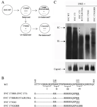
[image:2.585.131.446.68.413.2]are dependent on the presence of encapsidated RNA (22). Upon treatment with micrococcal nuclease, encapsidated RNAs were digested and lost, leading to capsid disassembly. In this study, we demonstrated that mutant 173GG maintained capsid stability upon the loss of encapsidated RNA, probably due to the loss of intersubunit positive charge repulsion at ARD IV. Interestingly, when mutation E117A was introduced into the context of mutant 173GG, the capsid stability of mu-tant capsids E117A/173GG was once again dependent on the FIG. 1. Arginine-deficient HBc mutants SVC173GG and SVC173/R157A/R158A exhibited a short DNA phenotype by complementation and Southern blot analysis. (A) To test the balanced electrostatic-interaction hypothesis, we engineered various HBc mutants with different arginine contents. The experimental approach is illustrated in the cartoon. (B) Amino acid sequence comparisons among the WT and HBc mutants. The hyphens represent amino acid sequences identical to that of the parental WT HBc. Roman numbers I to IV indicate the four different ARDs at the C terminus of HBc. The names SVC 173RR and SVC173 are used interchangeably in this paper. ARD IV mutant 173GG contains two arginine-to-glycine substitutions at positions 172 and 173, while ARD II mutant SVC173/R157A/R158A contains two arginine-to-alanine substi-tutions at positions 157 and 158. To further test the balanced electrostatic-interaction hypothesis, we restored arginines at the new positions 174 and 175 in mutant 175GGRR. (C, top) Plasmid 1903, an HBV genomic dimer containing an ablated core AUG initiation codon, was cotransfected with WT or various mutant core expression plasmids into Huh7 cells. HBV core-associated DNAs were analyzed by Southern blot analysis. The positive control mutant SVC173RR was WT-like in viral DNA synthesis (17, 20). More mature RC DNA of HBV was almost undetectable in mutants 173GG and 173/R157A/R158A, even after very long exposure to X-ray film. The replication defect of mutant 173GG could be rescued in mutant 175GGRR. The asterisk highlights the defect in synthesizing full-length viral DNA. In contrast, the black dots in lane SVC175GGRR highlight the functional rescue of full-length HBV DNA synthesis. SS, ssDNA replicative intermediate. (Bottom) Capsids collected from transfected cell lysates were measured by Western blot analysis using rabbit anti-core antibody.
on November 8, 2019 by guest
http://jvi.asm.org/
encapsidated RNA in a manner similar to that of its parental mutant, 173. Further studies also revealed that the truncated HBc mutant 172, like mutant 173, is sufficient for HBV DNA replication. Taken together, these results lend strong support to the balanced electrostatic-interaction hypothesis of HBV DNA replication.
MATERIALS AND METHODS
Plasmids.The core expression vectors SVC WT, SVC 169, SVC 171, SVC 173GG, SVC 173RR, and SVC 173RR/R157A/R158A were used as templates for mutagenesis (17, 22). The truncated core mutant 172 was made by introduc-ing a stop codon at amino acid 173 in SVC WT. Mutations E40A, E40R, E46A, E46R, E113A, E117A, E46A/E113A, E46A/E117A, E113A/E117A, E46A/ E113A/E117A, and R157A/R158A were introduced by site-directed mutagene-sis. The sequences of mutagenesis primers are not shown here. All mutants were confirmed by sequencing. Plasmid 1903 contains a core AUG knockout mutation in the context of the pWT genomic dimer (a replicon) and is unable to synthesize HBc (41).
Transfection, core particle isolation, core-associated HBV DNA extraction, and Southern blotting.Wild-type and mutant core expression plasmids were cotransfected with plasmid 1903 into Huh7 cells using Genejuice (EMD Bio-sciences) and following the manufacturer’s protocol. Intracellular core particles were isolated as previously described (41). Core-associated HBV DNAs were prepared by protease treatment of isolated core particles, followed by extraction with phenol-chloroform-isoamyl alcohol and precipitated using ethanol. Core-associated HBV DNA from transfected cells was subjected to Southern blotting as previously described using a vector-free HBV DNA probe.
Native agarose gel and Western blotting of core particles.Nuclear pellets were spun down from cells treated with lysis buffer (0.25% NP-40, 10 mM Tris, 1 mM EDTA, 50 mM NaCl, 8% sucrose) 5 days posttransfection. The supernatant was transferred into another tube. Ten microliters of sample (out of 100 microliters) was mixed with loading dye (40% sucrose, 0.25% bromophenol blue) and elec-trophoresed through a 1% agarose gel in 1⫻TBE buffer (89 mM Tris-borate, 2 mM EDTA, pH 8.0) for 1 h at 80 V. Core particles were then transferred by capillary action onto nitrocellulose membranes overnight in 2⫻SSC (0.3 M sodium chloride, 30 mM sodium citrate). The nitrocellulose membrane was blocked with 5% skim milk for 1 h, followed by incubation with rabbit anti-core polyclonal antibody (3) for 1 h. The membrane was then washed three times with phosphate-buffered saline (PBS) for 10 min each time and incubated in goat anti-rabbit horseradish peroxidase-conjugated antibody for 1 h. The membrane was again washed three times with PBS, and the proteins were visualized using enhanced chemiluminescence (Amersham Co.).
Mutant capsids in E. coli.HBc mutants 173GG, 173GG/E46A, 173GG/ E113A, and 173GG/E117A were created with a Site-Directed Mutagenesis Kit (Stratagene Co.), using the full-length wild-type (subtypeadr) plasmid pET-HBc183 as a template. The sequences of the mutagenesis primers are not shown here. All mutants were confirmed by DNA sequencing.
Capsid disassembly by micrococcal nuclease treatment.Approximately 0.25
g of purified capsids (2l of a 0.125-mg/ml stock in Tris-buffered saline [TBS] [0.1 M NaCl, 2 mM KCl, 25 mM Tris {pH 7.4}]) was diluted in 16l of distilled H2O (dH2O) before incubation overnight at 37°C with micrococcal nuclease at
1 agarose gel unit per 1 nanogram capsid protein (Roche Applied Science Co. or New England BioLabs) in the presence of 8 mM CaCl2and 5 mM dithiothreitol
(DTT). The extent of micrococcal nuclease digestion was evaluated on 1% native agarose gels sequentially stained with SYBR green II for encapsidated nucleic acids and with Sypro Ruby for capsid proteins (22). The images were scanned with a Typhoon 9410 ModeImager (Amersham BioScience, Piscataway, NJ), and quantitation of the intensity of the images was done with ImageJ (National Institutes of Health).
RESULTS
Arginine-deficient and replication-defective core mutants 173GG and 173RR/R157A/R158A.Previously, we and others demonstrated that C-terminally truncated HBc mutant 173 (also named 173RR here) exhibited the same DNA replica-tion phenotype as the wild-type HBV with full-length HBc 183 (17, 20). Therefore, the last 10 amino acids of HBc (HBc 174-183) are not essential to HBV replication in tissue
cul-ture. Here, we chose 173RR as a backbone plasmid to create derivatives to test our balanced electrostatic-interaction hy-pothesis in this study. Mutant 173GG contains arginine-to-glycine substitutions at positions 172 and 173 and exhibits a shorter-than-full-length DNA phenotype by Southern blot analysis (Fig. 1).
Unlike some of the previously reported duck hepatitis B virus (DHBV) class II core mutants (38), this shorter DNA phenotype is not due to digestion of core-associated HBV DNA by exogenous nuclease treatment during the DNA prep-aration procedure (13). The same shorter DNA phenotype as in mutant 173GG was observed when viral DNAs were pre-pared with or without exogenous nuclease treatment (refer-ence 17 and data not shown). By both Northern blot and primer extension analyses, mutant 173GG could encapsidate full-length pgRNA at an efficiency similar to or often higher than that of WT HBV (17). Therefore, the shorter DNA phe-notype of mutant 173GG appears to originate from a defect in DNA synthesis.
Rationale 1. To test the balanced electrostatic-interaction hypothesis of HBV DNA synthesis, we asked if one could induce a shorter DNA phenotype similar to that of mutant 173GG by removing arginines elsewhere within the arginine-rich domain of HBc. In addition, we asked if the replication defect of mutant 173GG could be restored by adding back positive charges at ectopic positions (other than at amino acid 172 and 173) within the C terminus of HBc.
Charge effect from the C terminus of HBc on viral DNA synthesis.As shown in Fig. 1A, we tested if R-157 and R-158 within the second stretch of ARD II are important for viral DNA synthesis. Furthermore, we asked if R-174 and R-175 in mutant 175GGRR could rescue the replication defect of mutant 173GG (Fig. 1A and B). Previously, we constructed the mutant HBV genome 1903, which is replication defec-tive due to the ablated AUG initiation codon of HBc at nucleotide 1903. As shown in Fig. 1C, the genomic dimer of mutant 1903 was cotransfected into Huh7 cells with HBc expression vectors, such as SVC WT, SVC173, and SVC175GGRR. In these three cotransfections, Southern blot analysis of core-associated DNA revealed full-length HBV DNA synthesis. In contrast, cotransfection with either mutant SVC 173GG or mutant SVC173/R157A/R158A (Fig. 1B) resulted in a shorter-than-full-length DNA genome (Fig. 1C). ARD III mutant SVC173/R165A/R166A has a DNA phenotype similar to that of mutant SVC173/R157A/ R158A (data not shown). While mutant SVC173/R157A/ R158A exhibited a reduced steady-state level of capsids, mutant 173GG appeared to have abundant capsids in the transfected cell lysate by Western blotting (Fig. 1C, bottom). Therefore, the short DNA phenotype is not correlated with any apparent capsid instability, which in turn affects viral DNA synthesis. Furthermore, R-157 and R-158 appeared to have stronger effects on capsid stability than R-172 and R-173, while R-174 and R-175 displayed an effect similar to those of R-172 and R-173 on viral DNA synthesis (Fig. 1C). Taken together, these results support the notion that the total amount of positive charges from the C terminus of HBc 173 core protein is important for viral DNA synthesis.
Rationale 2.Encouraged by the results shown in Fig. 1, we next asked if the N-terminal assembly domain (HBc 1-149)
on November 8, 2019 by guest
http://jvi.asm.org/
could also contribute to adequate electrostatic interaction. In our prototype hypothesis, we assumed that negative charges involved in the electrostatic interactions are entirely from the encapsidated RNA or DNA (17, 22). Here, we tested the possibility that part of the negative charges involved in the electrostatic interaction could originate from the N-terminal domain of HBc. If this is indeed the case, then perhaps one can repair “charge imbalance” caused by arginine deficiency (i.e., negative charge in excess) by reducing the negative charges from those acidic amino acids involved in the electrostatic interaction.
Acidic residues 40, 46, 113, and 117.Using the Swiss PDB viewer program to visualize the core dimer structure, we iden-tified 4 amino acids for further studies. Amino acid E40 on helix␣2b has its side chain pointing sideways from the capsid interior, while amino acids E46, between helices␣2b and␣3, and E113 and E117, on helix␣5, have their side chains pointing into the capsid interior (Fig. 2A, top) (6, 7, 36). E40 appeared to be at a position farther away from the capsid interior than E46, E113, and E117 (Fig. 2A, bottom). As shown in Fig. 2B, all four acidic residues are evolutionarily conserved among the
primate hepadnaviruses. Unlike position 113, the other three positions, E40, E46, and E117, are even conserved in rodent hepadnaviruses. Our approach here was to ask if the replica-tion defect of mutant 173GG could be rescued via a second mutation by reducing the negative charges at E40, E46, E113, and E117 (Fig. 2C).
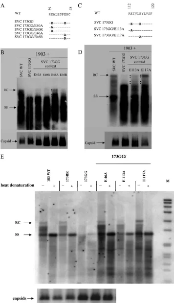
[image:4.585.45.537.65.381.2]To this end, mutations E40A, E40R, E46A, and E46R were individually introduced into the SVC 173GG context via site-directed mutagenesis (Fig. 3A). A complementation experi-ment similar to that shown in Fig. 1 was conducted. As shown in Fig. 3B, mutants 173GG/E40A and 173GG/E40R had the same DNA phenotype as the parental 173GG, indicating that these two mutants did not rescue the shorter DNA phenotype. Most interestingly, mutant 173GG/E46A exhibited a WT-like DNA phenotype with higher molecular weight and full-length RC DNA (Fig. 3B). Although mutant 173GG/E46R seemed to have some HBV-specific signals with increased DNA size, its full-length RC signal was weaker. These results suggest that the side chain of core amino acid E46 could be involved in viral DNA replication by contributing to adequate electrostatic in-teraction. We also observed that all the mutant capsids from FIG. 2. Experimental design of a charge rebalance approach by converting acidic residues at specific positions in the assembly domain of HBc into neutral amino acids. (A, top) Stereo view of the HBV capsid protein dimer (PDB 1QGT) (6, 7, 36). Four residues, E40, E46, E113, and E117, are shown as yellow, pink, green, and cyan sticks, respectively. Unlike that of E40, the side chains of glutamic acids at E46, E113, and E117 point toward the capsid interior. E40 is farther away from the capsid interior than E46, E113, and E117, as is more clearly shown in the illustration of a hexamer (bottom). This dimer structure was created with Discovery Studio 2.0, and the hexamer structure was generated by using Insight II (Accelrys Software Inc.). (B) Sequence alignment of primate and rodent hepadnavirus core protein amino acids 36 to 49 and 110 to 122, with amino acids 40, 46, 113, and 117 in boldface. Acidic residues at 40, 46, and 117 are evolutionarily conserved. (C) To take a new approach to testing the balanced electrostatic-interaction hypothesis, we engineered E-to-A and E-to-R core mutants in the context of an arginine-deficient mutant, 173GG.
on November 8, 2019 by guest
http://jvi.asm.org/
FIG. 3. The replication defect of arginine-deficient core mutant SVC173GG can be rescued by reducing the negative charge in the assembly domain via E46A, E113A, or E117A mutations. (A) Mutations E40A, E40R, E46A, and E46R were introduced into the ARD IV mutant SVC173GG. (B) These E40 and E46 core mutants were tested for the ability to rescue plasmid 1903 by cotransfection and Southern blotting, as described in the legend to Fig. 1C. Unlike mutants E40A, E40R, and E46R, mutant E46A, in the context of 173GG, can result in a WT-like DNA
on November 8, 2019 by guest
http://jvi.asm.org/
cell lysates were stable, as determined by native agarose gel electrophoresis and Western blotting (Fig. 3B, bottom). As a side note, the immunoblot signals of several mutant capsids were stronger than those of wild-type capsids. The negative results of mutations at amino acid 40 served as a good control, indicating that not all acidic residues were involved in the electrostatic interaction and DNA replication. As a control experiment, we introduced mutations E40A, E40R, E46A, and E46R individually into the SVC WT context and detected no significant effect on HBV DNA replication (data not shown). Mutations E113A and E117A were also created in the 173GG context (Fig. 3C), and were cotransfected into Huh7 cells with plasmid 1903. Southern blot analysis of core-associ-ated HBV DNA showed that 173GG/E117A resulted in a WT-like DNA phenotype (Fig. 3D), with a significantly in-creased amount of mature RC form DNA. In contrast, mutant 173GG/E113A exhibited a significantly reduced amount of full-length RC form DNA compared to SVC WT or mutant 173GG/E117A. We also found no significant adverse effect on DNA replication when either the E113A or E117A mutation was placed in the WT core context (reference 16 and data not shown). Mutant capsids from cell lysates were stable by Western blot analysis (Fig. 3D, bottom). To better resolve HBV ssDNA from RC DNA synthesis, we performed heat denaturation of extracted HBV DNA before agarose gel electrophoresis (Fig. 3E). While ARD IV mutant 173GG is highly attenuated in over-all DNA synthesis, it exhibited very weak full-length ssDNA signal and lack of full-length RC DNA. These DNA replication defects could be rescued by either E46A or E117A mutation. Although E113A could efficiently rescue the full-length ssDNA synthesis, it could not rescue full-length RC DNA synthesis as efficiently as E117A or E46A.
Rationale 3. The short DNA phenotype was observed in arginine-deficient mutants created by R-to-A or R-to-G sub-stitutions at ARD II or ARD IV (Fig. 1). Since ARD IV mutant 173GG could be at least partially rescued by an E-to-A mutation at amino acid 46, 113, or 117 (Fig. 3), we asked if ARD II mutant 173/R157A/R158A could also be rescued in a similar manner (Fig. 4).
When the single mutations E46A and E117A were placed in the context of SVC173/R157A/R158A, significant increases in ssDNA and RC DNA synthesis could be detected (Fig. 4A). Again, the rescue effect of E113A appeared to be limited to ssDNA, and the full-length RC DNA signal remained weak. In contrast, the rescue effect by double or triple rescue mutations was not as expected. Combinations of rescue mutations ap-peared to result in more attenuated replication than a single rescue mutation (Fig. 4A). For example, the triple rescue mu-tant E46A/E113A/E117A was always very poor in replication in at least 3 independent experiments. We also noted that the
double rescue mutant E46A/E113A did not exhibit any detect-able full-length RC form DNA in at least 3 independent ex-periments. Similar to the single rescue results in Fig. 3E and 4B, E117A was always the most efficient for the rescue of full-length RC DNA, while E113A was always the least effi-cient for the recovery of full-length RC DNA irrespective of the context of 173GG (Fig. 3E) or 173RR/R157A/R158A (Fig. 4A and B). In general, ARD IV mutant 173GG seemed to have stronger residual replication activity than the ARD II mutant 173RR/R157A/R158A (data not shown).
To better resolve the ssDNA from RC DNA synthesis, we performed heat denaturation experiments. As shown in Fig. 4B, ARD II mutant 173RR/R157A/R158A exhibited reduced replication and the absence of mature RC DNA. Similar to the results in Fig. 3E, both E46A and E117A could efficiently rescue both ssDNA and RC DNA synthesis, while E113A could efficiently rescue only ssDNA synthesis.
Rationale 4.The truncated core mutant SVC 171 has exactly the same number of charged residues as SVC 173GG; how-ever, the former is shorter than the latter by 2 glycine residues at the truncated C terminus. Since mutation E46A can partially restore the DNA replication defect of mutant SVC 173GG (Fig. 3B), we asked if mutation E46A could also rescue the shorter DNA phenotype of mutants SVC 171 and SVC 169 (Fig. 5A).
The length effect of the HBc C terminus on DNA replication in the context of mutant E46A.As shown in Fig. 5B, neither mutant SVC 171/E46A nor SVC 169/E46A could synthesize the full-length mature form of RC DNA. Even though mutant SVC 169/E46A seemed to display weaker DNA replication activity than mutant SVC 171/E46A, we found that SVC 169/ E46A capsids were stable and more abundant than SVC WT by Western blotting of intracellular capsids on native agarose gel electrophoresis (Fig. 5B, bottom). These results suggest that, in addition to electrostatic interaction, the length of the core proteinper seis also important for WT-like DNA repli-cation. It should be noted here that such a length effect of the HBc C terminus on DNA replication is dependent on the presence of an E46A mutation. In the WT HBc context with-out an E46A mutation, SVC171 and SVC173GG exhibited no difference in HBV DNA synthesis (17).
Rationale 5.By replacing one glutamic acid with one alanine at amino acid 46, 113, or 117 in mutant 173GG, we were essentially removing one negative charge (Fig. 3B). The fact that removing only one negative charge at position 46 or 117 was sufficient to rescue its shorter DNA phenotype suggests that such a replication defect could be caused by a shortage of only one positive charge in mutant 173GG. In other words, we speculated that a minimum length of HBc 1-172, instead of HBc 1-173, could be sufficient for WT-like DNA replication.
phenotype. The black dots highlight the successful rescue of the replication defect of mutant 173GG. (Bottom) All mutant capsids in cell lysates were shown to be stable by Western blotting. (C) Mutations E113A and E117A were introduced into core mutant SVC173GG. (D) Better efficiency of rescue was observed in mutant E117A. The black dots highlight the rescue of more mature RC form DNA synthesis. (Bottom) Mutant capsids collected from cell lysates were as stable as wild-type capsids by Western blot analysis. (E) Heat denaturation experiments revealed that all three rescued mutants can display strong signals at the full-length ssDNA position; however, the signal of full-length RC DNA of mutant 173GG/E113A is relatively very weak. The parental mutant 173GG exhibited weak signals at the ssDNA position and no detectable signal at the full-length RC position. M, size marker.
on November 8, 2019 by guest
http://jvi.asm.org/
FIG. 4. Like that of the ARD IV mutant 173GG (Fig. 3), the replication activity of ARD II mutant SVC173/R157A/R158A can be enhanced by the single mutations E46A, E113A, and E117A. The experimental design of transfection and Southern blot analysis was similar to that described in the legends to Fig. 1 and 3. (A) Combinations of these mutations did not further enhance the HBV DNA signal. On the contrary, triple rescue with E46A, E113A, and E117A reduced the replication activity in at least three independent experiments. (B) Heat denaturation experiments revealed that, despite the absence of full-length RC DNA signal, a weak signal of full-length SS DNA of the parental mutant 173RR/R157A/R158A could be detected after heat denaturation. Unlike the other two rescued mutants, 173RR/R157A/R158A/E46A and 173RR/R157A/R158A/E117A, mutant 173RR/R157A/R158A/E113A displayed no signals at the full-length RC DNA position, while its ssDNA synthesis appeared to be rescued efficiently.
on November 8, 2019 by guest
http://jvi.asm.org/
HBc 172 is indeed wild type-like.To test this idea, we cre-ated a trunccre-ated core mutant, SVC 172 (Fig. 6A), and cotrans-fected it with plasmid 1903 into Huh7 cells. As shown in the Southern blot in Fig. 6B, mutant SVC 172 indeed had a WT-like DNA phenotype similar to those of the full-length SVC WT and the truncated core mutant SVC 173. We also noted that the pattern of replicative intermediates of SVC171 ap-peared to be a “shift down” from the wild-type HBV pattern. A similar shift-down pattern was noted in mutants E46A/ SVC171 and E46A/SVC169 (Fig. 5B). This kind of shift-down pattern was observed before in mutant 164, which could pref-erentially package spliced viral RNA for reverse transcription (17). Again, all mutant capsids from cell lysates were found to be stable by Western blot analysis (Fig. 6B, bottom). In sum-mary, the minimum length of HBc protein required for full-length WT-like DNA synthesis was 172 amino acids. It remains to be investigated whether the phosphorylation potential at serine-170 could explain the lack of full-length RC DNA syn-thesis in mutant 171.
Rationale 6.We demonstrated previously that balanced elec-trostatic interaction is important for RNA packaging and capsid stabilityin vivoandin vitro(17, 22). Here, arginine-deficient ARD IV and ARD II mutants 173GG and 173RR/R157A/R158A dis-played DNA replication defects (Fig. 1), which could be rescued by a charge rebalance approach (Fig. 3 and 4). We therefore
asked if such a replication defect or its charge-rebalanced rescue could be a direct consequence of capsid stability, which may not be detectable by Western blot analysis of intracellular capsids in cell lysates. To address this issue, we performed anin vitrocapsid disassembly/reassembly assay as reported previously (22).
In vitro capsid disassembly/reassembly. We prepared E. coli-expressed wild-type and mutant capsids, including HBc 183, 173RR, 173GG, 173GG/E46A, 173GG/E113A, and 173GG/E117A. As shown in Fig. 7, these capsids were digested with or without micrococcal nuclease. The capsid-associated RNA in the agarose gel was stained with SYBR green II (top), and the capsid-associated protein of the same gel was later restained with Sypro Ruby (bottom). The encapsidated RNA
ofE. coli origin disappeared after nuclease treatment (top),
and capsid stability with or without encapsidated RNA was measured by the ratio of Sypro Ruby staining with or without nuclease treatment.
[image:8.585.113.485.68.365.2]As reported previously (22), capsid disassembly due to the putative positive-charge repulsion was observed in capsids 183 and 173RR (Fig. 7, bottom). The number 0.1728 indicates that only 17.28% of the wild-type capsids survived the nuclease treatment (i.e., 82.72% of capsids 183 fell apart upon the loss of RNA). Similarly, 64.02% of mutant 173RR capsids re-mained intact after nuclease treatment, indicating that approx-imately 47% (64%⫺17%) of charge repulsion was from HBc FIG. 5. In addition to electrostatic interaction, the size (length) of the HBV core protein, in the context of E46A mutation, can also affect viral DNA replication. (A) The same mutation, E46A, was introduced into different core contexts, SVCWT, SVC173GG, SVC171, and SVC169. Although SVC171 has the same charge content as SVC173GG, the former is shorter than the latter by two glycine residues. (B) The mutants were assayed by cotransfection and Southern blot analysis as described in the legend to Fig. 1C. The sizes of RC DNA products appeared to gradually increase when the length of HBc was increased. (Bottom) Mutant capsids collected from cell lysates were as stable as wild-type capsids by Western blot analysis.
on November 8, 2019 by guest
http://jvi.asm.org/
amino acids 174 to 183. In contrast to wild-type 183 and mutant 173RR capsids, approximately 94% of 173GG capsids re-mained intact, despite the complete digestion of their en-capsidated RNA. This was most likely due to the loss of proposed intersubunit positive-charge repulsion between ARD IV of 173GG icosahedral particles in close contact (22). When 173GG capsids with a second E-to-A mutation at position 46, 113, or 117 were added, only 173GG/E117A capsids reverted to a pattern similar to that of 173RR cap-sids (i.e., capsid stability partially depends on the presence of encapsidated RNA). Similar to 173GG capsids, capsids 173GG/E113A and 173GG/E46A maintained similar Sypro Ruby patterns with or without nuclease treatment, indicat-ing that their capsid stability did not depend on the presence of encapsidated RNA.
Relative to mutations E46A and E113A, mutation E117A is the most efficient in the rescue of full-length relaxed-circle DNA synthesis (Fig. 3 and 4). Coincidentally, mutation E117A is also the one that can restore the RNA-dependent capsid stability of mutant 173GG/E117A in a manner similar to those of the replication-competent wild type and mutant 173RR (Fig. 7, bottom).
DISCUSSION
Serine phosphorylation of HBc protein.HBc is known to be phosphorylatedin vivo(26, 29). Serine phosphorylation at the C terminus of the hepadnaviral core protein is associated with conformational changes at the C terminus (39) and is well known to influence several important viral functions. For ex-ample, phosphorylation of human HBc is important for RNA encapsidation (11, 15) and DNA replication (14, 17, 19, 39, 40). Serine dephosphorylation has also been reported to be crucial for mature RC form DNA accumulation (2). It is known that extracellular capsids are hypophosphorylated while intracellu-lar capsids are hyperphosphorylated (24, 26). Most likely, de-phosphorylation of HBc could contribute to the genome mat-uration signal for virion release (33).
If phosphorylation of HBc plays such a central role in the HBV life cycle as summarized above, then what could be the relationship between serine phosphorylation and our proposed hypothesis of balanced electrostatic interaction? Previously, we hypothesized that serine phosphorylation or dephosphoryla-FIG. 6. HBc 1-172 is sufficient for a WT-like DNA phenotype.
(A) The truncated core mutant SVC172 contains one arginine at position 172. (B) HBc mutants 171, 172, and 173 and SVC WT were assayed as described in the legend to Fig. 1C. Mutant SVC 172 dis-played a WT-like DNA phenotype. (Bottom) Mutant capsids collected from cell lysates were as stable as wild-type HBV capsids by Western blot analysis.
FIG. 7. Capsid stabilities of wild-type and various HBc mutant cap-sids isolated fromE. coliwere compared before and after micrococcal nuclease digestion by native agarose gel electrophoresis.⫹, with mi-crococcal nuclease digestion;⫺, no micrococcal nuclease digestion. (Top) SYBR green II staining for encapsidated RNA.(Bottom) The same gel was destained and restained with Sypro Ruby for HBc capsid protein. The assay procedures are described in detail in reference 22. The numbers represent the ratios of signals of Sypro Ruby staining for capsid particle-associated proteins with (⫹) versus without (⫺) micro-coccal nuclease treatment. These numbers reflectin vitrocapsid sta-bility in the absence of encapsidated RNA. The results indicated that the capsid stability of 183 WT, 173RR, and 173GG/E117A depends on the presence of encapsidated RNA, while the capsid stability of 173GG, 173GG/E113A, and 173GG/E46A does not depend on the presence of encapsidated RNA.
on November 8, 2019 by guest
http://jvi.asm.org/
[image:9.585.300.545.65.354.2]tion, which creates or removes negative charges within the arginine-rich domain, could help modulate electrostatic inter-action and thus alter capsid stability or dynamics (17, 22).
During the first-strand (minus-strand) DNA synthesis after pgRNA encapsidation, the RNase H in the polymerase con-comitantly degrades the pregenomic RNA, and the total amount of negative charge from the nucleic acid (RNA plus DNA) remains more or less constant. However, during the second-strand (plus-strand) synthesis, the total amount of neg-ative charge from the relaxed-circle DNA is significantly in-creased. It is perhaps at this point that serine dephosphoryla-tion occurs in order to counterbalance the increase in negative charge from the nascent plus-strand DNA synthesis within the capsid. Up to a point, when the increasing negative charge from the elongating plus-strand DNA can no longer be offset by dephosphorylation, capsid instability and conformational change will follow. There are two potential outcomes from this point on. One is to trigger the genome maturation signal (33), which presumably can be transduced from the capsid interior to the exterior via a hydrophobic pocket (23, 30), leading to virion secretion. The other outcome is for the capsids with a charge imbalance to undergo further conformational rear-rangement and exposure of the nuclear localization signal (NLS). These metastable capsids could then traffic to the nu-cleus and dock on nuclear pores, eventually leading to capsid disassembly and discharge of the more mature relaxed circular genome into the nucleus for synthesis of covalently closed circular DNA (cccDNA).
Nuclear localization signals. The C terminus of HBc has been localized to the capsid interior (42), but the structure of HBc 144-183 is not known (6, 7, 32, 36). The ARD of HBc 150-183 bears amino acid sequence similarity to NLS motifs. In the DHBV transfection system, a defect in virus production has been attributed to a defective NLS (18). Therefore, the replication defect of our arginine-deficient mutants (Fig. 1) could be interpreted as being due to a disrupted NLS of HBc capsids instead of being due to charge imbalance.
We consider this interpretation highly unlikely for the fol-lowing reasons. (i) If the short DNA phenotype is a direct consequence of the loss of NLS, it is difficult to imagine how E46A, E113A, and E117A could independently restore or re-generatede novoa functional NLSin situ. (ii) We noted that in the previous report (18), the NLS knockout mutant of DHBV changed HBc amino acids from RRK to GGE. Therefore, the replication defect of this particular NLS mutant could equally be interpreted as being due to charge imbalance. (iii) DNA replicating capsids are known to be localized in the cytoplasm (19, 31). The event of HBc trafficking to the nucleus could be more closely related to cccDNA synthesis, rather than having any immediate effect on the overall cytoplasmic HBV DNA synthesis in the transient-transfection system. (iv) SVC 172 is replication competent (Fig. 6B), despite the fact that its ARD IV of HBc 172-175 is not intact (Fig. 6A). (v) To date, the existing reports about the exact locations of the HBc NLS remain contradictory (9, 28, 37).
Negative- versus positive-charge repulsion. Previously, we hypothesized the existence of intersubunit positive-charge re-pulsion between the ARDs of HBc in close contact in the assembled icosahedral particles (22). In addition, we demon-strated that polyanions, irrespective of their secondary
struc-tures and chemical nastruc-tures, can induce efficientin vitrocapsid reassembly (22). Here, based on the experimental results of our charge rebalance approach (Fig. 3 and 4), we proposed the exis-tence of negative-charge repulsion between the acidic side chains of amino acids 46, 113, and 117 and the negatively charged phos-phate groups of the elongating HBV DNA (or, less likely, the phosphorylated serines of HBc). In the case of wild-type HBV, such a negative-charge repulsion between viral DNA and glu-tamic acids of the HBc protein can be minimized in the presence of sufficient positive charge from the ARD. However, in the cases of arginine-deficient mutants (e.g., SVC173GG and SVC173/ R157A/R158A), when the truncated arginine tail contains insuf-ficient positive charge, DNA synthesis is disturbed by negative-charge repulsion, and neither ssDNA nor RC DNA can be elongated efficiently, leading to a short DNA phenotype (Fig. 1C; 3B, D, and E; 4A and B; 5B; and 6B).
Charge imbalance: excessive positive charge can be better tolerated than excessive negative charge.How can the charge balance hypothesis explain the fact that mutants E46A, E113A, and E117A, in the context of wild-type HBc, exhibited no apparent defect in replication? Based on our working hypoth-esis, we speculate that HBc capsids can better tolerate positive charge in excess than negative charge in excess. For example, the full-length wild-type HBV is replication competent, despite the fact that it contains three arginines more than the replica-tion-competent HBc mutant 173RR (Fig. 1, 3, 4, and 6) (17, 20). In other words, wild-type HBV has positive charge in excess. Similarly, mutants E46A, E113A, and E117A, after the loss of only one negative charge, have even more positive charge in excess than wild-type HBV.
Dynamic electrostatic interaction between the ARD and the acidic residues in the assembly domain? It is tempting to speculate that arginine residues at the C terminus of HBc could interact electrostatically with glutamic acids at positions 46, 113, and 117. Indeed, consistent with our current results, HBV replication has been shown to be tolerant of substitutions with neutral amino acids at positions 113 and 117 (16). In contrast, the glutamic acid-to-lysine mutations E113K and E117K appeared to be lethal and exhibited neither RNA en-capsidation nor DNA synthesis (16). These results lend further support to our speculation of potential electrostatic interac-tions between glutamic acids 46, 113, and 117 and arginines in the ARD of WT HBc during RNA encapsidation and DNA synthesis.
We further speculate that such a charge-charge interaction or binding could be specific and reversible, depending on the dynamic charge content in the capsid interior. For example, during the plus-strand DNA synthesis, the amount of negative charge gradually builds up. Serine dephosphorylation from the ARD, as well as the dissociation of binding between the ARD and the acidic residues in the assembly domain, may be in-volved, possibly sequentially, in buffering excessive negative charges in the capsid interior.
Capsid stability and replication activity.In this paper, cap-sid stability was measured by thein vitronuclease treatment assay (Fig. 7) and thein vivoWestern blot analysis (Fig. 1C; 3B and D; 4; 5B; and 6B, bottom). The short DNA phenotype of ARD mutants had no apparent correlation within vivocapsid stability or instability by Western blot analysis of transfected cell lysates. For example, mutant 173GG is replication
on November 8, 2019 by guest
http://jvi.asm.org/
tive, yet the steady-state level of its capsids was higher than that of wild-type HBV (Fig. 1C and 3B, D, and E). On the other hand, mutant 173RR/R157A/R158A is replication defec-tive, and its steady-state level of intracellular capsids was sig-nificantly reduced (Fig. 1 and 4).
We can also examine the relationship between HBV DNA replication activity andin vitrocapsid stability after nuclease treatment. Wild-type HBc capsids exhibited capsid instability after the removal of encapsidated RNA by nuclease treatment (Fig. 7) (22). As shown in Fig. 3, we observed that E117A could rescue the RC DNA synthesis of mutant 173GG, while as shown in Fig. 7, we observed that E117A could restore RNA-dependent capsid stabilityin vitro. Therefore, in the case of mutant 173GG/E117A, the rescued replication activity and the restored RNA-dependent capsid stability appeared to be cor-related. On the other hand, in the case of mutant 173GG/ E46A, the rescue of replication activity (Fig. 3) did not have any apparent correlation with the RNA-dependent capsid sta-bility (Fig. 7). It is noteworthy that thein vitronuclease treat-ment/capsid stability assay has no polymerase, authentic pre-genomic RNA, and host factors required for viral DNA synthesisin vivo. Therefore, thein vivoDNA replication and genome maturation assay of Huh7 cells (Fig. 3 and 4) cannot be fairly compared with thein vitrocapsid stability system using
E. coli-expressed capsids (Fig. 7).
Our experimental results reported here provided support for the notion that balanced electrostatic interaction is crucial for full-length HBV DNA synthesis. Previously, we also reported that balanced electrostatic interactions are crucial for capsid assembly, stability, and RNA encapsidationin vitroandin vivo
(17, 22). It remains to be tested whether dynamic electrostatic interactions could affect capsid conformation, serine dephos-phorylation, capsid trafficking, nuclear import, and virion release.
ACKNOWLEDGMENTS
P.K.C. is a McLaughlin Postdoctoral Fellow. This work was sup-ported by U.S. NIH RO1 grants; a UTMB John Sealy Foundation grant; the Summit-III Project of Academia Sinica, Taiwan; and the National Science Council of the Republic of China (C.S.).
We thank Yi-Chih Mao and Ming-Jing Hwang for their help in the preparation of figures.
REFERENCES
1.Abraham, T. M., E. B. Lewellyn, K. M. Haines, and D. D. Loeb.2008. Characterization of the contribution of spliced RNAs of hepatitis B virus to DNA synthesis in transfected cultures of Huh7 and HepG2 cells. Virology
379:30–37.
2.Basagoudanavar, S. H., D. H. Perlman, and J. Hu.2007. Regulation of hepadnavirus reverse transcription by dynamic nucleocapsid phosphoryla-tion. J. Virol.81:1641–1649.
3.Beames, B., and R. E. Lanford.1993. Carboxy-terminal truncations of the HBV core protein affect capsid formation and the apparent size of encap-sidated HBV RNA. Virology194:597–607.
4.Birnbaum, F., and M. Nassal.1990. Hepatitis B virus nucleocapsid assembly: primary structure requirements in the core protein. J. Virol.64:3319–3330. 5.Blumberg, B. S.1997. Hepatitis B virus, the vaccine, and the control of primary cancer of the liver. Proc. Natl. Acad. Sci. U. S. A.94:7121–7125. 6.Bo¨ttcher, B., S. A. Wynne, and R. A. Crowther.1997. Determination of the
fold of the core protein of hepatitis B virus by electron cryomicroscopy. Nature386:88–91.
7.Conway, J. F., N. Cheng, A. Zlotnick, P. T. Wingfield, S. J. Stahl, and A. C. Steven.1997. Visualization of a 4-helix bundle in the hepatitis B virus capsid by cryo-electron microscopy. Nature386:91–94.
8.Crowther, R. A., N. A. Kiselev, B. Bottcher, J. A. Berriman, G. P. Borisova, V. Ose, and P. Pumpens.1994. Three-dimensional structure of hepatitis B virus core particles determined by electron cryomicroscopy. Cell77:943–950.
9.Eckhardt, S. G., D. R. Milich, and A. McLachlan.1991. Hepatitis B virus core antigen has two nuclear localization sequences in the arginine-rich carboxyl terminus. J. Virol.65:575–582.
10.Gallina, A., F. Bonelli, L. Zentilin, G. Rindi, M. Muttini, and G. Milanesi.
1989. A recombinant hepatitis B core antigen polypeptide with the prot-amine-like domain deleted self-assembles into capsid particles but fails to bind nucleic acids. J. Virol.63:4645–4652.
11.Gazina, E. V., J. E. Fielding, B. Lin, and D. A. Anderson.2000. Core protein phosphorylation modulates pregenomic RNA encapsidation to different ex-tents in human and duck hepatitis B viruses. J. Virol.74:4721–4728. 12.Hatton, T., S. Zhou, and D. N. Standring.1992. RNA- and DNA-binding
activities in hepatitis B virus capsid protein: a model for their roles in viral replication. J. Virol.66:5232–5241.
13.Kock, J., S. Wieland, H. E. Blum, and F. von Weizsacker.1998. Duck hepatitis B virus nucleocapsids formed by N-terminally extended or C-ter-minally truncated core proteins disintegrate during viral DNA maturation. J. Virol.72:9116–9120.
14.Kock, J., M. Nassal, K. Deres, H. E. Blum, and F. von Weizsacker.2004. Hepatitis B virus nucleocapsids formed by carboxy-terminally mutated core proteins contain spliced viral genomes but lack full-size DNA. J. Virol.
78:13812–13818.
15.Lan, Y. T., J. Li, W. Liao, and J. Ou.1999. Roles of the three major phosphorylation sites of hepatitis B virus core protein in viral replication. Virology259:342–348.
16.Lee, S. M., S. G. Park, E. Park, J. Y. Lee, and G. Jung.2003. The 113th and 117th charged amino acids in the 5th alpha-helix of the HBV core protein are necessary for pgRNA encapsidation. Virus Genes27:227–235. 17.Le Pogam, S., P. K. Chua, M. Newman, and C. Shih.2005. Exposure of RNA
templates and encapsidation of spliced viral RNA are influenced by the arginine-rich domain of human hepatitis B virus core antigen (HBcAg 165-173). J. Virol.79:1871–1887.
18.Mabit, H., K. M. Breiner, A. Knaust, B. Zachmann-Brand, and H. Schaller.
2001. Signals for bidirectional nucleocytoplasmic transport in the duck hep-atitis B virus capsid protein. J. Virol.75:1968–1977.
19.Melegari, M., S. K. Wolf, and R. J. Schneider.2005. Hepatitis B virus DNA replication is coordinated by core protein serine phosphorylation and HBx expression. J. Virol.79:9810–9820.
20.Nassal, M.1992. The arginine-rich domain of the hepatitis B virus core protein is required for pregenome encapsidation and productive viral posi-tive-strand DNA synthesis but not for virus assembly. J. Virol.66:4107–4116. 21.Newman, M., F.-M. Suk, M. Cajimat, P. K. Chua, and C. Shih.2003. Stability and morphology comparisons of self-assembled virus-like particles from wild-type and mutant human hepatitis B virus capsid proteins. J. Virol.
77:12950–12960.
22.Newman, M., P. K. Chua, F. M. Tang, P. Y. Su, and C. Shih.2009. Testing an electrostatic interaction hypothesis of hepatitis B virus capsid stability using an in vitro capsid disassembly/reassembly system. J. Virol.83:10616– 10626.
23.Ning, B., and C. Shih.2004. Nucleolar localization of human hepatitis B virus capsid protein. J. Virol.78:13653–13668.
24.Perlman, D. H., E. A. Berg, P. B. O’Connor, C. E. Costello, and J. Hu.2005. Reverse transcription-associated dephosphorylation of hepadnavirus nucleo-capsids. Proc. Natl. Acad. Sci. U. S. A.102:9020–9025.
25.Petit, M. A., and J. Pillot.1985. HBc and HBe antigenicity and DNA-binding activity of major core protein P22 in hepatitis B virus core particles isolated from the cytoplasm of human liver cells. J. Virol.53:543–551.
26.Pugh, J., A. Zweidler, and J. Summers.1989. Characterization of the major duck hepatitis B virus core particle protein. J. Virol.63:1371–1376. 27.Purcell, R. H.1994. Hepatitis viruses: changing patterns of human disease.
Proc. Natl. Acad. Sci. U. S. A.91:2401–2406.
28.Rabe, B., A. Vlachou, N. Pante, A. Helenius, and M. Kann.2003. Nuclear import of hepatitis B virus capsids and release of the viral genome. Proc. Natl. Acad. Sci. U. S. A.100:9849–9854.
29.Roossinck, M. J., and A. Siddiqui.1987. In vivo phosphorylation and protein analysis of hepatitis B virus core antigen. J. Virol.61:955–961.
30.Roseman, A. M., J. A. Berriman, S. A. Wynne, P. J. Butler, and R. A. Crowther.2005. A structural model for maturation of the hepatitis B virus core. Proc. Natl. Acad. Sci. U. S. A.102:15821–15826.
31.Seeger, C., and W. S. Mason.2000. Hepatitis B virus biology. Microbiol. Mol. Biol. Rev.64:51–68.
32.Steven, A. C., J. F. Conway, N. Cheng, N. R. Watts, D. M. Belnap, A. Harris, S. J. Stahl, and P. T. Wingfield.2005. Structure, assembly, and antigenicity of hepatitis B virus capsid proteins. Adv. Virus Res.64:125–164. 33.Summers, J., and W. S. Mason.1982. Replication of the genome of a
hepatitis B-like virus by reverse transcription of an RNA intermediate. Cell
29:403–415.
34.Wieland, S. F., and F. V. Chisari.2005. Stealth and cunning: hepatitis B and hepatitis C viruses. J. Virol.79:9369–9380.
35.Wingfield, P. T., S. J. Stahl, R. W. Williams, and A. C. Steven.1995. Hep-atitis core antigen produced in Escherichia coli: subunit composition, con-formational analysis, and in vitro capsid assembly. Biochemistry34:4919– 4932.
on November 8, 2019 by guest
http://jvi.asm.org/
36.Wynne, S. A., R. A. Crowther, and A. G. Leslie.1999. The crystal structure of the human hepatitis B virus capsid. Mol. Cell3:771–780.
37.Yeh, C. T., Y. F. Liaw, and J. H. Ou.1990. The arginine-rich domain of hepatitis B virus precore and core proteins contains a signal for nuclear transport. J. Virol.64:6141–6147.
38.Yu, M., and J. Summers.1991. A domain of the hepadnavirus capsid protein is specifically required for DNA maturation and virus assembly. J. Virol.
65:2511–2517.
39.Yu, M., and J. Summers.1994. Phosphorylation of the duck hepatitis B virus capsid protein associated with conformational changes in the C terminus. J. Virol.68:2965–2969.
40.Yu, M., and J. Summers.1994. Multiple functions of capsid protein phos-phorylation in duck hepatitis B virus replication. J. Virol.68:4341–4348. 41.Yuan, T. T., G. K. Sahu, W. E. Whitehead, R. Greenberg, and C. Shih.1999.
The mechanism of an immature secretion phenotype of a highly frequent naturally occurring missense mutation at codon 97 of human hepatitis B virus core antigen. J. Virol.73:5731–5740.
42.Zlotnick, A., N. Cheng, S. J. Stahl, J. F. Conway, A. C. Steven, and P. T. Wingfield.1997. Localization of the C terminus of the assembly domain of hepatitis B virus capsid protein: implications for morphogenesis and organization of encapsidated RNA. Proc. Natl. Acad. Sci. U. S. A.94:
9556–9561.