Domains of Prohead RNA in
29 DNA Packaging
Wei Zhao,aMitul Saha,bAilong Ke,cMarc C. Morais,bPaul J. Jardine,aand Shelley Grimesa
Department of Diagnostic and Biological Sciences and Institute for Molecular Virology, University of Minnesota, Minneapolis, Minnesota, USAa; Department of
Biochemistry and Microbiology, University of Texas Medical Branch at Galveston, Galveston, Texas, USAb; and Department of Molecular Biology and Genetics, Cornell
University, Ithaca, New York, USAc
The double-stranded-DNA bacteriophages employ powerful molecular motors to translocate genomic DNA into preformed
cap-sids during the packaging step in phage assembly.Bacillus subtilisbacteriophage29 has an oligomeric prohead RNA (pRNA)
that is an essential component of its packaging motor. The crystal structure of the pRNA-prohead binding domain suggested that a three-helix junction constitutes both a flexible region and part of a rigid RNA superhelix. Here we define the functional role of the three-helix junction in motor assembly and DNA packaging. Deletion mutagenesis showed that a U-rich region com-prising two sides of the junction plays a role in the stable assembly of pRNA to the prohead. The retention of at least two bulged residues in this region was essential for pRNA binding and thereby subsequent DNA packaging. Additional deletions resulted in the loss of the ability of pRNA to multimerize in solution, consistent with the hypothesis that this region provides the flexibility required for pRNA oligomerization and prohead binding. The third side of the junction is part of a large RNA superhelix that spans the motor. The insertion of bases into this feature resulted in a loss of DNA packaging and an impairment of initiation complex assembly. Additionally, cryo-electron microscopy (cryoEM) analysis of third-side insertion mutants showed an in-creased flexibility of the helix that binds the ATPase, suggesting that the rigidity of the RNA superhelix is necessary for efficient motor assembly and function. These results highlight the critical role of the three-way junction in bridging the prohead binding and ATPase assembly functions of pRNA.
D
uring the assembly of the double-stranded-DNA (dsDNA)bacteriophages, the genomic DNA is packaged into a pre-formed protein shell (prohead). During this process, the DNA is driven into the prohead by a powerful ATP-dependent molecular
motor and is compacted to a near-crystalline density (5,29). In
general, the motor is assembled at the unique portal vertex of the head and is comprised of the head-tail connector and a packaging
ATPase that is a member of a large class of ring ATPases (4,5,29).
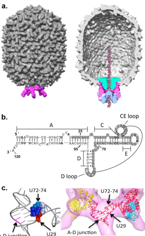
Bacillus subtilisbacteriophage29 is unusual in that an RNA
mol-ecule is also an essential component of the packaging motor (Fig.
1a) (16). Since the dsDNA phages are thought to employ a similar
translocation mechanism, it is likely that the roles played by
pro-head RNA (pRNA) in29 are carried out by subdomains of the
larger protein subunits of packaging motors in other dsDNA
phages (29).
pRNA is a virus-encoded 174-base phage transcript; a 120-base
form lacking the 3=-terminal 54 bases is fully functional inin vitro
assays (Fig. 1b) (13,15). RNase-treated proheads are inactive in
packaging, but packaging can be fully restored by the addition of
in vitro-transcribed pRNA, allowing us to test the activity of pRNA mutants. Mutagenesis studies show that pRNA has two functional
domains (Fig. 1b): a prohead binding domain (bases 25 to 95) and
a helical domain where mutations affect aspects of the packaging process other than prohead binding (the A-helix). In the motor, pRNA forms an oligomeric ring via intermolecular base pairing between the CE-and D-loops of adjacent pRNAs; this structure is
essential for DNA packaging (18,37). Cryo-electron microscopy
(cryoEM) analyses of proheads have shown pRNA appearing as a ring encircling the portal vertex with five spokes protruding from
the head (Fig. 1a) (27,28,33). These spokes are comprised of the
pRNA A-helix, providing the site for ATPase binding (27).
Recently, the crystal structure for the prohead binding domain
of pRNA (bases 25 to 95) was determined (9). This RNA construct
crystallized as a tetrameric ring, revealing in detail the intermolec-ular interaction that drives ring formation. pRNA has an extended appearance dominated by secondary structure. A region contain-ing flexible residues is found at the junction of the three main
helices in pRNA (A-, C-, and D-helices) (Fig. 1b), as evidenced by
the high-temperature factors and nonstandard geometry of both a single-U-base bulge (base 29, termed the first side of the junction
proceeding from 5=to 3=) and a tri-U linker (bases 72 to 74, the
second side). At the third side of the junction, the A-helix of the ATPase binding domain coaxially stacks underneath the D-helix of the prohead binding domain. This arrangement is common in three-helix junctions found in other RNAs, where three helices are organized such that two dsRNA segments are stacked and a third segment is connected by a small number of single-stranded bases
(8,22).
The fitting of an excised pRNA monomer from the crystal structure into the pentameric cryoEM envelope yielded insight into the pRNA structure in the context of the packaging motor (Fig. 1c) (9). The flexible single-U bulge and tri-U linker are prox-imal to each other, and their location in the ring suggested that they provide conformational flexibility that may be needed for the intermolecular interaction to occur on the prohead during pRNA assembly. The third side is part of an RNA superhelix that is
Received1 June 2012Accepted8 August 2012
Published ahead of print15 August 2012
Address correspondence to Shelley Grimes, [email protected].
Copyright © 2012, American Society for Microbiology. All Rights Reserved. doi:10.1128/JVI.01370-12
on November 7, 2019 by guest
http://jvi.asm.org/
formed by the alignment of multiple helical elements from two neighboring pRNAs, including the base-paired intermolecular
in-teraction (Fig. 1c). The superhelix would connect the head shell
and the protein components of the motor, potentially providing a
means of communication between these elements (9).
Previously, a mutational analysis of the three-helix junction showed that the single-base bulge U29 was dispensable for DNA
packaging but that the deletion of the tri-U linker (bases 72 to 74)
destroys DNA packaging activity (31). However, the deletion of
the tri-U linker is tolerated in the DNA packaging assay when a circularly permuted RNA is used to introduce a strand breakage at this site, consistent with the notion that a certain degree of
flexi-bility in this region needs to be maintained (36). Furthermore, the
tri-U linker can be replaced with three adenines, and the resulting
pRNA construct still supports⬃25% packaging activity (9).
Here we determined the functional significance of the three-helix junction of pRNA. Using deletion or insertion mutagenesis, we probed all three sides of the junction and determined the as-pects of assembly and pRNA function that are disrupted. The loss of flexibility due to the deletion of the unpaired U segments in the junction impaired prohead binding and motor assembly. Con-versely, the disruption of the superhelix by the insertion of just one base into the third side of the junction caused a decrease in DNA packaging, and the insertion of two bases abolished packag-ing. CryoEM analysis showed that the base insertions in the three-helix junction influence the flexibility of the pRNA spokes upon which the ATPase binds, suggesting that rigidity in the superhelix is required for pRNA function.
MATERIALS AND METHODS
Production of pRNA and pRNA mutants.Mutant sequences containing the proposed changes in pRNA were folded by using the RNA folding program mfold (version 3.2) (24,40) to confirm that the predicted sec-ondary structure was not disrupted by the mutation. Mutant pRNAs were then constructed in plasmid pRT72, which encodes the wild-type29 pRNA gene (30), by inverse PCR based on a method described previously by Wang and Wilkinson (35). Plasmid DNA was sequenced to verify the presence of the correct mutation. Wild-type and mutant pRNAs were produced byin vitrotranscription using T7 RNA polymerase and purified by denaturing urea-PAGE as described previously (30).
Production of packaging components. Proheads were produced from a 900-16-14-mutant infection (defective in the head fibers and the packaging ATPase) ofBacillus subtilisRD2. Infected cells were harvested at 65 min postinfection and lysed, and particles were purified from the lysate on sucrose gradients and concentrated by ultracentrifugation, as described previously (39). The particles were resuspended in 1⫻TMS buffer (50 mM Tris [pH 7.8], 10 mM MgCl2, 100 mM NaCl).
RNA-free particles were produced by RNase A treatment of purified proheads, followed by the repurification of the RNA-free particles by ul-tracentrifugation, as described previously (39). Particles were reconsti-tuted with wild-type or mutant pRNA by incubating particles with pRNA at a molar ratio of 1:10 in 0.5⫻TMS buffer prior to packaging (39).
DNA-gp3 and [3H]DNA-gp3 were extracted from phage and isolated on CsCl density gradients in 0.5⫻TE buffer (25 mM Tris [pH 7.8], 5 mM EDTA), as described previously (12).
The packaging ATPase was produced inBacillus subtilisfrom plasmid pSACB-gp16 and purified by chromatography, as described previously (19,39).
DNA packaging assay.Thein vitroDNA packaging assay is based on a DNase protection assay and was performed as described previously (39). Briefly, reconstituted proheads (8.3 nM), DNA-gp3 molecules (4.2 nM), and gp16 molecules (166 to 208 nM) were mixed together in 0.5⫻TMS buffer in 20l and incubated for 5 min at room temperature. ATP was then added to 0.5 mM to initiate packaging, and the mixture was incu-bated for 15 min. DNase I was added to 1g/ml, and the mixture was incubated for 10 min to digest unpackaged DNA. EDTA (25 mM final concentration) and proteinase K (500-g/ml final concentration) were then added to the reaction mixture, and the mixture was incubated for 30 min at 65°C to inactivate the DNase I and release the protected, packaged DNA from particles. The packaged DNA was analyzed by agarose gel
FIG 1The DNA packaging motor of bacteriophage29. (a) CryoEM recon-struction of the29 prohead (left) and with the packaging motor complex (right, cutaway), showing the molecular envelopes of the motor components. The head/tail connector is in green, the pentameric pRNA is in pink, and the packaging ATPase gp16 is in blue. A DNA molecule has been placed into the channel for reference. (Reproduced from reference14with permission from Elsevier.) (b) Predicted secondary structure of the 120-base pRNA (2). The helices and loops of pRNA are denoted by letters. Bases 25 to 95 comprise the prohead binding domain that was solved by X-ray crystallography (9). The shaded region represents the footprint of the prohead on bound pRNA (30). Oligomerization of pRNA occurs via intermolecular base pairing between the CE- and D-loops of adjacent pRNA molecules (18,37). The 3-way helix junc-tion is comprised of the single-base bulge U29, the tri-U linker (bases 72 to 74), and the intersection where the A- and D-helices meet (between bases U91 and C92). (c, left) Close-up of the crystal structure (PDB accession number 3R4F) (9) of the three-helix junction in a pRNA monomer, with bases U72 to U74 in blue, U29 in red, and the A-D junction in magenta. (Right) Model of the crystal structure of the pRNA-prohead binding domain flexibly docked into the pen-tameric cryoEM map of the pRNA density (9,26).
on November 7, 2019 by guest
http://jvi.asm.org/
[image:2.585.47.283.63.446.2]electrophoresis. The DNA packaging efficiency was quantified by densi-tometry using a UVP Gel Documentation system.
Prohead/gp16/DNA-gp3 ternary complex formation.Proheads with wild-type or mutant pRNAs were tested for their ability to support ternary complex formation by using a pulldown assay described previously (39). Briefly, proheads (8.3 nM), [3H]DNA-gp3 (4.2 nM), gp16 (166 to 208 nM), and␥-S-ATP (250M) were incubated together for 15 min. Mag-netic beads coated with anti-29 antisera were then added to capture the viral heads. The antiserum does not contain antibodies against the pack-aging ATPase gp16. After the beads were washed, the amount of gp16-dependent [3H]DNA-gp3 associated with the beads was measured by liq-uid scintillation counting. The efficiency of complex formation was calculated by the ratio of bead-bound cpm to the total cpm (unbound DNA-gp3, washes, and bound DNA-gp3). Controls where either pro-heads or the packaging ATPase was omitted from the reaction mixture were performed to determine background levels not related to ternary complex formation.
ATP hydrolysis during DNA packaging.Piproduction during DNA packaging was measured by the quantification of inorganic phosphate using the EnzChek phosphate assay kit (Invitrogen). A reaction mixture containing proheads with wild-type or mutant pRNA (4.2 nM), [3H]DNA-gp3 (4.2 nM), gp16 (125 nM), and 0.2 mM MESG (2-amino-6-mercapto-7-methylpurine riboside) was preincubated at room temper-ature for 10 min in the presence of PNP (purine nucleoside phosphory-lase) (0.1 units) in 0.5⫻TMS buffer in a final volume of 100l. To initiate DNA packaging, ATP (1 mM) was added and mixed well, and the absor-bance at 360 nm was measured every 15 s during the 10-min incubation period for DNA packaging.
Determination of the ratio of packaged DNA to hydrolyzed ATP.To quantify the ratio of base pairs of DNA packaged/ATP hydrolyzed, Pi production and DNA packaging were assessed by using the same reaction mixture (6). DNA packaging reaction mixtures containing proheads re-constituted with wild-type or mutant pRNAs, the packaging ATPase gp16, and [3H]DNA-gp3 were assembled as described above in a total volume of 40l and incubated for 5 min. Ten microliters of the sample was then removed to assess background levels of phosphate in the mixture. ATP was then added to 500M to initiate DNA packaging. Packaging was stalled at 5 min to trap actively packaging complexes by the addition of ␥-S-ATP to 250M to the reaction mixture. Ten microliters of sample was then removed to quantify ATP hydrolysis during the reaction by measuring the production of inorganic phosphate using the malachite green Lanzetta assay (20) and by reference to a standard curve. To deter-mine the amount of DNA packaged, the remaining reaction mixture was treated with 10g/ml DNase I and incubated for 5 min. Ten microliters of sample was diluted into 100l 0.5⫻TMS and layered on top of a 5 to 20% sucrose gradient in 0.5⫻TMS buffer. The gradients were spun in an SW55 rotor at 35,000 rpm at 20°C for 30 min. The gradients were fractionated, and the3H in fractions that represented DNase-protected packaged DNA were quantified by liquid scintillation counting and converted to base pairs packaged by using the specific activity of [3H]DNA-gp3.
Size determination of the right-end DNA-gp3 ApaLI restriction fragment.The integrity of the packaged right-end DNA, the last end packaged (3), in particles containing wild-type or mutant pRNAs was determined as described previously (14). Briefly,␥-S-ATP was added to the packaging reaction mixtures after a 15-min incubation to trap any incomplete packaging events. Unpackaged DNA was then digested with DNase I, and the packaged DNA was isolated and digested with ApaLI. The integrity of the 1.1-kbp DNA-gp3 right-end fragment was analyzed by agarose gel electrophoresis.
Electron microscopy.Prohead particles engineered to contain an in-sertion of either a single or two uracils at the U-rich junction were flash-frozen on holey grids in liquid ethane. Images were recorded at a⫻50,000 magnification with a JEM2200 FS field emission gun microscope equipped with an in-column energy filter. The electron dose for each micrograph was approximately 20 e⫺/Å2. All micrographs were collected
on a charge-coupled-device (CCD) camera with a step size of 2.0 Å per pixel.
Individual particle images were boxed, floated, and preprocessed to normalize mean intensities and variances and to remove linear back-ground gradients. Structure factor phases were modified as indicated by the parameters of the contrast transfer function. Reference projections of a previously reported prohead reconstruction (26) were used to initially classify particles for three-dimensional reconstruction. The resulting model was used to recalculate reference projections for better particle classification. Several cycles of iterative particle classification and recon-struction were performed until convergence had been reached. Fivefold symmetry was assumed in all stages of the reconstruction procedure. The resolutions of the final reconstruction of the U92 insertion mutant, in-cluding 1,871 particles, were 17 Å for the 5-fold-averaged map and 30.7 Å for the asymmetric reconstruction; the resolutions resulting from 2,134 U92-93 insertion mutant particles were 17.3 Å for the 5-fold-averaged map and 31.1 Å for the asymmetric reconstruction. All resolutions were determined by the Fourier shell correlation method using a correlation of 0.5 between independent half data sets as the cutoff criterion. All steps of the reconstruction process, including determinations of the contrast transfer function parameters, were performed with the program EMAN (23).
Accession numbers.The cryoEM maps for the proheads with pRNA containing the U92 insertion (accession numberEMD-5451) and the U92-93 insertion (accession numberEMD-2162) have been deposited in the EM Database.
RESULTS
Deletion mutagenesis of the U-rich region of the three-helix
junction and effect on DNA packaging.Since the crystal structure
of the pRNA-prohead binding domain suggests that the four U residues in the junction (U29 and U72-74) provide a region of flexibility, we targeted these residues for sequential deletion. All U residues at this junction can be simultaneously replaced with A
residues and retain biological activity (9), suggesting that the size
of the bulge rather than the sequence may be key to its function. RNA-free proheads were reconstituted with either wild-type or
mutant pRNA and tested for their ability to supportin vitroDNA
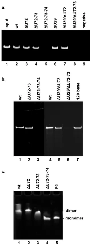
packaging (Fig. 2a). pRNAs with a deletion of one (Fig. 2a, lane 3)
or two (lane 4) U residues from the tri-U linker supported near-wild-type levels of DNA packaging. In contrast, as reported
previ-ously (31), the deletion of all three U residues abolished DNA
packaging (Fig. 2a, lane 5). To determine if the single-base bulge
U29 also impacts packaging, mutant pRNAs with a deletion of
U29 alone (⌬U29) (Fig. 2a, lane 6) or in combination with
dele-tions of residues in the tri-U linker (lanes 7 and 8) were tested. The
deletion of U29 alone (Fig. 2a, lane 6) as well as in combination
with a single-base deletion in the tri-U linker (lane 7) showed activity similar to that of wild-type pRNA (lane 2). However, the deletion of U29 in conjunction with two residues from the tri-U
linker resulted in no detectable packaging (Fig. 2a, lane 8). Since
the deletion of two U residues in the linker alone supported
pack-aging (Fig. 2a, lane 4), the loss of activity when combined with
⌬U29 suggests that these two sides of the junction are both
im-portant for packaging activity. The minimal tolerance for function is 2 residues: either 2 residues retained in the tri-U linker (i.e.,
⌬U29/⌬U72) or the U29 residue in conjunction with one tri-U
linker residue (i.e.,⌬U72-73).
Prohead binding of U deletion mutants.Having established
that both sides of the U-rich region impact DNA packaging, we sought to determine the nature of the defect in these deletion mutants. The pRNA function begins with its interaction/binding
on November 7, 2019 by guest
http://jvi.asm.org/
to the prohead (13,15). To test if mutations in the U-rich region affect prohead binding, RNA-free proheads were incubated with wild-type or mutant pRNA, the particles were repurified to re-move unbound pRNA, and the RNA content of particles was
an-alyzed by urea-PAGE (Fig. 2b). Double-U-deletion mutants that
supported packaging and inactive triple-U-deletion mutants were
tested. Particles incubated with the double-U deletions (⌬U72-73
[Fig. 2b, lane 2] or⌬U29/⌬U72 [lane 5]) showed wild-type (lanes 1 and 4) levels of pRNA binding. In contrast, particles incubated
with the triple-U deletions (⌬U72-73-74 [Fig. 2b, lane 3] or
⌬U29/72-73 [lane 6]) showed no pRNA binding. Thus, the loss of
packaging activity observed for mutants lacking three of the four U residues in the U-rich region is attributable to a defect in pro-head binding.
Previously, it was shown that multiple interactions are essential for successful pRNA-prohead binding. These include the posi-tively charged RKR residues of the N terminus of the connector
(1), complementary sequences on the CE- and D-loops that form
the intermolecular interaction (7,18,37), and a contact between
the E-loop of pRNA (bases 53 to 58) and the capsid shell (9). None
of these binding interactions were specifically targeted in our mu-tants, yet the manipulation of the U-rich region led to the loss of binding activity. Since these residues were hypothesized to be a flexible hinge in pRNA, this may impact multimerization even though the complementary bases needed for pairing are present. Therefore, we tested the ability of free mutant pRNA to multim-erize in solution.
Wild-type or mutant pRNAs were incubated in the presence of
Mg2⫹, and their oligomeric state was analyzed by native PAGE
(Fig. 2c). pRNA mutant F6 served as a monomeric pRNA control, as bases 45 to 48 of the intermolecular contact are mutated,
thereby preventing multimerization (Fig. 2c, lane 5) (37).
Wild-type pRNA migrates as a mixture of closed dimers and a
higher-multimer form on the gel (Fig. 2c, lane 1). The ⌬U72 and
⌬U72-73 mutants formed dimers (Fig. 2c, lanes 2 and 3,
respec-tively), while the⌬U72-73-74 triple deletion mutant migrated as a
monomer (lane 4). Thus, the triple deletion mutant that was de-fective in prohead binding also failed to multimerize in solution, while the binding-competent single and double deletion mutants were able to dimerize. The lack of dimerization in solution sug-gests that without the flexible U residues, the pRNA conformation has been altered such that intermolecular base pairing no longer occurs. Likely, this altered conformation also prohibits intermo-lecular base pairing in the context of the motor proteins on the viral head as well. Taken together, the data support the U-rich region having a functional role in productive prohead binding by facilitating the oligomerization, and thereby the ring formation, of pRNA on the prohead.
Insertion mutagenesis of the third side of the three-helix
junction and effect on DNA packaging.In contrast to the U-rich
region of the junction, the third side of the junction lacks bulged
residues and connects the A- and D-helices of pRNA (Fig. 1b).
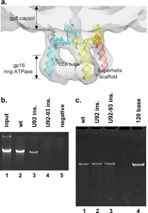
This region is part of the RNA superhelix observed in the crystal
structure (Fig. 1c). When the residues of the A-helix (omitted
from the crystallization construct) are modeled into the structure, the superhelical scaffold appears to be extensive. When viewed in the context of the cryoEM map of the prohead/ATPase motor complex, the superhelix serves to connect all the protein compo-nents, from the head shell, through the connector, to the ATPase (Fig. 3a) (9). This suggests that the RNA superhelix is likely
im-FIG 2Analysis of the U-rich region (U29 and U72-74) of the three-helix junction of pRNA. (a)In vitroDNA packaging by proheads containing pRNA with deletions in the U-rich junction. To assess packaging efficiency, the pack-aged DNA that was protected from DNase treatment was extracted from par-ticles and analyzed by agarose gel electrophoresis. Packaging was assessed by using proheads reconstituted with wild-type (wt) pRNA (lane 2), pRNA with deletions in the tri-U linker (bases 72 to 74) (lanes 3 to 5), a deletion of the single-base U29 bulge (lane 6), or deletions in both regions (lanes 7 and 8). Input represents DNA added to the packaging reaction mixture (lane 1). In the negative control, ATP was omitted from the packaging reaction mixture (lane 9). (b) Prohead binding by deletion mutants in the U-rich region. RNA-free proheads were mixed with wild-type or mutant pRNA, and the prohead-pRNA complexes were purified away from unbound prohead-pRNA. The RNA con-tents of these particles were analyzed on a denaturing urea-PAGE gel. Shown are the RNA contents of proheads with wild-type pRNA (lanes 1 and 4), pRNA with deletions in the tri-U linker (bases 72 to 74) (lanes 2 and 3), and pRNA with deletions of U29 and tri-U-linker bases (lanes 5 and 6). The 120-base pRNA is a standard to mark the position of pRNA (lane 7). (c) Multimeriza-tion of pRNA deleMultimeriza-tion mutants. Free pRNA was incubated in the presence of Mg2⫹, and its oligomeric state was assessed on a native-PAGE gel. Wild-type pRNA (lane 1) and deletions in the tri-U linker (lanes 2 to 4) were analyzed. pRNA mutant F6, which has mutated residues that prevent multimerization, was used as a control to mark the position of monomeric pRNA (lane 5) (37).
on November 7, 2019 by guest
http://jvi.asm.org/
[image:4.585.83.240.66.490.2]portant and may have a functional role, such as in communication between motor components.
In three-helix junctions in general, the junction between the
stacked helices usually contains no or very few unpaired bases (8,
22). Since the29 pRNA lacks unpaired residues at this third side
of the three-way junction, U residues were inserted here as a means of potentially weakening the relatively rigid superhelix, and
the effect on DNA packaging was examined (Fig. 3b). The
inser-tion of one U residue decreased packaging by⬃75% (Fig. 3b, lane
3), while the introduction of two U residues resulted in no detect-able DNA packaging (lane 4). Thus, like the U-rich region, this side of the three-helix junction also impacts the DNA packaging process.
Prohead binding of U insertion mutants.To identify the
de-fect in packaging, third-side insertion mutants were screened in a battery of assays. Prohead binding activity was assessed, and both single and double insertion mutant pRNAs bound the prohead
similarly to the wild type (Fig. 3c), even though the two-U
inser-tion did not support DNA packaging (Fig. 3b). Therefore, in
con-trast to the U-rich region, this side of the junction does not appear to influence prohead binding, and the defect must lie elsewhere in the packaging process.
Effect of U insertions on ternary complex formation.Since
prohead binding was unaffected by base insertions, we assessed the abilities of the mutants to support the coassembly of the pro-head, the packaging ATPase, and DNA-gp3 into a complex in preparation for the initiation of packaging. This ternary complex formation is ATPase dependent, as proheads and DNA-gp3 do
not interact in its absence (12,17). Proheads with wild-type or
mutant pRNA were mixed with the packaging ATPase gp16,
[3H]DNA-gp3, and␥-S-ATP (␥-S-ATP is required for stable
ter-nary complex formation [39]). Complexes were detected by a
pulldown assay, where beads coated with anti-29 antibodies
cap-ture the phage head, and the gp16-dependent [3H]DNA bound to
the beads was quantified. As a control for the nonspecific binding of DNA-gp3 to the beads, reactions were set up where either the prohead or the ATPase was omitted from the reaction mixture. All
negative-control combinations bound⬃3% of the [3H]DNA-gp3
in the reaction mixture (Fig. 4a). In the complete reaction
mix-ture, proheads with wild-type pRNA bound⬃20% of the
DNA-gp3. For the insertion mutants, DNA binding was decreased, with
the single insertion mutant binding⬃8% and the double insertion
mutant binding⬃6% of the DNA-gp3. This corresponds to a 3- to
4-fold reduction in ternary complexes for the mutants compared to wild-type pRNA. Whether this reduction is due to an altered ability to form ternary complexes or a result of the reduced stabil-ity of complexes once they form cannot be discerned by this assay.
ATPase activity of insertion mutants.Since both the single
and double insertion mutants support motor assembly, albeit with reduced efficiency, we next tested if they affect motor oper-ation. While the double insertion mutant had no detectable pack-aging, this result could be due to an inactive motor or an uncou-pling of ATP hydrolysis from translocation; i.e., the motor hydrolyzes ATP but fails to translate this into DNA movement. To assess ATP hydrolysis during packaging, packaging complexes
were assembled, and ATP hydrolysis was measured by detecting Pi
production during the 10-min packaging assay (Fig. 4b). To
en-sure that the ATPase activity meaen-sured was related to packaging, buffer conditions that suppress non-packaging-related ATPase activity resulting from prohead/gp16 complexes that may form in the reaction (i.e., DNA-independent ATPase activity) were used
(6). As controls, a prohead/ATPase sample and an ATPase-alone
sample were included in each experiment. The U92-93 insertion mutant sample had ATPase activity similar to that of the back-ground controls, indicating that the motor was not functional. The U92 insertion mutant sample had reduced levels of ATP hy-drolysis compared to those of the wild type but consistent with the amount of DNA packaged in each sample: the ratios of base pairs
packaged/ATP hydrolyzed were 1.95⫾0.16 for the wild type and
1.68⫾0.13 for the U92 insertion mutant (see Materials and
Meth-ods). Thus, the insertion mutants yielded two phenotypes, one where assembly is affected but motor function is retained (U92 insertion) and the other where assembly is similarly affected but motor function is impaired (U92-93 insertion).
FIG 3Analysis of the third side of the three-helix junction. (a) Model of the pentameric 120-base pRNA, created from the crystal structure of the prohead binding domain with a modeled A-helix, fit into the cryoEM map of the pro-head-pRNA-ATPase motor complex (9). The pRNA superhelical scaffold would be extensive, connecting the shell, connector, and ATPase. (Repro-duced from reference9with permission of the publisher.) (b)In vitroDNA packaging by proheads containing pRNA insertion mutants. Packaging was assessed by using proheads reconstituted with wild-type pRNA (lane 2) or U insertions (ins.) between bases 91 and 92 (lanes 3 and 4). (c) Prohead binding by insertion mutants of pRNA. Proheads were reconstituted with wild-type pRNA (lane 1) or U insertion mutant pRNA (lanes 2 and 3) and purified away from unbound pRNA, and the RNA content was analyzed by denaturing urea-PAGE. The 120-base pRNA is a standard to mark the position of pRNA (lane 4).
on November 7, 2019 by guest
http://jvi.asm.org/
[image:5.585.44.284.68.414.2]For the U92 insertion mutant, which retains motor function, we also determined whether the packaging process is completed such that the entire genome becomes DNase resistant. If the last bit of DNA is not stably packaged, the right end of the genome (the
last end packaged) (3) will be sensitive to DNase treatment and
will yield a heterogeneous right-end fragment upon restriction
digestion of the packaged DNA (14). After packaging,␥-S-ATP
was added to the reaction mixture to trap any packaging interme-diates. The reaction mixture was then treated with DNase to digest any DNA that remained outside the head, and the packaged DNA was then extracted and digested with ApaLI in order to reveal the integrity of the last 6% of the genome. The U92 insertion had an intact right-end DNA fragment, similar to wild-type particles, in-dicating the completion of packaging (data not shown). Thus, complexes that begin the packaging process can complete it, indi-cating that the motor is functional throughout the event of trans-location.
CryoEM analysis of insertion mutants.The intended design
of the insertion mutants was to disrupt the superhelix. To deter-mine if there was an alteration in the pRNA scaffold that could be visualized, cryoEM three-dimensional (3D) image reconstruction was carried out for proheads with the insertion mutant pRNAs
(Fig. 5). Previously, we had shown that we could detect structural changes in the spokes of pRNA (A-helix) upon the deletion of the
essential CCA bulge (bases 18 to 20) (39) (Fig. 1b). This deletion
causes the spokes of pRNA to twist and bend relative to wild-type
pRNA (compareFig. 5btoa), although the global features of the
pRNA are retained. Hence, 3D reconstructions of proheads bound to either the single- or double-U insertion mutant were determined to evaluate the gross structural effects of these muta-tions. The resulting maps indicated that the apparent density of the pRNA spokes in both maps is greatly reduced, whereas the
density of the pRNA ring remains strong (Fig. 5candd). This
result likely reflects that the positions of the A-helices are different in individual particles such that this feature is washed out during averaging between particles. Similarly, because 5-fold symmetry averaging was imposed on both maps, this result would also be seen if the mutations introduced asymmetry into the pRNA ring such that individual A-helices were not aligned during 5-fold av-eraging. Hence, asymmetric reconstructions were also calculated; the resulting maps also showed no density for the pRNA A-helices (data not shown). Thus, taken together with the functional anal-ysis, these structural results argue for the importance of rigidity
FIG 4Motor function of insertion mutants. (a) Ternary complex formation by insertion mutants. Proheads with wild-type or mutant pRNAs were incu-bated with the packaging ATPase gp16 and [3H]DNA-gp3, and the complexes
were isolated by use of magnetic beads coated with anti-29 antibodies. Error bars represent standard deviations (n⫽3). (b) ATPase activity during DNA packaging. Packaging complexes with wild-type or insertion mutant pRNAs were assembled, and Piproduction due to ATPase activity was monitored via a
colorimetric change detected at anA360. P⫹gp16 is a DNA-free mixture
containing proheads with U92 insertion pRNA and gp16, representing ATPase activity not associated with DNA packaging. gp16 alone lacks proheads, and DNA-gp3 and represents the basal ATPase activity. Shown is a trace represen-tative of data from 3 experiments. OD 360, optical density at 360 nm.
FIG 5CryoEM 3D reconstruction of pRNA insertion mutants on the third side of the three-helix junction. Proheads contained (a) wild-type pRNA (26); (b) pRNA with a deletion of the essential CCA bulge demonstrates the detec-tion of an altered A-helix of pRNA (39); (c) U92 insertion pRNA; and (d) U92-93 insertion pRNA. CryoEM maps of proheads with wild-type pRNA and CCA deletion pRNA (a and b, respectively) were low-pass filtered to 18 Å for comparison with the reconstructions of the U92 and U92-93 insertion mutants.
on November 7, 2019 by guest
http://jvi.asm.org/
[image:6.585.301.541.65.391.2] [image:6.585.42.281.66.350.2]along the superhelices of the RNA scaffold for the DNA packaging process.
DISCUSSION
The29 DNA packaging motor has been shown to be a complex
and highly coordinated machine. Single-molecule studies re-vealed that the DNA is translocated by a two-step cyclic mecha-nism: a dwell phase, during which at least four ATPases in the ring bind ATP, and a burst phase, where 10 bp of DNA is translocated
into the head via four 2.5-bp substeps (25). Even at limiting ATP
concentrations, the motor retains this coordinated burst behavior involving multiple ATPase subunits rather than breaking down into individual substeps. This degree of coordination suggests a
need for communication within the motor. Additionally,29 and
other phage packaging motors have been shown to respond to the amount of DNA packaged in the head by slowing as the head fills
and the pressure within builds (11,34), further suggesting
com-munication in the motor. An understanding of coordination and communication during packaging is therefore an integral part of determining the mechanism of DNA packaging.
pRNA is located at the center of the29 packaging motor,
between the protein ring components, the head-tail connector,
and the packaging ATPase (Fig. 1a) (27). This location enables
pRNA to participate in motor assembly and to provide a potential role in communication during motor function. It is clear that pRNA has a role as a scaffold for motor assembly, as the A-helix provides the binding site for the packaging ATPase, attaching this catalytic, and likely force-generating, component of the motor to
the prohead (19,21,27). It is also possible that pRNA has other
roles in motor function or communication. Of note is that the arrangement of pRNA in the motor provides the opportunity for communication in two ways: via the RNA superhelices that extend over the length of the motor assemblage, linking the protein
com-ponents (head shell, connector, and ATPase) (Fig. 3a), and via the
unique ring structure created by the intermolecular base pairing between pRNA monomers that provides a continuous linkage be-tween motor subunits of the ring ATPase. These two potential modes of communication differ in directionality. The superhelix would provide communication that parallels the translocating DNA (i.e., “vertical”), while communication through intermolec-ular base pairing would be perpendicintermolec-ular to the translocating DNA (i.e., “horizontal”). Here we examined the role of the 3-helix junction of pRNA that lies at the center of these putative interac-tions.
Residues in the U bulges (U29 and U72-74), located at two sides of the junction, are shown to be involved in prohead binding and impact DNA packaging activity. A minimum of 2 residues, distributed between the two bulges, are required for pRNA
bind-ing to the head and are also sufficient for DNA packagbind-ing (Fig. 2a
andb). The inability of triple-U-deletion mutants to oligomerize
in solution or bind to the prohead supports the hypothesis, de-rived from the crystal structure of the pRNA-prohead binding
domain (9), that these bulged residues provide a flexible joint that
would facilitate the intermolecular base pairing of pRNA that is
required for binding to the head (Fig. 2bandc).
Regarding the third side of the junction, part of the contiguous strand of the superhelix, we showed that base insertions at this site inhibit DNA packaging. While pRNA-prohead binding activity is retained in these mutants, the ability to assemble the ternary ini-tiation complex is impaired, thus identifying a role in motor
as-sembly. Additionally, motors with one insertion at this site sup-ported ATP hydrolysis, whereas those with two insertions were inactive, suggesting that ATPase and translocation functions may also be affected. CryoEM reconstructions of these insertion mu-tants showed a lack of density for the entire A-helix. The A-helices of wild-type pRNA are easily visualized by using identical
recon-struction methods (Fig. 5), arguing that the A-helix is a rigid
ex-tension of the RNA superhelix. Modifications of the A-helix, such as with the deletion of the CCA bulge structure, alter the confor-mation of the helix without diminishing its density in cryoEM maps. The disappearance of the entire A-helix from reconstruc-tions with inserreconstruc-tions in the superhelix backbone at the 3-way junc-tion provides strong evidence that inserjunc-tions at this side of the junction mobilize the distal domain of the superhelix. Taken to-gether, the impaired activity of these mutants is consistent with a disruption of the integrity of the superhelix and argues that this higher-order structure is important for packaging. Future single-molecule laser tweezers analyses of the U92 insertion mutant will allow us to study this motor in action in order to probe if and how motor function and coordination are influenced by the altered superhelical scaffold.
This relatively small region thus serves to organize different functional aspects of pRNA. It appears that the structure of the junction is designed to impart a range of flexibility in the “hori-zontal” direction that is needed to form the essential intermolec-ular base pairing on the head while simultaneously providing the degree of rigidity in the “vertical” direction that is required for superhelix function, including initiation complex formation and motor activity. The presence of bulged residues on two sides of the three-helix junction serves to project the C-helix away from the A-and D-helices without an interruption of the pRNA superhelix. As a result, the CE-loop is positioned to base pair with the D-loop of the neighboring pRNA, becoming part of the adjacent superhelix. The lack of bulges on the third side provides the relative rigidity needed for the contiguous stacking of the A- and D-helices that participate in superhelix formation and function.
A recent electron paramagnetic resonance (EPR) spectroscopic analysis of the global shape of the three-way junction for a pRNA dimer in solution found that the A- and D-helices had a kinked
conformation (38), rather than being stacked, as found in the
crystal structure (9). This difference may reflect the experimental
design of the EPR study, where the tri-U linker at the three-way junction (bases 72 to 74) was absent in one of the experimental constructs such that the 3-way junction was unconstrained on one side. Interestingly, this type of modification is tolerated in pack-aging in the case of circularly permuted pRNAs of the same design
(36). In this case, interactions with the protein components of the
motor likely constrain this RNA into the proper conformation, while in solution, those constraints are lacking.
pRNA displays general features common to RNA three-way
junctions (8, 22) but with variations unique to pRNA. For
in-stance, three-way junctions often mediate the stacking of two of
the participating helices. In the29 pRNA, the stacked A- and
D-helices at the three-way junction actually become part of an extensive superhelix that involves the neighboring pRNA in the
ring (9). The intermolecular interaction unique to pRNA creates
an additional helical segment that results in an alignment of the stacked helices of the three-way junction with the E-helix of the adjoining pRNA. Also, three-helix junctions often mediate
inter-nal tertiary interactions that are important for function (8,22). In
on November 7, 2019 by guest
http://jvi.asm.org/
contrast, the pRNA three-helix junction facilitates a quaternary interaction that is essential for pRNA binding to the prohead and the superhelices spanning the motor assemblage that affect motor
assembly and operation. A recent analysis of the29 pRNA
three-way junction showed that it was a highly stable structure and sug-gested that the unique properties of this junction can be harnessed
for nanotechnology applications (32).
The dsDNA phages, such as T4, lambda, SPP1, and P22, are thought to share commonalities in their mechanism of DNA
packaging (5, 29). As mentioned previously, although an RNA
motor component has been demonstrated only for29, the
func-tions provided by pRNA in29 DNA packaging likely reside in
subdomains of the packaging proteins in these other phages (29).
While it remains to be determined if other phage motors have a
level of coordination in their motors similar to that seen for29,
it is possible that a subdomain of the packaging ATPase complex of other phages also provides a relatively rigid anchor to the shell or connector that facilitates the movement of DNA under the high
forces against which the motor works during DNA packaging (10,
11,34).
ACKNOWLEDGMENTS
We thank Rockney Atz for helpful discussions, Michael Sherman for as-sistance in preparing cryoEM samples, and the Sealy Center for Structural and Computational Biology at the University of Texas Medical Branch.
This work was supported by Public Health Service grants GM059604 and GM095516 from the National Institutes of Health.
REFERENCES
1.Atz R, Ma S, Gao J, Anderson DL, Grimes S.2007. Alanine scanning and Fe-BABE probing of the bacteriophage29 prohead RNA-connector in-teraction. J. Mol. Biol.369:239 –248.
2.Bailey S, et al.1990. Phylogenetic analysis and secondary structure of the
Bacillus subtilisbacteriophage RNA required for DNA packaging. J. Biol. Chem.265:22365–22370.
3.Bjornsti MA, Reilly BE, Anderson DL.1983. Morphogenesis of bacte-riophage29 ofBacillus subtilis: oriented and quantizedin vitropackaging of DNA-gp3. J. Virol.45:383–396.
4.Burroughs A, Iyer L, Aravind L.2007. Comparative genomics and evo-lutionary trajectories of viral ATP dependent DNA-packaging systems, p 48 – 65.InVolff J-N (ed), Gene and protein evolution. Genomic dynamics, vol 3. Karger, Basel, Switzerland.
5.Casjens SR.2011. The DNA-packaging motor of tailed bacteriophages. Nat. Rev. Microbiol.9:647– 657.
6.Chemla YR, et al.2005. Mechanism of force generation of a viral DNA packaging motor. Cell122:683– 692.
7.Chen C, Sheng S, Shao Z, Guo P.2000. A dimer as a building block in assembling RNA. J. Biol. Chem.275:17510 –17516.
8.de la Pena M, Dufour D, Gallego J.2009. Three-way junctions with remote tertiary contacts: a recurrent and highly versatile fold. RNA15: 1949 –1964.
9.Ding F, et al.2011. Structure and assembly of the essential RNA ring component of a viral DNA packaging motor. Proc. Natl. Acad. Sci. U. S. A.
108:7357–7362.
10. Fuller DN, Raymer DM, Kottadiel VI, Rao VB, Smith DE.2007. Single phage T4 DNA packaging motors exhibit large force generation, high ve-locity, and dynamic variability. Proc. Natl. Acad. Sci. U. S. A.104:16868 – 16873.
11. Fuller DN, et al.2007. Measurements of single DNA molecule packaging dynamics in phage lambda reveal high forces, high motor processivity, and capsid transformations. J. Mol. Biol.373:1113–1122.
12. Grimes S, Anderson D.1997. The bacteriophage phi29 packaging pro-teins supercoil the DNA ends. J. Mol. Biol.266:901–914.
13. Grimes S, Jardine PJ, Anderson D.2002. Bacteriophage29 DNA pack-aging. Adv. Virus Res.58:255–294.
14. Grimes S, Ma S, Gao J, Atz R, Jardine PJ.2011. Role of29 connector channel loops in late-stage DNA packaging. J. Mol. Biol.410:50 –59. 15. Guo P.2002. Structure and function of29 hexameric RNA that drives
the viral DNA packaging motor. Prog. Nucleic Acid Res. Mol. Biol.72: 415– 472.
16. Guo P, Erickson S, Anderson D.1987. A small viral RNA is required for
in vitropackaging of bacteriophage29 DNA. Science236:690 – 694. 17. Guo P, Peterson C, Anderson DL.1987. Initiation events in in vitro
packaging of bacteriophage29 DNA-gp3. J. Mol. Biol.197:219 –228. 18. Guo P, Zhang C, Chen C, Garver K, Trottier M. 1998. Inter-RNA
interaction of phage29 pRNA to form a hexameric complex for viral DNA transportation. Mol. Cell2:149 –155.
19. Koti J, et al.2008. DNA packaging motor assembly intermediate of bac-teriophage29. J. Mol. Biol.381:1114 –1132.
20. Lanzetta PA, Alvarez LJ, Reinach PS, Candia OA.1979. An improved assay for nanomole amounts of inorganic phosphate. Anal. Biochem.100: 95–97.
21. Lee TJ, Guo P. 2006. Interaction of gp16 with pRNA and DNA for genome packaging by the motor of bacterial virus phi29. J. Mol. Biol.
356:589 –599.
22. Lescoute A, Westhof E.2006. Topology of three-way junctions in folded RNAs. RNA12:83–93.
23. Ludtke SJ, Baldwin PR, Chiu W.1999. EMAN: semiautomated soft-ware for high-resolution single-particle reconstructions. J. Struct. Biol.
128:82–97.
24. Mathews DH, Sabina J, Zuker M, Turner DH.1999. Expanded sequence dependence of thermodynamic parameters improves prediction of RNA secondary structure. J. Mol. Biol.288:911–940.
25. Moffitt JR, et al.2009. Intersubunit coordination in a homomeric ring ATPase. Nature457:446 – 450.
26. Morais MC, et al.2005. Conservation of the capsid structure in tailed dsDNA bacteriophages: the pseudoatomic structure of phi29. Mol. Cell
18:149 –159.
27. Morais MC, et al.2008. Defining molecular and domain boundaries in the bacteriophage29 DNA packaging motor. Structure16:1267–1274. 28. Morais MC, et al.2001. Cryo-EM image reconstruction of symmetry
mismatches in bacteriophage29. J. Struct. Biol.135:38 – 46.
29. Rao VB, Feiss M.2008. The bacteriophage DNA packaging motor. Annu. Rev. Genet.42:647– 681.
30. Reid RJD, Bodley JW, Anderson DL. 1994. Characterization of the prohead-pRNA interaction of bacteriophage29. J. Biol. Chem.269: 5157–5162.
31. Reid RJD, Zhang F, Benson S, Anderson D.1994. Probing the structure of bacteriophage29 prohead RNA with specific mutations. J. Biol. Chem.269:18656 –18661.
32. Shu D, Shu Y, Abdelmawla S, Guo P.2011. Thermodynamically stable RNA three-way junction for constructing multifunctional nanaoparticles for delivery of therapeutics. Nat. Nanotechnol.6:658 – 667.
33. Simpson AA, et al.2000. Structure of the bacteriophage29 DNA pack-aging motor. Nature408:745–750.
34. Smith DE, et al.2001. The bacteriophage29 portal motor can package DNA against a large internal force. Nature413:748 –752.
35. Wang J, Wilkinson MF.2000. Site-directed mutagenesis of large (13-kb) plasmids in a single-PCR procedure. Biotechniques29:976 –978. 36. Zhang CL, Tellinghuisen T, Guo P.1997. Use of circular permutation to
assess six bulges and four loops of DNA-packaging pRNA of bacterio-phage phi29. RNA3:315–322.
37. Zhang F, et al.1998. Function of hexameric RNA in packaging of bacte-riophage29 DNAin vitro.Mol. Cell2:141–147.
38. Zhang X, et al.2012. Global structure of a three-way junction in a phi29 packaging RNA dimer determined using site-directed spin labeling. J. Am. Chem. Soc.134:2644 –2652.
39. Zhao W, Morais MC, Anderson DL, Jardine PJ, Grimes S.2008. Role of the CCA bulge of prohead RNA of bacteriophage29 in DNA packaging. J. Mol. Biol.383:520 –528.
40. Zuker M.2003. Mfold Web server for nucleic acid folding and hybridiza-tion predichybridiza-tion. Nucleic Acids Res.31:3406 –3415.