0022-538X/07/$08.00⫹0 doi:10.1128/JVI.02207-06
Copyright © 2007, American Society for Microbiology. All Rights Reserved.
Ty3 Capsid Mutations Reveal Early and Late Functions of the
Amino-Terminal Domain
䌤
Liza S. Z. Larsen,
1,3† Min Zhang,
2† Nadejda Beliakova-Bethell,
2Virginia Bilanchone,
2Anne Lamsa,
2Kunio Nagashima,
4Rani Najdi,
1Kathryn Kosaka,
1Vuk Kovacevic,
2Jianlin Cheng,
3‡ Pierre Baldi,
3G. Wesley Hatfield,
1,3and Suzanne Sandmeyer
1,2,3*
Departments of Microbiology and Molecular Genetics1and Biological Chemistry2 and School of Information and
Computer Sciences and Institute for Genomics and Bioinformatics,3University of California, Irvine, California 92697,
Image Analysis Laboratory, NCI-Frederick, SAIC-Frederick, Inc., Frederick, Maryland 217024
Received 8 October 2006/Accepted 26 March 2007
The Ty3 retrotransposon assembles into 50-nm virus-like particles that occur in large intracellular clusters in the case of wild-type (wt) Ty3. Within these particles, maturation of the Gag3 and Gag3-Pol3 polyproteins by Ty3 protease produces the structural proteins capsid (CA), spacer, and nucleocapsid. Secondary and tertiary structure predictions showed that, like retroviral CA, Ty3 CA contains a large amount of helical structure arranged in amino-terminal and carboxyl-terminal bundles. Twenty-six mutants in which alanines were substituted for native residues were used to study CA subdomain functions. Transposition was measured, and particle morphogenesis and localization were characterized by analysis of protein processing, cDNA production, genomic RNA protection, and sedimentation and by fluorescence and electron microscopy. These measures defined five groups of mutants. Proteins from each group could be sedimented in a large complex. Mutations in the amino-terminal domain reduced the formation of fluorescent Ty3 protein foci. In at least one major homology region mutant, Ty3 protein concentrated in foci but no wt clusters of particles were observed. One mutation in the carboxyl-terminal domain shifted assembly from spherical particles to long filaments. Two mutants formed foci separate from P bodies, the proposed sites of assembly, and formed defective particles. P-body association was therefore found to be not necessary for assembly but correlated with the production of functional particles. One mutation in the amino terminus blocked transposition after cDNA synthesis. Our data suggest that Ty3 proteins are concentrated first, assembly associated with P bodies occurs, and particle morphogenesis concludes with a post-reverse transcription, CA-dependent step. Particle formation was gen-erally resistant to localized substitutions, possibly indicating that multiple domains are involved.
Retrotransposons occur widely in eukaryotic species. Some classes, such as the Ty3/gypsy-like elements (Metaviridae), are similar to retroviruses in organization and even in specific sequence features of structural proteins (30). However, these mobile elements typically do not encode matrix or envelope functions, and they assemble and disassemble within a single host cell. Compared to retroviruses, relatively little is known about where they assemble and what protein domains are involved in this process. The present study was undertaken to examine the role of Ty3 capsid (CA) subdomains in localiza-tion, assembly, reverse transcriplocaliza-tion, and transposition.
Although many questions remain concerning the roles of specific retrovirus CA subdomains, knowledge of retrovirus particle structure (21) can inform the design of experiments to define the process of retrotransposon assembly. The major retrovirus structural polyprotein is encoded by the first, orgag, open reading frame (ORF). Although the Gag sequence is not highly conserved, its derivative matrix (MA), CA, and nucleo-capsid (NC) proteins in different viruses share motifs related
to common functions. The pol reading frame is typically ex-pressed as part of a Gag-Pol fusion protein that includes domains for protease (PR), reverse transcriptase (RT), and integrase (IN).
In the immature, spherical retrovirus particle, Gag mole-cules are aligned side by side and extend radially from the NC domain in the particle interior to the MA domain on the exterior (33, 102). MA proteins of retroviruses are implicated in localization of assembly to membranes or to the microtubule organizing complex (74, 82, 85, 103). Particle formation is dependent on the carboxyl-terminal domain (CTD) of CA and spacer and the I domain of NC, with contributions from dif-ferent domains dominating in difdif-ferent viruses (1, 23, 32, 73, 95, 96, 99; reviewed in references 3, 64, 82, 98, and 100). During assembly, PR is activated upon dimerization (14) and, coordinately with budding, polyprotein precursors are pro-cessed into mature forms.
After proteolysis, the enveloped virion reorganizes into its characteristic spherical, conical, or cylindrical mature form, the outer shell of which is composed of CA. Studies of the human T-cell lymphotrophic virus type 1 (HTLV-1), Rous sarcoma virus (RSV), human immunodeficiency virus type 1 (HIV-1), and equine infectious anemia virus (EIAV) CA proteins report a common structure, including an amino-terminal domain (NTD) consisting of a hairpin followed by a bundle of ␣ helices linked through an unstructured region to a CTD of a smaller␣-helical bundle terminating in an unstructured region * Corresponding author. Mailing address: Department of Biological
Chemistry, University of California, Irvine, CA 92697. Phone: (949) 824-7571. Fax: (949) 824-2688. E-mail: [email protected].
† Liza S. Z. Larsen and Min Zhang contributed equally to this work. ‡ Present address: School of Electrical Engineering and Computer Science, University of Central Florida, Orlando, FL 32816-2362.
䌤Published ahead of print on 18 April 2007.
6957
on November 8, 2019 by guest
http://jvi.asm.org/
(8, 10, 17, 20, 34, 35, 39, 48, 50, 51, 62, 63). Electron micro-scopic (EM) images of assemblies of Gag and CA in lipid monolayers and virus core particles indicate that these mole-cules assemble into hexameric lattices (4, 5, 12, 36, 58, 67, 102). A recent X-ray crystal analysis shows that the N-tropic murine leukemia virus (MLV) CA NTD forms a hexamer with the amino-terminal three helices mediating intermolecular inter-actions (65). Consistent with that key position in the structure, Ala-scanning mutagenesis of HIV-1 CA supports a role for the NTD in the transition from immature to mature particle mor-phology (37, 93, 94) and particle stability (31). The major homology region (MHR), a 19-amino-acid (aa) motif, begins at the distal end of the linker region between the NTD and CTD
␣-helical regions and overlaps the first helix of the CTD. Mu-tations within the MHR have pleiotropic effects, including disruption of particle formation and cDNA synthesis, but its precise molecular function remains unknown (100).
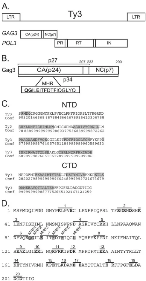
Ty3 is 5.4 kb in length and is comprised of 340-bp long terminal repeats (LTRs) flankingGAG3andPOL3ORFs (re-viewed in reference 81) (Fig. 1A).GAG3encodes CA, spacer, and NC domains (41, 54, 55, 70) (Fig. 1B).POL3encodes PR, RT, and IN domains. Gag3 is translated from the first ORF, and, as in many retroviruses, Gag3-Pol3 is produced via a programmed frameshift. The 290-aa Gag3 polyprotein is pro-cessed by Ty3 PR (53) into a 206-aa CA (lacking the initiation Met) and a 57-aa NC (53, 55). A 26-aa spacer is inferred to be generated from these cleavages but has not yet been observed. The CA domain contains a copy of the MHR motif, and mu-tations in this motif have pleiotropic effects (69). Ty3 CA has a copy of the late motif YPXL (26, 64), but its function is not known. Ty3 NC contains one zinc binding domain, which is required for virus-like particle (VLP) formation (70). Cells in which Ty3 is expressed accumulate VLPs that are roughly 50 nm in diameter and have features of icosahedral symmetry (55). It is presumed that, similar to what occurs in retrovirus maturation, incorporation of Gag3-Pol3 into particles is re-quired for proteolytic maturation. Processing bodies (P bodies) are sites of nontranslating RNAs associated with decapping, degradation, and RNA storage in yeast (11, 86). Ty3 transpo-sition frequency and localization are affected by deletion of the DHH1andXRN1genes, which encode RNA P-body nents, and Ty3 CA and RNA colocalize with P-body compo-nents. P bodies are therefore proposed to be sites of VLP assembly (6).
Ty3 is the sole representative of theMetaviridaein Saccha-romyces cerevisiae.OtherS. cerevisiae retrotransposons (Ty1, Ty2, Ty4, and Ty5) are members of the copia-like class (Pseudoviridae). The Ty3 VLP has an external structure similar to what has been observed for the Ty1 VLP (55, 77). However, Ty3 is more similar to retroviruses than to Ty1 in organization and sequence of coding domains (30).
[image:2.585.298.542.87.555.2]The present study was undertaken to define the functional subdomains of Ty3 CA by using 26 Ala substitution mutants. Mutants were characterized for VLP formation, RNA packag-ing, cDNA production, transposition, and Ty3 protein and particle localization. All but a few of the mutants were able to form particles. However, most were affected in some aspect of particle function. Mutant phenotypes implicated Ty3 CA in processes including localization of Gag3, packaging of genomic
FIG. 1. Ty3 Gag3. (A) Ty3 genome and polyprotein organization. (B) Ty3 Gag3 processing. PR processing of Gag3 results in p34 (aa 1 to 290), p27 (aa 1 to 233), and CA p24 (aa 1 to 207). The conserved MHR domain is indicated in the lower box. The final Phe is a weakly conserved hydrophobic/Ala residue in retrovirus MHR sequences. Ty3 MHR sequences are shown (aa 86 to 104). (C) Predicted helical do-mains of Ty3 CA. Consensus predictions of Ty3 CA NTD and CTD helical regions were made with SSpro4, PSIPRED, ProfSec, SAM-T99, and SABLE2. The numbers on the “Conf” line are the confidence scores of secondary structure predictions given by PSIPRED. The magnitude of the number correlates with confidence. The helical re-gions are shaded gray; all nonshaded rere-gions are loops. (D) Ala-scanning mutagenesis of Ty3 CA. Ala-Ala-scanning windows containing two or more charged residues are overlined. Mutations 1 to 20 are shown with the substituted positions in bold type, and the six single replacement MHR mutations are shown boxed in gray.
on November 8, 2019 by guest
http://jvi.asm.org/
RNA, proteolytic maturation, and even retrotransposition of cDNA.
MATERIALS AND METHODS
Strains and culture conditions forS. cerevisiaeandEscherichia coli.Yeast strain yTM443 (MATaura3-52 trp1-H3 his3-⌬200 ade2-101 lys2-1 leu1-12 can1
-100 bar1::hisGTy3 null) (61) was used unless otherwise specified. For EM and fluorescence microscopy, yeast strain BY4741 was used (MATahis3-⌬1 leu2⌬0 met15⌬0 ura3⌬0) (Open Biosystems, Huntsville, AL). Yeast transformations were performed by the lithium acetate procedure (84). Rich medium forS. cerevisiaecultures was YPD (1% yeast extract–2% peptone–2% dextrose). In order to select for cells with particular prototrophic markers, synthetic dextrose (SD) medium (0.67% yeast nitrogen base–2% dextrose), synthetic galactose (SG) medium (0.67% yeast nitrogen base–2% galactose), or raffinose medium (0.67% yeast nitrogen base–1% raffinose–2% [vol/vol] glycerol–2% [vol/vol] lac-tic acid) containing complete amino acids minus the appropriate amino acid was used. Yeast growth was as described previously (85a).E. colistrain DH5␣ [F⫺80lacZ⌬M15⌬(lacZYA-argF)U169 deoR recA1 endA1 hsdR17(rK⫺mK⫹) phoA supE44 thi-1 gyrA96 relA1⫺] (Invitrogen, Carlsbad, CA) was used for DNA cloning and plasmid preparation.
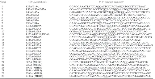
Plasmid constructions and mutagenesis.All complementary primers contain-ing site-directed mutations (Table 1) were purchased from Integrated DNA Technology (Coralville, IA). All mutants were created in the pGEM3Zf(⫹ )-GAG3 plasmid, which was constructed by ligation of the 2,235-bp BamHI-KpnI Ty3 fragment of plasmid pDLC201 (formerly pEGTy3-1, 2,URA3, galactose-inducible Ty3-1) (42) into the BamHI-KpnI sites of pGEM3Zf(⫹) (Promega, Madison, WI). The pGEM3Zf(⫹)-GAG3 plasmid was isolated from DH5␣cells and used as a template for QuikChange mutagenesis, according to the manu-facturer’s instructions (Stratagene Inc., Cesar Creek, TX). The sequences of mutated plasmids were determined (Cogenics Inc., Houston, TX). pGEM3Zf(⫹ )-GAG3 mutants with sequence-verified mutations were selected and digested with BamHI and KpnI and ligated into the BamHI-KpnI sites of pDLC201. Each mutation was verified after recloning into pDLC201.
In order to localize mutant Ty3 relative to P bodies, the putative assembly site, Ty3 marked with red fluorescent protein (RFP) was expressed in a strain in which a P-body marker protein was fused to green fluorescent protein (GFP). This strategy was previously described and used to show that wild-type (wt) Ty3 proteins are concentrated in P bodies. In the Ty3-RFP construct previously
described (6), the coding region for RFP optimized for expression inS. cerevisiae
(Coda Genomics, Inc., Laguna Hills, CA) was fused in frame to the end of the Ty3POL3reading frame in the high-copy-number expression vector pDLC201. Ty3-RFP expression and VLP formation result in PR processing of Gag3-Pol3 and generation of an IN-RFP fusion. Twenty-one Ty3-RFP CA mutant variants were created by substituting the BamHI-KpnI fragment containing the mutation in the respective pDLC201-based plasmid for the wt version of this fragment in Ty3-RFP. The presence of the mutation in the full-length Ty3 was reconfirmed by DNA sequence analysis of the region containing the mutation (Cogenics Inc.). Structure predictions.The secondary structure of the Ty3 CA protein was predicted by a consensus approach. The secondary structures of Ty3 CA are consensus predictions made by SSpro4 (19), PSIPRED (13), ProfSec (76), SAM-T99 (49), and SABLE2 (2). As the accuracy of these state-of-the-art secondary structure predictors is high (about 75 to 80%), but not perfect, the confidence scores of the secondary structure predictions generated by PSIPRED were in-cluded. The confidence scores for each position range from 0 to 9 (low confi-dence to high). The secondary structure prediction showed that, like retrovirus CA proteins, Ty3 CA is high in helical structure.
The tertiary structure of Ty3 CA was also predicted, based on the assumption that it shares its structural fold with retrovirus CA proteins. To test the validity of this assumption, the Ty3 CA was submitted to several recognition (or thread-ing) servers to look for similar structures. Surprisingly, one server, FFAS03 (47), ranked HTLV-1 (50), HIV-1 (8, 20, 63), RSV (17), and EIAV (10) CA proteins within the top 10 templates, despite the lack of sequence similarity between Ty3 CA and the other CA proteins. More specifically, the CA proteins of EIAV and HIV-1 ranked first and second, respectively. Two other recognition servers, Meta-BASIC (38) and FOLDpro (18), also ranked one of the retrovirus proteins within the top templates. Although not all recognition servers returned one or more retrovirus CA proteins as templates, the overlap between the independent predictions made by FFAS03, MetaBASIC, and FOLDpro provides some evi-dence that Ty3 CA may have a nontrivial degree of structural similarity with other retrovirus CA proteins.
[image:3.585.45.547.82.338.2]To further assess the tertiary structure of Ty3 CA, the sequences and struc-tures of the CA proteins of HTLV-1 (1QRJ) (50), RSV (1D1D) (17, 51), EIAV (1IEA) (10), and HIV-1 (1E6J) (8, 39, 63) were extracted from the Protein Data Bank (7). Because sequence similarity between the Ty3 CA protein and other retroviruses is very low, profile-profile alignment methods, which are more sen-sitive than sequence-sequence or profile-sequence alignment methods, were used to align the Ty3 CA protein with the four retrovirus proteins. The profiles of Ty3 TABLE 1. Primers used to create CA mutations
Primer Ty3 CA mutation(s) 5⬘–3⬘(forward) sequencea
1 K15A/E19A GGAGGAAATTATCCAGCACTCCCAGTAGCATGCCTTCCTAAC
2 R33A/R35A/D37A CCAACCATCTTTGACCTTCGCAGGTGCAAATGCCTCGCATAAAC
3 K40A/K42A CAGAGGTAGAAATGACTCGCATGCACTGGCAAACTTTATCTCCG
4 D60A/R63A GATATCTTGGCCGAATGCTGCCAGTGCTATTGTGTACTGCAG
5 R68A/R69A CAGTCGTATTGTGTACTGCGCAGCACATTTATTAAACCCCGCTGC
6 D81A/E85A GCTCAGTGGGCTAATGCCTTTGTACAAGCACAAGGTATAC
7 E90A/D94A GAACAAGGTATACTTGCAATAACATTCGCCACATTCATACAAGG
8 K106A/D109A GGATTATATCAGCATTTCTATGCCCCACCAGCTATCAATAAAATC
9 E122A/K124A GCAATCACGCAACTTTCCGCAGCTGCACTTGGTATTGAGCGTCTC
10 E128A/R129A CCGAAGCTAAACTTGGTATTGCCGCTCTCAACCAACGATTCAG
11 R133A/R135A/K136A GCGTCTCAACCAAGCATTCGCAGCCATTTGGGACAGAATGCCACC
12 D139A/R140A GATTCAGAAAGATTTGGGCCGCAATGCCACCAGACTTCATGACCG
13 E148A/K149A GCCACCAGACTTCATGACCGCAGCAGCTGCCATAATGAC
14 K161A/E162A CTAGGCTATTGACAGCCGCAACCTATAATATTGTCAGAATGCAC
15 K171A/E173A GTCAGAATGCACGCACCAGCCACATTAAAAGACGCCATGGAAGAG
16 K176A/D177A GCACAAACCAGAGACATTAGCAGCCGCCATGGAAGAGGCTTACC
17 E180A/E181A GAGACATTAAAAGACGCCATGGCAGCCGCTTACCAGACAACTGC
18 E190A/R191A CCAGACAACTGCACTAACTGCAGCATTCTTCCCAGGATTCG
19 E197A/D199A CCCAGGATTCGCACTTGCTGCTGATGGAGACACTATCATCGG
20 D201A/D203A CGAACTTGATGCTGCTGGAGCCACTATCATCGGTGCCAC
21 Q86A (MHR1) GCTCAGTGGGCTAATGACTTTGTACAAGAAGCAGGTATACTTG
22 G87A (MHR2) GACTTTGTACAAGAACAAGCTATACTTGAAATAACATTCGACAC
23 E90A (MHR3) GTACAAGAACAAGGTATACTTGCAATAACATTCGACACATTC
24 F93A (MHR4) GAACAAGGTATACTTGAAATAACAGCCGACACATTCATACAAGG
25 F96A (MHR5) CATTCGACACAGCCATACAAGGATTATATCAGCATTTCTATAAGC
26 L100A (MHR6) CGACACATTCATACAAGGAGCATATCAGCATTTCTATAAGCCACC
aMutated codons are underlined.
on November 8, 2019 by guest
http://jvi.asm.org/
CA and the other four proteins were generated by using PSI-BLAST to search the nonredundant protein sequence database. The profile of the Ty3 CA protein was aligned with those of the four retroviruses by ClustalW (90) and PALIGN (68). ClustalW and PALIGN generated almost exactly the same alignments for Ty3 and HIV-1 and very similar alignments for Ty3 and EIAV, but the align-ments with HTLV-1 and RSV were more divergent. Thus, we chose to use the HIV-1 CA template to predict the structure of Ty3 CA. Based on the alignment of HIV-1 CA and Ty3 CA generated by ClustalW, Modeler (79) was used to generate the energy minimized tertiary structure of Ty3 CA.
Transposition assays.Transposition was measured with a target plasmid assay described previously (52). Transposition was induced from Ty3 under transcrip-tional control of theGAL1-10upstream activation sequence (UAS) on a plasmid by growth on galactose-containing medium. Transposition of Ty3 into the low-copy-number target plasmid pCH2bo19V activates expression of a suppressor tRNA gene. Transposition is scored by growth dependent on suppression of the ochre nonsense allelesade2-101andlys2-1in yeast strain yTM443 (52, 61). Transposition assays were performed at 24°C and 30°C. Under these conditions, four independent transformants for each mutant were patched onto SD-Ura-His medium. After 1 day, cells were replica plated to SG-Ura-His to induce trans-position. After 2 days, cells were replica plated to minimal medium supple-mented with Leu and Trp to select for Ade⫹and Lys⫹colonies.
Immunoblot analysis of Ty3 intermediates in extracts of cells expressing Ty3. Two transformants containing pDLC201 wt or mutant derivative plasmid and target plasmid were grown overnight at 30°C in 3 ml of noninducing synthetic raffinose (SR)-Ura medium. Five milliliters of SR-Ura was added to the 3-ml overnight culture. Cells were grown for 5 h, diluted into inducing medium (2% SG-Ura) to a final optical density at 600 nm (OD600) of 0.35, and grown for 24 h
at 24°C. Whole-cell extracts (WCEs) were prepared from 5-ml cultures by vor-texing with glass beads in the presence of denaturing buffer (9 M urea–5 mM EDTA) (Continental Labs, Carlsbad, CA), as previously described. WCEs were fractionated by electrophoresis in a 4 to 20% sodium dodecyl sulfate (SDS)-polyacrylamide gel (Pierce Biotechnology, Inc., Rockford, IL). Proteins were transferred to Immobilon-P membranes (Millipore, Bedford, MA) and were incubated with rabbit polyclonal anti-Ty3 CA or anti-Ty3 IN antibodies diluted 1:20,000 and 1:2,000, respectively (61). Rabbit immunoglobulin G was visualized with horseradish peroxidase-conjugated secondary antibody and ECL Plus (Am-ersham, Buckinghamshire, United Kingdom), according to the manufacturer’s instructions. The blot used to probe for IN was also used to probe for phospho-glycerate kinase (PGK) by using anti-phosphophospho-glycerate kinase immunoglobulin G (Invitrogen, Carlsbad, CA) (data not shown).
Southern blot analysis of Ty3 cDNA in extracts of cells expressing Ty3.Cells of yeast strain yTM443 transformed with pDLC201 mutant derivative plasmids and the target plasmid pCH2bo19V were grown as described above for immu-noblot analysis. Five milliliters of induced cells was suspended in extraction buffer (10 mM Tris-HCl [pH 8.0], 1 mM EDTA, 100 mM NaCl, 2% Triton X-100, 1% SDS) and vortexed vigorously with glass beads in the presence of phenol-chloroform-isoamyl alcohol (25:24:1). Nucleic acid was precipitated from the aqueous phase with ethanol, redissolved, and digested with RNase A (43). RNA-free DNA (1.0g) was digested with EcoRI, fractionated by electrophore-sis in a 0.8% agarose gel, transferred to a Duralon UV membrane (Stratagene, La Jolla, CA), and immobilized by UV cross-linking in a Stratalinker 1800 instrument (Stratagene). Hybridization was performed with a32P-labeled
inter-nal BglII fragment of Ty3, which hybridizes with the full-length 5.4-kbp Ty3 cDNA, as well as Ty3 donor plasmid and chromosomal Ty3 elements. The filters were washed and exposed to a Personal Molecular Imager FX phosphorimager screen (Bio-Rad Inc., Richmond, CA), and hybridization signals were quanti-tated by Quantity One software (Bio-Rad Inc.).
RNA protection assays.Yeast strain yTM443 was grown under conditions of Ty3 expression for 24 h. Five milliliters of induced cells was suspended in 100l of extraction buffer (100 mM KCl, 10 mM Tris-HCl [pH 7.5], 10 mM EDTA, 2.1
g/ml aprotinin, 6.6g/ml pepstatin, 2 mM phenylmethylsulfonyl fluoride, 0.66
g/ml leupeptin, and 5.3 U RNase inhibitor) and vortexed vigorously with sterile glass beads. An additional 150l of extraction buffer was added, and WCEs were cleared by centrifugation at 500⫻gfor 5 min at 4°C. WCEs were divided into three fractions of 65l each. In order to test for protection of genomic RNA, extracts were incubated with the nuclease benzonase. Ty3 control RNA was generated in vitro by T7 RNA polymerase transcription of a Ty3 template plasmid linearized at the downstream end of Ty3 by digestion with SnaBI. WCEs were processed by a modification of the benzonase assay previously described (59), except that 5g of in vitro-transcribed Ty3 RNA was added per reaction. Two of the fractions were incubated, one on ice and one at 24°C, with 15l of RNase-free benzonase buffer (100 mM Tris [pH 7.5], 100 mM MgCl2) for 7 min.
The third fraction was incubated at 24°C for 1 min, followed by the addition 5l
of benzonase buffer and 36 U of benzonase and incubation for 6 min. The benzonase reaction was terminated by the addition of 5l of 0.5 M EDTA, followed by 300l of BSE buffer (10 mM Tris [pH 7.5], 10 mM EDTA, 2% SDS), and RNA was extracted with 300l of phenol-chloroform-isoamylalcohol (25: 24:1). This extraction was repeated three more times, and the aqueous layer was precipitated with 2 volumes of ethanol. The pellet was resuspended in 25l of diethyl pyrocarbonate-treated water with 10 U of RNase inhibitor. Two micro-liters was denatured in 6.7% glyoxal for 1 h at 50°C and resolved by agarose gel electrophoresis in a 1% agarose gel in 10 mM sodium phosphate buffer (pH 7.0). The RNA was transferred to a Duralon UV membrane (Stratagene, La Jolla, CA) and immobilized by UV cross-linking in a Stratalinker 1800 instrument (Stratagene). Hybridization was performed with a 32P-labeled internal BglII
fragment of Ty3. Blots were processed as described previously (80).
Particle analysis by equilibrium gradient centrifugation and sedimentation. Conditions for fractionation of Ty3 VLP forms were derived empirically to maximize the separation of immature and mature VLP components (V. Bilan-chone, unpublished data). In the protocol, cell extracts are prepared by lysis under nondenaturing conditions and are fractionated by centrifugation to equi-librium at 100,000⫻gon 20% to 60% (wt/wt) sucrose gradients for 17 h at 4°C. Extracts of cells induced for short times (e.g., 2 h at 24°C) show Gag3 protein near the bottom of the gradient, as do extracts of cells expressing PR mutants that fail to process (V. Bilanchone, unpublished). Extracts of cells induced to express Ty3 for longer times (e.g., 6 h) show processed forms of Gag3 near the middle of the gradient. CA that is no longer associated with VLPs migrates at the top of these gradients.
YTM443 cells transformed with wt and representative mutant derivative Ty3 plasmids were induced for Ty3 expression by growth in galactose for 24 h at 24°C. Ten milliliters of induced cells was resuspended in 300l of STEK10 buffer (10% [wt/wt] sucrose, 10 mM Tris-HCl [pH 7.5], 10 mM EDTA, 100 mM KCl [pH 7.5]) with protease inhibitors (1 mM phenylmethylsulfonyl fluoride, 1g/ml pepstatin, 2.1g/ml aprotinin, 16 U RNase inhibitor) and vortexed vigorously with sterile glass beads. Three hundred microliters of cleared extract was fractionated by equilibrium sedimentation over a 20/30/60% (wt/wt) linear sucrose gradient by centrifugation in a SW55 Ti rotor at 100,000⫻gfor 17 h at 4°C. Fourteen 340-l fractions were successively removed from the top of the gradient. For each fraction, 70-l aliquots were used for protein analysis. The 70-l aliquot of each fraction was trichloroacetic acid precipitated in the presence of 0.013% deoxycholate and fractionated by electrophoresis in a 4 to 20% SDS-polyacryl-amide gel (Pierce Biotechnology Inc., Rockford, IL) for immunoblot analysis as described above.
In order to further test for particle formation, WCEs were subjected to velocity sedimentation. Cells of yeast strain yTM443 transformed with pDLC201 mutant derivative plasmids and the target plasmid pCH2vo19V were grown as described above for immunoblot analysis. WCEs from 5 ml of induced cells were prepared as described for RNA protection assays. Cleared WCEs were divided into two portions: one untreated and the other adjusted to a final concentration of 1% Triton X-100. Both were incubated for 10 min at 24°C, followed by centrifugation in a TLA 100.3 rotor at 100,000⫻gfor 30 min. Pellets were resuspended in equal volumes of supernatants, and fractions for both treated and untreated samples were fractionated by electrophoresis in a 4 to 20% SDS-polyacrylamide gel and analyzed by immunoblotting with anti-Ty3 CA and anti-Ty3 IN, as described previously.
Fluorescence microscopy.The proposed assembly site for Ty3 is the P body. The ability of different CA mutants to localize to the P body was therefore determined. Fluorescence microscopy of wt and mutant variants of Ty3-RFP in a strain in which the P body was visualized by the P-body marker protein Dhh1-GFP was as previously described (6). Induction of Ty3 expression was as described above for immunoblot analysis except that inductions were from 18 to 24 h. One hundred fifty cells representing each of two independent transformants were observed for 21 mutants that were defective for transposition and 2 that transposed at wt frequencies.
EM. Cells of yeast strain BY4741 transformed with pDLC201 or mutant derivative plasmids were grown in noninducing SR-Ura medium until early log phase, induced by the addition of galactose to 2%, and grown for 6 h at 24°C. Cells (12 OD units) were prepared for EM as described previously (89) except that cell walls were digested with 2 ml potassium phosphate buffer, 0.25 mg/ml Zymolyase 100T (U.S. Biological, Swampscott, MA), and 90 mM -mercapto-ethanol for 20 min at 30°C. Cells were imaged with a transmission electron microscope (H7600; Hitachi, Tokyo, Japan). Approximately 100 cells were ob-served per mutant transformant. Cells induced for this experiment were firmed to have been induced and to have a pattern of protein expression con-sistent with previously described transformants by immunoblot analysis using anti-CA (data not shown).
on November 8, 2019 by guest
http://jvi.asm.org/
RESULTS
Ty3 CA structure model.A consensus approach was used to generate a secondary structure prediction for Ty3 CA. Pre-dicted helical regions of Ty3 CA account for 58% of the entire sequence (Fig. 1C). This is comparable to the structure-based helical compositions of HTLV-1, RSV, EIAV, and HIV-1, which are 57%, 58%, 60%, and 63%, respectively (8, 10, 17, 20, 34, 35, 39, 48, 50, 51, 62, 63). In the case of each retrovirus CA, helices are separated into NTD and CTD by a flexible loop region. In Ty3 CA, a 7-aa loop region separates the predicted NTD (aa 2 to 139) and CTD (aa 148 to 207) helical clusters. No structure could be predicted for amino-terminal residues 7 to 17 and carboxyl-terminal residues 193 to 207. It is presumed that they are flexible or adopt multiple conformations. In this prediction, the Ty3 MHR spans a loop-helix junction in the NTD. This differs from the retroviral CA MHR, which over-laps the beginning of the CTD.
Ala-scanning mutagenesis strategy.Residues of Gag3 within hydrophilic surface patches potentially mediate interactions with other Gag3 molecules, genomic RNA, or host proteins that are important for VLP assembly. An Ala-scanning mu-tagenesis strategy was used to specifically address the roles of these residues in assembly (Fig. 1D). A total of 18 double and 2 triple mutants were generated, resulting in a total of 42 residues converted to Ala. In addition, the six conserved amino acids in the Ty3 MHR (residues 86 to 104) were individually converted to Ala. Mutants were characterized by transposition, immunoblot, Southern blot, RNA protection, equilibrium velocity gradient, particle sedimentation, fluorescence
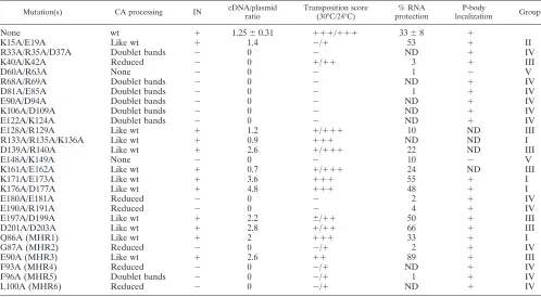
micros-copy, and EM analyses. The properties of each mutant are summarized in Table 2.
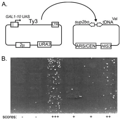
Retrotransposition of Ala-scanning mutants. A qualitative genetic assay was performed to determine the effects of CA mutations on retrotransposition frequency (52) (Fig. 2; Table 2). Two independent transformants were tested for wt Ty3 and each mutant at 24°C and 30°C.
Four of the 26 mutants [R133A/R135A/K136A, K171A/ E173A, K176A/D177A, and Q86A (MHR1)] had frequencies of transposition that were indistinguishable from that of the wt at both temperatures. Seven of the mutants displayed signifi-cant levels of transposition but were defective at one or both temperatures [K40A/K42A, E128A/R129A, D139A/R140A, K161A/E162A, E197A/D199A, D201A/D203A, and E90A (MHR3)]. Fifteen of the mutants were severely blocked for transposition at both temperatures [K15A/E19A, R33A/R35A/ D37A, D60A/R63A, R68A/R69A, D81A/E85A, E90A/D94A, K106A/D109A, E122A/K124A, E148A/K149A, E180A/E181A, E190A/R191A, G87A (MHR2), F93A (MHR4), F96A (MHR5), and L100A (MHR6)]. Of the 18 mutants with mutations in the amino-terminal 140 residues of CA, 12 were severely disrupted for transposition. The strongest temperature dependence was observed for three mutants with Ala substitutions near the predicted interdomain loop region (E128A/R129A, D139A/ R140A, and K161A/E162A).
[image:5.585.43.541.81.355.2]Polyprotein processing. YTM443 transformants carrying pDLC201, the Ty3 expression plasmid, with wt Ty3 or mutant derivatives were grown in SR-Ura medium. Cultures were in-duced for Ty3 expression by the addition of galactose to 2% TABLE 2. Ty3 CA mutant phenotypesa
Mutation(s) CA processing IN cDNA/plasmid ratio
Transposition score (30°C/24°C)
% RNA protection
P-body
localization Group
None wt ⫹ 1.25⫾0.31 ⫹⫹⫹/⫹⫹⫹ 33⫾8 ⫹
K15A/E19A Like wt ⫹ 1.4 ⫺/⫹ 53 ⫹ II
R33A/R35A/D37A Doublet bands ⫺ 0 ⫺ ND ⫹ IV
K40A/K42A Reduced ⫺ 0 ⫹/⫹⫹ 3 ⫹ III
D60A/R63A None ⫺ 0 ⫺ 1 ⫺ V
R68A/R69A Doublet bands ⫺ 0 ⫺ ND ⫹ IV
D81A/E85A Doublet bands ⫺ 0 ⫺ 1 ⫹ IV
E90A/D94A Doublet bands ⫺ 0 ⫺ ND ⫹ IV
K106A/D109A Doublet bands ⫺ 0 ⫺ ND ⫹ IV
E122A/K124A Doublet bands ⫺ 0 ⫺ ND ⫹ IV
E128A/R129A Like wt ⫹ 1.2 ⫹/⫹⫹⫹ 10 ND III
R133A/R135A/K136A Like wt ⫹ 0.9 ⫹⫹⫹ ND ND I
D139A/R140A Like wt ⫹ 2.6 ⫹/⫹⫹⫹ 22 ND III
E148A/K149A None ⫺ 0 ⫺ 10 ⫺ V
K161A/E162A Like wt ⫹ 0.7 ⫹/⫹⫹⫹ 24 ND III
K171A/E173A Like wt ⫹ 3.6 ⫹⫹⫹ 55 ⫹ I
K176A/D177A Like wt ⫹ 4.8 ⫹⫹⫹ 48 ⫹ I
E180A/E181A Reduced ⫺ 0 ⫺ 2 ⫹ IV
E190A/R191A Reduced ⫺ 0 ⫺ 4 ⫹ IV
E197A/D199A Like wt ⫹ 2.2 ⫾/⫹⫹ 50 ⫹ III
D201A/D203A Like wt ⫹ 2.8 ⫹/⫹⫹ 66 ⫹ III
Q86A (MHR1) Like wt ⫹ 2 ⫹⫹⫹ 33 ⫹ I
G87A (MHR2) Reduced ⫺ 0 ⫺/⫹ 2 ⫹ IV
E90A (MHR3) Like wt ⫹ 2.6 ⫹⫹ 89 ⫹ III
F93A (MHR4) Reduced ⫺ 0 ⫺/⫹ ND ⫹ IV
F96A (MHR5) Doublet bands ⫺ 0 ⫺/⫹ 1 ⫹ IV
L100A (MHR6) Reduced ⫺ 0 ⫺/⫹ ND ⫹ IV
a
ND, not determined.
on November 8, 2019 by guest
http://jvi.asm.org/
(SG-Ura) and were grown for 24 h at 24°C. Proteins were extracted under denaturing conditions, and WCE were sub-jected to immunoblot analysis using antibodies against CA (Fig. 3A) or IN (Fig. 3B). Most mutants produced significant amounts of Ty3 protein, indicating that in most cases, Gag3 stability was not the only factor accounting for decreased trans-position. In cells expressing Ty3 for long periods of time, dur-ing which they enter stationary phase, Gag3 also undergoes limited background proteolysis by cytoplasmic proteases to produce products similar in size to p27 and CA (data not shown). Therefore mutations that fully blocked PR activation could not be unambiguously identified. Nevertheless, the
ex-tent of Gag3 processing correlated well with the severity of mutant transposition phenotypes.
Extracts of cells expressing wt Ty3 and each of the four mutants with wt levels of transposition [R133A/R135A/K136A, K171A/E173A, K176A/D177A, and Q86A (MHR1)] showed similar amounts of Gag3 and p27 and a significantly greater amount of CA than either other species (Fig. 3A). These ex-tracts showed similar amounts of IN (Fig. 3B).
Seven mutants showed significant, but attenuated, transpo-sition at one or both temperatures [K40A/K42A, E128A/ R129A, D139A/R140A, K161A/E162A, E197A/D199A, D201A/ D203A, and E90A (MHR3)]. These mutants generally showed levels of CA approaching that found in wt and detectable IN. Only the mutant that was lowest for transposition (K40A/ K42A) had amounts of Gag3 greater than CA and no detect-able IN.
Fifteen mutants were severely reduced in transposition. Sur-prisingly, one of these (K15A/E19A) showed a wt pattern of protein maturation for Gag3 and IN. Fourteen mutants showed reduced or no Gag3 processing and no detectable IN. These mutants were differentiated into three classes based on the pattern of Gag3 and IN immunoblot analysis: (i) lim-ited and aberrant processing (R33A/R35A/D37A, R68A/R69A, D81A/E85A, E90A/D94A, K106A/D109A, and E122A/K124A); (ii) severely reduced processing [E180A/E181A, E190A/R191A, G87A (MHR2), F93A (MHR4), F96A (MHR5), and L100A (MHR6)], and (iii) no detectable processing (D60A/R63A and E148A/K149A). Overall, based on inferences of apparent pro-cessing of Gag3, multimerization was robust. The extracts of mutants in the first and second classes generally had higher levels of Gag3 than were observed in extracts of cells express-ing wt Ty3, suggestexpress-ing that Gag3 was accumulatexpress-ing in a stable form. Only two mutants (D60A/R63A and E148A/K149A) showed Gag3 but completely lacked detectable smaller species. In contrast to extracts of cells expressing other mutants with severely reduced processing, which showed accumulation of Gag3, these extracts had amounts of Gag3 similar to extracts of cells expressing wt Ty3, consistent with reduced levels of ex-pression or instability.
Based on the foregoing analysis of transposition and protein processing, mutants were classified as follows: group I, not measurably affected [R133A/R135A/K136A, K171A/E173A, K176A/D177A, and Q86A (MHR1)]; groups II and III, mod-FIG. 2. Analysis of Ty3 retrotransposition. (A) Strategy. Ty3 and
[image:6.585.64.260.67.261.2]mutant variants were transformed into yTM443 and expressed on a high-copy-numberURA3-marked plasmid. Transposition into the low-copy-number target plasmid activates expression of thesup2boochre suppressor required for growth on selective medium. LTRs are indi-cated as boxes; theGAL1-10UAS is indicated as a gray portion where it replaces part of the upstream LTR to confer galactose regulation. (B) Qualitative transposition assay. Activation of suppression by trans-position into the target allows colony growth on minimal medium supplemented with Leu and Trp. Cells with transpositions become papillations on selective medium, and phenotype is scored for several independent mutants (⫺,⫾,⫹,⫹⫹, and⫹⫹⫹). Patterns are shown for a representative sample of mutants at 24°C.
FIG. 3. Immunoblot analysis of Ty3 proteins in WCE. (A) Immunoblot analysis of Ty3 Gag3 proteins. Cultures were induced for Ty3 expression for 24 h at 24°C, and WCEs were prepared under denaturing conditions as described in Materials and Methods. Equivalent volumes of WCEs from cells expressing the indicated mutations were fractionated by electrophoresis on a 4 to 20% SDS-polyacrylamide gel and analyzed by immuno-blotting using polyclonal anti-CA. The arrow indicates a lower-molecular-weight protein that appears at later times of expression. (B) Immunoblot analysis of Ty3 IN. WCEs were prepared and processed for immunoblotting as described for panel A except that polyclonal antibody against IN was used.
on November 8, 2019 by guest
http://jvi.asm.org/
[image:6.585.42.540.581.668.2]est to no detectable defects in protein processing [K15A/E19A, K40A/K42A, E128A/R129A, D139A/R140A, K161A/E162A, E197A/D199A, D201A/D203A, and E90A (MHR3)]; group IV, severely blocked prior to or during protein processing [R33A/R35A/D37A, R68A/R69A, D81A/E85A, E90A/D94A, K106A/D109A, E122A/K124A, E180A/E181A, E190A/R191A, G87A (MHR2), F93A (MHR4), F96A (MHR5), and L100A (MHR6)]; and group V, present in small amounts with no protein processing (D60A/R63A and E148A/K149A). Mutant K15A/E19A was later designated group II based on its normal processing and cDNA pattern, which indicated a later defect in transposition than appeared to be the case for the other mu-tants in group III.
Reverse transcription. Mutations in CA produced a wide range of Gag3 and Gag3-Pol3 processing phenotypes, and many mutants that displayed some processing were signifi-cantly attenuated in transposition. In order to further differ-entiate the effects of these mutations, Ty3 extrachromosomal cDNA was measured in cells induced as described above (Fig. 4). Group I mutants all produced cDNA. The group II mutant, which failed to transpose but displayed normal processing, had a cDNA-to-plasmid ratio in the same range as wt Ty3. The seven group III mutants had detectable cDNA, with the ex-ception of mutant K40A/K42A, which also showed less Gag3 processing. None of the group IV or V mutants had detectable cDNA.
Overall, these results showed clear and parallel effects of mutations in CA on Gag3 processing and accumulation of reverse transcripts. As was observed for protein processing, clusters of mutations with similar effects on cDNA production appeared to define CA subdomains. Examples of mutations defining such subdomains were 12 mutations in the proximal domain of the NTD between residues 33 and 124 (negative for cDNA), 7 mutations between residues 128 and 177 (with one exception, positive for cDNA), E180A/E181A and E190A/ R191A (negative for cDNA), and E197A/D199A and D201A/ D203A (positive for cDNA). Overall, mutations that attenu-ated processing resulted in complete loss of cDNA. This is consistent with the drastic reduction in reverse transcription activity previously observed for a Ty3 PR catalytic mutant (53).
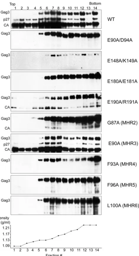
Complex formation.Mutants that produced no cDNA showed a range of processing phenotypes. In order to better correlate the protein processing phenotypes with assembly, extracts of cells expressing representative mutants from groups III to V were subjected to equilibrium gradient sedimentation analysis. As expected, extracts of cells expressing wt Ty3 contained a mixture of immature and mature VLP forms. As with obser-vations in previous experiments (unpublished data), Gag3 was distributed from a density of 1.13 mg/ml to 1.23 mg/ml (Fig. 5), whereas mature CA was more concentrated, from 1.13 to 1.16 mg/ml. This region of the gradient is presumed to include mature VLPs. The group III mutant [E90A (MHR3)], which was transposition competent, showed a pattern very similar to that found in cells expressing wt Ty3.
The seven group IV mutants, which were severely reduced in transposition, protein processing, and amount of cDNA, showed patterns on the equilibrium gradients similar to those of a PR catalytic site mutant, which has been shown to be competent to assemble particles (55). This pattern was also similar to that of immature particles observed in extracts of cells expressing Ty3 for only 2 h (unpublished data). Immuno-blot analysis of cells expressing mutant E180A/E181A (Fig. 5) showed a peak of Gag3 in the bottom-most fraction, with detectable amounts extending upward in the gradient. Mutant E190A/R191A also showed the greatest amount of Gag3 in the bottom of the gradient but in addition showed small amounts of processed forms in the same region as CA in extracts of cells expressing wt Ty3. Interestingly, extracts of the four MHR mutants in this group, which were among the most severely attenuated for protein processing, showed that mutant Gag3 accumulated at densities similar to those of immature wt par-ticles.
The two group V mutants showed only Gag3, indicating that they failed to process and suggesting that they were potentially defective in some early aspect of particle formation. In the equilibrium gradient, Ty3 protein was undetectable in the ly-sates of cells expressing D60A/R63A (data not shown). Gag3 was also not detected in the pellet of the low-speed clarifica-tion spin that preceded gradient analysis (data not shown). Extracts of cells expressing the other group V mutant, E148A/ FIG. 4. Southern blot analysis of mutant Ty3 cDNA. Total nucleic acid was purified from cells induced for Ty3 expression as described in the legend to Fig. 3, and DNA was cleaved with EcoRI to linearize the Ty3 expression plasmid. Southern blot analysis was performed with a32P-labeled Ty3-specific probe, which hybridizes with the full-length 5.4-kbp Ty3 cDNA (cDNA), as well as with Ty3 donor plasmid (plasmid). The Ty3 cDNA/plasmid ratio for four independent wt cultures was 1.3⫾0.3. Measurements were normalized to this ratio.
on November 8, 2019 by guest
http://jvi.asm.org/
K149A, showed very small amounts of Gag3 precursor migrat-ing similarly to Gag3 in wt particles (Fig. 5).
Equilibrium gradient centrifugation profiles of WCEs of cells expressing wt and group III, IV, and V mutants suggested that most or all were able to form particles. Although equilib-rium gradient analysis showed that mutant Gag3 could be sedimented, it was possible that it was not particulate but associated with cellular membranes, causing it to sediment. In order to investigate this possibility, production of pelletable material in the presence and absence of detergent was also tested. WCEs obtained under nondenaturing conditions from cells expressing representative group IV and V mutants [E148A/
K149A, E190/R191A, G87A (MHR2), F93A (MHR4), and F96A (MHR5)] were incubated in the presence or absence of 1% Tri-ton X-100 and centrifuged at 100,000⫻gfor 30 min. In all cases examined, Ty3 Gag3 species sedimented, indicating that sedi-mentation was not Triton-soluble membrane dependent (Fig. 6). Examination of pellets and supernatants for Gag3-Pol3-related species using anti-IN antibody showed that processed IN was present in significant amounts only in the wt. Much lower amounts of partially processed Gag3-Pol3 species were present in the extracts of cells expressing each of the three MHR mutants (Fig. 6). Extracts of cells expressing E148A/ K149A and E190A/R191A showed no detectable Gag3-Pol3 (IN)-related species.
Protection of genomic RNA.Gradient analysis indicated that most mutants were probably competent for some level of par-ticle formation but did not differentiate among them with re-spect to particle integrity. Benzonase is a nuclease that de-grades RNA and DNA. Genomic RNA of Tf1 retrotransposon VLPs was previously shown to be protected against benzonase digestion, making resistance to digestion an indirect measure of packaging (59). Wt Ty3 and Ty3 PR mutant (immature) VLPs have been similarly shown to protect Ty3 RNA (unpub-lished data). The benzonase protection assay was used to mon-itor Ty3 genomic RNA packaging.
Lysates of cells expressing wt or mutant Ty3 were supple-mented with control in vitro-generated Ty3 transcripts and treated with benzonase as described in Materials and Methods. Nucleic acid in these extracts was analyzed by Northern blot-ting using a Ty3-specific probe. Under these conditions, 33% (⫾8%) of wt Ty3 RNA was protected from benzonase diges-tion compared to untreated control extracts incubated on ice or at 24°C. Control in vitro Ty3 transcripts added to the cell extracts were completely sensitive to degradation (Fig. 7). A subgenomic 3.1-kb Ty3 RNA was previously reported (9) and was also observed in these experiments. It showed a pattern of protection similar to that of the genomic Ty3 RNA. However, it fractionated with mobility similar to that of 25S rRNA, which is degraded in the incubation. This loss of RNA in the same region of the gel affected the mobility and efficiency of filter transfer of the associated Ty3 RNA and made its measurement less accurate (data not shown).
[image:8.585.48.283.67.500.2]Eighteen mutants representing groups I to V were tested for protection of Ty3 RNA from benzonase digestion (Fig. 7 and Table 2). The group I mutants tested [K171A/E173A, K176A/ D177A, and Q86A (MHR1)] showed protection of RNA from 33% to 55%, the group II mutant (K15A/E19A) showed 53% protection, and group III mutant [E90A (MHR3)] showed 89% protection. These results were consistent with the ability of these mutants to produce cDNA. The K40A/K42A mutant, which showed the least transposition and the most-defective Gag3 processing pattern of the group III mutants and no detectable cDNA, showed only 3% protection. The five group IV mutants that were tested (including two MHR single-sub-stitution mutants) showed protection of only 1% to 4% of the Ty3 RNA. This result was consistent with the severe defect in transposition and the uniform absence of detectable cDNA for cells expressing group IV mutants. The two group V mutants, which were the most severely affected by all other criteria, were also defective in genomic RNA protection. Mutant D60A/ R63A protected 1% of the Ty3 RNA and mutant E148A/ FIG. 5. Equilibrium gradient analysis of Ty3 wt and representative
mutant WCEs. Cultures were induced for Ty3 expression as described in the legend to Fig. 3. WCEs were prepared under native conditions as described in Materials and Methods and were fractionated over a 20 to 60% (wt/wt) linear sucrose gradient and centrifuged to equilibrium. Fourteen 340-l fractions were collected and examined by immunoblot analysis using anti-CA, as described in the legend to Fig. 3.
on November 8, 2019 by guest
http://jvi.asm.org/
K149A protected 10% of the RNA. These results showed a strong correlation between the ability of particles to protect genomic RNA and the presence of cDNA.
Northern analysis of untreated RNA samples showed that less RNA was recovered in the case of group V mutants D60A/ R63A and E148A/K149A than for the other mutants. Investi-gation of localization of Ty3 proteins (see below) showed that a smaller proportion of cells was positive for expression of these mutants than for the wt. However, the proportion was similar to that for some other mutants that produced substantial amounts of RNA (data not shown). RNA was also extracted under denaturing conditions from cells
ex-pressing these mutants, and Northern analysis confirmed that Ty3 RNA was reduced (data not shown). Overall, group V mutants had reduced RNA and Gag3 protein and defec-tive packaging.
[image:9.585.43.542.68.311.2]Localization of assembly.Ty3 RNA, protein, and VLPs con-centrate in association with P-body components (6). We have proposed that Ty3 assembles in association with P bodies, potentially as a mechanism for sequestering RNA packaging from translation. In order to test for a role for CA in Ty3 localization to P bodies, fragments containing 21 CA mutations representing groups I to V were swapped into Ty3-RFP and expressed under control of theGAL1-10 UAS from a high-FIG. 6. Particle formation by representative mutants. Cells were induced and WCEs were prepared as described in the legend to Fig. 5. Aliquots of extracts were either analyzed directly or mixed with Triton X-100 for a final concentration of 1%. Extracts were analyzed by immunoblotting using anti-CA and anti-IN as described in the legend to Fig. 3. PR, similar amounts of extracts of a Ty3 PR catalytic site mutant which also produces substantial amounts of Gag3-Pol3 fusion protein. The arrow indicates a 17-kDa protein that appeared at later times in extracts of the wt and transposition-competent mutants.
FIG. 7. Assay for protection of Ty3 genomic RNA in cells expressing the wt and CA mutants. Cultures were induced for Ty3 expression as described in the legend to Fig. 3, and WCEs were prepared under native conditions as described in the legend to Fig. 5. Positive control in vitro-transcribed Ty3 RNA (Ty3 runoff RNA) was added to each WCE. Untreated control samples were maintained at 4°C and at 24°C for 6 min (lanes 1 and 2, respectively, in each set). WCEs were treated with benzonase for 6 min at 24°C as described in Materials and Methods (lane 3 in each set). RNA was extracted and analyzed by Northern blotting using a32P-labeled Ty3-specific probe. Two or more independent experiments were performed for each mutant, and a representative example is shown. Ty3-related 3.1-kb RNA is indicated by the unlabeled arrow. Percent protection of RNA is indicated under each set.
on November 8, 2019 by guest
http://jvi.asm.org/
[image:9.585.47.542.551.659.2]copy-number plasmid (6). In this context, RFP is produced fused to the Ty3 IN domain. This fusion does not grossly interfere with assembly or Gag3 processing, and an IN-RFP fusion of the expected size is produced (6). Cells were induced for expression of Ty3-RFP or mutant derivatives for 24 h at 24°C. Protein was extracted, and immunoblot analysis was per-formed with anti-CA and anti-IN. This analysis showed that maturation of Gag3 and IN-RFP species in the Ty3-RFP de-rivative mutant was similar to that observed for Gag3 and IN, respectively, as observed for the untagged Ty3 mutant, except that the presence of low-mobility Gag3-Pol3-RFP species could not be assessed for mutants with little or no processing (data not shown). P-body localization was monitored as de-scribed previously by using a GFP reporter fused to the P-body protein Dhh1 (6, 86).
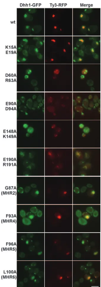
The 21 Ty3-RFP mutants were induced for 18 to 24 h at 24°C, and Ty3-RFP was localized relative to Dhh1-GFP for two independent transformants of each mutant (Fig. 8 and data not shown). Under these conditions, Ty3-RFP and Dhh1-GFP typically localize to a single large focus per expressing cell. Localization was determined for three of the four group I mutants [K171A/E173A, K176A/D177A, and Q86A (MHR1)]. Each produced a pattern of Ty3-RFP and Dhh1-GFP indistin-guishable from that observed for the wt (Fig. 8 and data not shown). The late-acting group II mutant (K15A/E19A) also showed a pattern of Ty3-RFP and P-body fluorescence similar to that in cells expressing wt Ty3-RFP (Fig. 8).
Three group III mutants were examined. Mutant K40A/ K42A, which was defective in protein processing and RNA protection, showed enlarged and faint RFP foci, which colo-calized with Dhh1-GFP (data not shown). The other two group III mutants [E197A/D199A and E90A (MHR3)], which showed wt processing and 50% and 89% protection of RNA, respectively, were similar to wt Ty3-RFP in colocalization with the P-body marker protein (data not shown). These mutants all showed amounts of IN-RFP fusion protein similar to that ob-served in cells expressing wt Ty3-RFP (data not shown).
Inspection of all 12 group IV mutants with severely reduced, aberrant patterns of processing and little or no transposition showed differing patterns in Ty3-RFP. In some cases, even among transformants of a single mutant, there was a range of phenotypes. Mutant R33A/R35A/D37A showed large, faint clusters of Ty3-RFP. Mutant K106/D109A showed normal-sized, faint clusters, as well as some cytoplasmic fluorescence. Clusters in both cases colocalized with Dhh1-GFP (data not shown). Other group IV mutants (R68A/R69A, D81A/E85A, E90A/D94A, K106A/D109A, E122A/K124A, and E180A/E181A) showed more diffuse cytoplasmic RFP (e.g., Fig. 8, E90A/ D94A, and data not shown). Even some group IV mutants that showed unusually diffuse patterns retained subsets of cells that showed colocalization of Ty3 and P-body markers. For exam-ple, Ty3-RFP and Dhh1-GFP formed elongated, polar foci in subpopulations of cells expressing the group IV mutants R68A/ R69A, E90A/D94A, and E180A/E181A (data not shown).
A significant fraction of cells expressing mutant E190A/ R191A showed colocalization of Ty3-RFP with Dhh1-GFP in a remarkable network of streaks (Fig. 8). Dhh1 is a candidate protein for shuttling of RNA into P bodies (11, 22). Thus, it was possible that Dhh1-GFP was somehow diverted away from P bodies in cells expressing this mutant. In order to investigate
this possibility, a second strain, in which P bodies were labeled with Xrn1/Kem1-GFP, was used to express E190A/R191A (86). The resulting pattern was similar to that observed with Dhh1-GFP (data not shown). These results suggested that overexpression of aberrant Ty3 Gag3 causes relocalization of P-body components.
[image:10.585.335.506.65.551.2]The four single MHR replacement mutants [G87A (MHR2), FIG. 8. Visualization of wt and mutant Ty3 and P bodies. CA mutations were introduced to Ty3-RFP and expressed in a strain that expresses a Dhh1-GFP marker under the native promoter. Mutants shown are representative of two independent transfor-mants of each type. Cells were induced for 18 to 24 h as described in the legend to Fig. 3 (6). The scale bar at the lower right repre-sents 5.0m.
on November 8, 2019 by guest
http://jvi.asm.org/
F93A (MHR4), F96A (MHR5), and L100A (MHR6)] formed a distinctive subset. They showed particulate material according to one or both of the sedimentation tests, but they were the most severely defective in protein processing of the group IV mutants. These mutants produced what in some cases ap-peared to be enlarged Ty3-RFP clusters, which colocalized with the P-body marker, or diffuse red fluorescence. In one case [F93A (MHR4)], there was even diffuse Dhh1-GFP flu-orescence in addition to Dhh1-GFP colocalization with RFP (Fig. 8 and data not shown).
Cells expressing group V mutant Ty3-RFP showed low levels of red fluorescence in foci (e.g., Fig. 8, D60A/R63A and E148A/K149A). The low level of fluorescence was consistent with the low level of Gag3 observed with immunoblot analysis. The Dhh1-GFP pattern of multiple small foci in the cells expressing these mutants resembled the pattern in cells not expressing Ty3 (6). In addition, the green and red fluorescence foci did not overlap.
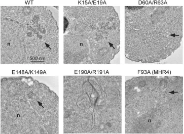
Electron microscopy of cells expressing Ty3 mutants.Group IV and V mutants failed to process Gag3 and showed variable patterns of localization, indicating defects in particle assembly. However, most mutant proteins separated from soluble pro-tein fractions in equilibrium sedimentation and pelleting as-says. Thus, it was possible that Ty3 proteins were associated with some larger structure or formed defective particles. In order to differentiate these possibilities, EM of cells expressing wt Ty3 and representative mutants was undertaken. Expression of wt Ty3 results in multilobed clusters of⬃50-nm VLPs (Fig. 9) (41). Cells expressing the late-acting group II mutant K15A/ E19A produced particles, but these were more loosely
[image:11.585.111.474.69.334.2]associ-ated than were wt particles (Fig. 9 and data not shown). Group IV mutant E190A/R191A, which showed some processing but no cDNA synthesis, formed bizarre filamentous structures (Fig. 9 and data not shown). These structures were consistent with what was observed by fluorescence microscopy with the RFP-tagged Ty3 mutant. MHR mutants G87A (MHR2) and F93A (MHR4) were both severely deficient in processing and had no detectable cDNA. Fluorescence microscopy showed that G87A (MHR2) formed some clusters but that F93A (MHR4), in addition, had a considerable amount of cytoplas-mic fluorescence. Equilibrium gradient analyses showed the presence of sedimentable material. Examination of over 100 cells expressing these mutants (Fig. 9 and data not shown) showed no cells with particles similar to those of the wt but a number of cells with regions containing a low concentration of particle-like structures, which appeared to be in a ribosome-free area. Although this was a negative result, it suggested that these MHR mutants are defective in particle formation. Group V was severely affected by the criteria of protein production, processing, and RNA protection and did not colocalize with P bodies. Only mutant E148A/K149A produced sufficient pro-tein to be readily analyzed by sedimentation, and it showed sedimentable material. Both mutant D60A/R63A and mutant E148A/K149A produced small clusters of particles. Particles produced by expression of the former mutant appeared more heterogeneous, while those of the latter mutant were relatively uniform and compact (Fig. 9 and data not shown). Thus, sur-prisingly, group V mutants were found to be competent to assemble particle structures.
FIG. 9. EM of cells expressing Ty3 CA mutants. Cells transformed with the wt and Ty3 mutants K15A/E19A, D60A/R63A, E148A/K149A, E190A/R191A, and F93A (MHR4) were grown to log phase and induced for Ty3 expression for 6 h. Cultures were processed as described in Materials and Methods. Cells were imaged at 100 kV and⫻5,000 direct magnification.
on November 8, 2019 by guest
http://jvi.asm.org/
DISCUSSION
The secondary structure of the Ty3 CA protein was pre-dicted by a consensus of several structure prediction algo-rithms to be similar to retroviral CA protein in that it is rich in
␣-helical structure. In order to understand CA subdomain functions, 22 Ala substitution mutants defective in transposi-tion were characterized. Mutants were divided into five groups based on CA function. Four mutants (group I) were similar to the wt. One mutant (group II) was wt in Gag3 processing, RNA protection, and cDNA production but failed to trans-pose, indicating that the requirement for Ty3 CA function occurred after reverse transcription. Seven mutants (group III) had intermediate transposition phenotypes. This group was heterogeneous in biochemical properties. Twelve mutants (group IV), including four MHR single-substitution mutants, showed no transposition, severe reductions in Gag3 process-ing, and no cDNA. Representative mutants formed defective particles that did not protect genomic RNA. Localization of these mutants suggested that they were arrested in particle morphogenesis through different mechanisms. Two mutants (group V) produced small amounts of Gag3 and were de-fective by most criteria. Particle production was minimal or nonexistent.
In our study, residues and subdomains of Ty3 CA that con-tributed to localization of Ty3 components, integrity of RNA packaging, fidelity of protein processing, reverse transcription, and post-reverse transcription stages of transposition were identified. However, this analysis showed that particle forma-tion per se is relatively resistant to mutaforma-tions in CA and that multiple Gag3 domains are therefore likely to be involved.
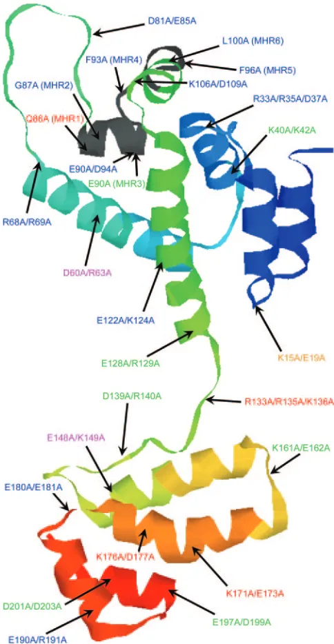
Prediction of the Ty3 CA tertiary structure. In order to better relate the outcome of our mutagenesis study to what is known about the structure and function of retrovirus CA, a tertiary structure was predicted for Ty3 CA. A protein thread-ing approach (see Materials and Methods) and the known structures of HTLV-1 (50), HIV-1 (8, 20, 63), EIAV (10), and RSV (17) were used to generate this prediction. The predicted Ty3 CA has a large bundle of seven ␣ helices in the NTD separated from a smaller CTD bundle by an unstructured loop (Fig. 10). As in the case of retroviruses, a disparity in volume of the predicted CA domains could contribute to curvature of the mature VLP surface. The position of the Ty3 MHR over-lapped a loop-helix junction in the Ty3 CA NTD. It differs from the position of the retroviral MHR, which overlaps the NTD-CTD junction and the first helix of the CTD. The thread-ing prediction differed from the secondary structure prediction in the terminal regions. This may indicate that these regions can adopt more than one conformation.
Localization of VLP assembly.Cells expressing Ty3 colocal-ize Ty3 and P-body components. Because Ty3 lacks a discrete MA domain and because CA is important for particle function, we hypothesize that CA drives localization of Ty3 components. Mutations in CA had diverse effects on colocalization of VLP and P-body components. Mutants in group IV, none of which displayed significant transposition, were mostly of two types: those that formed large Ty3-RFP-Dhh1-GFP foci and those that showed diffuse cytoplasmic RFP. Five group IV mutants had mutations that affected localization of the P-body marker, including three mutants that showed cells with
elon-gated cap-like structures or streaks of RFP and GFP and one MHR mutant that showed more diffuse cytoplasmic GFP than did the wt. These mutations indicate that the colocalization of Ty3 VLP and P-body components is based on physical inter-actions. This study did not indicate whether those interactions are direct or indirect.
[image:12.585.299.541.68.535.2]Cells in which group V mutants were expressed showed multiple small Dhh1-GFP foci, typical of cells not expressing Ty3. Strikingly, the RFP Ty3 marker of the group V mutants FIG. 10. Ribbon representation of modeled tertiary structure of Ty3 CA. The predicted Ty3 CA tertiary structure is comprised of NTD and CTD bundles of seven and four␣helices, respectively. Mutations indicated in the modeled structure are color coded according to the mutant phenotype group (see Table 2). In order of least to most deficient: group I (red), group II (ochre), group III (blue), group IV (blue), and group V (fuschia).
on November 8, 2019 by guest
http://jvi.asm.org/
was present in smooth round foci, which did not colocalize with the Dhh1-GFP marker, and potentially represented sequestra-tion of Ty3-RFP or RFP alone in some vesicular compartment. In order to examine this possibility, cells were stained with a fluorescent lipophilic dye, FM4-64 (92). Because FM4-64 flu-oresces in the red wavelength overlapping RFP, group V mu-tations were individually introduced into Ty3-GFP. Cells ex-pressing either group V mutant for 6 h showed a severely retarded endosomal uptake of dye. A previous screen of the yeast knockout collection showed that several ESCRT traffick-ing components contribute positively to Ty3 transposition (45). One possibility is that Ty3 Gag3 normally interacts transiently with a trafficking component and that this interaction is dis-rupted in the case of the group V mutant Gag3, thus interfer-ing with function of a traffickinterfer-ing factor.
Examination of these cells by EM showed that Ty3 particles occurred in clusters similar to what was suggested by the flu-orescence study. This result implicates the CA domain in co-localization with P-body components, as opposed to a model in which P-body colocalization is solely determined by Ty3 RNA. In addition, this result argues against the possibility that P-body components are needed simply for multimerization of particles.
Results of the present study argue that (i) Ty3 can multi-merize prior to or in the absence of P-body localization, (ii) the CA domain of Gag3 contributes to typical P-body colocaliza-tion and may even interact directly with P-body components, and (iii) Gag3 localization to P bodies is not sufficient to guarantee protection of genomic RNA. We failed to observe mutants that did not associate with the P-body marker and were transposition competent. Thus, our results are consistent with, but do not demonstrate a role for P-body association in, Ty3 VLP assembly.
Even if Ty3 is shown to assemble in P bodies, the overall impact of P-body association on transposition is likely to be complex. The extent to which cells overexpressing Ty3 accu-mulate VLPs within a single cluster suggests that this structure could become limiting for transposition. We speculate that this phenomenon contributes to copy number control, which has been described for Ty1 (24) and also occurs for Ty3 (unpub-lished observations).
Because retrovirus targeting to assembly sites is specified by MA and envelope, the extent to which our findings bear upon retrovirus assembly and CA function in particular is not clear. However, similar to our results, recent studies of retroviral assembly have shown that intracellular complexes smaller than cores form prior to budding from the assembly site (56, 91). It is even possible that there is some specialization in these pre-assembly multimerization sites. For example, HIV-1 passes through a perinuclear compartment prior to delivery to the multivesicular body (71). Whether that compartment is related to the P body is not known; however, APOBEC3G, a host restriction factor for HIV-1, was recently reported to localize to P bodies (97). It has also been reported that the endogenous retrovirus related to Jaagsiekte sheep retrovirus and the en-dogenous musD mouse retrovirus accumulate Gag proteins at intracellular foci (66, 75).
Structural role of CA in VLP assembly.Particle production was observed for Ty3 CA mutants representing each group. Thus, it could be that multiple subdomains of CA or other
domains of Gag3, such as spacer and NC, make important contributions to multimerization. In contrast, Ty3 particle function was relatively sensitive to mutation. Interestingly, 12 of 18 Ty3 CA NTD mutants had reduced particle functionality. The extreme amino-terminal group II mutant (K15A/E19A), which produced cDNA but failed to transpose, was among these. Additional mutagenesis has shown that individual sub-stitutions at positions 15 and 16 affect clustering and assembly, respectively, further supporting a key role for the NTD in Ty3 VLP assembly (unpublished observations). Consistent with the NTD interface of the hexameric retrovirus CA, Ala-scanning mutagenesis of HIV-1 CA showed that the NTD influences the shape and stability of cores and may even influence nuclear entry (27, 31, 37).
The Ty3 MHR is located in the NTD rather than at the NTD-CTD junction, as occurs in retrovirus CA. Therefore, differences between MHR functions in retrovirus and Ty3 par-ticle assembly may exist. Depending on the severity of the mutation, disruption of the MHR can cause defects in particle formation, infectivity, and cDNA synthesis (16, 28, 60, 88). At the mechanistic level, the MHR has been implicated in mem-brane association (29), incorporation of Gag-Pol into particles (44, 87), and productive assembly of Gag (72). It has been suggested that the MHR exerts its effect on assembly by me-diating a CTD swap, which stabilizes a dimer interface (46).
In a previous study, we mutated three of the six conserved positions in the Ty3 MHR (aa 86, 87, and 90). The results of that study were consistent with the results of this study in that the effects of substitutions at aa 86 (Q86R) and 90 (E90D, E90K, and E90N) were less severe than at aa 87 (G87A and G87V). The present study showed that some MHR mutants, which are defective in protein processing and transposition, do localize to P bodies. However, at least one had a more cyto-plasmic localization. In addition, our data suggest that at least some residues in the MHR are critical for assembly or stability of particles. Two of the severely defective mutants that were examined by EM (G87A and F93A) were decreased in VLP formation, consistent with defects in RNA protection. In ad-dition, although MHR mutant Gag3 appeared to be stable, Gag3-Pol3-derived IN was reduced or absent in extracts of cells expressing MHR mutants and the ratio of Gag3-Pol3 to Gag3 species in pelletable material was severely reduced. Since at least one mutant competent for cluster formation was none-theless defective in particle assembly by EM, these results suggest that the MHR could participate in more than one phase of particle assembly.
The group V mutant D60A/R63A is mutated at a position similar to the Asp that is conserved among retrovirus CA proteins and which participates in a salt bridge with a con-served amino-terminal Pro residue after Gag maturation. Re-placement of this residue has been shown to destabilize retro-virus cores. Although Ty3 does not have an amino-terminal Pro, it is possible that the Asp at position 60 of the Ty3 CA performs a similar stabilization function. The group V mutant (E148A/K149A) bridges the NTD-CTD loop and the first CTD helical region. This is the position of the MHR in retroviral CA. The fact that mutations in conserved residues of the MHR, as well as mutations in the NTD-CTD linker, have effects on transposition suggests that functions performed by the MHR linker in retrovirus CA could be contributed by