0022-538X/08/$08.00⫹0 doi:10.1128/JVI.02021-07
Copyright © 2008, American Society for Microbiology. All Rights Reserved.
Role of Cellular Phosphatase cdc25C in Herpes Simplex
Virus 1 Replication
䌤
Benjamin A. Smith-Donald, Lizette O. Durand, and Bernard Roizman*
Marjorie B. Kovler Viral Oncology Laboratories, The University of Chicago, 910 East 58th Street, Chicago, Illinois 60637
Received 13 September 2007/Accepted 5 February 2008
Earlier studies have shown that in herpes simplex virus 1-infected cells, ICP22 upregulates the accumulation
of a subset of␥2proteins exemplified by the products of the UL38, UL41, and US11 genes. The ICP22-dependent
process involves degradation of cyclins A and B1, the stabilization and activation of cdc2, physical interaction
of activated cdc2 with the UL42 DNA synthesis processivity factor, and recruitment and phosphorylation of
topoisomerase II␣by the cdc2/UL42 complex. Activation of cdc2, the first step in the process, is a key function
of the mitotic phosphatase cdc25C. To define the role of cdc25C, we probed some features of the
ICP22-dependent pathway of upregulation of␥2genes in cdc25Cⴚ/ⴚcells and in cdc25Cⴙ/ⴙcells derived from sibling
mice. We report that cyclin B1 turned over in cdc25Cⴙ/ⴙ or cdc25Cⴚ/ⴚ cells at the same rate, that cdc2
increased in amount, and that US11 and UL38 proteins and infectious virus accumulated in smaller amounts
than in wild-type infected cells. The reduction in UL38 protein accumulation and virus was greater in
cdc25Cⴚ/ⴚcells infected with virus lacking ICP22 than in cells infected with wild-type virus. We conclude that
cdc25C phosphatase plays a role in viral replication and that this role extends beyond its function of activating
cdc2 for initiation of the ICP22-dependent cascade for upregulation of␥2gene expression.
The studies reported here stemmed from the discovery that herpes simplex virus 1 (HSV-1) activates and diverts the mi-totic kinase cdc2 (also known as cdk1) to enhance the expres-sion of a subset of late genes exemplified by UL38, UL41, and
US11. Specifically, activation of cdc2 during viral replication requires the regulatory protein ICP22, a product of the␣22 gene, and the viral protein kinase encoded by the UL13 gene. In the process, the natural partners of cdc2, cyclins A and B1, are degraded. Activated cdc2 physically interacts with UL42, an HSV protein known primarily as a DNA synthesis processivity factor (12). UL42-bound cdc2 kinase is redirected to phosphor-ylate UL42 and to recruit and phosphorylate topoisomerase
II␣for post-DNA synthesis expression of the subset of␥2late genes listed above (1, 3–5).
In uninfected interphase cells, cdc2 is inactive, in part as a consequence of phosphorylation of threonine 14 and tyrosine 15 (6). Activation of cdc2 requires phosphorylation of threo-nine 161 and removal of phosphates from threothreo-nine 14 and tyrosine 15 by cdc25C. In this process, the role of cdc25C is critical. cdc25C has been characterized as the “mitotic trigger” for its ability to rapidly activate cdc2 (13); consequently, the activation of cdc25C is tightly regulated by the cell. The growth suppressor p53 inhibits transcription of cdc25C mRNA and directly binds to the cdc25C protein to prevent entry into mitosis (20).
In infected cells, activation of cdc2 during infection takes place concurrently with the down-regulation of the cdc2 inhib-itor, wee1 kinase, and increased activity of the cdc2 activator, cdc25C phosphatase, as measured by a generic phosphatase
assay (1; S. Advani and B. Roizman, unpublished observa-tions).
The sequence of events described above raises the question as to the role of cdc25C phosphatase in the course of the HSV-1 replicative cycle. To address this question, we took advantage of the availability of murine cdc25C⫺/⫺cells and
cdc25C⫹/⫹sibling cells (10). The cdc25C⫺/⫺cells were derived
by targeted disruption of exon 3 of endogenous cdc25C, result-ing in viable mouse and murine embryonic fibroblasts that, surprisingly, had no apparent defect in growth or cell cycle checkpoint (10).
In this report, we examine the role of the cdc2 activator cdc25C in the replication of HSV-1. We show that in cdc25C⫺/⫺cells, the rate of degradation of cyclin B1 remained
similar to that of wild-type cells, whereas the amounts of cdc2 actually increased. Concurrently, the accumulation of viral DNA, the US11 and UL38 proteins, and infectious virus was reduced. However, cdc25C was not required for the virally mediated increase in cdc2 kinase activity and phosphorylation of topoisomerase II␣.
MATERIALS AND METHODS
Cells and viruses.The HEp-2 and Vero cell lines were initially obtained from the American Type Culture Collection. cdc25C⫺/⫺and cdc25C⫹/⫹murine em-bryonic fibroblast (MEF) cell lines (10) were a kind gift of H. Piwnica-Worms (Washington University, St. Louis, MO). Telomerase-transformed human em-bryonic lung fibroblasts (HEL cells) were a gift of T. E. Shenk (Princeton University, Princeton, NJ). Cell lines were grown in Dulbecco’s modified Eagle medium supplemented with 5% newborn calf serum (HEp-2 and Vero), 10% fetal bovine serum (HEL cells), or 10% fetal bovine serum, 1%L-glutamine, and 1% nonessential amino acids (cdc25C⫺/⫺and cdc25C⫹/⫹MEF cells). HSV-1(F) is the prototype HSV-1 wild-type strain used in this laboratory (11). R325 (in
which ␣22 does not encode the C-terminal domain [CTD] of ICP22
[␣22⌬CTD]), R7356 (UL13⫺), and R7802 (␣22⫺US1.5⫺) have been previously described (16, 18).
Plasmids.pRB143, containing the terminal BamHI S fragment of the HSV-1 genome, has been described previously (15) and was used in Southern blotting as described below.
* Corresponding author. Mailing address: The University of Chi-cago, Marjorie B. Kovler Viral Oncology Laboratories, 910 East 58th St., Chicago, IL 60637. Phone: (773) 702-1898. Fax: (773) 702-1631. E-mail: [email protected].
䌤Published ahead of print on 13 February 2008.
4527
on November 8, 2019 by guest
http://jvi.asm.org/
Cell infection. Cultures of the indicated cell type were infected with the appropriate virus at an appropriate multiplicity of infection in medium 199V (mixture 199 supplemented with 1% calf serum) on a rotary shaker at 37°C. After 2 h, the inoculums were replaced with fresh growth medium and culture flasks were incubated at 37°C until cells were harvested. For infection times delineated in the figures, time zero indicates when infection was initiated. Cells were har-vested by scraping into their own medium, pelleted by low-speed centrifugation, washed twice in phosphate-buffered saline (PBS) A (0.14 M NaCl, 3 mM KCl, 10 mM Na2HPO4, 1.5 mM KH2PO4), and then lysed in the appropriate buffer.
One-dimensional electrophoretic separation and immunoblotting.Cell pellets were lysed and denatured in disruption buffer (50 mM Tris [pH 7.0], 2.75% sucrose, 5%-mercaptoethanol, 2% sodium dodecyl sulfate). Protein samples were boiled for 5 min and then were electrophoretically separated in a 10% denaturing polyacrylamide gel and electrically transferred to a nitrocellulose sheet. The membrane was then blocked with 5% nonfat milk and reacted with primary antibody followed by an appropriate secondary antibody conjugated to alkaline phosphatase (Bio-Rad Laboratories) or horseradish peroxidase (Sigma). Immunoblots were developed either with 5-bromo-4-chloro-3-indolylphosphate– nitroblue tetrazolium (Sigma) or through enhanced chemiluminescence (ECL; Amersham Biosciences) and exposed to film or by ECL Plus (Amersham Bio-sciences) followed by quantification with the aid of a Molecular Dynamics Storm imaging system.
Two-dimensional electrophoretic analysis of topoisomerase II␣. Two-dimen-sional electrophoresis was performed using an immobilized pH gradient for first-dimension isoelectric focusing (9). A protocol previously described (8) was modified. Briefly, 150-cm2flasks of cdc25C⫹/⫹or cdc25C⫺/⫺MEF cells, mock or HSV-1(F) infected, were harvested for two-dimensional gel electrophoresis as previously described (2). After electrophoretic separation, proteins were trans-ferred and immunoblotted as described above.
Antibodies.The antibodies to cellular proteins used in these studies were antiactin (catalog no. A4700; Sigma), anti-cdc2 (catalog no. sc-54; Santa Cruz), anti-cyclin B1 (catalog no. sc-245; Santa Cruz), anti-TopoII (catalog no. Ab-1; Oncogene), all monoclonal, and polyclonal anti-TopoII alpha (catalog no. A300-054A; Bethyl Labs). Additionally, the polyclonal anti-cdc2 (catalog no. 9112; Cell Signaling) antibody was used at a dilution of 1:1,000 and the monoclonal anti-cyclin B1 (catalog no. 4135; Cell Signaling) antibody was used at a dilution of 1:2,000. To detect viral proteins, we used the monoclonal antibodies anti-ICP0, anti-ICP4, and anti-US11 and polyclonal antibodies anti-ICP22 and anti-UL38 (Goodwin Cancer Research Institute.). Anti-mouse immunoglobulin G (IgG)-peroxidase (catalog no. A4416; Sigma), anti-rabbit IgG-(IgG)-peroxidase (catalog no. A0545; Sigma), anti-mouse IgG-alkaline phosphatase conjugate (catalog no. 170-6520; Bio-Rad), and anti-rabbit IgG-AP conjugate (catalog no. 170-6518; Bio-Rad) were used as secondary antibodies for immunoblotting.
Histone H1 kinase assay.The histone H1 kinase assay was performed as previously described (1).
Southern blotting.For analysis of DNA synthesis, cultures in 25-cm2flasks of Vero cells or cdc25C⫺/⫺or cdc25C⫹/⫹MEF cells were infected with 5 PFU of HSV-1(F) per cell and harvested at 20 h after infection. Total viral DNA was isolated as previously described (7, 17), digested with BamHI restriction endo-nuclease, subjected to electrophoresis on 0.7% agarose gels, transferred to zeta-probe membranes, and hybridized with the radiolabeled plasmid pRB143, which contains the terminal BamHI S fragment (15). The bound probe was visualized by autoradiography. The probe hybridized with both the junction fragment (BamHI SP) and the terminal fragment (BamHI S).
Infectious virus yield in MEF cells.Replicate cdc25C⫺/⫺or cdc25C⫹/⫹MEF cells were exposed to 0.01 PFU per cell of R7356, R7802, R325, or wild-type HSV-1(F) virus in 1 ml medium 199V while on a rotating shaker at 37°C. After 2 h, the inoculum was removed, cells were rinsed three times, and 2.5 ml medium 199V was added. Cells were harvested at 2 and 24 h after infection by scraping directly into the medium. The sample was frozen and thawed three times and then sonicated before being titered in Vero cells.
RESULTS
HSV-1(F)-mediated changes in the levels of cdc2 in MEFs. The purpose of this series of experiments was to determine whether cdc25C affects the levels of cdc2 and cyclin B in in-fected cells. Figure 1 shows immunoblots of cyclin B1 and of cdc2 in mock-infected and infected cdc25C⫹/⫹or cdc25C⫺/⫺
MEFs. The cells were infected with wild-type HSV-1(F) or mock infected and harvested at 6, 12, and 24 h after infection.
Electrophoretically separated proteins from cellular lysates were immunoblotted for cyclin B1 and cdc2. As reported ear-lier for HSV-1(F)-infected HeLa cells (1), the cyclin B1 pro-tein largely disappeared from HSV-1(F)-infected cdc25C⫹/⫹
MEFs (Fig. 1A, lanes 1, 3, 5, and 7). Cyclin B1 also decreased in cdc25C⫺/⫺ cells but at a rate similar to that of wild-type
sibling cells (Fig. 1A, lanes 2, 4, 6, and 8). cdc2 levels tended to decrease at least during the first 12 h after infection of cdc25C⫹/⫹cells (Fig. 1B, lanes 1, 3, 5, and 7). In contrast, there
was a consistent net increase in the amount of cdc2 in infected cdc25C⫺/⫺cells (Fig. 1B, lanes 2, 4, 6, and 8). The results of
this and other experiments in this series suggest that cdc25C expression was necessary for the maintenance of or actual increase in the levels of the cdc2 protein in HSV-1(F)-infected
FIG. 1. The metabolism of cdc2 and cyclin B1 proteins in MEFs infected with wild-type HSV-1(F). cdc25C⫹/⫹or cdc25C⫺/⫺MEF cells
were mock or HSV-1(F) infected for the indicated amount of time, and cell lysates were electrophoretically separated and immunoblotted. The blots were scanned with the aid of a Molecular Dynamics PhosphorImager and normalized with respect to the amounts detected in corresponding mock-infected cells. (A) Immunoblot and results of scanning of cyclin B1. (B) Immunoblot and results of scanning of cdc2.
4528 SMITH-DONALD ET AL. J. VIROL.
on November 8, 2019 by guest
http://jvi.asm.org/
cells. The absence of cdc25C played no role in the disappear-ance of cyclin B1.
HSV-1(F)-mediated increase in cdc2 activity does not
re-quire presence of cdc25C.To determine the functional
rele-vance of cdc25C for the cdc2 kinase activity in mock-infected and infected MEFs, cdc25C⫹/⫹ and cdc25C⫺/⫺ MEFs were
infected and maintained for 16 h and then harvested and lysed. cdc2 complex immunoprecipitated with antibody against cyclin B1 or cdc2 was reacted with purified histone H1 in a [␥-32P]ATP-labeled kinase assay. As previously reported for
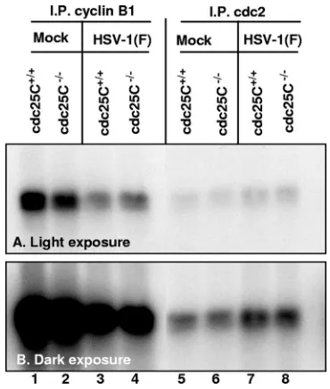
HeLa cells (1), in cdc25C⫹/⫹MEFs the kinase activity of the
complex immunoprecipitated by antibody against cyclin B1 decreased after infection (Fig. 2, lanes 1 and 3) whereas the kinase activity of the complex immunoprecipitated by antibody against cdc2 was increased after infection (Fig. 2, lanes 5 and 7). The same trends were observed in cdc25C⫺/⫺MEFs.
We conclude that the activation of cdc2 is not significantly affected by the absence of cdc25C in MEFs.
Topoisomerase II␣ is posttranslationally modified in
in-fected cdc25Cⴚ/ⴚ
and cdc25Cⴙ/ⴙ
cells. As reported earlier,
cdc2 binds the UL42 protein and this complex recruits, phos-phorylates, and activates topoisomerase II␣(4). To define the role of cdc25C in the regulation of topoisomerase II␣, cdc25C⫹/⫹or cdc25C⫺/⫺MEF or HEL cells were mock
in-fected or inin-fected with R325 (␣22 deletion mutant) or HSV-1(F). The cells were harvested, lysed, subjected to electro-phoresis, and reacted with antibody to topoisomerase II␣. Human topoisomerase II␣migrates more slowly in the pres-ence of HSV-1(F) infection than with mock infection or infec-tion with R325 virus (data not shown), which is consistent with the earlier report (4) that intact, functional ICP22 is required for the posttranslational modification of topoisomerase II␣. In
contrast, there was no detectable shift in electrophoretic mo-bility of murine topoisomerase II␣ in mock-infected, R325-infected, or HSV-1(F)-infected cdc25C⫹/⫹or cdc25C⫺/⫺MEF
cells (data not shown).
In order to detect a modification of topoisomerase II␣that did not affect its linear gel migration, the lysates of cdc25C⫺/⫺
or cdc25C⫹/⫹MEFs harvested 12 h after mock infection or
infection with HSV-1(F) were subjected to both one-dimen-sional separation (Fig. 3A) and two-dimenone-dimen-sional separation (Fig. 3B). The one-dimensional separation (Fig. 3A, lanes 1 to 4) revealed an increase in the amounts of topoisomerase II␣ in infected cells over those in uninfected cdc25C⫹/⫹ or
cdc25C⫺/⫺MEFs. The two-dimensional gel analysis showed
that following infection, most of the topoisomerase II␣shifted to the right, indicating an increasing negative charge (Fig. 3B).
FIG. 2. cdc25C is not required for increase in cdc2 kinase activity after HSV-1(F) infection. cdc25C⫹/⫹ or cdc25C⫺/⫺ MEF cells were
mock or HSV-1(F) infected for 16 h. cdc2 kinase complex immuno-precipitated using antibody against cyclin B1 (lanes 1 to 4) or against cdc2 (lanes 5 to 8) was incubated with purified histone H1 in a [␥-32P]ATP-labeled kinase assay. (A) Light exposure of
[image:3.585.70.255.67.285.2]autoradio-gram. (B) Dark exposure of same autoradioautoradio-gram.
FIG. 3. cdc25C is not required for modification of TopoII␣to neg-atively charged forms. cdc25C⫹/⫹ and cdc25C⫺/⫺ MEF cells were
mock infected or infected with 10 PFU/cell HSV-1(F) for 12 h. Cell lysates were separated electrophoretically for linear analysis or sepa-rated by charge followed by electrophoresis for two-dimensional anal-ysis. (A) Linear separation, immunoblot for topoisomerase II␣. (B) Two-dimensional separation, immunoblot for topoisomerase II␣. Linear migration is marked with an arrowhead; charge distribution is indicated with a line.
on November 8, 2019 by guest
http://jvi.asm.org/
This is in contrast to results for mock-infected cells, in which topoisomerase II␣ varied more extensively with respect to charge. The results obtained from infected cdc25C⫹/⫹ and
cdc25C⫺/⫺cell lines indicate that topoisomerase II␣ is
post-translationally modified after HSV-1 infection and that the absence of cdc25C does not affect the apparent level of mod-ification of topoisomerase II␣. We should note, however, that although the data are consistent with the earlier report that in HSV-1-infected cells topoisomerase II␣is posttranslationally modified, we present no evidence to support the hypothesis that the modification of topoisomerase II␣in HSV-1-infected murine cells is similar to or as extensive as that seen in infected human cells (4). Topoisomerase II␣is highly conserved among humans and mice, although several sites of phosphorylation have been found in the human protein while the mouse protein has not been extensively mapped (14).
The production of viral DNA during HSV-1(F) infection was
lower in cdc25Cⴚ/ⴚcells than in cdc25Cⴙ/ⴙMEF cells.Advani
et al. (4) suggested that cdc2 may play a role in HSV-1 DNA synthesis. To test the hypothesis that cdc25C plays a role in viral DNA accumulation, total DNA was purified from HSV-1(F)-infected Vero cells or MEFs, digested with BamHI re-striction endonuclease, electrophoretically separated in a 0.7% agarose gel, transferred to a membrane, and hybridized with a labeled BamHI S DNA fragment. This fragment is derived from the terminus of the L component of linearized DNA and is also present in the BamHI SP fragment present at the junc-tion between the L and S components in both linear and circular or concatemeric DNA. DNA extracted from mock-infected or HSV-1(F)-mock-infected Vero cells served as negative and positive controls for the assay (Fig. 4, lanes 1 and 2).
As shown in Fig. 4, lanes 3 and 4, the amounts of BamHI SP and BamHI S DNA fragments, reflecting linear and concate-meric DNAs and ends of linearized DNA, respectively, that accumulated in cdc25C⫺/⫺ MEFs were smaller than those
present in cdc25C⫹/⫹MEFs. These results suggest that cdc25C
plays a role in viral DNA accumulation in infected cells.
Accumulation of a subset of ␥2 proteins is dependent on
presence of cdc25C.A subset of ␥2 proteins depend on cdc2
kinase activity and ICP22 for their accumulation in infected cells (5, 18). In order to evaluate the role of cdc25C in the accumulation of these␥2proteins, cdc25C⫹/⫹and cdc25C⫺/⫺
MEFs were mock infected or infected with HSV-1(F) (Fig. 5A and B) or R325 (Fig. 5B) and harvested at times shown in Fig. 5. The electrophoretically separated proteins were reacted with antibodies to ICP4 (panel A), ICP0 (panels A and B), ICP22 (panel A), UL38 (panels A and B), US11 (panels A and
B), and actin (panel B). The results were as follows. (i) The accumulation of ICP22 and ICP0 was not significantly affected by the absence of cdc25C. (ii) In the absence of cdc25C, there was a decrease in the accumulation of ICP4, US11, and UL38.
(iii) As expected, MEFs infected with R325 mutant virus ac-cumulated less of the UL38 or US11 protein than
correspond-ing cells infected with wild-type virus (compare Fig. 5B, lanes 15 and 16, with lanes 17 and 18). In addition, cdc25C⫺/⫺MEFs
infected with the mutant virus accumulated less of the UL38 protein than infected cdc25C⫹/⫹cells (compare lanes 15 and
16 in panel B). The accumulation of US11 was less affected by the absence of cdc25C than by that of the UL38 protein.
We conclude that cdc25C is required for optimal accumu-lation of at least a subset of HSV-1 proteins and that the combined absence of both intact ICP22 and cdc25C has a more drastic effect on the accumulation of UL38, one member of the
subset of␥2proteins regulated by ICP22.
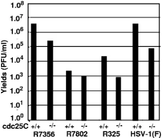
HSV-1 accumulates at lower levels in absence of cdc25C.In
this series of experiments, replicate cultures of cdc25C⫹/⫹and
cdc25C⫺/⫺MEFs were exposed to 0.1 PFU/cell of HSV-1(F),
R325, R7802 (⌬␣22), or R7356 (⌬UL13). The cells were har-vested at 24 h after infection, and titers of virus were deter-mined on Vero cells. The results, depicted in Fig. 6, were as follows. (i) As predicted, the R325 mutant, lacking the carbox-yl-terminal 220 residues of ICP22, replicated less well than the wild-type virus. The R7802 mutant, lacking the entire ICP22 open reading frame, barely replicated in wild-type murine cells. (ii) For each virus that replicated in murine cells, the yields in infected cdc25C⫺/⫺MEF cells were at least 10-fold
lower than those obtained from infected cdc25C⫹/⫹MEFs. We
conclude that cdc25C plays a role in viral replication.
DISCUSSION
In earlier studies, this laboratory reported that after infec-tion, cyclins A and B1 are degraded, the residual cdc2 kinase binds UL42, and this complex recruits and phosphorylates to-poisomerase II␣(1, 3, 4). This chain of events is mediated by ICP22 and is essential for optimal expression of a subset of␥2 genes exemplified by US11, UL38, and UL41 and production of
[image:4.585.70.254.67.275.2]infectious progeny (5, 18). A key step in the activation of cdc2 is the removal of inhibitory phosphates by the cdc25C phos-phatase. To clarify the partial stabilization and activation of cdc2, we examined the role of cdc25C in key aspects of the
FIG. 4. cdc25C is required for maximal production of HSV-1(F) DNA. Total viral and cellular DNA was purified from Vero and MEF cells 20 h after mock or HSV-1(F) infection. DNA was digested with BamHI restriction endonuclease, electrophoretically separated in a 0.7% agarose gel, transferred to a membrane, and labeled with a probe specific for the genomic junction region designated BamHI fragment S. Digested genomic concatemers and circles migrate more slowly and are identified as BamHI SP. Digested genomic terminal fragments migrate more quickly and are identified as BamHI S.
4530 SMITH-DONALD ET AL. J. VIROL.
on November 8, 2019 by guest
http://jvi.asm.org/
pathway leading to the expression of the ICP22-regulated genes by taking advantage of the availability of cdc25C⫺/⫺
MEFs and wild-type cdc25C⫹/⫹MEFs derived from a sibling
mouse. A priori we should note that the phenotype of cdc25C⫺/⫺ mice was not significantly different from that of
wild-type mice, suggesting that the functions of cdc25C were taken over by other genes or metabolic pathways (10).
Our results may be summarized as follows. (i) In the cell lines tested, cyclin B1 was degraded at the same rate in both cdc25C⫺/⫺and in the wild-type sibling cdc25C⫹/⫹MEFs. Of
particular significance is the observation that the cdc2 protein was actually upregulated in the course of infection of cdc25C⫺/⫺cells compared to results with cdc25C⫹/⫹ MEFs.
(ii) cdc2 kinase was more active in infected cdc25C⫹/⫹and
cdc25C⫺/⫺MEFs than in uninfected cells. (iii) Topoisomerase
II␣ was posttranslationally modified in both cdc25C⫹/⫹and
cdc25C⫺/⫺MEFs infected with wild-type virus. As noted in the
results, however, we have no data on the nature of the modi-fication or its identity to that observed in human cells. (iv) The accumulation of representative␥2proteins regulated by ICP22
and virus yields were reduced in cdc25C⫺/⫺MEFs from those
in cdc25C⫹/⫹MEFs. Equally significantly, the decrease in virus
yields and in the accumulation of at least the UL38 protein observed in cdc25C⫺/⫺MEFs infected with wild-type virus was
also observed in cells infected with a virus mutant lacking the CTD of ICP22 and which yields less virus and accumulates smaller amounts of ICP22-dependent␥2 proteins. This
sug-gests a role for cdc25C that is not initiated solely by ICP22. One strategy of HSV takeover of the infected cells is to sequester and redirect cellular proteins to perform functions similar to or very different from those performed by the same protein in uninfected cells (19). For heuristic reasons, it seems appropriate to consider two hypotheses. The first is that cdc25C plays a role consistent with its normal function in uninfected cells. The alternative hypothesis is that the func-tions of cdc25C are not related to activation of cdc2.
The scenario consistent with both hypotheses is that the fraction of active cdc2 corresponding to the protein band that remains in infected cdc25C⫺/⫺ MEFs binds to U
L42 even
[image:5.585.107.480.68.339.2]though cyclin B1 is not degraded and topoisomerase II␣ is
FIG. 5. Accumulation of a subset of␥2proteins is depends on cdc25C and functional ICP22. cdc25C⫹/⫹and cdc25C⫺/⫺MEF cells were mock
infected or infected with HSV-1(F) (A and B) or R325 (B) and harvested at the times indicated. Electrophoretically separated proteins were immunoblotted for ICP4 (A), ICP0 (A and B), ICP22 (A), UL38 (A and B), US11 (A and B), or actin (B).
FIG. 6. cdc25C and␣22 are each important for virus growth at a low multiplicity of infection. cdc25C⫹/⫹ and cdc25C⫺/⫺ MEF cells
were infected with 0.01 PFU of R7356 (⌬UL13), R7802 (⌬␣22), R325
(␣22⌬CTD), or wild-type HSV-1(F) per cell. At 24 h after infection, the cells were harvested, the virus was extracted by repeated freeze thawing, and titers were determined on Vero cells.
on November 8, 2019 by guest
http://jvi.asm.org/
[image:5.585.84.242.533.668.2]modified by the cdc2/UL42 complex. The discriminating datum is the observation that cdc25C plays a role even in MEFs infected with an ICP22 mutant incapable of upregulating the accumulation of the representative␥2proteins. Since all
avail-able data support the conclusion that ICP22 regulates the accumulation of the␥2proteins, exemplified by UL38, UL41,
and US11, loss of any component of the pathway regulated by ICP22 should have the same effect as loss of ICP22. This is clearly not the case. By necessity, we conclude that cdc25C phosphatase acts in addition to its role in activating cdc2. The precise mechanism remains to be elucidated.
Of note, a mild defect in accumulation of the immediate-early proteins ICP4 and ICP22 was observed in cdc25C⫺/⫺
MEFs. Can this explain the defects seen in infection of this cell line—DNA replication, late gene expression, and viral yields? It is critical to note that the defect seen in infection of these cells is not merely a delay in the actions of the immediate-early proteins. If that were the case, the ICP22 mutant should have the same phenotype, due to a functional overlap, in the pres-ence or abspres-ence of cdc25C. In contrast, the ICP22 mutant in combination with the absence of cdc25C demonstrated a cu-mulative decrease in the accrual of the ␥2 genes UL38 and US11 and in the replication of the virus.
ACKNOWLEDGMENTS
We thank Sunil Advani for invaluable discussions, Alice Poon for technical advice about Southern blotting, and H. Piwnica-Worms for providing cdc25C⫺/⫺and cdc25C⫹/⫹MEF cells.
B.S.-D. was supported by UC MSTP. These studies were aided by National Cancer Institute grants CA115662, CA83939, CA71933, CA78766, and CA88860.
REFERENCES
1.Advani, S. J., R. Brandimarti, R. R. Weichselbaum, and B. Roizman.2000. The disappearance of cyclins A and B and the increase in activity of the G2/M-phase cellular kinase cdc2 in herpes simplex virus 1-infected cells require expression of the␣22/US1.5 and UL13 viral genes. J. Virol.74:8–15. 2.Advani, S. J., R. Hagglund, R. R. Weichselbaum, and B. Roizman.2001. Posttranslational processing of infected cell proteins 0 and 4 of herpes simplex virus 1 is sequential and reflects the subcellular compartment in which the proteins localize. J. Virol.75:7904–7912.
3.Advani, S. J., R. R. Weichselbaum, and B. Roizman. 2001. cdc2 cyclin-dependent kinase binds and phosphorylates herpes simplex virus 1 UL42 DNA synthesis processivity factor. J. Virol.75:10326–10333.
4.Advani, S. J., R. R. Weichselbaum, and B. Roizman.2003. Herpes simplex virus 1 activates cdc2 to recruit topoisomerase II alpha for post-DNA syn-thesis expression of late genes. Proc. Natl. Acad. Sci. USA100:4825–4830. 5.Advani, S. J., R. R. Weichselbaum, and B. Roizman.2000. The role of cdc2 in the expression of herpes simplex virus genes. Proc. Natl. Acad. Sci. USA 97:10996–11001.
6.Atherton-Fessler, S., F. Liu, B. Gabrielli, M. S. Lee, C. Y. Peng, and H. Piwnica-Worms.1994. Cell cycle regulation of the p34cdc2 inhibitory ki-nases. Mol. Biol. Cell5:989–1001.
7.Baines, J. D., A. P. Poon, J. Rovnak, and B. Roizman.1994. The herpes simplex virus 1 UL15 gene encodes two proteins and is required for cleavage of genomic viral DNA. J. Virol.68:8118–8124.
8.Berkelman, T., and T. Stenstedt.1998. 2-D electrophoresis using immobi-lized pH gradients: principles and methods. Amersham Pharmacia Biotech, Piscataway, NJ.
9.Bjellqvist, B., K. Ek, P. G. Righetti, E. Gianazza, A. Gorg, R. Westermeier, and W. Postel.1982. Isoelectric focusing in immobilized pH gradients: prin-ciple, methodology and some applications. J. Biochem. Biophys. Methods 6:317–339.
10.Chen, M. S., J. Hurov, L. S. White, T. Woodford-Thomas, and H. Piwnica-Worms.2001. Absence of apparent phenotype in mice lacking Cdc25C pro-tein phosphatase. Mol. Cell. Biol.21:3853–3861.
11.Ejercito, P. M., E. D. Kieff, and B. Roizman.1968. Characterization of herpes simplex virus strains differing in their effects on social behaviour of infected cells. J. Gen. Virol.2:357–364.
12.Gottlieb, J., A. I. Marcy, D. M. Coen, and M. D. Challberg.1990. The herpes simplex virus type 1 UL42 gene product: a subunit of DNA polymerase that functions to increase processivity. J. Virol.64:5976–5987.
13.Hutchins, J. R., and P. R. Clarke.2004. Many fingers on the mitotic trigger: post-translational regulation of the Cdc25C phosphatase. Cell Cycle3:41–45. 14.Isaacs, R. J., S. L. Davies, M. I. Sandri, C. Redwood, N. J. Wells, and I. D. Hickson.1998. Physiological regulation of eukaryotic topoisomerase II. Bio-chim. Biophys. Acta1400:121–137.
15.Mocarski, E. S., and B. Roizman.1982. Structure and role of the herpes simplex virus DNA termini in inversion, circularization and generation of virion DNA. Cell31:89–97.
16.Ogle, W. O., and B. Roizman.1999. Functional anatomy of herpes simplex virus 1 overlapping genes encoding infected-cell protein 22 and US1.5 pro-tein. J. Virol.73:4305–4315.
17.Poon, A. P., and B. Roizman.1993. Characterization of a temperature-sensitive mutant of the UL15 open reading frame of herpes simplex virus 1. J. Virol.67:4497–4503.
18.Purves, F. C., W. O. Ogle, and B. Roizman.1993. Processing of the herpes simplex virus regulatory protein alpha 22 mediated by the UL13 protein kinase determines the accumulation of a subset of alpha and gamma mRNAs and proteins in infected cells. Proc. Natl. Acad. Sci. USA90:6701–6705. 19.Roizman, B., D. M. Knipe, and R. J. Whitley.2007. The replication of herpes
simplex viruses, p. 2501–2601.InD. M. Knipe, P. Howley, D. E. Griffin, R. A. Lamb, M. A. Martin, B. Roizman, and S. E. Straus (ed.), Fields virology, 5th ed. Lippincott-Williams and Wilkins, New York, NY.
20.Ruppenthal, S. L., A. Noll, C. Gotz, and M. Montenarh.2007. Interference between p53 and cdc25C in cell cycle regulation. Int. J. Oncol.31:345–352.
4532 SMITH-DONALD ET AL. J. VIROL.