Hierarchical and Redundant Roles of
Activating Fc
␥
Rs in Protection against
Influenza Disease by M2e-Specific IgG1
and IgG2a Antibodies
Silvie Van den Hoecke,
a,bKatrin Ehrhardt,
cAnnasaheb Kolpe,
a,bKarim El Bakkouri,
a,b*
Lei Deng,
a,b*
Hendrik Grootaert,
a,dSteve Schoonooghe,
e,fAnouk Smet,
a,bMostafa Bentahir,
a,b*
Kenny Roose,
a,bMichael Schotsaert,
a,b*
Bert Schepens,
a,bNico Callewaert,
a,dFalk Nimmerjahn,
gPeter Staeheli,
cHartmut Hengel,
cXavier Saelens
a,bMedical Biotechnology Center, VIB, Ghent, Belgiuma; Department of Biomedical Molecular Biology, Ghent
University, Ghent, Belgiumb; Institute of Virology, Medical Center University of Freiburg, Freiburg, Germanyc;
Department of Biochemistry and Microbiology, Ghent University, Ghent, Belgiumd; Laboratory of Myeloid Cell
Immunology, VIB, Brussels, Belgiume; Laboratory of Cellular and Molecular Immunology, Vrije Universiteit
Brussel, Brussels, Belgiumf; Department of Biology, University of Erlangen-Nuremberg, Erlangen, Germanyg
ABSTRACT
The ectodomain of matrix protein 2 is a universal influenza A virus
vac-cine candidate that provides protection through antibody-dependent effector
mech-anisms. Here we compared the functional engagement of Fc
␥
receptor (Fc
␥
R) family
members by two M2e-specific monoclonal antibodies (MAbs), MAb 37 (IgG1) and
MAb 65 (IgG2a), which recognize a similar epitope in M2e with similar affinities. The
binding of MAb 65 to influenza A virus-infected cells triggered all three activating
mouse Fc
␥
receptors
in vitro
, whereas MAb 37 activated only Fc
␥
RIII. The passive
transfer of MAb 37 or MAb 65 in wild-type,
Fcer1g
⫺/⫺,
Fcgr3
⫺/⫺, and
Fcgr1
⫺/⫺Fcgr3
⫺/⫺BALB/c mice revealed the importance of these receptors for protection
against influenza A virus challenge, with a clear requirement of Fc
␥
RIII for IgG1 MAb
37 being found. We also report that Fc
␥
RIV contributes to protection by
M2e-specific IgG2a antibodies.
IMPORTANCE
There is increased awareness that protection by antibodies directed
against viral antigens is also mediated by the Fc domain of these antibodies. These
Fc-mediated effector functions are often missed in clinical assays, which are used,
for example, to define correlates of protection induced by vaccines. The use of
anti-bodies to prevent and treat infectious diseases is on the rise and has proven to be a
promising approach in our battle against newly emerging viral infections. It is now
also realized that Fc
␥
receptors significantly enhance the
in vivo
protective effect of
broadly neutralizing antibodies directed against the conserved parts of the influenza
virus hemagglutinin. We show here that two M2e-specific monoclonal antibodies
with close to identical antigen-binding specificities and affinities have a very
differ-ent
in vivo
protective potential that is controlled by their capacity to interact with
activating Fc
␥
receptors.
KEYWORDS
influenza A virus, M2e, viral infection, Fc
␥
receptors, mechanism of
protection, IgG antibody isotype
T
he ectodomain (M2e) of the influenza virus membrane protein M2 is an interesting
candidate for a universal influenza vaccine. M2e vaccine-induced protection against
influenza A viruses (IAV) is mainly conferred by antibodies (1–3). Although only some
M2 molecules are incorporated into the virion, M2 is abundantly expressed on the
Received4 January 2017Accepted5 January 2017
Accepted manuscript posted online11 January 2017
CitationVan den Hoecke S, Ehrhardt K, Kolpe A, El Bakkouri K, Deng L, Grootaert H, Schoonooghe S, Smet A, Bentahir M, Roose K, Schotsaert M, Schepens B, Callewaert N, Nimmerjahn F, Staeheli P, Hengel H, Saelens X. 2017. Hierarchical and redundant roles of activating FcγRs in protection against influenza disease by M2e-specific IgG1 and IgG2a antibodies. J Virol 91:e02500-16.https:// doi.org/10.1128/JVI.02500-16.
EditorStacey Schultz-Cherry, St. Jude Children's Research Hospital
Copyright© 2017 American Society for Microbiology.All Rights Reserved.
Address correspondence to Xavier Saelens, xavier.saelens@vib-ugent.be.
*Present address: Karim El Bakkouri, King Fahad Medical City, Riyadh, Kingdom of Saudi Arabia; Lei Deng, Department of Microbiology and Immunology, School of Medicine, Emory University, Atlanta, Georgia, USA; Mostafa Bentahir, Center for Applied Molecular Technologies, Cliniques Universitaires Saint-Luc, Université Catholique de Louvain, Institut de Recherche Expérimentale et Clinique, Brussels, Belgium; Michael Schotsaert, Department of Microbiology, Icahn School of Medicine at Mount Sinai, New York, New York, USA.
PATHOGENESIS AND IMMUNITY
crossm
on November 7, 2019 by guest
http://jvi.asm.org/
surface of infected cells (4, 5). Hence, these cells are the most likely
in vivo
targets of
M2e-based immune protection.
Influenza A virus infection elicits poor serum antibody responses against M2e (6, 7).
Immunization with M2e fused to a heterologous carrier, however, readily induces
M2e-specific antibody responses in animal models (1, 4, 5, 8–12). Some M2e vaccine
candidates have reached early-stage clinical testing, which showed their safety and
immunogenicity (11, 13). Despite these developments, there is still confusion about the
mechanism of protection of M2e-specific responses. For example, a role for
comple-ment, natural killer cells, and alveolar macrophages has been proposed (2, 10, 14, 15).
It is important to understand this
in vivo
mechanism in order to anticipate the potential
immune evasion strategies of influenza viruses under the selection pressure of
M2e-based immunity and to establish correlates of protection that are measurable by
in vitro
assays.
Fc
␥
receptor (Fc
␥
R) family members are crucial for protection by M2e-specific and
broadly neutralizing hemagglutinin (HA)-specific IgG (2, 14–16). The mouse Fc
␥
R family
comprises four members: three activating Fc
␥
Rs (Fc
␥
RI, Fc
␥
RIII, and Fc
␥
RIV) and one
inhibitory Fc
␥
R (Fc
␥
RIIB) (14, 15). We showed that polyclonal IgG1 isotype antibodies
purified from mouse M2e immune serum required Fc
␥
RIII for immune protection and
that the protection of
Fcgr3
-deficient mice could be restored by an IgG fraction
containing M2e-specific IgG1 and IgG2a antibodies (2). That study, however, did not
address the affinity of the purified IgG subclasses for M2e, nor did it address the
protection conferred by IgG2a antibodies alone. Moreover, the possible role of mouse
Fc
␥
RIV in the protection provided by anti-M2e IgG was unknown.
Here we compared the protective potential of two mouse monoclonal antibodies
(MAbs) with very similar affinities for M2e but different Fc domains: MAb 37 (IgG1) and
MAb 65 (IgG2a). Our data show that M2e-specific IgG1 requires Fc
␥
RIII, whereas IgG2a
isotype antibodies protect against influenza A virus challenge via any of the three
activating Fc
␥
Rs.
RESULTS
M2e-specific MAbs 37 and 65 have comparable target specificities and
affini-ties.
Our aim was to compare the capacity of two M2e-specific monoclonal antibodies
that bind a similar epitope with similar affinities but are of a different isotype, to engage
Fc
␥
Rs
in vitro
and
in vivo
. For this comparison, we used two M2e-specific monoclonal
antibodies: MAb 37 (IgG1) and MAb 65 (IgG2a). Ala scan mutagenesis of M2e revealed
that Ala substitutions introduced at positions Thr5, Glu6, Pro10, Ile11, and Trp15 and,
to a lesser extent, positions Val7, Glu8, Thr9, and Glu14 affected the recognition of M2
by MAb 37 and MAb 65 (Fig. 1A). The involvement of these amino acid residues in M2e
binding is in line with the cocrystal structure of the Fab fragment of MAb 65 with an
M2e peptide that we recently reported (17). Next, the affinity of the two MAbs for M2e
was determined from surface plasmon resonance measurements, in which the
anti-bodies were immobilized on the surface of a flow cell and M2e peptide was added at
different concentrations in solution. This showed that MAb 37 and MAb 65 bound the
M2e peptide with similar equilibrium dissociation constants (
K
ds) (Table 1). We also
calculated the affinity for the immobilized M2e peptide by enzyme-linked
immunosor-bent assay (ELISA). However, a higher affinity for MAb 65 (0.929 nM) than for MAb 37
(2.762 nM) was observed using this technique (Fig. 1B). Therefore, the affinity of MAbs
37 and 65 for M2 on virus-infected cells, where M2 assembles as a tetrameric
mem-brane protein, was also determined. MAbs 37 and 65 bound to M2 expressed on the
surface of A/Puerto Rico/8/34 (H1N1) (PR8) virus-infected cells with estimated
K
ds of
3.480 nM and 3.946 nM, respectively, on the basis of a cellular ELISA (Fig. 1C). The
estimated
K
dvalues deduced from flow cytometry analysis of PR8 virus-infected cells for
MAbs 37 and 65 were also in the same range: 16.260 nM and 13.200 nM, respectively
(Fig. 1D). From the results of these
in vitro
binding studies, we conclude that the two
M2e-specific MAbs bind to the M2e peptide (as determined by surface plasmon
resonance) and PR8 virus-infected cells with comparable affinities.
Van den Hoecke et al. Journal of Virology
on November 7, 2019 by guest
http://jvi.asm.org/
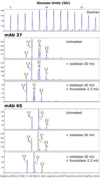
Murine and human IgGs have a conserved N-glycosylation site in their Fc region (18),
where the associated N-glycan plays an important role in Fc-dependent effector
functions (19). Core fucosylation especially strongly influences the binding of
antibod-ies to Fc
␥
Rs and the subsequent mediated cellular cytotoxicity or
antibody-dependent cellular phagocytosis (20–23). We therefore profiled the N-glycans of MAbs
37 and 65 using 8-amino-1,3,6-pyrenetrisulfonic acid (APTS) labeling followed by
capillary electrophoresis, using a labeled dextran ladder and N-glycans from bovine
RNase B as references (Fig. 2). The two antibodies contained comparable levels of total
terminal galactose residues (55.67% for MAb 37 and 65.52% for MAb 65) and were
completely core fucosylated (100% for both MAb 37 and MAb 65) (Fig. 2).
MAbs 37 and 65 differentially activate Fc
␥
Rs
in vitro
.
To compare the potencies
of MAb 37 and MAb 65 for the activation of individual Fc
␥
Rs
in vitro
, a recently
developed cell-based activation assay was applied (24, 25). We compared the
engage-FIG 1MAb 37 and MAb 65 recognize a similar epitope in M2e and bind with similar affinities to M2e and M2. (A) Western blot analyses of lysates of HEK293T cells that had been transfected with Flag-tagged M2 wild type (WT) and M2e Ala scan mutants. The blot was probed with anti-Flag MAb, MAb 37, and MAb 65. Lanes: NC, lysate from nontransfected cells; S2A, serine at position 2 in M2e changed to alanine; L3A, leucine at position 3 in M2e changed to alanine, etc. The results shown for the anti-Flag MAb and MAb 65 are republished from reference 17. (B) M2e peptide ELISA using a high-performance liquid chromatography (HPLC)-purified M2e peptide (SLLTEVETPIRNEWG CRCNDSSD) for coating. Binding of MAb 37 or MAb 65 to M2e-coated wells was determined using a secondary HRP-conjugated anti-mouse immunoglobulin antibody. (C) MDCK cells were infected with A/Puerto Rico/8/34 virus. Twenty-four hours later, the cells were incubated with a dilution series of MAb 37 or MAb 65, followed by fixation with 4% paraformaldehyde and detection by a cellular ELISA. (D) HEK293T cells were infected with A/Puerto Rico/8/34 virus. Sixteen hours later, the cells were incubated with a dilution series of MAb 37 or MAb 65, followed by fixation with 4% paraformaldehyde, permeabilization, and staining with goat anti-influenza virus RNP polyclonal serum. Binding of MAb 37 and MAb 65 was detected with donkey anti-mouse IgG coupled to Alexa Fluor 488, and binding of anti-influenza virus RNP was detected with donkey anti-goat IgG coupled to Alexa Fluor 647. Theyaxis depicts the median fluorescence intensity (MFI), which corresponds to the median fluorescence of binding of MAb 37 or MAb 65 to infected cells from which the median fluorescence of uninfected cells bound by MAb 37 or MAb 65 has been subtracted. The graphs in panels B and C are representative of those from one out of three repeat experiments. Data points represent averages of triplicates (B and C) or duplicates (D), and error bars represent standard deviations. The equilibrium dissociation constants (Kds) of MAb 37 or MAb 65 in panels B and C are averages from three
[image:3.585.46.475.73.291.2]independent experiments, together with their standard deviations.
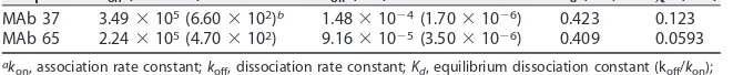
TABLE 1Affinities of anti-M2e antibodies for the M2e consensus sequence measured by surface plasmon resonancea
Sample kon(Mⴚ1sⴚ1) koff(sⴚ1) Kd(nM) 2(RU2)
MAb 37 3.49⫻105(6.60⫻102)b 1.48⫻10⫺4(1.70⫻10⫺6) 0.423 0.123 MAb 65 2.24⫻105(4.70⫻102) 9.16⫻10⫺5(3.50⫻10⫺6) 0.409 0.0593 ak
on, association rate constant;koff, dissociation rate constant;Kd, equilibrium dissociation constant (koff/kon); 2, goodness of fit; RU, resonance units.
bThe data in parentheses represent the standard error calculated on the basis of measurements obtained at
seven different peptide concentrations.
Fc␥R–Anti-M2e IgG Interactions Journal of Virology
on November 7, 2019 by guest
http://jvi.asm.org/
[image:3.585.40.375.668.702.2]FIG 2N-glycan profiling of MAb 37 and MAb 65. DNA sequencer-assisted fluorophore-assisted capillary electro-phoresis chromatograms of the fluorescently labeled dextran ladder (top) and N-glycans on MAb 37 and MAb 65 (bottom). For each MAb, the upper panel provides the chromatogram for the untreated sample, which was subjected to exoglycosidase digests using an␣-2,3/6/8-sialidase alone or␣-2,3/6/8-sialidase combined with an ␣-1,2/3/4/6-fucosidase, as indicated. The glucose units from the ladder were annotated using N-glycans from bovine RNase B (not shown), and the annotated structures were confirmed using additional exoglycosidase digests. Blue squares,N-acetylglucosamine residues; green circles, mannose residues; yellow circles, galactose residues; red triangles, fucose residues.
Van den Hoecke et al. Journal of Virology
on November 7, 2019 by guest
http://jvi.asm.org/
[image:4.585.62.405.67.664.2]ment of individual Fc
␥
Rs by graded concentrations of MAb 37 and MAb 65 bound to
PR8 virus-infected MDCK cells. Control IgG1 and IgG2a monoclonal antibodies did not
activate any of the Fc
␥
R-
s in this assay (Fig. 3). IgG2a MAb 65 activated all Fc
␥
R-
s with
very similar dose-response curves, ranging from 0.0065 nM MAb to plateau values at 6.7
nM antibody (Fig. 3). In contrast, MAb 37 (IgG1) triggered only the inhibitory Fc
␥
RII-
and the activating Fc
␥
RIII-
and completely failed to activate Fc
␥
RI and Fc
␥
RIV even at
very high concentrations of opsonizing MAb. The latter result is in line with the report
that monomeric mouse IgG1 binds very poorly to Fc
␥
RI and -IV (26). MAbs 37 and 65
activated Fc
␥
RIII-
equally well, but MAb 37 was approximately 10-fold more potent in
activating Fc
␥
RII-
than MAb 65 (Fig. 3), which accords well with the reported higher
affinity of monomeric IgG1 for Fc
␥
RII (27). Taken together, MAb 65 and MAb 37
exhibited similar F(ab)
2-mediated M2e-binding characteristics and had clearly distinct
Fc-mediated functions
in vitro
.
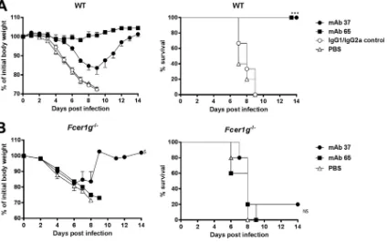
Protection by MAb 37 and MAb 65 requires Fc
␥
Rs.
Next, we compared the
protective efficacy of passively transferred MAbs 37 and 65 against a potentially lethal
influenza A virus challenge (4 50% lethal doses [LD
50]) of BALB/c mice with X47
(A/Victoria/3/75 [H3N2]
⫻
PR8), a mouse-adapted H3N2 virus (1). Compared to the
protection provided by isotype control MAbs, both MAb 37 and MAb 65 protected
the animals from lethal infection (Fig. 4A). However, mice that were treated with the
specific IgG2a MAb 65 displayed significantly less body weight loss than
M2e-specific IgG1 MAb 37 recipients (
P
⬍
0.001) (Fig. 4A). This correlated with the lung viral
load, since MAb 65 treatment resulted in significantly lower lung viral titers than MAb
FIG 3MAb 37 and MAb 65 differentially activate Fc␥Rsin vitro. MDCK cells were infected with A/Puerto Rico/8/34 virus (MOI, 5) for 1 h, washed, and cocultured overnight with the Fc␥R-expression reporter cells and graded concentrations of the anti-M2e MAbs or the corresponding isotype controls. The IL-2 produced was quantified by sandwich ELISA and represents a measure of the magnitude of Fc␥R activation. Data points are averages from triplicates, and error bars represent 95% confidence intervals. Two-way ANOVA with the Sidak correction was used for multiple comparisons (**,P⬍0.01;***,P⬍0.001; ns, not significant). The graphs are representative of those from one out of three repeat experiments. OD, optical density.
Fc␥R–Anti-M2e IgG Interactions Journal of Virology
on November 7, 2019 by guest
http://jvi.asm.org/
[image:5.585.40.491.69.388.2]37 treatment (
P
⫽
0.0004, unpaired
t
test) (Fig. 5C). The protection provided by MAb 37
and MAb 65 was dose dependent (unpublished result).
Next, we evaluated the
in vivo
requirement of FcR
␥
s for protection by the two MAbs.
To define the requirement for one or more activating Fc
␥
Rs for
in vivo
protection
mediated by the two anti-M2e MAbs, we performed passive transfer experiments in
Fcer1g
⫺/⫺mice, which lack the common
␥
chain and cannot express any functional
activating Fc
␥
R. At 24 h after antibody administration, the mice were challenged with
4 LD
50of X47.
Fcer1g
⫺/⫺mice that received MAb 65, MAb 37, or the isotype controls
showed no significant difference in body weight loss (
P
⫽
0.541) (Fig. 4B). Except for
one mouse in the MAb 37 recipient group, all
Fcer1g
⫺/⫺mice succumbed to the
challenge infection (Fig. 4B), demonstrating that Fc
␥
Rs are essential for protection
against influenza A virus challenge by both M2e-specific MAbs.
MAb 65 can protect in the absence of Fc
␥
RI and Fc
␥
RIII, and the protection
provided by MAb 65 involves Fc
␥
RIV.
MAb 37 largely failed to protect
Fcgr3
⫺/⫺mice,
although the difference in the survival rates of challenged mice that had been treated
with the irrelevant control antibody was significant (
P
⫽
0.0289) (Fig. 5A). MAb 65
performed significantly better (
P
⬍
0.001) than MAb 37 and completely protected
Fcgr3
⫺/⫺mice from lethal infection and severe morbidity (Fig. 5A). To further narrow
down the differential requirement for the activating Fc
␥
Rs, we performed challenge
FIG 4M2e-specific IgG2a protects better than M2e-specific IgG1, and protection depends on FcR␥. (A) M2e-specific IgG2a protects against influenza A virus challenge better than M2e-specific IgG1. Wild-type (WT) BALB/c mice received 5 mg/kg of either MAb 37 (IgG1) or MAb 65 (IgG2a) by intraperitoneal injection. The IgG1/IgG2a control group was treated with 2.5 mg/kg of control IgG1 and 2.5 mg/kg of control IgG2a per mouse. Twenty-four hours later, the mice were challenged with 4 LD50of mouse-adapted X47 virus. Body weight (left) and survival (right) were monitored for 2 weeks after challenge. In the left-hand graph, data points represent averages, and error bars represent the standard errors of the means. Differences in body weight between the negative-control groups, on the one hand, and the MAb 37-and MAb 65-treated groups, on the other h37-and, were statistically significant (P⬍0.001, REML variance components analysis;n⫽6 mice per group on day 0). The differences in body weight curves between groups that received MAb 37 or MAb 65 were statistically significant (P⬍0.001, REML variance components analysis). The survival rate of the mice that received MAb 65 or MAb 37 was significantly different from that of mice in the control groups (***,P⬍0.001, log-rank test). The results represent those from one out of two repeat experiments, which had similar results. (B) M2e-specific MAbs fail to protect mice lacking the Fc common␥chain.Fcer1g⫺/⫺BALB/c mice were treated with 5 mg/kg of MAb 37, MAb 65, or PBS by intraperitoneal injection. Twenty-four hours later, the mice were challenged with 4 LD50of mouse-adapted X47 virus. Body weight (left) and survival (right) were monitored for 2 weeks after challenge. In the left-hand graph, data points represent averages and error bars represent the standard errors of the means. $, starting from day 9 after infection, data were based on only one surviving mouse. The survival rates of the groups that received MAb 37 or MAb 65 were not significantly different from those for the control groups (P⬎0.05, log-rank test;n⫽5 mice per group on day 0). NS, not significant.
Van den Hoecke et al. Journal of Virology
on November 7, 2019 by guest
http://jvi.asm.org/
[image:6.585.45.470.72.337.2]FIG 5Protection provided by IgG2a subclass antibodies in the absence of Fc␥RI and Fc␥RIII (A) The protection provided by M2e-specific IgG1 MAb depends on Fc␥RIII.Fcgr3⫺/⫺BALB/c mice (n⫽6 per group) received 5 mg/kg of MAb 37, MAb 65, negative-control IgG1 or IgG2a MAb, or PBS by intraperitoneal injection. Twenty-four hours later, the mice were challenged with 4 LD50of mouse-adapted X47 virus and body weight (left) and survival (right) were monitored. In the left-hand graph, data points represent averages and error bars represent the standard errors of the means. The body weight changes of the group that received MAb 65 were significantly different from those of mice that received MAb 37 or control MAbs (P⬍0.001, REML variance components analysis). The survival rate of the mice that received MAb 65 was significantly different from that of the mice that received MAb 37 (*,P⫽0.0178, log-rank test) and from that of the mice in the control group (***,P⬍0.001, log-rank test). The survival rate of the MAb 37 recipient mice was significantly different from that of the mice in the control groups (*,P⬍0.05, log-rank test). (B) IgG2a MAb 65 protects against influenza A virus challenge in the absence of Fc␥RI and -III.Fcgr1⫺/⫺Fcgr3⫺/⫺BALB/c mice (n⫽6 mice per group, except for the MAb 37 group, which had 7 mice) were treated as described in the legend to panel A, and morbidity and mortality were monitored for 2 weeks after challenge. Differences in body weight between the group that received MAb 65 and the group that received MAb 37 were significant (P⬍0.001, REML variance components analysis). The survival rate of the group that received MAb 65 was significantly different from that of the group that received MAb 37 and the control group (***,P⬍0.001, log-rank test). The graphs are representative of those from one out of two repeat experiments with similar results. (C) Treatment with M2e-specific IgG1 or IgG2a MAb decreases the lung viral load. Wild-type mice (n⫽4) orFcgr1⫺/⫺Fcgr3⫺/⫺mice (n⫽4 mice per group, except for the IgG control group, which had 3 mice) received 5 mg/kg of MAb 37 or MAb 65 or a control IgG mix containing 2.5 mg/kg of IgG1 control MAb and 2.5 mg/kg of IgG2a control MAb. Twenty-four hours later, the mice were challenged with 4 LD50of mouse-adapted X47 virus, and the lungs were harvested at 6 days postinfection, followed by viral titration by plaque assay using MAb 37. Treatment of wild-type orFcgr1⫺/⫺Fcgr3⫺/⫺mice with MAb 37 or MAb 65 resulted in a significant decrease in viral lung titers (P⫽0.03 andP⫽0.0003, respectively, for BALB/c mice andP⫽0.0107 andP⫽0.0005, respectively, for mice deficient inFcgr1andFcgr3;Pvalues were determined by an unpairedttest) compared to those in mice receiving the control IgG treatment. The differences in lung viral loads between mice receiving MAb 37 and mice receiving MAb 65 were statistically significant for wild-type BALB/c mice (P⫽0.0004, unpairedttest) and mice deficient inFcgr1andFcgr3(P⫽0.0112, unpairedttest). Differences in lung viral loads in mice receiving the IgG control treatment or MAb 65 were statistically significant between wild-type mice and mice deficient inFcgr1andFcgr3(for IgG control treatment,P⫽0.0060; for MAb 37,P⫽0.0976; for MAb 65,P⫽0.0054;Pvalues were determined by an unpairedttest).
Fc␥R–Anti-M2e IgG Interactions Journal of Virology
on November 7, 2019 by guest
http://jvi.asm.org/
[image:7.585.40.422.71.468.2]studies in mice deficient in
Fcgr1
and
Fcgr3
. In these double-deficient mice, MAb 65
provided full protection against virus challenge, whereas all control-treated and MAb
37-treated mice died (Fig. 5B). MAb 65 treatment partially protected against body
weight loss after challenge of
Fcgr1
⫺/⫺Fcgr3
⫺/⫺mice, whereas MAb 37 failed to do so.
This difference in protection against morbidity correlated with a significantly lower
lung virus load in the
Fcgr1
⫺/⫺Fcgr3
⫺/⫺mice that had been treated with MAb 65
compared with that in mice that had been treated with MAb 37 (
P
⫽
0.0112;
unpaired
t
test) (Fig. 5C).
We hypothesized that Fc
␥
RIV could be responsible for the protection provided by
MAb 65 in the absence of Fc
␥
RI and Fc
␥
RIII. To test this, wild-type and
Fcgr4
⫺/⫺C57BL/6 mice were treated with MAb 65 or MAb 37 and infected with a lethal dose of
X31 (H3N2). Both MAb 37 and MAb 65 protected the mice, whereas control-treated
wild-type and
Fcgr4
⫺/⫺C57BL/6 mice did not survive the virus challenge (Fig. 6). Similar
to wild-type mice,
Fcgr4
⫺/⫺mice treated with MAb 65 displayed significantly less body
weight loss than MAb 37 recipients (
P
⬍
0.001) (Fig. 6). The body weight loss after
challenge was the same in
Fcgr4
⫺/⫺and wild-type C57BL/6 mice treated with MAb 37
(
P
⫽
0.114), in line with a lack of Fc
␥
RIV engagement by mouse IgG1 isotype antibodies
(Fig. 3 and 6). The protection provided by MAb 65 was partially dependent on Fc
␥
RIV,
since after infection
Fcgr4
⫺/⫺mice showed a body weight loss significantly different
from that for wild-type mice (
P
⬍
0.001) (Fig. 6). Taken together, these results suggest
that Fc
␥
RIV contributes to the protection provided by M2e-specific IgG2a.
DISCUSSION
We isolated and characterized a pair of M2e-specific murine MAbs with similar
affinities for M2e and comparable N-glycan profiles of their Fc parts. We compared the
potency of activation of individual Fc
␥
Rs
in vitro
by this antibody pair in the context of
FIG 6Fc␥RIV contributes to protection by M2e-specific IgG2a antibodies against influenza A virus challenge. (A) Reduced protection provided by IgG2a MAb 65 inFcgr4⫺/⫺mice. Wild-type orFcgr4⫺/⫺mice were treated with 5 mg/kg of mouse IgG1 MAb 37, IgG2a MAb 65, an isotype control MAb, or PBS 24 h before intranasal infection with 4 LD50of influenza A virus strain X31. Wild-type andFcgr4⫺/⫺mice were treated with antibody and challenged in parallel in the same experiment. (A) Curves of the body weights of mice treated with MAb 37 (left) or MAb 65 (right); (B) survival curves for mice treated with MAb 37 (left) or MAb 65 (right). Data points represent averages, and error bars represent the standard errors of the means. The data from two independent experiments were pooled. In total, 6 mice were used in the groups treated with PBS and 8 or 9 mice were used in the groups treated with an MAb. The differences in the change in body weight between wild-type andFcgr4⫺/⫺mice that had received MAb 65 prior to challenge were statistically significant (P⬍0.001, REML variance components analysis).
Van den Hoecke et al. Journal of Virology
on November 7, 2019 by guest
http://jvi.asm.org/
[image:8.585.45.542.71.335.2]a viral infection and their protective potential in wild-type and Fc
␥
R-deficient mice.
MAb 65 activated all Fc
␥
R-
s in the presence of influenza A virus-infected cells. In
contrast, MAb 37 triggered only the activating Fc
␥
RIII-
and the inhibitory Fc
␥
RII-
, with
the level of activation of the latter being remarkably higher than that by MAb 65. The
two M2e-specific MAbs thus differentially activate Fc
␥
Rs
in vitro
. These
in vitro
results
also highlight that Fc
␥
RIV can contribute to anti-M2e immune complex recognition on
influenza A virus-infected target cells.
The results of
in vitro
experiments correlated surprisingly well with the results of
in
vivo
experiments in which we compared the protection provided by MAbs 37 and 65
against influenza A virus challenge in mice with different deficiencies in their Fc
␥
R
compartments. The M2e-specific IgG2a antibody protected better against influenza A
virus challenge than the M2e-specific IgG1 antibody, presumably because, as detected
in vitro
, MAb 65 could engage all three activating receptors, which are expressed on
natural killer cells, neutrophils, monocytes, and macrophages (28, 29). This is in
agree-ment with the finding that active vaccination strategies with M2e fusion constructs that
predominantly induce M2e-specific IgG2a/c antibodies result in better protection
against challenge (30, 31). In addition, we observed that MAb 65 protected mice from
mortality even at a concentration as low as 0.3125 mg/kg of body weight (unpublished
result).
In the absence of Fc
␥
RIII, the IgG1 MAb largely failed to protect against influenza A
virus challenge, while MAb 65 was still protective. Therefore, Fc
␥
RIII is not strictly
required for MAb 65-mediated protection but significantly contributes to MAb
37-mediated protection. This also suggests that natural killer cells, which in mice express
only Fc
␥
RIII (28), do not play an indispensable role in the M2e-based immune
protec-tion provided by IgG2a isotype antibodies. In the absence of both Fc
␥
RI and Fc
␥
RIII,
mice were still fully protected against death caused by influenza virus challenge.
However,
Fcgr1
⫺/⫺Fcgr3
⫺/⫺mice exhibited significantly more body weight loss than
wild-type mice (Fig. 5). The possible contribution of Fc
␥
RIV to the protection provided
by M2e-specific antibodies has not yet been reported. We observed that
Fcgr4
⫺/⫺mice
are protected by MAb 65 but displayed significantly more body weight loss after
influenza A virus challenge than type mice. In contrast, MAb 37 protected
wild-type and
Fcgr4
⫺/⫺mice equally well, although it performed worse than MAb 65 in
terms of weight loss (Fig. 6). These results also accord with the finding that Fc
␥
RIV,
which is expressed on monocytes, macrophages, and neutrophils, plays an important
role in IgG2a-dependent effector activities
in vivo
, including IgG2a-mediated killing of
tumor cells (32). Both Fc
␥
RI and Fc
␥
RIV are expressed on alveolar macrophages, which
play an essential role in M2e antibody-mediated immune protection (2, 27, 33, 34). In
our model, we found a contribution of Fc
␥
RIV but not a determining role for Fc
␥
RIV in
the protection provided by M2e-specific IgG2a against influenza A virus challenge.
Future studies comparing Fc
␥
RI-deficient mice and mice lacking all three activating
receptors, Fc
␥
RI, Fc
␥
RIII, and Fc
␥
RIV, will be required to determine the relative
contri-bution of the two high-affinity activating Fc receptors Fc
␥
RI and Fc
␥
RIV.
What are the implications of our findings for the clinical development of M2e-based
vaccines? M2e immunity appears to operate in the absence of demonstrable
virus-neutralizing activity but, rather, engages Fc
␥
receptor-expressing myeloid cells. Human
IgG1 and IgG3 isotype antibodies can be considered the counterparts of mouse IgG2a
antibodies. Therefore, vaccine formulations that promote the induction of
antigen-specific IgG1 and IgG3 in humans should be used. The MF59, AS03, and AS04 adjuvants
promote such a Th1-specific response (35). Human IgG1 and IgG3 have the highest
affinity for Fc
␥
RI, which, as in mice, has a broad expression pattern (dendritic cells,
monocytes, and macrophages) (27). The sequence of mouse Fc
␥
RIV suggests that it is
related to human Fc
␥
RIIIA (expressed on natural killer cells, monocytes, and
macro-phages) (26, 34). Therefore, M2e-specific IgG antibodies could possibly provide
protec-tion through multiple effector cells that are resident at or recruited to the site of
infection.
We still know surprisingly little about how effective antimicrobial vaccines work. It
Fc␥R–Anti-M2e IgG Interactions Journal of Virology
on November 7, 2019 by guest
http://jvi.asm.org/
was reported that Fc
␥
receptor-dependent phagocytosis of influenza A virus virions
opsonized with HA-specific antibodies is a strong contributor to the protection
pro-vided by a conventional influenza A vaccine (36). In addition, the protection against
influenza A virus infection provided by broadly neutralizing antibodies directed against
the HA stalk largely depends on Fc
␥
Rs (16). Recently, another report even suggested
that broadly neutralizing anti-influenza virus antibodies that lack hemagglutination
inhibition activity require interaction with Fc
␥
Rs to mediate
in vivo
protection (37).
Therefore, future developments toward antibody-based universal influenza vaccines
should consider the role of the Fc
␥
receptor repertoire in vaccine efficacy.
MATERIALS AND METHODS
Ethics statement.All animal experiments described in this study were conducted according to national legislation (Belgian laws 14/08/1986 and 22/12/2003, Belgian Royal Decree 06/04/2010) and European legislation (EU Directives 2010/63/EU and 86/609/EEC). All experiments on mice and animal protocols were approved by the ethics committee of Ghent University (permit numbers LA1400091 and EC2012-034).
Monoclonal antibodies and their epitope specificity.Hybridomas that produce M2e-specific MAbs 37 and 65 were isolated as described previously (17). After subcloning, these two hybridoma cultures were scaled up and MAbs 37 and 65 were purified from the culture supernatant with protein A-Sepharose (GE Healthcare). M2e Ala scan analysis was performed as described previously (17), and the results were visualized by Western blotting using MAb 37, MAb 65, or anti-Flag (Sigma-Aldrich) antibody followed by horseradish peroxidase (HRP)-conjugated sheep anti-mouse IgG (GE Healthcare). Isotype control MAbs directed against the hepatitis B virus core (IgG1) or the small hydrophobic protein of human respiratory syncytial virus (IgG2a) were produced and purified as described above.
Affinity measurement by ELISA and flow cytometry.The affinity of MAb 37 and MAb 65 for M2e was determined by ELISA with an M2e peptide (SLLTEVETPIRNEWGCRCNDSSDSG, used at 2g/ml in 50 l/well), as described in reference 17, or MDCK cells infected with A/Puerto Rico/8/34 (H1N1) (PR8). A dilution series of MAb 65 or MAb 37 was applied to infected cells on ice. Subsequently, cells were fixed with 4% paraformaldehyde and antibody binding was detected using HRP-conjugated sheep anti-mouse IgG (GE Healthcare).
For flow cytometry analysis, PR8 virus-infected human embryonic kidney 293T (HEK293T) cells were incubated on ice with a dilution series of MAb 65 or MAb 37 in 0.5% bovine serum albumin (BSA) in phosphate-buffered saline (PBS) and subsequently fixed with 4% paraformaldehyde (PFA). After permea-bilization (10⫻permeabilization buffer diluted in double-distilled water; eBioscience), cells were stained with 1/2,000-diluted polyclonal goat anti-influenza virus ribonucleoprotein (RNP; catalog number NR-4282; Biodefense and Emerging Infections Resources Repository, NIAID, NIH). Binding of primary anti-bodies was revealed with donkey anti-mouse IgG coupled to Alexa Fluor 488 (1/600; Invitrogen) and donkey anti-goat IgG coupled to Alexa Fluor 647 (1/600; Invitrogen). The median fluorescence intensity (MFI) of the cells was determined with an LSRII HTS flow cytometer (BD). The influenza virus-specific MFI was calculated by subtracting the MFI of MAb 37- or MAb 65-positive cells in the RNP-negative population (uninfected HEK293T cells stained with 10g/ml MAb 37 or MAb 65 and anti-RNP) from the MFI of MAb 37- or MAb 65-positive cells in the RNP-positive population (infected HEK293T cells stained with a dilution series of MAb 37 or MAb 65 and RNP).
Affinity measurement by surface plasmon resonance.The affinities of MAbs 37 and 65 for the M2e peptide were determined using a Biacore T200 instrument (GE Healthcare). Anti-M2e MAbs were immobilized on the flow cells of a CM5 sensor chip (GE Healthcare) by amine coupling according to the manufacturer’s instructions. A flow cell blocked with ethanolamine served as a reference. The M2e peptide in HBS-EP buffer (0.01 M HEPES, pH 7.4, 0.15 M NaCl, 3 mM EDTA, 0.005% [vol/vol] surfactant P20) was added at concentrations of 0.39, 0.78, 3.13, 6.25, 12.5, 25, and 50 nM. The samples were injected at 50l/min for 180 s, after which dissociation was monitored for 1,000 s. The sensor chip surface was regenerated by injecting 10 mM HCl for 60 s and 20 mM HCl for another 60 s. Biacore T200 evaluation software (v1.0) was used to calculate the association rate constant (kon), the dissociation rate constant (koff), and the equilibrium dissociation constant (Kd; which is equal tokoff/kon) by fitting a 1:1 binding model with a drifting baseline.
N-glycan analysis.N-linked oligosaccharides were isolated, derivatized with APTS, and analyzed by capillary electrophoresis on an ABI 3130 capillary DNA sequencer using in-house-produced peptide-N -glycosidase F (15.4 mU/l) as described previously (38). As electrophoretic mobility references, a labeled dextran ladder and N-glycans from bovine RNase B were included. The data were analyzed using GeneMapper software (Applied Biosystems), and N-glycan profiles were exported as scalable vector graphics (svg) for manual alignment and annotation in Inkscape software (v0.91). Exoglycosidase treatments to determine the degree of fucosylation on labeled glycans were carried out overnight at 37°C in 20 mM sodium acetate (pH 5.0) using 20 mU Arthrobacter ureafaciens ␣-2,3/6/8-sialidase (produced in-house), 2.2 mU␣-1,2/3/4/6-fucosidase from bovine kidney (Prozyme), or both.
In vitroFc␥R activation assay.Fc␥R activation by MAbs 37 and 65 was compared using a recently developedin vitroFc␥R activation assay (24, 25). Cloning of Fc␥R-constructs and the generation of Fc␥R-BW5147 reporter cells were performed as reported previously (24, 25). Activation of stably transduced Fc␥R-BW5147 reporter cells by immune complexes results in the production of interleukin-2 (IL-2), which was quantified by ELISA (24, 25). Madin-Darby canine kidney (MDCK) cells were seeded in
Van den Hoecke et al. Journal of Virology
on November 7, 2019 by guest
http://jvi.asm.org/
96-well flat-bottom plates and infected with PR8 virus (multiplicity of infection [MOI], 5). After 1 h of incubation at 37°C, unbound virus particles were removed by washing, and serial dilutions of the MAbs were added, followed by the addition of 1.5⫻105Fc␥R-BW5147 reporter cells in a total volume of 200
l RPMI with 10% fetal calf serum per well. Cocultures were incubated overnight at 37°C in a 5% CO2 atmosphere. To increase the release of produced IL-2 from reporter cells, 100l PBS with 0.1% Tween was added to each well, and 150l from each well was used in an anti-IL-2 sandwich ELISA as described previously (24, 25).
Challenge experiments in mice.BALB/c mice were purchased from Harlan (The Netherlands) or Charles River (France);Fcer1g⫺/⫺BALB/c mice, which lack the common␥chain and, consequently, the ability to express all activating Fc␥Rs, were purchased from Taconic (Denmark); and C57BL/6 mice were purchased from Charles River (France). Fcgr3⫺/⫺BALB/c mice,Fcgr1⫺/⫺Fcgr3⫺/⫺BALB/c mice, and Fcgr4⫺/⫺C57BL/6 mice were bred in-house under specific-pathogen-free (SPF) conditions. Mice were used at the age of 7 to 8 weeks and were housed in individually ventilated cages in a temperature-controlled environment with 12-h light and 12-h dark cycles and food and waterad libitum. To evaluate protection, mice were injected intraperitoneally with 5 mg/kg of MAb 37 or MAb 65 (unless otherwise stated) or negative-control MAbs. Twenty-four hours later, the mice were anesthetized with a mixture of ketamine (10 mg/kg) and xylazine (60 mg/kg) and challenged by intranasal administration of 50l PBS containing 4 LD50of mouse-adapted X47 (A/Victoria/3/75 [H3N2]⫻PR8) influenza A virus for wild-type BALB/c,Fcer1g⫺/⫺BALB/c,Fcgr3⫺/⫺BALB/c, andFcgr1⫺/⫺Fcgr3⫺/⫺BALB/c mice. Four LD
50of mouse-adapted X31 (A/Aichi/2/68 [H3N2]⫻PR8) influenza A virus was used to challenge wild-type C57BL/6 and Fcgr4⫺/⫺C57BL/6 mice. C57BL/6 mice were challenged with X31 because the LD
50of this virus was established for this mouse strain. However, X31 and X47 have an identical gene segment 7. The body weights and rates of survival of the mice were monitored for 2 weeks after challenge, and animals that had lost more than 25% of their body weight compared to that at the time of challenge were euthanized. To determine the effect of MAb 37 and MAb 65 treatment on lung viral load, complete lungs were harvested at 6 days postinfection and homogenized in 1 ml of PBS using metal beads, followed by 5 min of centrifugation at 450⫻g. The supernatants were stored at⫺80°C. The lung viral titer was determined using a plaque assay, where plaques were stained with MAb 37, followed by secondary anti-mouse IgG HRP-linked antibody (GE Healthcare) and visualization using TrueBlue peroxidase substrate (KPL).
Statistics.The statistical significance of the difference in the results obtained with the different MAbs in the Fc␥R activation assay was analyzed using two-way analysis of variance (ANOVA) with the Sidak correction for multiple comparisons. Statistical analysis of the differences in survival rates was performed by comparing Kaplan-Meier curves using the log-rank test. Statistical analysis of the differences in lung viral titers was performed using the unpairedttest. These tests were performed in GraphPad Prism (v6.07) software for Windows (GraphPad Software, San Diego, CA). Statistical comparison of the differ-ences in body weight loss was performed using restricted maximum likelihood (REML) variance com-ponents analysis in Genstat software (64 bit, v18.1). The following linear mixed model (random terms are underlined) was fitted to most of the data:Yijkt⫽⫹genotypej⫹treatmentk⫹timet⫹(genotype.
treatment)jk⫹(genotype.time)jt⫹(treatment.time)kt⫹(genotype.treatment.time)jkt⫹(mouse.time)it⫹
residualijkt, whereYijktis the relative body weight of theith mouse with genotypejtreated withkfor
which the results were measured at time pointt(wheretis equal to 1 to 14 days, equally spaced), andis the overall mean calculated for all mice considered across all time points. To compare the morbidity of wild-type BALB/c mice andFcgr1⫺/⫺Fcgr3⫺/⫺BALB/c mice after MAb 65 treatment, the following model was fitted:Yijkt⫽⫹genotypej⫹timet⫹(genotype.time)jt⫹residualijt, where Yijtis the relative body weight of theith mouse with genotypejfor which the results were measured
at time pointt(wheretis equal to 1 to 14 days, equally spaced), andis the overall mean calculated for all mice considered across all time points. A first-order autoregressive covariance structure was used to model the within-subject correlation and allowed heterogeneity across time. The signifi-cance of the fixed main and interaction effects was assessed by anFtest. APvalue ofⱕ0.05 was considered statistically significant.
ACKNOWLEDGMENTS
We are grateful to Sjef Verbeek (Leiden University Medical Center, Leiden, The
Netherlands) for providing
Fcgr3
⫺/⫺and
Fcgr1
⫺/⫺Fcgr3
⫺/⫺BALB/c mice and to Helmut
Hanenberg (Heinrich Heine University Düsseldorf, Düsseldorf, Germany) for providing
lentiviral plasmids. We thank Marnik Vuylsteke for performing the statistical analysis.
We thank Frederik Vervalle for excellent technical assistance and Céline Steyt for animal
care. We thank Jan Spitaels for help with the flow cytometric analysis.
We declare no competing financial interests.
This work was supported by the Fonds voor Wetenschappelijk Onderzoek (project
grant G052412N), the Ghent University Industrial Research Fund (IOF08/STEP/001), and
the Ghent University Special Research Fund (project BOF12/GOA/014) to X.S. S.V.D.H. is
a Ph.D. fellow and B.S. a postdoctoral fellow at the Fonds voor Wetenschappelijk
Onderzoek. L.D. was supported by the State Scholarship Fund (file no. 2011674067)
from the China Scholarship Council and by IUAP-BELVIR p7/45. A.K. is supported by FP7
ITN UniVacFlu, and K.R. is supported by FP7 Collaborative Project FLUNIVAC. K.E. was
Fc␥R–Anti-M2e IgG Interactions Journal of Virology
on November 7, 2019 by guest
http://jvi.asm.org/
supported by the Graduate School Molecules of Infection, Heinrich Heine University
Düsseldorf.
REFERENCES
1. Neirynck S, Deroo T, Saelens X, Vanlandschoot P, Jou WM, Fiers W. 1999. A universal influenza A vaccine based on the extracellular domain of the M2 protein. Nat Med 5:1157–1163.https://doi.org/10.1038/13484. 2. El Bakkouri K, Descamps F, De Filette M, Smet A, Festjens E, Birkett A, Van
Rooijen N, Verbeek S, Fiers W, Saelens X. 2011. Universal vaccine based on ectodomain of matrix protein 2 of influenza A: Fc receptors and alveolar macrophages mediate protection. J Immunol 186:1022–1031.
https://doi.org/10.4049/jimmunol.0902147.
3. Treanor JJ, Tierney EL, Zebedee SL, Lamb RA, Murphy BR. 1990. Passively transferred monoclonal antibody to the M2 protein inhibits influenza A virus replication in mice. J Virol 64:1375–1377.
4. Lamb RA, Zebedee SL, Richardson CD. 1985. Influenza virus-M2 protein is an integral membrane-protein expressed on the infected-cell surface. Cell 40:627– 633.https://doi.org/10.1016/0092-8674(85)90211-9. 5. Zebedee SL, Lamb RA. 1988. Influenza A virus M2 protein: monoclonal
antibody restriction of virus growth and detection of M2 in virions. J Virol 62:2762–2772.
6. Feng J, Zhang M, Mozdzanowska K, Zharikova D, Hoff H, Wunner W, Couch RB, Gerhard W. 2006. Influenza A virus infection engenders a poor antibody response against the ectodomain of matrix protein 2. Virol J 3:102.https://doi.org/10.1186/1743-422X-3-102.
7. Zhong W, Reed C, Blair PJ, Katz JM, Hancock K. 2014. Serum antibody response to matrix protein 2 following natural infection with 2009 pandemic influenza A(H1N1) virus in humans. J Infect Dis 209:986 –994.
https://doi.org/10.1093/infdis/jit811.
8. De Filette M, Martens W, Roose K, Deroo T, Vervalle F, Bentahir M, Vandekerckhove J, Fiers W, Saelens X. 2008. An influenza A vaccine based on tetrameric ectodomain of matrix protein 2. J Biol Chem 283:11382–11387.https://doi.org/10.1074/jbc.M800650200.
9. Fan J, Liang X, Horton MS, Perry HC, Citron MP, Heidecker GJ, Fu TM, Joyce J, Przysiecki CT, Keller PM, Garsky VM, Ionescu R, Rippeon Y, Shi L, Chastain MA, Condra JH, Davies ME, Liao J, Emini EA, Shiver JW. 2004. Preclinical study of influenza virus A M2 peptide conjugate vaccines in mice, ferrets, and rhesus monkeys. Vaccine 22:2993–3003. https:// doi.org/10.1016/j.vaccine.2004.02.021.
10. Jegerlehner A, Schmitz N, Storni T, Bachmann MF. 2004. Influenza A vaccine based on the extracellular domain of M2: weak protection mediated via antibody-dependent NK cell activity. J Immunol 172: 5598 –5605.https://doi.org/10.4049/jimmunol.172.9.5598.
11. Schotsaert M, De Filette M, Fiers W, Saelens X. 2009. Universal M2 ectodomain-based influenza A vaccines: preclinical and clinical develop-ments. Expert Rev Vaccines 8:499 –508.https://doi.org/10.1586/erv.09.6. 12. Wang BZ, Gill HS, Kang SM, Wang L, Wang YC, Vassilieva EV, Compans RW.
2012. Enhanced influenza virus-like particle vaccines containing the extra-cellular domain of matrix protein 2 and a Toll-like receptor ligand. Clin Vaccine Immunol 19:1119 –1125.https://doi.org/10.1128/CVI.00153-12. 13. Turley CB, Rupp RE, Johnson C, Taylor DN, Wolfson J, Tussey L, Kavita U,
Stanberry L, Shaw A. 2011. Safety and immunogenicity of a recombinant M2e-flagellin influenza vaccine (STF2.4xM2e) in healthy adults. Vaccine 29:5145–5152.https://doi.org/10.1016/j.vaccine.2011.05.041.
14. Wang R, Song A, Levin J, Dennis D, Zhang NJ, Yoshida H, Koriazova L, Madura L, Shapiro L, Matsumoto A, Mikayama T, Kubo RT, Sarawar S, Cheroutre H, Kato S. 2008. Therapeutic potential of a fully human monoclonal antibody against influenza A virus M2 protein. Antiviral Res 80:168 –177.https://doi.org/10.1016/j.antiviral.2008.06.002.
15. Schmitz N, Beerli RR, Bauer M, Jegerlehner A, Dietmeier K, Maudrich M, Pumpens P, Saudan P, Bachmann MF. 2012. Universal vaccine against influenza virus: linking TLR signaling to anti-viral protection. Eur J Im-munol 42:863– 869.https://doi.org/10.1002/eji.201041225.
16. DiLillo DJ, Tan GS, Palese P, Ravetch JV. 2014. Broadly neutralizing hemagglutinin stalk-specific antibodies require FcgammaR interactions for protection against influenza virusin vivo. Nat Med 20:143–151.
https://doi.org/10.1038/nm.3443.
17. Cho KJ, Schepens B, Seok JH, Kim S, Roose K, Lee JH, Gallardo R, Van Hamme E, Schymkowitz J, Rousseau F, Fiers W, Saelens X, Kim KH. 2015. Structure of the extracellular domain of matrix protein 2 of influenza A
virus in complex with a protective monoclonal antibody. J Virol 89: 3700 –3711.https://doi.org/10.1128/JVI.02576-14.
18. Krapp S, Mimura Y, Jefferis R, Huber R, Sondermann P. 2003. Structural analysis of human IgG-Fc glycoforms reveals a correlation between glycosylation and structural integrity. J Mol Biol 325:979 –989.https:// doi.org/10.1016/S0022-2836(02)01250-0.
19. Jefferis R, Lund J, Pound JD. 1998. IgG-Fc-mediated effector functions: molecular definition of interaction sites for effector ligands and the role of glycosylation. Immunol Rev 163:59 –76.https://doi.org/10.1111/j.1600 -065X.1998.tb01188.x.
20. Forthal DN, Gach JS, Landucci G, Jez J, Strasser R, Kunert R, Steinkellner H. 2010. Fc-glycosylation influences Fcgamma receptor binding and cell-mediated anti-HIV activity of monoclonal antibody 2G12. J Immunol 185:6876 – 6882.https://doi.org/10.4049/jimmunol.1002600.
21. Herter S, Birk MC, Klein C, Gerdes C, Umana P, Bacac M. 2014. Glycoen-gineering of therapeutic antibodies enhances monocyte/macrophage-mediated phagocytosis and cytotoxicity. J Immunol 192:2252–2260.
https://doi.org/10.4049/jimmunol.1301249.
22. Shields RL, Lai J, Keck R, O’Connell LY, Hong K, Meng YG, Weikert SH, Presta LG. 2002. Lack of fucose on human IgG1 N-linked oligosaccharide improves binding to human Fcgamma RIII and antibody-dependent cellular toxicity. J Biol Chem 277:26733–26740.https://doi.org/10.1074/ jbc.M202069200.
23. Shinkawa T, Nakamura K, Yamane N, Shoji-Hosaka E, Kanda Y, Sakurada M, Uchida K, Anazawa H, Satoh M, Yamasaki M, Hanai N, Shitara K. 2003. The absence of fucose but not the presence of galactose or bisecting N-acetylglucosamine of human IgG1 complex-type oligosaccharides shows the critical role of enhancing antibody-dependent cellular cyto-toxicity. J Biol Chem 278:3466 –3473. https://doi.org/10.1074/jbc .M210665200.
24. Corrales-Aguilar E, Trilling M, Hunold K, Fiedler M, Le VT, Reinhard H, Ehrhardt K, Merce-Maldonado E, Aliyev E, Zimmermann A, Johnson DC, Hengel H. 2014. Human cytomegalovirus Fcgamma binding proteins gp34 and gp68 antagonize Fcgamma receptors I, II and III. PLoS Pathog 10:e1004131.https://doi.org/10.1371/journal.ppat.1004131.
25. Corrales-Aguilar E, Trilling M, Reinhard H, Merce-Maldonado E, Widera M, Schaal H, Zimmermann A, Mandelboim O, Hengel H. 2013. A novel assay for detecting virus-specific antibodies triggering activation of Fcgamma receptors. J Immunol Methods 387:21–35. https://doi.org/10.1016/ j.jim.2012.09.006.
26. Nimmerjahn F, Bruhns P, Horiuchi K, Ravetch JV. 2005. FcgammaRIV: a novel FcR with distinct IgG subclass specificity. Immunity 23:41–51.
https://doi.org/10.1016/j.immuni.2005.05.010.
27. Guilliams M, Bruhns P, Saeys Y, Hammad H, Lambrecht BN. 2014. The function of Fcgamma receptors in dendritic cells and macrophages. Nat Rev Immunol 14:94 –108.https://doi.org/10.1038/nri3582.
28. Bruhns P. 2012. Properties of mouse and human IgG receptors and their contribution to disease models. Blood 119:5640 –5649.https://doi.org/ 10.1182/blood-2012-01-380121.
29. Nimmerjahn F, Ravetch JV. 2011. FcgammaRs in health and disease. Curr Top Microbiol Immunol 350:105–125.https://doi.org/10.1007/82_2010_86. 30. Ibanez LI, Roose K, De Filette M, Schotsaert M, De Sloovere J, Roels S, Pollard C, Schepens B, Grooten J, Fiers W, Saelens X. 2013. M2e-displaying virus-like particles with associated RNA promote T helper 1 type adaptive immunity against influenza A. PLoS One 8:e59081.https:// doi.org/10.1371/journal.pone.0059081.
31. Song JM, Van Rooijen N, Bozja J, Compans RW, Kang SM. 2011. Vacci-nation inducing broad and improved cross protection against multiple subtypes of influenza A virus. Proc Natl Acad Sci U S A 108:757–761.
https://doi.org/10.1073/pnas.1012199108.
32. Nimmerjahn F, Lux A, Albert H, Woigk M, Lehmann C, Dudziak D, Smith P, Ravetch JV. 2010. FcgammaRIV deletion reveals its central role for IgG2a and IgG2b activity in vivo. Proc Natl Acad Sci U S A 107: 19396 –19401.https://doi.org/10.1073/pnas.1014515107.
33. Lee SM, Gardy JL, Cheung CY, Cheung TK, Hui KP, Ip NY, Guan Y, Hancock RE, Peiris JS. 2009. Systems-level comparison of host-responses elicited by avian H5N1 and seasonal H1N1 influenza viruses in primary
Van den Hoecke et al. Journal of Virology
on November 7, 2019 by guest
http://jvi.asm.org/
human macrophages. PLoS One 4:e8072. https://doi.org/10.1371/ journal.pone.0008072.
34. Mancardi DA, Iannascoli B, Hoos S, England P, Daeron M, Bruhns P. 2008. FcgammaRIV is a mouse IgE receptor that resembles macrophage Fcep-silonRI in humans and promotes IgE-induced lung inflammation. J Clin Invest 118:3738 –3750.https://doi.org/10.1172/JCI36452.
35. Coffman RL, Sher A, Seder RA. 2010. Vaccine adjuvants: putting innate immunity to work. Immunity 33:492–503. https://doi.org/10.1016/ j.immuni.2010.10.002.
36. Huber VC, Lynch JM, Bucher DJ, Le J, Metzger DW. 2001. Fc
receptor-mediated phagocytosis makes a significant contribution to clearance of influenza virus infections. J Immunol 166:7381–7388.https://doi.org/ 10.4049/jimmunol.166.12.7381.
37. DiLillo DJ, Palese P, Wilson PC, Ravetch JV. 2016. Broadly neutralizing anti-influenza antibodies require Fc receptor engagement forin vivo protection. J Clin Invest 126:605– 610. https://doi.org/10.1172/ JCI84428.
38. Laroy W, Contreras R, Callewaert N. 2006. Glycome mapping on DNA sequencing equipment. Nat Protoc 1:397– 405.https://doi.org/10.1038/ nprot.2006.60.
Fc␥R–Anti-M2e IgG Interactions Journal of Virology
on November 7, 2019 by guest
http://jvi.asm.org/