Functional Incompatibility between the Generic NF-
B Motif and a
Subtype-Specific Sp1III Element Drives the Formation of the HIV-1
Subtype C Viral Promoter
Anjali Verma,aPavithra Rajagopalan,aRishikesh Lotke,aRebu Varghese,aDeepak Selvam,aTapas K. Kundu,bUdaykumar Rangaa
HIV-AIDS Laboratory, Molecular Biology and Genetics Unit, Jawaharlal Nehru Centre for Advanced Scientific Research, Bangalore, Indiaa
; Transcription Biology Laboratory, Molecular Biology and Genetics Unit, Jawaharlal Nehru Centre for Advanced Scientific Research, Bangalore, Indiab
ABSTRACT
Of the various genetic subtypes of human immunodeficiency virus types 1 and 2 (HIV-1 and HIV-2) and simian immunodefi-ciency virus (SIV), only in subtype C of HIV-1 is a genetically variant NF-B binding site found at the core of the viral promoter in association with a subtype-specific Sp1III motif. How the subtype-associated variations in the core transcription factor bind-ing sites (TFBS) influence gene expression from the viral promoter has not been examined previously. Usbind-ing panels of infectious viral molecular clones, we demonstrate that subtype-specific NF-B and Sp1III motifs have evolved for optimal gene expression, and neither of the motifs can be replaced by a corresponding TFBS variant. The variant NF-B motif binds NF-B with an affin-ity 2-fold higher than that of the generic NF-B site. Importantly, in the context of an infectious virus, the subtype-specific Sp1III motif demonstrates a profound loss of function in association with the generic NF-B motif. An additional substitution of the Sp1III motif fully restores viral replication, suggesting that the subtype C-specific Sp1III has evolved to function with the variant, but not generic, NF-B motif. A change of only two base pairs in the central NF-B motif completely suppresses viral transcription from the provirus and converts the promoter into heterochromatin refractory to tumor necrosis factor alpha (TNF-␣) induction. The present work represents the first demonstration of functional incompatibility between an otherwise functional NF-B motif and a unique Sp1 site in the context of an HIV-1 promoter. Our work provides important leads as to the evolution of the HIV-1 subtype C viral promoter with relevance for gene expression regulation and viral latency.
IMPORTANCE
Subtype-specific genetic variations provide a powerful tool to examine how these variations offer a replication advantage to spe-cific viral subtypes, if any. Only in subtype C of HIV-1 are two genetically distinct transcription factor binding sites positioned at the most critical location of the viral promoter. Since a single promoter regulates viral gene expression, the promoter variations can play a critical role in determining the replication fitness of the viral strains. Our work for the first time provides a scientific explanation for the presence of a unique NF-B binding motif in subtype C, a major HIV-1 genetic family responsible for half of the global HIV-1 infections. The results offer compelling evidence that the subtype C viral promoter not only is stronger but also is endowed with a qualitative gain-of-function advantage. The genetically variant NF-B and the Sp1III motifs may be respond differently to specific cell signal pathways, and these mechanisms must be examined.
B
ased on genetic variation, human immunodeficiency virustype 1 (HIV-1) is classified into four distinct groups (M, N, O, and P), and group M is subclassified into nine molecular subtypes (A, B, C, D, F, G, H, J, and K) and numerous circulating
recom-binant forms, including A/E (1). The global distribution of HIV-1
subtypes is uneven, with C, A, and B being the most widespread subtypes. Subtype C is predominant in southern and eastern Af-rican countries, India, and Nepal and in recombinant forms in China, and it is currently emerging in southern Brazil. Subtype C is responsible for approximately half of the global HIV-1
infec-tions and more than 95% of the infecinfec-tions in India (2). The factors
that contribute to the widespread expansion of subtype C are not well understood. Diverse viral subtypes differ from one another
up to 30 to 35% in certain gene segments, such as the envelope (3).
A genetic variation to this large an extent is expected to have a significant impact on the biological properties of the subtypes in-fluencing their relative fitness properties. Subtype-specific genetic variations in elements such as the viral promoter (enhancer and other regulatory elements), regulatory proteins, and structural proteins may underlie the biological differences, although
draw-ing such a correlation between these factors may not always be possible.
The individual components of the viral promoter, including the modulator region, the enhancer, and the core promoter, are characterized by several subtype-specific molecular variations. Such differences have been mapped to many transcription factor binding sites (TFBS), including USF, c-Myb, AT, Ap-1,
NF-B, and Sp1, and regulatory elements such as the TATA box and
Received17 February 2016 Accepted12 May 2016
Accepted manuscript posted online18 May 2016
CitationVerma A, Rajagopalan P, Lotke R, Varghese R, Selvam D, Kundu TK, Ranga U. 2016. Functional incompatibility between the generic NF-B motif and a subtype-specific Sp1III element drives the formation of the HIV-1 subtype C viral promoter. J Virol 90:7046 –7065.doi:10.1128/JVI.00308-16.
Editor:F. Kirchhoff, Ulm University Medical Center
Address correspondence to Udaykumar Ranga, [email protected].
P.R. and R.L. contributed equally to this work.
Copyright © 2016, American Society for Microbiology. All Rights Reserved.
on November 7, 2019 by guest
http://jvi.asm.org/
the TAR element (4,5). Of these differences, the genetic sequence
variations and copy number differences in the NF-B and Sp1
binding motifs are of importance especially in the context of
sub-type C. The Sp1 and NF-B motifs are the key players in regulating
the inducible and basal levels of transcription from the long
ter-minal repeat (LTR), respectively (6–8).
In the HIV-1 LTR, the binding sites of Sp1 and NF-B are
characterized by several unique qualities. First, the binding sites of
Sp1 and NF-B are present in multiple copies, typically arranged
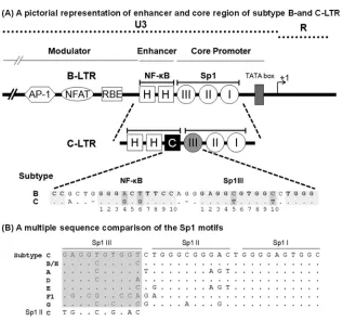
in a tandem fashion and in proximity to any two adjacent binding sites separated by very short spacer sequences of only 1 to 3 bases (Fig. 1A). While a large number of HIV-1 and HIV-2 subtypes contain three copies of Sp1 binding sites in the core promoter, a few simian immunodeficiency virus (SIV) strains contain four
copies (9). Likewise, while a large number of HIV-1 subtypes (A,
B, D, F, G, H, J, and K) and several subtypes of SIV contain two
copies of NF-B binding sites in the enhancer, subtype A/E of
HIV-1 (10,11), all HIV-2 subtypes, and the other strains of SIV
contain a single NF-B binding site. Subtype C of HIV-1 is the
only viral family that contains three binding sites for NF-B (12–
15). Second, a great sequence variation characterizes the Sp1
bind-ing sites, especially in Sp1III and Sp1II sites, with only the Sp1I motif being highly conserved among most of the HIV-1 subtypes
(16). In particular, the genetic variation of the Sp1III motif located
adjacent to the NF-B sites is so great that no two major subtypes
of HIV-1 share the same sequence (Fig. 1B). Of the three motifs,
the Sp1III site plays a more deterministic role in gene expression from the LTR, as Sp1 protein binding at this site must physically
interact with the p65 protein of NF-B recruited to the H-B site
located immediately upstream (17). Thus, the Sp1III site critical
for the regulation of gene expression for the viral promoter dem-onstrates subtype-specific sequence variations, and the influence
of such variations has not been adequately examined (18,19).
Third, in contrast to the large genetic variation of the Sp1 sites, the
sequence context of the NF-B binding sites is invariable in the
viral strains regardless of the copy number difference. Although
the NF-B binding sequence typically is degenerate (5=-GGGPuN
NTTCC-3=), a single NF-B motif is present in all the HIV-1,
HIV-2, and SIV strains, and several canonical and noncanonical binding motifs have been identified in various cellular and viral
promoters. The unique NF-B motif that we refer to as the H-B
site (5=-GGGACTTTCC-3=) is present in all of the enhancers of
HIV-1, HIV-2, and SIV subtypes (12,20), alluding to the
biolog-ical significance of this element in these promoters. In the
back-drop of the universal presence of the H-B site in all of these viral
strains, subtype C of HIV-1 alone stands out as an exception in
containing a variable, but canonical, NF-B element, which we
refer to as the C-B site (5=-GGGGCGTTCC-3=; differences from
FIG 1(A) Schematic diagram comparing the TFBS of the enhancer and core promoter elements of B- and C-LTRs. An outline of the prototype B LTR with some of the basic transcription factor binding sites is shown at the top (not to scale). The enhancer region of the B-LTR contains twoB motifs (HH), in contrast to the C-LTR, which contains threeB elements (HHC). The additional NF-B motif is genetically distinct and is referred to here as the C-B site (filled square box). Note that the enhancer-proximal Sp1III site is also characterized by subtype-specific genetic variations, as shown at the bottom. The central TFBS sequences are shaded, and the genetic variations between the subtypes are highlighted by the darker shading. Additionally, the spacer length between the two TFBS is comprised of only two bases in subtype C, unlike the three bases in subtype B. (B) Multiple-sequence comparison of the Sp1 motifs. The nucleotide sequences of Sp1III, Sp1II, and Sp1I motifs representing the consensus sequences of the major HIV-1 genetic subtypes are aligned. Dots in the alignment represent sequence identity. The sequence alignment of the Sp1III motif is highlighted in gray. The Sp1II sequence of subtype C is also included in the alignment of Sp1III sequences for comparison.
on November 7, 2019 by guest
http://jvi.asm.org/
[image:2.585.134.451.59.355.2]the H-B motif are underlined), in addition to the presence of two
copies of the generic H-B site (HHC). Lastly, the two TFBS, the
C-B and Sp1III motifs that contain subtype-specific variations in
the subtype C LTR (C-LTR), are invariably positioned at the cen-tral location where these two binding sites, in the context of sub-type B, have been shown to permit physical association between
the host factors binding to the respective sites (17,21). Of note, in
almost all of the sequences of subtype C viral strains available in
the extant databases, the C-B motif is present, invariably
posi-tioned at the central location and never swapped with the H-B
sequence, alluding to the critical role the variantB motif may be
playing in this genetic subtype (Fig. 2A).
Some studies previously examined the regulation of gene ex-pression from the HIV-1 LTR primarily in the context of subtype B (B-LTR). In previous studies, an active functional interaction in the B-LTR was demonstrated between the centrally positioned
NF-B and Sp1III sites in such a way that not only the spacing
between the sites but also the relative orientation of each site with respect to the other cannot be altered, suggesting that the host factors must bind the specific elements in a specific orientation
(17,21). Furthermore, the centrally positioned NF-B and Sp1III
motifs play a role, more critical than that of the flanking sites (one
upstream H-B site and two downstream Sp1 motifs), in
control-ling viral gene expression (18,19). Given the special significance
attached to the central NF-B and the Sp1III motifs in the HIV-1
LTR, it is enigmatic that a genetically different NF-B site (the
C-B motif) is inserted in the subtype C-LTR, disrupting the
orig-inal association between the H-B and Sp1III motifs.
Addition-ally, in the C-LTR, the Sp1III motif is subjected to subtype-specific
genetic variation (4), alluding to a newly formed association
be-tween the newly inserted C-B site and the subtype-specific Sp1
motif.
FIG 2Genetic variation within the C-B and Sp1III motifs of subtype C LTR. (A) Multiple-sequence alignment of the enhancer and core promoter sequences of the subtype C LTR. The LTR sequences comprising the three NF-B sites and three Sp1 motifs of a few representative subtype C strains deposited during the years 1989 to 2010 were downloaded fromhttp://www.hiv.lanl.gov/. The sequence of subtype C prototype Indie C1 (GenBank accession numberAB023804) was used as the reference sequence. Dots and dashes represent the sequence identity and deletions, respectively. The C-B and Sp1III motifs are highlighted by shading and the other TFBS by square boxes. (B) Position weight matrix of the C-B motif. A total of 1,091 subtype C U3 sequences available at the HIV sequence database (http://www.hiv.lanl.gov/) were downloaded. The sequence logo was generated using the WebLogo tool (http://weblogo.berkeley.edu/logo.cgi). (C) Position weight matrix of the Sp1III motif. The sequence logo was generated as described above.
on November 7, 2019 by guest
http://jvi.asm.org/
[image:3.585.71.519.72.456.2]Several publications previously demonstrated that an HIV-1
LTR containing three NF-B binding sites is a stronger viral
pro-moter than the one that contains only two such sites (7,16,22,23).
It was further proposed that the stronger viral promoter contrib-uted significantly to the widespread global expansion of subtype C
(5,24). These publications, however, evaluated only the
quantita-tive gain-of-function properties of the C-LTR that contained an
additional NF-B binding site but ignored the significance of
ge-netic variation of this motif that may confer an additional quali-tative gain of function on the promoter. If acquiring a stronger promoter is the only objective, the subtype C-LTR could have
duplicated an existing H-B motif, thus attaining a promoter
con-figuration of three H-B sites, HHH. However, the C-LTR
se-quences are characterized exclusively by the NF-B configuration
of HHC, where the presence of the C-B site at the central location
appears to be critical, and no exceptions to this condition appear to be permissible.
Here, we attempted to evaluate the quantitative as well as the qualitative differences that may contribute to transcription differ-ences in the C-LTR. Although genetic differdiffer-ences in the centrally
located NF-B and Sp1 elements have been reported previously
(5,12,25), the present study represents the first analysis to
exam-ine the functional implications of these genetic variations to sub-type C. The primary objective of the present work was to examine if there exists a newly established functional association between
the HIV-1 subtype C unique NF-B binding site (the C-B site)
and the proximal and subtype-specific Sp1III motif in the C-LTR. A functional association between the two adjacent motifs is pected and remains to be demonstrated. We used a range of ex-perimental strategies to study the functional association between the two regulatory elements in the C-LTR. Importantly, we
dem-onstrate that the creation of a variant NF-B motif (the C-B
motif) was necessary for subtype C, as the subtype-specific Sp1III
site cannot function in association with the generic H-B element.
To the best of our knowledge, in the context of a mammalian
promoter, this is the first demonstration that a unique NF-B
motif and a specific Sp1 site are functionally incompatible with each other, although each TFBS is biologically functional in a dif-ferent context.
MATERIALS AND METHODS
Cell culture.T-cell lines (Jurkat and CEM-CCR5 cells) were cultured in RPMI 1640 medium (catalog no. R4130; Sigma, St. Louis, MO) supple-mented with 10% fetal bovine serum (catalog no. RM10435; HiMedia Laboratories, Mumbai, India), 2 mM glutamine (catalog no. G8540; Sigma, St. Louis, MO), 100 U/ml penicillin G (catalog no. P3032; Sigma, St. Louis, MO), and 100g/ml streptomycin (catalog no. S9137; Sigma, St. Louis, MO). The peripheral blood mononuclear cells (PBMC) were isolated from 10 ml of fresh blood, collected from healthy donors, by density gradient centrifugation using HiSep LSM-1077 (catalog no. LS001; HiMedia Laboratories, Mumbai, India). The CD8 cells were de-pleted from the PBMC using the StemSep human CD8⫹depletion kit (catalog no. 14662; Stem Cell Technologies). PBMC were cultured in complete RPMI medium supplemented with 20 U/ml of interleukin-2 (IL-2; catalog no. H7041; Sigma, St. Louis, MO) and 5g/ml of PHA-P (catalog no. L1668; Sigma) for 72 h. PBMC were subsequently used either for nucleofection or viral infection in a medium supplemented with 20 U/ml IL-2 (catalog no. H7041; Sigma, St. Louis, MO). The human embry-onic kidney 293T and 293 cells were grown in Dulbecco’s modified Eagle’s medium (DMEM; catalog no. D1152; Sigma, St. Louis, MO).
Construction of the reporter vectors and viral molecular clones.A dual-reporter viral vector, LTR-sLuc-IRES-GFP, that simultaneously
ex-presses two different reporter genes, secretedGaussiaLuciferase (sLuc) and green fluorescent protein (GFP), was constructed using a parental vector, pCMV-sLuc-IRES-GFP, reported previously (12,26). The cyto-megalovirus (CMV) promoter was replaced by the full-length HIV-1 sub-type C LTR of 634 bp, amplified from the Indie C1 subsub-type C molecular clone (GenBank accession numberAB023804) using primers N1515 (5= -GGATCCACGCGTTGGAAGGGTTAATTTACTG-3=) and N1359 (5=-C CGCGGGAATTCCTGCTAGAGATTTTCC-3=). The PCR amplicon was cloned directionally using the restriction sites MluI and EcoRI. Subse-quently, a series of isogenic variant LTR reporter vectors was generated from the parental vector, LTR-sLuc-IRES-GFP, using the overlap PCR strategy. Transcription factor binding sites were inactivated by mutating critical residues using site-directed mutagenesis, including the H-B binding site (GGGACTTTCC to TCTACTTTCC), the C-B binding site (GGGGCGTTCC to GTCTCATTCA), Sp1III (GAGGTGTGGT to GATTCGTGGT), Sp1II (TGGGCGGGAC to TGTTCGGGAC), and Sp1I (TGGGAGTGGT to TGTTAGTGGT).
Truncated LTR vectors.A panel of minimal LTR reporter vectors expressing luciferase was constructed by deleting the 313-bp upstream modulator region from the viral promoter using the BspEI and MluI restriction sites in the pLTR-sLuc-IRES-GFP vector. The vector was self-ligated to generate pmLTR-sLuc-IRES-GFP vector. The panel of vectors consisted of three different LTRs, HHC:321 (wild type), HHC:231, and HHC:213, with the numerals representing the Sp1 motifs.
The pLGITCseries of vectors.A minivirus reporter plasmid, pLGIT (a
kind gift from David Schaffer, University of California), expressed en-hanced GFP (EGFP) and Tat under the HIV-1 LTR (18). This vector was modified further in two successive steps to replace Tat and the LTR of subtype B origin with the subtype C counterparts. First, the subtype B Tat expression segment was replaced with that of subtype C (GenBank acces-sion numberFJ765005.1). C-Tat was amplified using pcTat.BL43.CS as a template and primers N1140 (5=-TCCAGTCCACAACCATGGATGGAG CCAGTAGATCCTAAC-3=) and N1141 (5=-GGGCCCCTCGAGCTAGT CGAAGGGGTCTGTCTC-3=), and the amplified fragment was cloned between BstXI and XhoI sites on pLGIT to give rise to the vector pLGITC.
Second, the subtype B LTR in pLGITCvector was replaced with the
sub-type C LTR (Indie C1; GenBank accession numberAB023804). The sub-type C LTR was amplified using primers N1145 (5=-GGCGCCCTCGAG ACGCGTTGGAAGGGTTAATTTACTCC-3=) and N1146 (5=-TCTA GAGTTTAAACGCGGCCGCTGCTAGAGATTTTTCCCACACT AC-3=) and cloned between XhoI and PmeI restriction sites to generate the pcLGIT vector. Further, the 3=-LTR of the pcLGIT vector was sub-jected to additional mutations in the NF-B and Sp1 binding sites to generate a panel of reporter viral vectors with isogenic LTRs analogous to the panel of reporter plasmid vectors described above.
Infectious viral strains.The viral molecular clones Indie HHC:C, HHC:B, HHH:C, and HHH:B were constructed using the pIndie FHHC molecular clone reported previously from our laboratory (12). Using overlap PCR, variant LTRs were generated and substituted for the original FHHC LTR at the 3=end of the virus between the MluI and SacII sites.
Reporter gene expression analysis.Jurkat cells were transiently trans-fected with the reporter expression vectors using the Lipofectamine 2000 transfection reagent (catalog no. 11668-019; Invitrogen BioServices). The cells were seeded into 48-well plates at a density of 0.2⫻106cells/well in
250l of RPMI 1640 medium supplemented with 10% fetal calf serum. A plasmid DNA pool of 500 ng, containing 400 ng of one of the reporter plasmids and 100 ng of pGL3 vector expressing Firefly luciferase (catalog no. E1741; Promega Corporation), included as a control for the transfec-tion efficiency, was prepared in 50l of serum-free RPMI medium. One microliter of Lipofectamine was diluted with 49l of serum-free RPMI medium to prepare the lipid transfection reagent. The plasmid pool was mixed with the lipid reagent, and the plasmid-lipid mix was incubated for 20 min at room temperature and then added to appropriate wells. Four hours following the transfection, 350l of 10% RPMI with or without tumor necrosis factor alpha (TNF-␣; 200 ng/ml; catalog no. T0157;
on November 7, 2019 by guest
http://jvi.asm.org/
Sigma, St. Louis, MO) was added to the wells. The plates were incubated, and the reporter gene expression was monitored periodically. A BioLux
GaussiaLuciferase assay kit (catalog no. E3300L; New England BioLabs, Ipswich, MA) was used for monitoring the levels ofGaussiaLuciferase secreted into the culture supernatant. The luciferase assay was performed using a SpectraMax L luminescence 96-well microplate reader (model no. s/n Lu 03094; MDS, Inc.) by mixing 20l of the culture supernatant and an equal volume of the 1⫻BioLuxGLuc substrate reagent. The assays were performed in triplicate wells, and every experiment was repeated at least two times. Expression of Firefly luciferase in the cell extracts was mea-sured using a Bright-Glo luciferase assay kit (catalog no. E2620; Promega Corporation, Madison, WI) per the manufacturer’s instructions. The pri-mary data for each well were normalized for transfection efficiency.
Preparation of viral stocks and estimation of viral titer.Viral stocks of different molecular clones were generated in HEK 293T cells using the standard calcium-phosphate protocol. Briefly, cells were seeded in a 100-mm dish at a density of 3⫻106cells and transfected with 20g of the
viral vector DNA along with 0.2g of the CMV-red fluorescent protein (RFP) expression vector as an internal control for transfection efficiency. Six hours posttransfection, medium was replaced with complete DMEM. Culture supernatants were harvested at 48 h, filtered through a 0.22-m filter, and stored in a deep freezer in multiple aliquots of 1 ml. The p24 levels of the viral stocks were measured using a p24 enzyme-linked immu-nosorbent assay (ELISA) kit (4th generation p24 ELISA kit; J. Mitra and Co. Pvt. Ltd., New Delhi, India) per the manufacturer’s instructions. The 50% tissue culture infectious dose (TCID50) titer of the viral stocks was
determined using a-galactosidase assay in TZM-bl cells. Briefly, 104
TZM-bl cells were seeded in 100l of complete DMEM in a flat-bottom 96-well culture plate. After 12 h, 50l of serially diluted viral stocks (a serial 4-fold dilution) was added to appropriate wells in complete DMEM supplemented with 25 g/ml of DEAE-dextran (catalog no. D9885; Sigma, St. Louis, MO) (27). Six hours postinfection, medium in the wells was replaced with 100l of complete DMEM. The plates were incubated at 37°C in the presence of 5% CO2. To examine-galactosidase expres-sion, 48 h postinfection, cells in each well were washed using 1⫻ phos-phate-buffered saline (PBS; 137 mM NaCl, 2.7 mM KCl, 4.3 mM Na2HPO4, 1.47 mM KH2PO4, pH 7.4). After washing, cells were fixed
with 100l of a fixing solution (1.0% formaldehyde and 0.2% glutaralde-hyde in 1⫻PBS), and the plates were incubated for 5 min at room tem-perature. The cells were washed with 1⫻PBS and stained with 100l of the freshly prepared-galactosidase staining solution (4 mM potassium ferrocyanide, 4 mM potassium ferricyanide, 2 mM MgCl2, and 1 mM
5-bromo-4-chloro-3-indolyl--D-galactopyranoside [X-Gal] in PBS),
and the plates were incubated for 4 h at 37°C. The wells were washed with 1⫻PBS, and the stained cells were counted manually under a low-reso-lution microscope. The infectious titer of each viral stock was determined by multiplying the cell count by the dilution factor. Pseudotyped lentiviral vectors used for the chromatin immunoprecipitation (ChIP) analysis were packaged in HEK 293T cells (18). Briefly, cells were seeded in 100-mm culture dishes and transfected using the calcium phosphate transfection protocol with a total of 20g of plasmid DNA pool. The plasmid DNA pool consisted of four different vectors: 10g of pcLGIT reporter virus containing one of the 3=-LTR variant constructs, 5 g psPax2, 3.5g pVSV-G, and 1.5g pCMV-Rev. Culture supernatant was harvested 48 h following the transfection and stored in a deep freezer in 1-ml aliquots. The p24 levels and the TCID50titers of the viral stocks were
determined as described above.
Protein expression and purification.The expression plasmid pGEX CD-p50 was a kind gift from Neil D. Perkins (University of Dundee, Scotland, United Kingdom). The p50-glutathioneS-transferase (GST) fu-sion protein was expressed inEscherichia coliBL21(DE3) cells. The p50-GST fusion protein was purified as described previously (12), with a small modification. Briefly, the cells were grown in 250 ml of the LB broth supplemented with 100g/ml of ampicillin (catalog no. A9518; Sigma, St. Louis, MO) for 4 h at 28°C, until the optical density (OD) at 600 nm
reached 0.4. Protein expression was induced for 2 h by supplementing the medium with 1 mM isopropyl--D-thiogalactopyranoside (IPTG). The
bacterial cells were pelleted and resuspended in 10 ml of homogenization buffer (20 mM HEPES, pH 7.9, 10 mM MgCl2, 20% glycerol, 0.1%
ND-40, 1 mM dithiothreitol [DTT], 0.5 mM phenylmethylsulfonyl fluoride [PMSF], and 1⫻protease inhibitor cocktail [catalog no. P8454; Sigma, St. Louis, MO]). The resuspended cells were sonicated using a Vibra cell sonicator (Sonics and Materials Inc., Newtown, CT) at an amplitude of 35%, with 2 s on and 4 s off for 2 min. The sonicated bacterial cell lysate was centrifuged at 45,000⫻gfor 30 min, and to the supernatant 400l of prewashed GST-bind resin (catalog no. 70541; Novagen, Madison, WI) was added. The samples were incubated for 2 h at 4°C with constant mixing. The supernatant-resin suspension was centrifuged at 100⫻gfor 5 min, and the supernatant was carefully aspirated. The GST-resin pellet was washed three times with 10 ml of homogenization buffer. The washed GST-resin was transferred to a fresh 1.5-ml vial and incubated for 10 min with 200l of the elution buffer (50 mM Tris-Cl, pH 7.9, containing 10 mM reduced glutathione). The sample was centrifuged for 5 min at 100⫻
g, and the clear supernatant was transferred to a fresh vial. The superna-tant was dialyzed each time against 200 ml of dialysis buffer (5 mM HEPES, pH 7.6, 1 mM DTT, 1 mM EDTA, 10% glycerol, 1⫻protease inhibitor cocktail, and 0.2 mM PMSF) for 4 h at 4°C for three rounds and stored in a⫺20°C deep freezer.
Electrophoretic mobility gel-shift assay (EMSA) and supershift as-say.Jurkat cells (10⫻106cells), harvested and resuspend in 1 ml of ice-cold 1⫻PBS, were transferred to a 1.5-ml plastic vial and centrifuged at 500⫻gfor 5 min at 4°C. The buffer was aspirated and the nuclear extract was prepared using a commercial kit (cytoplasm and nuclear pro-tein extraction kit; catalog no. K0311; Fermentas Life Sciences, Waltham, MA). The protein concentration of the nuclear extracts was determined using a commercial bicinchoninic acid (BCA) protein assay kit (catalog no. 23227; Pierce Biotech, Waltham, MA). Aliquots of 50l of the nuclear extract were snap-frozen in liquid nitrogen and stored in a⫺80°C deep freezer until use.
A single-stranded DNA probe was radiolabeled using 5g of DNA with 1 U of T4 polynucleotide kinase (catalog no. M0201; New England BioLabs) in a solution containing 2Ci of [␥-32P]ATP. The reaction
mixture was incubated for 1 h at 37°C, and the enzyme was heat inacti-vated at 65°C for 20 min. The radiolabeled oligonucleotide was then hy-bridized to 10g of the complementary strand in the annealing buffer (50 mM NaCl, 20 mM Tris, pH 7.5, 10 mM MgCl2, 20M EDTA) by
incu-bating the reaction mixture for 5 min in boiling water, followed by gradual cooling at room temperature for 2 h. The radiolabeled annealed double-stranded probe was gel purified using a 6% (29:1) polyacrylamide gel. The gel was exposed to an X-ray film for 1 h to develop the autoradiogram. The developed autoradiogram was superimposed over the gel containing ra-diolabeled probed, and the corresponding gel piece was excised out using a scalpel. The radiolabeled probe was extracted from the excised gel piece using the crush-and-soak method with minor modifications (28). Briefly, the excised gel piece was placed in a fresh 1.5-ml plastic vial and crushed into fine pieces using a fresh needle. The weight of gel pieces was deter-mined and the gel pieces were soaked in 2 volumes of acrylamide gel extraction buffer (0.5 M ammonium acetate, 10 mM magnesium acetate, 1 mM EDTA), and the vial was incubated at 37°C for 6 to 8 h. The sample was vortexed briefly and centrifuged at 10,000 rpm for 5 min, and the supernatant was transferred to a fresh vial. The gel pieces were treated once again with a 0.5 volume of acrylamide extraction buffer, and the supernatant was pooled with the extraction solution from the previous step. Two volumes of 100% ethanol were added to the supernatant, the sample was incubated at⫺20°C for 1 h, and the vial was centrifuged at 10,000 rpm for 5 min at 4°C. The supernatant was carefully aspirated and the pellet was dissolved in 200l of 10 mM Tris-EDTA (pH 8.0). The probe was reprecipitated with 2 volumes of 100% ethanol, and the pellet was washed with 1 ml of 70% ethanol, air-dried, and resuspended in 50l of 10 mM Tris-EDTA buffer. The radioactivity counts of each probe were
on November 7, 2019 by guest
http://jvi.asm.org/
determined using a liquid scintillation counter (Wallac 1409; Perkin Elmer, Waltham, MA). Probes of the following sequences, sense and an-tisense in that order, were used in the assay. The uppercase letters in the sequences denote the respective transcription factor binding sites and the lowercase letters the native flanking sequences for the C-B probe (5=-cc actGGGGCGTTCCagga-3=and 5=-tcctGGAACGCCCCagtgg-3=), the H-B probe (5=-ccactGGGACTTTCCagga-3=and 5=-tcctGGAAAGTCCCagtg g-3=), the NFAT binding sequence from the IL-2 promoter (5=-tcgagccca aagaGGAAAatttgtttcatg-3=and 5=-catgaaacaaatTTTCCtctttgggctcga-3=), and a nonspecific oligonucleotide sequence (5=-ctactgtctcattaagaa-3=and 5=-ttcttaatgagacagtag-3=). For the EMSA, 25g of nuclear extract was mixed with 30,000 cpm of the labeled probe in the binding buffer (10 mM Tris, pH 7.9, 150 mM KCl, 1 mM DTT, 0.5 mM EDTA, 0.1% Triton X-100, and 10% glycerol) and supplemented with 1g poly(dI-dC) per reaction and 1g/ml of bovine serum albumin (BSA) in a reaction vol-ume of 30l. The binding reaction mixture was incubated for 20 min at room temperature and resolved on a 6% polyacrylamide gel (16 by 16 cm) at 250 V for 2.5 h at 4°C. The competition experiments were performed with a 5-, 10-, or 50-fold excess of unlabeled oligonucleotides added to the reaction mixture. For the supershift assay, the nuclear extracts were pre-incubated with 2 g of affinity-purified rabbit polyclonal antibodies raised against p50, p52, p65, RelB, or cRel proteins. For the supershift of NFAT complexes, antibodies specific for NFAT1 (catalog no. ab2722; Abcam, Cambridge, United Kingdom), NFAT2 (catalog no. ab2796; Ab-cam, Cambridge, United Kingdom), and NFAT5 (catalog no. ab3446; Abcam, Cambridge, United Kingdom) were used. A nonspecific antibody raised against HIV-1 p24 (generated in-house) was used as a negative control. Thirty minutes following incubation, radiolabeled probes were added to the mix and EMSA was performed as described above. Gels were dried and exposed to a Kodak Biomax film at⫺80°C. Quantitative EMSA was performed using recombinant p50 protein to estimate the affinity of the recombinant p50 protein for the C-B or H-B binding site (29).
Generation of the cell pools with stable viral integration.In a volume of 700l of 10% RPMI supplemented with 25g/ml DEAE-dextran in a 12-well tissue culture plate, 0.3⫻106Jurkat cells were infected at a
mul-tiplicity of infection (MOI) of 0.02 to 0.05 with pseudotyped viruses har-boring one of the LTR viral variants. The cells were washed after 6 h of incubation and resuspended in 1 ml of complete RPMI medium. Fifteen days following the viral infection, cells positive for GFP fluorescence were selected using a flow sorter (Aria III sorter; BD Biosciences, Franklin Lakes, NJ). Sorted cell pools were allowed to proliferate, and stable GFP expression was confirmed. To confirm comparable levels of integration with different viral variants, a quantitative real-time PCR was used to measure the mean number of integrated proviral copies in a fixed quantity of the genomic DNA extracted from the cells. A 129-bp fragment span-ning the R region to the U5 region (⫹18 to⫹147) was amplified using the primers N2208 [5=-GATCTGAGCC(T/C)GGGAGCTCTCTG-3=] and N2209 (5=-TCTGAGGGATCTCTAGTTACCAGAGTC-3=) in a real-time PCR that has the sensitivity to detect as few copies as 101in the assay.
Genomic DNA extracted from TZM-bl cells that harbor a single copy of the integrated virus per cell was used to construct the standard curve for the quantification of the number of integrated viral copies using regres-sion analysis. For normalization of the viral real-time PCR data, the glyc-eraldehyde-3-phosphate dehydrogenase (GAPDH) housekeeping gene was amplified using primers N2232 (5=-GAGCTGAACGGGAAGCTCAC TG-3=) and N2233 (5=-GCTTCACCACCTTCTTGATGTCATC-3=).
ChIP assay.The ChIP assay was performed using genomic DNA ex-tracted from Jurkat cell pools harboring integrated proviruses containing variations in the NF-B and/or Sp1 sites. Cells (20⫻106cells per assay)
were incubated in the absence or presence of TNF-␣(100 ng/ml) for 12 h. The cells were washed once with 5 ml of 1⫻PBS, resuspended in 1 ml of plain RPMI supplemented with 1% formaldehyde, and agitated in a shaker incubator for 10 min at room temperature. After 10 min, the cross-linking reaction was stopped by adding glycine to a final concentration of 0.125 M. The cells were centrifuged at 3,000 rpm for 5 min at 4°C,
super-natant was aspirated, and the cells were washed once with 1 ml of ice-cold 1⫻PBS. The cells were resuspended in 0.6 ml of the chilled lysis buffer [5 mM piperazine-N,N=-bis(2-ethanesulfonic acid) (PIPES), pH 8.0, 85 mM KCl, 0.5% NP-40] supplemented with the protease inhibitor cocktail (cat-alog no. 11836170001; Roche Applied Science) and incubated for 10 min on ice. The nuclear suspension was divided into 100-l fractions, and each fraction was subjected to 22 cycles of sonication at the high-level setting, using 30-s-on and 30-s-off pulses, in ice-chilled water using Bioruptor plus equipment (Diagenode, Liege, Belgium). The magnitude of DNA shearing was monitored in gel electrophoresis, and an average shearing size of 200 to 600 bp was targeted. The sonicated lysate was centrifuged at 14,000 rpm for 10 min at 4°C and the supernatant was collected. Immu-noprecipitation was performed using 2g of one of the antibodies against p50 (catalog no. ab7971; Abcam, Cambridge, United Kingdom), p65 (cat-alog no. ab 7970; Abcam, Cambridge, United Kingdom), Sp1 (cat(cat-alog no. CS200631; Upstate, Syracuse, NY), RNA polymerase II (Pol II; catalog no. C15200004; Diagenode, Seraing, Belgium), RNA Pol II CTD phospho-S2 (catalog no. ab5095; Abcam, Cambridge, United Kingdom), RNA Pol II phospho-S5 (catalog no. C15200007; Diagenode, Seraing, Belgium), HDAC1 (catalog no. C15410053; Diagenode, Seraing, Belgium), HDAC3 (catalog no. ab 7030; Abcam, Cambridge, United Kingdom), H3K9Ac (catalog no. ab441; Abcam, Cambridge, United Kingdom), or HIV-1 p24 (as a negative control; generated in-house). The immunoprecipitated DNA was subjected to PCR using the primer pair N1054 (5=-GAAGTAT TAAAGTGGAAGTTTGACATTC-3=) and N1056 (5=-AGAGACCCAGT ACAGGCGAAAAGC-3=), which amplified a 240-bp region flanking the B and Sp1 elements in the LTR. A 239-bp fragment was amplified in the gag region to observe the occupancy of RNA Pol II S5 (associated with transcription initiation) using primer pair N2467 (5=-GTAATAGAGGA GAAGGCTTTTAGCC-3=) and N2468 (5=-AACCAAGGGGAAGTGACA TAGC-3=). Occupancy of RNA Pol II S2 was observed by amplifying a 218-bp region in thetatgene using the primer pair N2469 (5=-GATCCT AACCTAGAGCCCTGGA-3=) and N2470 (5=-GTTCGGGGTAAGGGTT GCTTTG-3=). The amplified fragments were subjected to agarose gel elec-trophoresis to confirm the presence or absence of transcription factors over the transcription factor binding sites.
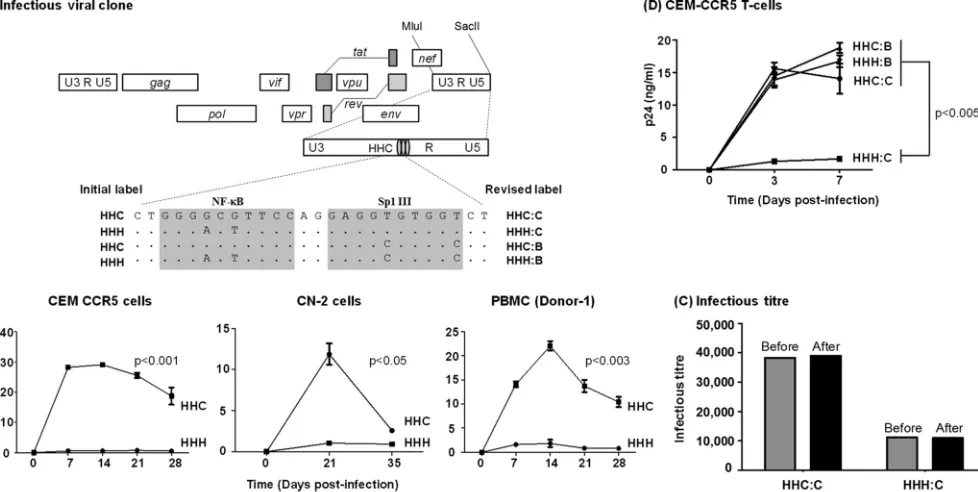
Viral proliferation and replication kinetics. CEM-CCR5 cells or PBMC (0.3⫻106) were infected with the virus Indie HHC3
C21, Indie
HHH3C21, Indie HHC3B21, or Indie HHH3B21 at approximately 500 IU. The infectious titers of the viral stocks were determined as described above. Prior to viral infection, the PBMC were CD8 cell depleted and activated with 5g/ml phytohemagglutinin (PHA) in complete RPMI medium supplemented with 20 U/ml of IL-2 for 72 h. Following this, the cells were incubated with the viruses in complete RPMI medium supple-mented with 10g/ml of DEAE-dextran (30) for 6 h at 37°C in the pres-ence of 5% CO2. Six hours following the infection, the cells were washed
three times in PBS, suspended in complete RPMI 1640 medium, and incubated. Supernatants from the third wash were saved as the day zero time point for p24 ELISA. The cells were monitored for p24 production at weekly intervals for a period of a month. The p24 ELISA was performed for each time point, and the viral growth curve was constructed for each monoinfection.
Postentry events of viral infection.Full-length Indie C1 viruses with variant LTRs (HHC3C21, HHH3C21, HHC3B21, and HHH3B21) were allowed to infect the CEM-CCR5 cells (1⫻106) at an infectious titer of
1,000 TCID50in a 6-well tissue culture plate. To analyze the late reverse
transcription (RT) products, the cellular DNA was isolated using the GenElute blood genomic DNA kit (catalog no. NA2020; Sigma, St. Louis, MO). Primer pair N1734 (5=-TGTGTGCCCGTCTGTTGT-3=) and N1735 (5=-GAGTCCTGCGTCTAGAGGATC-3=) was used to am-plify a 145-bp amplicon from the U5-region. A standard curve was constructed using a 10-fold serial dilution of the plasmid Indie-C1 (copy number ranging from 101to 105) using salmon sperm DNA (5 ng/l). The
real-time PCR was performed using a commercial kit (catalog no. BIO-94002; SensiFAST SYBR Mastermix kit; Bioline, London, United
on November 7, 2019 by guest
http://jvi.asm.org/
dom) and with 250 ng of the cellular DNA derived from the infection of each virus for the template.
To determine the rate of transcription of the viruses, real-time PCR analysis was performed for the proximal (TAR) and distal (Tat) tran-scripts. Forty-eight hours postinfection, cells were treated with 100 ng/ml of TNF-␣for 2 h or were left untreated. The total cellular RNA was ex-tracted from 1⫻106cells using TRIzol (catalog no. T9424; Sigma, St.
Louis, MO). The RNAs were subjected to cDNA synthesis using a com-mercial kit (catalog no. BIO-65042; Tetro cDNA synthesis kit; Bioline, London, United Kingdom). The cDNA was diluted 10-fold using salmon sperm DNA and subjected to real-time PCR analysis. Eighty-nine bp of TAR transcript was amplified using the primer pair N2367 (5=-GGTAGA CCAGATCTGAGCC-3=) and N2368 (5=-CTCAGAGCACTCAAGGCAA G-3=). The distal transcript of a 152-bp fragment was amplified in Tat using the primer pair N2270 (5=-TGGAGCCAGTAGATCCTAACCTAG AGCC-3=) and N1784 (5= -CTTCGTCGCTGTCTCCGCTTCTTCCTG-3=). For normalization, a transcript from the GAPDH gene was amplified using primers N2232 (5=-GAGCTGAACGGGAAGCTCACTG-3=) and N2233 (5=-GCTTCACCACCTTCTTGATGTCATC-3=). All of the time PCR analyses were performed using the Bio-Rad CFX96 Touch real-time PCR machine.
RESULTS
Physical proximity between the C-B and Sp1III motifs is crit-ical for optimal gene expression from subtype C LTR.Compared with the HIV-1 subtype B LTR (B-LTR), three important molec-ular differences could be seen in the subtype C LTR (C-LTR)
within the NF-B and Sp1 binding sites. First, the total number of
NF-B binding sites in the viral enhancer of C-LTR is three, as
opposed to only two in B-LTR. Second, the additional NF-B
binding site is invariably located immediately upstream of the Sp1
motifs and is genetically different (5=-GGGGCGTTCC-3=;
re-ferred to as the C-B site) from the two upstream generic motifs
(5=-GGGACTTTCC-3=; referred to as the H-B sites). Finally, the
Sp1III binding site located immediately downstream of the C-B
site contains subtype-specific variations. Thus, in C-LTR, the
vari-ant NF-B site and the subtype-specific Sp1III motif are invariably
juxtaposed with each other in the core of the viral promoter (Fig.
1A). Of note, while the subtype-specific C-B site and Sp1III
mo-tifs are highly conserved among subtype C strains (Fig. 2BandC),
a functional association between these two adjacent motifs is ex-pected and remains to be demonstrated. To delineate the nature of
the association between the C-B and proximal Sp1 sites, we
structed two different panels of expression vectors under the con-trol of C-LTR, using a parental bicistronic vector,
pLTR-sLuc-IRES-EGFP, reported previously (12). The initial rounds of
standardization did not show a significant difference in the profile of reporter gene expression whether a complete C-LTR or a
trun-cated one that lacked the upstream modulator region (⫺150 to
⫺463) was used in the assay (data not shown). In all subsequent
analyses in the present study, we used only the full-length
(634-bp) promoter (Indie C1; GenBank accession no.AB023804).
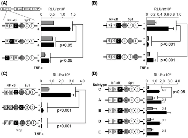
In one of the two panels of the reporter vectors (Fig. 3A),
con-sisting of three different promoters (HHC, HCH, and CHH) and
keeping the Sp1III site constant, the C-B motif was moved
pro-gressively upstream, thus increasing the distance between the
Sp1III site and the C-B element. In the other panel of three
vec-tors (Fig. 3B), doing the contrary, i.e., keeping the C-B site
con-stant, the subtype C-specific Sp1III site was moved progressively downstream, again increasing the distance between the elements. Of note, in all of the expression vectors, all 6 TFBS (three each
FIG 3C-B site is functionally associated with the Sp1III motif in HIV-1 subtype C LTR. Jurkat cells were transiently transfected with one of the reporter vectors, and 8 h following transfection the cells were exposed to TNF-␣activation (100 ng/ml) or left without activation (filled black and gray bars, respectively). The luciferase secretion was evaluated from the culture supernatant at 24 h following the activation or at later times. Gene expression was significantly attenuated when the subtype C-specific C-B site (A) or subtype C-specific Sp1 motif (B) was shifted away from their respective natural positions, when the orientation of the two motifs (middle construct) was changed, or when a 5-bp linker was inserted between the two elements (lower construct) (C). (D) The native Sp1III motif of subtype C was replaced by the heterologous Sp1 sequences from the other subtypes of HIV-1. Sequences used at motif Sp1III are depicted inFig. 1B. Two-way analysis of variance (ANOVA) was used for statistical analysis.
on November 7, 2019 by guest
http://jvi.asm.org/
[image:7.585.138.449.66.294.2]NF-B and Sp1 sites) remained intact except for the positional shifts described.
Jurkat cells were transiently transfected with the vectors of
panel 1 containing the positional shift of the C-B site. The cells
were activated with TNF-␣(100 ng/ml) or left without activation,
and the expression pattern of secreted luciferase or GFP (data not shown) was measured 24 h after activation and at later time
points. The wild-type LTR (HHC) demonstrated 588,286⫾428
relative light units (RLU) of luciferase activity that was
upregu-lated 2.3-fold (1,359,497⫾42,491 RLU) following TNF-␣
activa-tion. The basal and induced promoter activities were significantly reduced when the C-kB motif was progressively shifted upstream to one (HCH) or the other (CHH) position away from the original location. Although the loss of the promoter activity was
statisti-cally significant (P⬍0.05), gene expression was not completely
abrogated. These results suggested that although the H-B motif
can function from the central location, this motif is not an ideal
substitute for the natural C-B element located here in the C-LTR.
Comparable results were also obtained in HEK293 cells (data not presented). Further, in Jurkat cells, a loss of promoter activity was also manifested from the vectors in panel 2, where the subtype C-specific Sp1III motif was shifted progressively downstream
away from the C-B element (Fig. 3B). The loss of reporter
func-tion was quite severe, suggesting that the Sp1II motif is not a functional substitute for Sp1III in the C-LTR. Comparable results were obtained in HEK293 cells (data not presented). Collectively, the strategy of the TFBS shifting suggested that in the C-LTR, the
C-B and Sp1III motifs must be located proximal to each other
and the alternate TFBS are not functional substitutes for either of the two original regulatory motifs.
We generated an additional panel of reporter vectors by
flip-ping the C-B and Sp1III sites or inserting a five-residue linker
between the two sites (Fig. 3C). The vector in which the positions
of the C-B and Sp1III sites were interchanged did not show any
reporter activity in Jurkat cells (Fig. 3C, middle) or HEK293 cells
(data not shown). The DNA-binding domain of the p65 subunit can interact with the DNA-binding domain of Sp1 only when the
NF-B motif is present upstream of the Sp1 binding element, thus
juxtaposing the two factors in a specific fashion as demonstrated
previously (17). Likewise, when the spacer length between the two
sites was increased to 7 bp from the natural 2 bp (GGGGCGTTC CtgcagagGAGGTGTGGT; the lowercase letters represent the spacer sequence and the added bases are underlined), a complete
loss of reporter activity was observed in Jurkat (Fig. 3C, lower) or
HEK293 (data not shown) cells. Collectively, these data
ascer-tained that the association between the C-B and Sp1III motifs is
orientation and distance dependent.
C-B motif of subtype C can functionally associate with the heterologous Sp1III elements of other subtypes.It was unex-pected that the Sp1III site of subtype C could not be replaced by
Sp1II sequence of the same viral promoter (Fig. 3B, vectors II:III:I
and II:I:III). Of the three genetically diverse Sp1 sites in the core promoter of HIV-1, the Sp1III site is the most variable element,
demonstrating subtype-specific variations (Fig. 1B) (31).
Impor-tantly, experimental evidence is suggestive that the Sp1III site is functionally more important for the viral promoter than the other
two (18,19,32). Given the biological significance of the centrally
located Sp1III motif and the subtype-associated variations within this motif among HIV-1 subtypes, we asked if the heterologous Sp1III sequences derived from the other HIV-1 subtypes can
sub-stitute for the original Sp1III sequence in C-LTR. To this end, we generated a panel of reporter vectors by grafting subtype-specific
Sp1III sequences of subtypes A, B, D, and E at the NF-B proximal
location in the C-LTR. The expression of luciferase reporter was
determined in Jurkat (Fig. 3D) or HEK 293 (data not shown) cells.
The wild-type subtype C-LTR demonstrated 22,975⫾7,343 and
1.02⫻108⫾2.0⫻107RLU/s of reporter activity in Jurkat cells
under control and TNF-␣-induced conditions, respectively. The
gene expression profile from other chimeric viral promoters con-taining the heterologous Sp1III motifs was comparable to that of wild-type C-LTR with an important difference. Although the in-duced reporter activity of all of the different LTRs in the panel is comparable, the basal-level promoter function of the wild-type LTR was the lowest in both cell lines. Consequently, the fold trans-activation of the wild-type C-LTR was the highest (6- to 7-fold
versus 2- to 3-fold) and significantly different (P⬍0.05) among
all of the viral promoters compared. The biological significance of the low-basal-level activity of the wild-type C-LTR for the estab-lishment of viral latency needs further evaluation. In summary, regardless of the genetic variation, the heterologous Sp1III motifs of all four viral subtypes (A, B, D, and E) functioned in the context of C-LTR with comparable efficiency, suggesting that each of these
motifs can complement efficiently the C-B site. Thus, these
het-erologous Sp1III elements can serve as functional substitutes for the subtype C Sp1III motif.
Functional association between the C-B and Sp1III motifs in the absence of the flanking NF-B and Sp1 sites.Although the
wild-type subtype C-LTR contains three each of NF-B and Sp1
binding sites in the promoter, a direct association between the bound TF is possible only between the centrally positioned Sp1III
site and the C-B, but not the upstream H-B, motif. In the
con-text of the viral promoter, the relative magnitude of gene
expres-sion due to the specific interaction between the central C-B and
Sp1III sites is likely to be modulated by interference from the two
upstream NF-B sites and the two downstream Sp1 motifs. To
overcome this overlapping activity of the flanking TFBS and
ex-amine the direct influence of the C-B and Sp1III elements on
gene expression from the C-LTR, we introduced inactivating mu-tations into the four flanking TFBS sequences using site-directed
mutagenesis, leaving only the centrally positioned C-B and
Sp1III sites intact. By introducing mutations into critical residues of the corresponding motifs, we generated a panel of four reporter
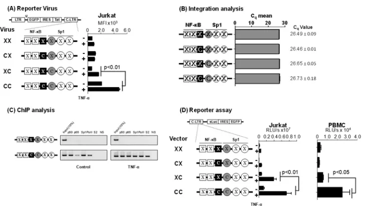
viral vectors, using the pcLGIT vector where the C-B and/or the
Sp1III sites were intact or both were inactivated (C:C, C:X, X:C,
and X:X, where C represents the subtype-specific NF-B or Sp1III
site and X the inactivated TFBS) (Fig. 4). To maintain the same
DNA helical conformation as that in the wild-type LTR, no se-quences were deleted from any of the vectors. Therefore, the over-all length of the promoter sequences remained constant.
Viruses pseudotyped with the vesicular stomatitis virus G gly-coprotein (VSV-G) envelope were used for the infection of Jurkat T cells at a low MOI of 0.01. Cell pools stably expressing EGFP were sorted 21 days after the viral infection and used in the assay
with or without TNF-␣activation. The three reporter viruses that
lacked one (X:C or C:X) or both (X:X) TFBS demonstrated a min-imal basal level of activity that did not increase significantly
fol-lowing TNF-␣activation (Fig. 4A). The reporter virus containing
both motifs (C:C) demonstrated the highest basal level of
fluores-cence (mean fluoresfluores-cence intensity [MFI], 2,128⫾9) that
in-creased 2.2-fold (MFI, 4,783⫾115;P⬍0.01) following TNF-␣
on November 7, 2019 by guest
http://jvi.asm.org/
activation. To rule out the possibility that different rates of viral infection influenced the assay outcome, we used a real-time PCR targeting the R-U5 region of the LTR to confirm equivalent levels
of viral infection (Fig. 4B). The results are highly reproducible
when virus-infected cell pools were established independently (data not shown).
To correlate the magnitude of gene expression from the inte-grated viral promoter with the occupancy of the host factors at the promoter, we performed a chromatin immunoprecipitation (ChIP) analysis using the cellular DNA extracted from Jurkat cells harboring integrated X:X or C:C reporter viral strains. Frag-mented chromatin was immunoprecipitated using antibodies specific to p50, p65, Sp1, RNA polymerase II (RNA Pol II), phos-phorylated RNA Pol II-Ser-2, or a nonspecific antibody (an
anti-body against the p24 antigen of HIV-1) as a negative control (Fig.
4C). The X:X viral promoter did not bind any of the tested host
factors regardless of TNF-␣activation, thus evidencing the lack of
viral proliferation. In contrast, the promoter containing intact
C-B and Sp1III sites, in the absence of activation, bound p50 and
Sp1 but little p65 or RNA polymerase II, suggesting the occupancy of the enhancer core by the repressive p50 homodimer. Following
TNF-␣activation, however, the promoter was occupied by p50,
p65, Sp1, and significantly higher quantities of RNA Pol II that was phosphorylated at serine 2. Thus, following activation, the sup-pressive p50:p50 homodimer was replaced by a transcription-sup-portive p50-p65 heterodimer complex at the viral enhancer.
Ad-ditionally, the presence of the phosphorylated RNA Pol II is suggestive of an efficient transcription elongation. The relative quantity of Sp1 immobilized on the promoter was not signifi-cantly different under the basal and induced conditions. The ChIP data not only are consistent with the proliferation analysis of the reporter viruses but also confirmed that regardless of the
subtype-specific genetic variations that characterize the C-B and Sp1III
motifs, these sequences regulate gene expression by recruiting the appropriate host factors. Additionally, comparable results were obtained in Jurkat cells or PBMC isolated from different healthy donors from a panel of plasmid reporter vectors (X:X, C:X, X:C, and C:C) that are analogous to the corresponding reporter viral
vectors (Fig. 4D). Collectively, the data evidenced the pivotal role
of the association between the C-B and Sp1III sites in regulating
gene expression from the subtype C viral promoter in the absence or presence of the flanking regulatory elements.
The C-B motif recruits the p50-p65 heterodimer.
Com-pared with the canonical H-B site, the C-B motif in C-LTR is
genetically distinct, with variations found at positions 4 (A to G)
and 6 (T to G) (Fig. 1A). Using gel shift analysis (cold competition
and supershift assays), a couple of publications previously claimed
the absence of NF-B binding to the C-B sequence with an
un-derlying implication that the motif is not biologically functional
(11,33). A close examination of the probes used in these reports
reveals variations in the probe sequences used or an improper
experimental design (33). Importantly, in contrast to the
above-FIG 4Gene expression in the absence of the flanking TFBS. (A) GFP expression profile of the promoter variant reporter viruses used in flow cytometry. A panel of four different reporter viruses with TFBS variations engineered in the 3=-LTR was constructed. Following viral infection, Jurkat cell pools harboring stable proviruses for each of the four viruses were generated. Jurkat cells were activated with TNF-␣(100 ng/ml) for 24 h or were left without activation, and the mean fluorescence intensity (MFI) of the EGFP was measured. Each assay was performed in duplicate wells. Data are representative of three independent experiments and are presented as MFI⫾standard deviations (SD). Two-way ANOVA was used for statistical analysis. (B) Comparable levels of viral integration in the four Jurkat stable pools. Genomic DNA was extracted from the cell pools used in panel A and subjected to quantitative real-time PCR, amplifying a 129-bp region spanning the R-U5 region in the LTR. Data are presented as mean quantification cycle (Cq)⫾SD. The data were normalized using the GAPDH housekeeping
gene. (C) ChIP analysis to examine the occupancy of transcription factors in the variant LTRs. The Jurkat stable cell pools harboring the C:C- or X:X-LTR reporter virus were activated with TNF-␣for 4 h or left without activation. The chromatin complexes were immune precipitated using 2g of antibodies specific to different cellular factors as shown. An antibody against HIV-1 p24 was used as a negative control in the experiment. The ChIP analysis was performed as described in Materials and Methods. One-tenth of the input chromatin was loaded as the input control. The data are representative of three independent experiments. (D) The C-B and Sp1III sites of subtype C are functionally associated when the flankingB and Sp1 sites are inactivated. A panel of reporter expression vectors was constructed that contained only the C-B site (C:X), Sp1III site (X:C), both (C:C), or none (X:X). While C represents subtype C-specificB or Sp1III sequences, X denotes a debilitating mutation in the corresponding motif. Luciferase activity was determined in Jurkat cells (left) or PMA-activated PBMC of a healthy subject (right) at 24 h following the transfection. The assay was performed using PBMC derived from three different subjects, and the data from one representative subject are presented. The assay is representative of three independent experiments.
on November 7, 2019 by guest
http://jvi.asm.org/
[image:9.585.112.476.62.268.2]described reports, two other publications demonstrated that the
C-B motif is biologically functional (5,16). With the backdrop of
this controversy, we set out to examine the ability of the C-B
probe to recruit NF-B from nuclear extracts. Using
double-stranded radiolabeled DNA probes comprised of either the
canon-ical H-B (5=-ccactGGGACTTTCCagga-3=; the flanking
se-quences are in lowercase and the differences are underlined), the
C-B (5=-ccactGGGGCGTTCCagga-3=), or a scrambled (5=-CTA
CTGTCTCATTAAGAA-3=) probe sequence, we compared the
binding of cellular factors from Jurkat cell nuclear extract in the
presence or absence of TNF-␣activation. Distinct cellular
com-plexes were recruited by both H- and C-B DNA probes under
no-activation conditions, and the intensity of the complexes
in-creased severalfold following TNF-␣activation (Fig. 5A, compare
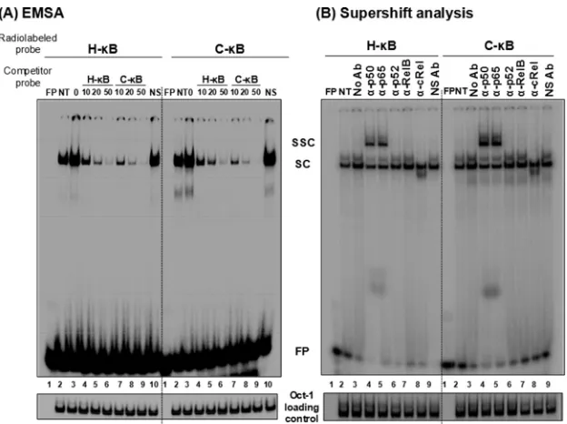
lanes 2 and 3). These specific complexes were outcompeted with progressively increasing concentrations of the cold probes (10-,
20-, and 50-fold molar excesses) representing H- or C-B
se-quences (Fig. 5A, lanes 4 to 6) but not with a nonspecific probe at
a 50-fold molar excess concentration (lanes 10). The nature and intensity of the shifted complexes appear to be comparable
be-tween the H- and C-B probes with no significantly visible
differ-ences. The binding of the nuclear complexes to the C-B DNA
probe was highly reproducible. To examine the nature of the host factors binding the DNA probes, we performed a supershift assay
using nuclear extract isolated from TNF-␣-activated Jurkat cells
and immunoaffinity-purified rabbit antibodies raised against each
of the five Rel family members (12). The analysis revealed the
presence of a p50-p65 heterodimer, but not the other members of the Rel family, in the recruited complexes. The slower-moving
complexes were evident only in the p50 and p65 lanes (Fig. 5B,
lanes 4 and 5). The presence of the p50-p65 heterodimer also was reproducible using the nuclear extract of HeLa cells (data not shown). Furthermore, the nature of the shifted and supershifted
complexes was comparable between the H- and C-B probes, with
a small difference. The intensity of the supershifted complexes was
clearly higher when the C-B probe was used in the assay rather
than the H-B probe, suggesting a higher affinity of binding of the
nuclear complexes to the C-B element (see below).
The C-B element demonstrates higher binding affinity for
NF-B.To compare the relative strengths of binding of the
cellu-lar complexes to the H- versus C-B probes, we performed a
quantitative cold competition assay (29) using Jurkat nuclear
ex-tract. The binding of 30,000 cpm of the labeled H- or C-B probe
to the host factors in 25g of nuclear extract was competed with
incrementally increasing concentrations (0-, 4-, 16-, 64-, 128-,
and 258-fold molar excesses) of cold H- or C-B probes. The band
intensities were measured using densitometry, and the percent binding was determined at each concentration of the competing probe considering the intensity of the probe binding as 100% in the absence of competition. The percent binding of the probe was plotted against the concentration of the competing probe. The amount of the competing probe required to reduce the probe binding to the host factors by half was determined using Scatchard
plot analysis (Fig. 6A). When the H-B probe was used in the
assay, 13.0 pmol of the cold H-B oligonucleotide was required
for a 50% reduction in probe binding, whereas approximately
only half of this quantity of the C-B cold probe, 6.6 pmol, was
sufficient for a comparable level of inhibition (Fig. 6A, left),
con-firming higher affinity of the C-B motif for NF-B. The results
were comparable when the binding of the C-B probe to the host
FIG 5C-B probe binds NF-B from the Jurkat nuclear extract. (A) Electrophoretic mobility shift analysis. Radiolabeled double-stranded probes (40,000 cpm) comprised of the H-B (left) or the C-B motif (right) were incubated with 30g of the Jurkat nuclear extract prepared from cells not activated or activated with TNF-␣for 2 h. Cold competition was performed by incubating the complexes with 0-, 10-, 20-, or 50-fold molar excesses of the cold probes (H-B or C-B probe). A nonspecific probe was used only at the highest concentration. (B) Supershift analysis. The nuclear extract prepared from the activated Jurkat cells was incubated with antibodies specific to each of the Rel family members before the addition of radiolabeled H-B or C-B probe. Oct-1 was used as a loading control in the experiment. FP, free probe; NS, nonspecific probe; NS Ab, nonspecific antibody (anti-p24 antibody); NT, no TNF-␣treatment; SC, shifted complex; SSC, supershifted complex. The data are representative of three independent experiments.
on November 7, 2019 by guest
http://jvi.asm.org/
[image:10.585.137.449.64.297.2]factors was outcompeted with the H- or C-B cold probe. For a 50% inhibition of the probe binding to the nuclear complexes,
35.2 and 18.0 pmol of H- and C-B competitors, respectively,
were required (Fig. 6A, right). NF-B DNA probes can bind
re-combinant p50 protein in the absence of posttranslation modifi-cation and other host factors. The recruitment of p50
ho-modimers to the NF-B motifs is critical for the establishment and
maintenance of viral latency (34). Therefore, we compared the
binding profile of recombinant p50 to those of H- and C-B
probes to determine the values of the affinity constant. We per-formed a quantitative EMSA using increasing amounts of recom-binant p50 protein (0, 0.6, 1.3, 2.6, 5.2, and 10.4 nM) incubated
with a constant amount of H- or C-B radiolabeled probe (60,000
cpm) (35,36). The quantities of free and complexed probes were
quantitated using densitometry and plotted against the probe concentration used in the assay. Using Scatchard plot analysis, the
dissociation constant (Kd) values for the H- and C-B probes for
the recombinant p50 protein were found to be 4.6 and 2.8 nM,
respectively (Fig. 6B). The data from the gel shift analyses
collec-tively demonstrate that compared to the H-B element, the C-B
motif contains an approximately 2-fold higher affinity for NF-B
present in the nuclear extract or the recombinant p50
ho-modimer. A higher binding affinity of the C-B site for NF-B
could have a significant impact on the functioning of the viral promoter in subtype C.
The C-B motif binds NFAT.The canonical H-B element contains an overlapping binding site for another important family of transcription factors called the nuclear factor of activated T cells (NFAT). The NFAT family contains four different members (NFAT1-NFAT4) and one calcium-independent transcription
factor (NFAT5) (37,38). The core sequence for NFAT binding is
GGAAA (38,39). Of the NFAT family members, the NFAT1
ho-modimer can occupy the complementary sequence of 5 bp in the
3=half of the H-B motif (GGGACTTTCC; the NFAT binding
sequence within the H-B motif is underlined) and modulate
HIV-1 gene expression (40,41). Due to a partial overlap in the
H-B motif, NFAT and NF-B could compete, leading to the
mutually exclusive binding and alternate transactivation by these
two factors (42). While NFAT1 binding to the H-B site has a
negative modulatory effect on NF-B-mediated transactivation
from the HIV-1 LTR, NFAT2 binding positively regulates HIV-1
LTR gene expression (43).
Importantly, it is not known if the T-to-G genetic variation at
position 6 of the C-B site (GGGGCGTTCC) has an impact on
NFAT binding, as only four residues at the 3=end of this element
will be available for the binding. To this end, we compared NFAT
binding to H- versus C-B probes in a gel shift assay.
Double-stranded radiolabeled DNA probe containing the NFAT binding sequence, derived from the IL-2 promoter, was incubated with the nuclear extract prepared from Jurkat cells induced with phorbol myristate acetate (20 ng/ml) and ionomycin (10 nM) for 1 h. The intensity of the shifted complexes increased following cell
induc-tion (Fig. 7A, compare lanes 2 and 3). In self-competition, the
NFAT cold probe outcompeted the probe binding to the nuclear
complexes at 5- and 25-fold molar excesses (Fig. 7A, compare lane
3 to lanes 4 and 5). Importantly, the C-B cold oligonucleotide
was as efficient as the H-B cold probe in inhibiting the binding of
the nuclear factors to the NFAT probe (Fig. 7A, compare lanes 4
and 5 to lanes 8 and 9). A scrambled cold DNA oligonucleotide, used as a negative control, was not able to compete in the assay,
confirming the specificity of the assay (Fig. 7A, lanes 10 and
11). Collectively, the data confirmed that the C-B sequence is
capable of binding NFAT efficiently regardless of the variation at position 6.
To determine the identity of the NFAT family members pres-ent in the nuclear complexes bound to the probes, we performed a supershift analysis using commercial antibodies specific to three of the important members of the family, NFAT1, NFAT2, and NFAT5. The resolved nuclear bands clearly demonstrated the presence of NFAT1 and NFAT2, but not NFAT5, in the nuclear
complexes recruited by all three DNA probes, including the C-B
probe (Fig. 7B). Although a supershifted complex was not visible
under the experimental conditions, even with the NFAT positive-control probe, the reduced intensity of the bands when specific
FIG 6C-B probe demonstrates a higher binding affinity for NF-B in the Jurkat nuclear extract as well as recombinant p50 protein. (A) Radiolabeled double-stranded DNA probe (40,000 cpm) comprised of the H-B or C-B probe was incubated with 30g of the Jurkat nuclear extract. The competition was performed with progressively increasing quantities (fold excess) of H- or C-B cold probe as shown. (Lower) The intensities of the DNA complexes were determined by densitometry. The percentage of the DNA band intensity was plotted against the concentration of competitor probe. The intercept on thexaxis is considered the affinity of binding of the probes for the nuclear factors. The data are representative of three independent experiments. (B) Radiolabeled double-stranded DNA probe (60,000 cpm) comprised of the H-B or C-B probe was incubated with 0 to 10.4 nmol of the recombinant p50 protein. The competition was performed with progressively increasing quantities (fold excess) of H- or C-B cold probe as shown. (Lower) The intensities of the DNA complexes were determined by densitometry. The per-centage of the DNA band intensity compared to that of the no-competition control was plotted as log(cold probes) versus log(FP/SC). The intercept on thexaxis is considered the affinity of binding of the probes for the nuclear factors. The data are representative of three independent experiments. FP, free probe; SC, shifted complex.