Contamination of Multiple Nasopharyngeal Carcinoma Cell Lines with
HeLa Cell Genomes
Michael J. Strong,aMelody Baddoo,aAsuka Nanbo,cMiao Xu,dAdriane Puetter,bZhen Lina
Tulane University Health Sciences Center and Tulane Cancer Center, New Orleans, Louisiana, USAa
; Department of Medicine, Section of Gastroenterology, Tulane University School of Medicine, New Orleans, Louisiana, USAb; Graduate School of Pharmaceutical Sciences, Hokkaido University, Sapporo, Hokkaido, Japanc; Sun Yat-Sen University Cancer Center, Guangzhou, People’s Republic of Chinad
ABSTRACT
In an attempt to explore infectious agents associated with nasopharyngeal carcinomas (NPCs), we employed our high-through-put RNA sequencing (RNA-seq) analysis pipeline, RNA CoMPASS, to investigate the presence of ectopic organisms within a number of NPC cell lines commonly used by NPC and Epstein-Barr virus (EBV) researchers. Sequencing data sets from both CNE1 and HONE1 were found to contain reads for human papillomavirus 18 (HPV-18). Subsequent real-time reverse transcrip-tion-PCR (RT-PCR) analysis on a panel of NPC cell lines identified HPV-18 in CNE1 and HONE1 as well as three additional NPC cell lines (CNE2, AdAH, and NPC-KT). Further analysis of the chromosomal integration arrangement of HPV-18 in NPCs re-vealed patterns identical to those observed in HeLa cells. Clustering based on human single nucleotide variation (SNV) analysis of two separate HeLa cell lines and several NPC cell lines demonstrated two distinct clusters with CNE1, as well as HONE1 clus-tering with the two HeLa cell lines. In addition, duplex-PCR-based genotyping showed that CNE1, CNE2, and HONE1 do not
have a HeLa cell-specific L1 retrotransposon insertion, suggesting that these three HPV-18ⴙNPC lines are likely products of a
somatic hybridization with HeLa cells, which is also consistent with our RNA-seq-based gene level SNV analysis. Taking all of these findings together, we conclude that a widespread HeLa contamination may exist in many NPC cell lines, and authentica-tion of these cell lines is recommended. Finally, we provide a proof of concept for the utility of an RNA-seq-based approach for cell authentication.
IMPORTANCE
Nasopharyngeal carcinoma (NPC) cell lines are important model systems for analyzing the complex life cycle and pathogenesis of Epstein-Barr virus (EBV). Using an RNA-seq-based approach, we found HeLa cell contamination in several NPC cell lines that are commonly used in the EBV and related fields. Our data support the notion that contamination resulted from somatic hybrid-ization with HeLa cells, likely occurring at the point of cell line establishment. Given the rarity of NPCs, the long history of NPC cell lines, and the lack of rigorous cell line authentication, it is likely that the actual prevalence and impact of HeLa cell contami-nation on the EBV field might be greater. We therefore recommend cell line authentication prior to performing experiments using NPC cell lines to avoid inaccurate conclusions. The novel RNA-seq-based cell authentication approach reported here can serve as a comprehensive method for validating cell lines.
N
asopharyngeal carcinoma (NPC) is an epithelial malignancy arising within the posterior nasopharynx with a high inci-dence among Southeast Asian, Alaskan Eskimo, Greenland, and Central and North African populations (1). The World Health Organization (WHO) categorizes NPCs into three histological subtypes: well-differentiated squamous cell carcinoma (WHO type I), nonkeratinizing carcinoma (WHO type II), and undiffer-entiated carcinoma (WHO type III) (2). Although the etiology of NPC is still unclear, both genetic and environmental factors have been linked to the development of NPC. Among the environmen-tal factors, infection with Epstein-Barr virus (EBV) has been ex-tensively studied and shown to play a critical etiological role in NPCs, particularly the undifferentiated nasopharyngeal carci-noma subtype (WHO type III). However, EBV is less commonly found in other types of NPC, such as WHO type I.Due to the rarity of this disease and the limited availability of pathological specimens, NPC cell lines have been important model systems to study its pathophysiology. The unique tropism of EBV to NPC tissues also makes NPC cell lines valuable systems
to study EBV’s biology and pathogenesis. The majority of NPC cell lines were established around 10 to 30 years ago, with very few NPC cell lines stably harboring natural EBV infection (e.g., c666-1). Although most of NPC cell lines used today are EBV negative, it is believed that they were once EBV positive and that the EBV genome was lost due to long-term culture (3). Whether other exogenous agents are present in these NPC cell lines has not yet been documented.
Received20 May 2014Accepted27 June 2014
Published ahead of print2 July 2014
Editor:R. M. Longnecker
Address correspondence to Zhen Lin, zlin@tulane.edu.
Supplemental material for this article may be found athttp://dx.doi.org/10.1128 /JVI.01457-14.
Copyright © 2014, American Society for Microbiology. All Rights Reserved.
doi:10.1128/JVI.01457-14
on November 7, 2019 by guest
http://jvi.asm.org/
The use of next-generation sequencing (NGS) technology has successfully been applied to the discovery and investigation of pathogens associated with cancer. This approach utilizes an unbi-ased method for the global assessment of all exogenous agents within a cancer sample with high sensitivity and specificity. Sev-eral laboratories have successfully utilized NGS and specifically high-throughput RNA sequencing (RNA-seq) for the discovery and investigation of exogenous agents associated with various cancers (4–11).
In this study, we utilized RNA-seq technology along with our computational analysis pipeline RNA CoMPASS (12) to explore the exogenous agents associated with nasopharyngeal carcinomas. To our surprise, most of the NPC cell lines analyzed were positive for human papillomavirus 18 (HPV-18). Further transcriptome and comparative analyses revealed that these HPV-18-positive NPC cell lines, CNE1, CNE2, HONE1, AdAH, and NPC-KT, are likely HeLa derivatives.
MATERIALS AND METHODS
Cell culture.CNE1 is a well-differentiated nasopharyngeal carcinoma cell line established by the Laboratory of Tumor Viruses of Cancer Institute in China (13) and was obtained from three independent sources: Jack Strominger (Harvard) for CNE1, Yixin Zeng (China) for CNE1-CN, and Asuka Nanbo (Japan) for CNE1-JPN. CNE2 is a poorly differentiated nasopharyngeal carcinoma cell line established by Gu and colleagues (14) and was obtained from Jack Strominger (Harvard). AdAH is a human nasopharyngeal adenoid epithelial cell line established by Toru Takimo-to’s group in Japan (15) and was obtained from Erik Flemington (Tulane). NPC-KT is an EBV-positive hybrid cell line which was generated by the fusion of EBV genome-positive NPC epithelial explant culture cells with AdAH cells in Takimoto’s laboratory in Japan (16) and was obtained from Erik Flemington (Tulane). HONE1 is a poorly differentiated nasopharyn-geal carcinoma cell line established by Ronald Glaser and colleagues (17) and was obtained from Asuka Nanbo (Japan). The cell line c666-1 is an EBV-infected undifferentiated NPC cell line established by Dolly Huang’s group in China (18) and was obtained from Bryan Cullen (Duke). HeLa is a well-known and characterized human cervical adenocarcinoma cell line (19) and was obtained from the ATCC and from Erik Flemington (Tu-lane).
All cells (except c666-1, which was cultured in RPMI 1640) were grown in Dulbecco modified Eagle medium (DMEM; Thermo Scientific, Waltham, MA) supplemented with10% fetal bovine serum (FBS; Invitro-gen-Gibco, Grand Island, NY) and 0.5% penicillin-streptomycin (Invit-rogen-Gibco). Cells were grown at 37°C in a humidified, 5% CO2 incu-bator. In our laboratory, we have never used or generated any HPV plasmid or HPV recombinant plasmid.
Sequencing data set acquisition.RNA-seq data sets from c666-1, HK1, NP460, and x666-1 were downloaded from the Gene Expression Omnibus under accession numberGSE54174. In addition, paired-end HeLa cells RNA-seq data sets were downloaded from the European Nu-cleotide Archive and the Sequence Read Archive under run accession numbers ERR049804 (20) and SRR309265 (21), respectively.
Sample preparation and next-generation sequencing.All cell lines were freshly thawed, grown to 90% confluence on a 10-cm plate, and harvested for RNA using 0.25% trypsin with 1 mM EDTA. Total RNA was extracted from each cell line culture using the miRNeasy minikit (Qiagen, Hilden, Germany) according to the manufacturer’s instructions. Three cell lines were selected for RNA-sequencing, including CNE1 (CNE1 cells from Harvard), CNE1-JPN (CNE1 cells from Japan), and HONE1. All cDNA libraries were prepared from ribosomal RNA-depleted (ribode-pleted) RNAs using the Illumina Truseq Stranded Total RNA Sample Prep kit (RS-122-2101). The ribodepleted RNA cDNA library was subjected to 1⫻100-base single-end strand-specific sequencing on an Illumina HiSeq 2000 instrument.
RNA CoMPASS analysis.RNA CoMPASS is an automated computa-tional pipeline that seamlessly analyzes RNA-seq data sets (12). Briefly, RNA-seq data sets were first aligned to the human reference genome, hg19 (Genome Reference Consortium GRCH37), plus a splice junction data-base (which was generated using the make transcriptome application from Useq [22]; splice junction radius set to the read length minus 4) using Novoalign version 3.00.05 (Novocraft) (-o SAM, default options). Nonmapped reads were isolated and subjected to consecutive BLAST ver-sion 2.2.27 (23) searches against the Human RefSeq RNA database and then to the NCBI NT database to identify reads corresponding to known exogenous organisms (24). Results from the NT BLAST searches were filtered to eliminate matches with an E value of greater than 10e⫺6. The results were then fed into the taxonomic classifier MEGAN 4 version 4.70.4 (25) for visualization of taxonomic classifications within the spec-imens being analyzed. RNA CoMPASS was run in parallel on three 2x2.66 GHz 12 core Intel Xeon Mac Pro computers with 64 to 96 GB of memory each.
Real-time RT-PCR analysis.Total RNA was reverse transcribed using the SuperScript III First-Strand Synthesis System for reverse transcrip-tion-PCR (RT-PCR; Invitrogen, Carlsbad, CA). Random hexamers were used along with 1g of RNA in a 20-l reaction volume according to the manufacturer’s instructions. For the incubation steps (25°C for 10 min followed by 50°C for 50 min), a Mastercycler ep (Eppendorf, Hamburg, Germany) was used. The resulting cDNA was subjected to quantitative (real-time) PCR using sequence-specific forward and reverse primers (In-tegrated DNA Technologies) (see Table S1 in the supplemental material). For real-time PCR, 1l of the resulting cDNA was used in a 10-l reaction volume that included 5l of SybrGreen and a 500 nM concentration each of forward and reverse primers. Amplification was carried out using the following conditions: 95°C for 3 min, followed by 40 cycles of 95°C for 15 s and 60°C for 60 s. Melt curve analysis was performed at the end of every quantitative RT-PCR (qRT-PCR) run. Samples were tested in triplicates. No-template controls were included in each PCR run. PCRs were per-formed on a Bio-Rad CFX96 Real Time System, and data analysis was performed using CFX Manager 3.0 software. Relative detection levels were calculated by normalizing with the glyceraldehyde-3-phosphate dehydro-genase (GAPDH) gene as a reference gene.
Viral transcriptome analysis.Raw sequence data were aligned to a reference genome containing a human genome (hg19; Genome Reference Consortium GRCH37) plus a library of virus sequences (including se-quences from all known human viruses documented by NCBI and PAvE (Papillomavirus Episteme Database [http://pave.niaid.nih.gov/]) (see Ta-ble S2 in the supplemental material). The alignments were performed using Spliced Transcripts Alignment to a Reference (STAR) aligner ver-sion 2.3.0 (--clip5pNbases 6, default options) (26) and were subjected to visual inspection using the Integrative Genomics Viewer (IGV) genome browser (27). Transcript data from STAR were subsequently analyzed using RSEM version 1.2.09 (28) for quantification of human and HPV-18 gene expression. Signal maps (i.e., the total number of reads covering each nucleotide position) were generated using IGV tools, and read coverage maps were visualized using the IGV genome browser (27).
Identification of HPV-18 integration sites and breakpoints.The ri-bodepleted single-end strand-specific reads aligned previously were ana-lyzed further to determine HPV-18 integration sites and breakpoints in NPCs. For each 100-base single-end read, we extracted the first 25-mers from both ends, and these paired 25-mer reads were then aligned inde-pendently of each other to a reference genome containing both human hg19 and HPV-18 genomes. If one 25-mer end was aligned to human and the other corresponding 25-mer end mapped to the viral genome, their corresponding single-end read represents a fusion read. The correspond-ing 25-mer read pairs from the identified fusion reads were collected and visualized in the IGV browser to pinpoint the viral integration sites in the host genome and to identify the breakpoints in the viral genome.
SNV analysis.The coverage of reads aligning to human and HPV-18 from the STAR aligner was visualized using IGV. Mutations in the HeLa Cell Contamination in Several NPC Cell Lines
on November 7, 2019 by guest
http://jvi.asm.org/
HPV-18 genome were color-coded and manually inspected. Human sin-gle nucleotide variants (SNVs) were identified using a detection pipeline consisting of samtools version 0.1.19 mpileup (-q 10, default parameters) (29) and VarScan version 2.3.6 (mpileup2snp --p-value 0.01 -output -vcf 1, default parameters) (30) to identify candidate SNVs and output in variant call format (VCF) format. In dealing with SNVs from RNA-seq data, the potential candidate SNVs not called may be due to lack of ex-pression or not meeting the default parameter thresholds. To overcome this obstacle, coverage maps for each chromosome were generated from expression data for each RNA-seq data set with a minimum of 10 reads using IGVtools (27). The coverage maps were compared to the candidate SNV output in order to determine SNVs that were from uniform gene expression for all RNA-seq data sets analyzed. This was a necessary step prior to cluster analysis using PLINK version 1.9 (https://www.cog -genomics.org/plink2) (31) to eliminate cluster bias based on SNVs orig-inating from genes expressed only in certain cell types. The human SNVs in VCF format from commonly expressed genes for each cell line were merged using vcftools version 0.1.11 (vcf-merge, default parameters) (32). The merged VCF file was converted to binary PED files using PLINK 1.9 (--vcf, --make-bed). The resulting binary PED files were subjected to clustering (-cluster) and a subsequent cluster dendrogram was generated in R using plot(hclust(dist(x))) (33).
Genomic DNA isolation and genotyping of the HeLa-specific L1 ret-rotransposon marker.Genomic DNA was isolated from freshly thawed NPC and HeLa cell lines using the DNeasy Blood & Tissue kit (Qiagen catalog no. 69504) by following the manufacturer’s protocol. DNA sam-ples were quantified using a NanoDrop 2000 spectrophotometer (Thermo Scientific).
To detect the L1 retotransposon marker, 500 ng of the genomic DNA was used in a 50-l reaction volume that included 1⫻standardTaq reac-tion buffer (NEB catalog no. B9014), 500 nM each primer (VM164A, VM164B, and RB164K2), 400M deoxynucleotide solution mix (NEB
catalog no. N0447), and 2.5 U ofTaqDNA polymerase (NEB catalog no. M0273). A list of primers can be found in Table S1 in the supplemental material. Amplification was carried out on a Mastercycler ep (Eppendorf, Hamburg, Germany), using the following conditions: 96°C for 30 s, fol-lowed by 30 cycles of 96°C for 30 s, 54°C for 30 s, and 72°C for 1 min, and then 1 cycle of 72°C for 5 min.
Thirty microliters of PCR product was run on a 1% agarose gel and visualized by ethidium bromide staining on a Bio-Rad gel documentation system. The product size was estimated by comparison to a Quick-Load 2-log DNA ladder (NEB N0469).
Nucleotide sequence accession numbers.Sequence data (RNA-seq) for CNE1, CNE1-JPN, and HONE1 can be retrieved from the National Center for Biotechnology Information Sequence Read Archive (accession no. SRP043686). Fasta files, bed format annotation files (for visualization of annotation data on a genome browser), and .gtf format annotation files (for quantification of RNA-seq data) are available on request.
RESULTS
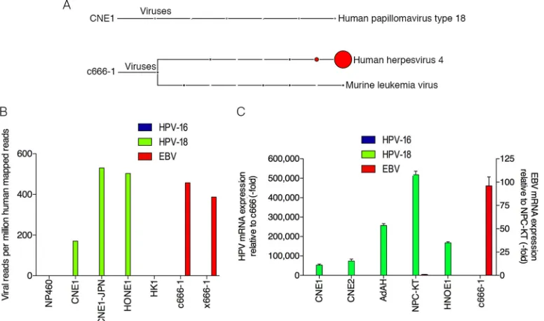
Detection of HPV-18 in nasopharyngeal carcinoma cells.To
elucidate the metatranscriptome of NPC cells that are commonly used in NPC and EBV research, RNA-seq data sets generated from our lab (CNE1 and HONE1) as well as from publicly available sequencing data sets (HK1, c666-1, x666-1, and NP460) were an-alyzed using RNA CoMPASS. Analysis of the virome component of RNA CoMPASS showed that these samples were free of most known DNA viruses (see Fig. S1 to S6 in the supplemental mate-rial). As expected, Epstein-Barr virus was detected in both c666-1
(Fig. 1A; see also Fig. S1) and x666-1 (see Fig. S2) but not in the
other data sets analyzed. In addition to the EBV findings, we were surprised to find high read numbers for the high-risk human pap-FIG 1Detection of human papillomavirus 18 in nasopharyngeal carcinomas. (A) RNA-seq data from each NPC sample was analyzed using RNA CoMPASS. A representative virome branch of the taxonomy tree (CNE1) for each HPV⫹sample was generated using the metagenome analyzer MEGAN 4. c666-1 was positive for both EBV (human herpesvirus 4) and MuLV. (B) In-depth analysis of the virome. Raw reads from each sample were aligned to the hg19 genome with the custom human virus genome using the genome aligner STAR. Reads are displayed as viral reads per million human mapped reads. Blue, HPV-16 reads; green, HPV-18 reads; and red, EBV reads. (C) Using quantitative real-time PCR, six NPC cell lines were surveyed for the presence of HPV-16 (blue columns), HPV-18 (green columns), and EBV (red columns). Five out of the six NPC cell lines demonstrated evidence of HPV-18, but none of the NPC cell lines was infected with HPV-16. Both NPC-KT and c666-1 were positive for EBV EBER. Samples were normalized to GAPDH and compared to c666-1 for HPV-16 or -18 expression and compared to NPC-KT for EBV expression.
on November 7, 2019 by guest
http://jvi.asm.org/
[image:3.585.100.488.68.297.2]illomavirus 18 (HPV-18) in the CNE1 (Fig. 1A; see also Fig. S3) and HONE1 (see Fig. S4) data sets.
The murine leukemia virus (MuLV), an RNA oncogenic virus, was detected in both the c666-1 and x666-1 cell lines (see Fig. S1, S2, and S7 in the supplemental material) and represented active infection (M. J. Strong and Z. Lin, unpublished data). To our knowledge no study has previously reported the detection of MuLV within these 2 NPC cell lines, and we reason that the infec-tion most likely occurred before 1999 at the source instituinfec-tion, since both cell lines were established through passage in labora-tory mice. MuLV was also detected in NP460 (see Fig. S7). De-tailed MuLV transcriptome analysis in this cell line revealed that the MuLV reads may be derived from MuLV-based retroviral ex-pression vectors since coverage was restricted to the LTR regions (Strong and Lin, unpublished).
To confirm the virus findings from RNA CoMPASS, the NPC RNA-seq data sets were aligned to a reference genome con-taining a human genome (hg19; Genome Reference Consortium GRCH37) plus a library of virus sequences (including sequences from all known human viruses documented by NCBI and PAvE) (see Table S2 in the supplemental material) using the aligner Spliced Transcripts Alignment to a Reference (STAR) version 2.3.0 with default options. Consistent with our RNA CoMPASS analysis, CNE1 and HONE1 data sets were positive for HPV-18 but not HPV-16, with viral reads per million human mapped reads of 170 and 503 for CNE1 and HONE1, respectively (Fig. 1B). Furthermore, c666-1 and x666-1 samples were negative for HPV-16 and HPV-18 but positive for EBV, with 457 and 387 viral reads per million human mapped reads, respectively. Finally, HK1 was negative for all known viruses (see Fig. S5 in the supplemental material).
The detection of HPV-18 in these NPC cell lines was unex-pected because HPV-18 typically infects the genital epithelium and is generally not associated with the head and neck region (34). To verify that the presence of HPV-18 in these cells was not due to contamination from a particular lab, CNE1 cells were obtained from two additional independent laboratories in China (CNE1-CN) and Japan (CNE1-JPN) and analyzed by real-time PCR using primers specific for HPV-18 and HPV-16. The results confirmed the presence of HPV-18 but not HPV-16 in these CNE1 cell lines (see Fig. S8 in the supplemental material). Furthermore, total RNA from CNE1 cells obtained from Japan was subjected to RNA sequencing and subsequent virome analysis (see Fig. S9). The re-sults were consistent with our first CNE1 RNA-seq data set, with more than 500 HPV-18 reads per million human mapped reads
(Fig. 1B).
An additional three NPC cell lines (CNE2, AdAH, and NPC-KT) were surveyed for the prevalence of HPV as well as EBV using real-time PCR. Cell lines (CNE1, HONE1, and c666-1) were also included in the assay to serve as positive controls and to further validate the RNA-seq results. As shown inFig. 1C, CNE1, CNE2, AdAH, NPC-KT, and HONE1 cells were all positive for HPV-18 but not HPV-16, with NPC-KT also positive for EBV. In contrast, c666-1 was negative for both HPV-18 and HPV-16 but positive for EBV.
HPV transcriptome analysis in NPC cells.The depth of
cov-erage in our NPC RNA-seq data sets were sufficient for HPV tran-scriptome analysis, which indicated robust expression of HPV-18 (see Fig. S10 in the supplemental material). Further transcriptome analysis revealed that the viral early genes E6 and E7 were actively
expressed at relatively high levels (see Table S3). As a comparison, two publicly available HeLa RNA-seq data sets from Nagaraj et al. (20) and Cabili et al. (21) were obtained and subjected to HPV transcriptome analysis. The average transcripts per million (TPM) for HPV-18 E6 in HeLa and HPV⫹NPC cell lines were 461.6 (20% of total transcripts) and 132.4 (23% of total transcripts), respec-tively. The average TPM for HPV-18 E7 in HeLa cells and HPV⫹ NPC cell lines were 1,228.4 (52% of total transcripts) and 297.7 (50% of total transcripts), respectively. Although there was abun-dant expression of the E1 gene (average TPM, 622.3 [26% of total transcripts] and 122.3 [21% of total transcripts] for HeLa cells and HPV⫹NPC cell lines, respectively), the expression of the E1 open reading frame (ORF) is disrupted, leading to a premature mRNA product (see Fig. S10).
Since the NPC cell lines CNE1, CNE1-JPN, and HONE1 were subjected to strand-specific RNA sequencing, we were able to dis-tinguish transcripts initiated from either the sense or antisense direction (strand-specific viral transcripts). In addition to the transcripts observed from the E6/E7 region, large numbers of reads were observed in the E1/E2 and L1/URR regions (see Fig. S10 in the supplemental material). The middle of the HPV-18 genome is void of coverage due to the viral genome rearrangement during integration into human chromosome 8 (see details below). Strand-specific transcriptome analysis further revealed that nearly all reads were derived from the sense strand of HPV-18. There were very low read levels (⬍15) from the antisense strand, possi-bly resulting from inefficiencies in the strand-specific sequencing approach (see Fig. S10).
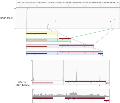
HPVⴙNPCs have a common pattern of viral and cellular
genomic rearrangement as HeLa cells.In HPV-associated
can-cers, the HPV genome is typically found to be integrated into the host genomes. We therefore examined the transcriptionally active integration sites in NPCs. Our single-end ribodepleted strand-specific RNA-seq data set was analyzed by extracting the first 25-mer reads from each end of the 100-base read and aligning them independently of each other to a reference genome containing both the human hg19 and HPV-18 genomes. Next, fusion reads were identified as those in which one 25-mer end mapped to one genome and the other corresponding 25-mer end mapped to the other genome. These fusion reads were then utilized to pinpoint the integration sites/breakpoints through visualizing the read cov-erage peaks in the IGV genomic browser. This analysis revealed that all examined HPV⫹NPCs demonstrated similar viral and cellular genomic rearrangements within a 12-kb fragile region of human chromosome 8 (Fig. 2). Further, the HPV-18 integrated in a reverse manner and exhibited five genome breakpoints (num-bered 1 to 5 with corresponding read coverage peaks) resulting in the four potential HPV-18 integration patterns (colored and boxed). For each HPV-18 integration pattern, there is a corre-sponding read coverage peak for chromosome 8 (labeled a to e). All HPV-18 integration patterns started with the L1 breakpoint and shared the same upstream insertion point on chromosome 8, as demonstrated by the high coverage peaks, respectively. Analysis of reads spanning the rearranged HPV-18 integrated genome demonstrated consistent coverage across all segments, suggesting active transcription in this rearranged configuration (Fig. 2).
Since HPV-18 has been shown to integrate in the same region of chromosome 8 in HeLa cells (35), and since HeLa cells have been plaguingin vitrocell cultures since the 1950s, we compared the precise integration sites and viral breakpoints between NPCs HeLa Cell Contamination in Several NPC Cell Lines
on November 7, 2019 by guest
http://jvi.asm.org/
and HeLa cells. Reference genomes that span the five HPV-18/ human chromosome 8 junctions in NPCs were generated, and reads from HPV⫹NPCs and the two HeLa data sets were aligned using the STAR aligner with default parameters. The results showed that all HPV-18/human chromosome 8 junctions in HPV⫹NPCs were identical (down to the nucleotide) to those observed in the two HeLa cells (see Fig. S11 to S15 in the supple-mental material). Furthermore, nucleotide insertions within a junction sequence specifically identified in HeLa cells (35) show-ing no homology to human or HPV-18 were also identified in analyzed HPV⫹NPCs (see Fig. S15).
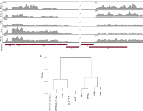
SNV analysis of NPCs further substantiates HeLa cell
con-tamination in NPC cell lines.To further elucidate the cell origins
of these HPV⫹NPCs, we investigated both viral and human single nucleotide variations (SNVs) in the three NPCs and the two HeLa data sets. Analysis of the viral SNV showed that all five data sets have the same integrated HPV-18 genome, exhibiting 29 identical SNVs relative to a reference HPV-18 consensus genome (Fig. 3A; see also Fig. S16 in the supplemental material). Additional SNVs observed in the HeLa-1 sample are likely from sequencing errors, since those were not preserved in the HeLa-2 sample.
Analysis of the human SNVs for all RNA-seq data sets was also performed. A total of 13,551 human SNVs were identified within genes commonly expressed in all nine data sets. These SNVs were subjected to clustering using PLINK (31). As shown inFig. 3B, two distinct clusters were identified. The two HeLa cell samples cluster FIG 2HPV⫹NPCs demonstrate same pattern of viral and cellular genomic rearrangement as HeLa cells. Strand-specific sequencing data from ribodepleted CNE1 RNA were analyzed for integration sites and breakpoints. First, 25-mer reads were extracted from both ends of each read and aligned to a reference genome containing both the human hg19 and HPV-CNE1 genomes. Next, fusion reads that were aligned to both human and HPV genomes were identified and visualized in the IGV genomic browser. Potential HPV integration patterns are represented as colored boxes. Read coverage peaks represent chromosome 8 integration sites (a to e) and HPV genome breakpoints (1 to 5). The rearranged HPV integrated genome is shown with breakpoints highlighted by numbers and corresponding read coverage.
on November 7, 2019 by guest
http://jvi.asm.org/
[image:5.585.45.544.66.493.2]tightly with CNE1, CNE1-JPN, and HONE1, forming one major cluster. In contrast, cell lines c666-1, x666-1, HK1, and NP460 formed a distinct major cluster. Notably, CNE1 and HONE1 are within the HeLa cell cluster but not tightly clustering with the other two HeLa cell samples, suggesting that these NPC cell lines were not entirely of HeLa cell origin.
In addition to cluster analysis, in-depth investigation of the SNV patterns at the gene level was performed. By comparing in-dividual SNVs, we found that HPV⫹NPC cells exhibited distinct SNV patterns in certain genomic regions compared to HeLa cells. For example, isoenzyme analysis of glucose-6-phosphate dehy-drogenase (G6PD) revealed G6PD type A for both HeLa cell sam-ples, a signature variant that has been previously used to identify HeLa cells (36). Interestingly, CNE1, CNE1-JPN, and HONE1 were all heterozygous for this G6PD type A variant (see Fig. S17A in the supplemental material), while HK1, c666-1, x666-1, and NP460 were homozygous for the wild-type G6PD type B variant.
Further, a unique SNV within the p53 gene was found in CNE1, CNE1-JPN, and HONE1 but not HeLa or the other NPC cell lines (see Fig. S17B). Finally, SNV analysis of phosphoglucomutase 1 (PGM1) showed a heterozygous variant in the 3=untranslated region in HeLa, CNE1, CNE1-JPN, and HONE1 but not in HK1, c666-1, x666-1, or NP460 (see Fig. S17C). Taken together, these data suggest that HPV⫹NPCs, CNE1 and HONE1, are likely de-rivatives of HeLa and another cell of unknown origin.
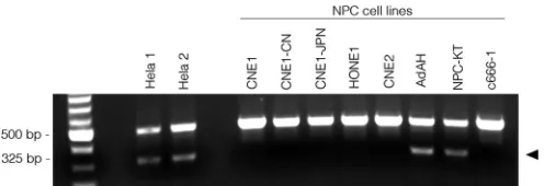
HeLa-specific L1 retrotransposon marker is present in some
NPC cell lines.To further confirm our findings, we screened the
six NPC cell lines with a single duplex detection PCR assay, which targets a HeLa-specific L1 retrotransposon insertion (37). As shown inFig. 4, a 325-bp insertion-specific amplicon was readily detected in HeLa cells. This marker was also detected in the AdAH and NPC-KT cells. In contrast, other HPV⫹NPCs such as CNE1, CNE1-JPN, CNE1-CN, and HONE1 were negative for this HeLa marker, indicating that these cells bear only part of the HeLa FIG 3Single nucleotide variant analysis of NPCs further substantiates HeLa cell contamination in NPC cell lines. (A) HPV-18 SNVs were analyzed in all HPV-18-positive NPCs and visualized using IGV. The nucleotide mutation coordinates and corresponding amino acid changes are represented in Fig. S16 in the supplemental material. Theyaxis represents the number of reads at each nucleotide position of the genome. The maximum scale for the coverage graphs is 4,950 reads for HeLa-1 and 1,650 reads for the other graphs. Coverage graph insets represent blowup regions of the genome with maximum scales of 60 reads for HeLa-1 and 200 reads for HeLa-2 and HONE1. (B) Universally expressed human-specific SNVs in each cell line were also identified and clustered using PLINK. CNE1 and HONE1 were clustered with HeLa cells in one major branch, whereas c666-1, x666-1, HK1, and NP460 were clustered in another branch.
HeLa Cell Contamination in Several NPC Cell Lines
on November 7, 2019 by guest
http://jvi.asm.org/
[image:6.585.48.542.67.452.2]genomic sequences (partial HeLa genome) and lowering the pos-sibility that these cell lines are simply cross-contaminants of HeLa cells.
DISCUSSION
CNE1 and other NPC cell lines serve as an essential widely used tool in both the NPC and the EBV research communities. Al-though the majority of NPC cell lines used today are currently EBV negative, it is believed that they were initially positive for EBV and that their EBV genomes were lost due to long culture (3). To date, whether other exogenous agents are present in NPC cell lines has not been determined. During our effort to fully assess the viromes of NPC cell lines, we identified active HPV-18 infection in several cell lines analyzed. Even though it is possible that these NPCs might have naturally acquired HPV infection (38), it is HPV-16, but not HPV-18, that is typically associated with tumors in the head and neck region (38). Considering the HPV-18 inte-gration pattern and the host and viral SNV analyses, along with the widespread contamination of HeLa in several tissue culture sys-tems, we reasoned that this HPV-18-positive cervical adenocarci-noma cell line is a likely source of contamination in these NPC cell lines.
Using our in-house RNA-seq based computational pipeline, we were able to identify the HPV/human genome rearrangements, which provided critical evidence to support the notion that CNE1 and HONE1 cells were of HeLa origin. Four complex integration patterns were detected in CNE1, HONE1, and HeLa data sets, which was consistent with a recent study showing similar integra-tion patterns of HPV-18 in HeLa cells (35). Despite the similarity in HPV-18 integration patterns observed in our data and those of Adey et al., we observed an additional HPV-18 breakpoint and insertion. As shown inFig. 2, there was a peak at the beginning of HPV-18 E1 (peak 3) indicating a pileup of fusion reads with the corresponding pileup shown as peak d on chromosome 8. This new observed breakpoint and integration pattern (see Fig. S13 in the supplemental material) may be due to differences in HeLa cell clones or may be allele specific, since we analyzed the average of both alleles, while Adrey et al. analyzed only a single allele from chromosome 8.
Our results indicated that several NPC cell lines analyzed (CNE1, CNE2, AdAH, NPC-KT, and HONE1) are contaminated with HeLa cells either during the initial establishment of these lines (e.g., CNE1) or during subsequent cell passage. Further, an earlier study identified the presence of HPV-18 E6 protein in AdAH and its variant (39), suggesting contamination with HeLa.
In addition, since NPC-KT is also HPV-18 positive and was estab-lished in the same laboratory as AdAH, it is likely that NPC-KT is also contaminated with HeLa, with contamination possibly oc-curring during the initial establishment of this cell line. It is likely that other NPC cell lines not analyzed in this study that are com-monly used in the field are also contaminated. For example, we detected HPV-18 in another NPC RNA-seq data set (SRA020971) (40) generated from 5-8F cells (a SUNE-1-derived cell line) (data not shown). Unfortunately, due to the nature of the experimental design (microRNA-seq study, which misses the majority of mRNA sequences), we do not have definitive evidence to call HeLa contamination in this cell line. Therefore, considering the rarity of NPCs, the long history of NPC cell lines, and the lack of rigorous authentication (since general cell line vendors such as the ATCC do not carry NPC cell lines), we strongly recommend cell line authentication prior to performing experiments. Otherwise, us-age of HeLa-contaminated NPC cell lines in both NPC- and EBV-related studies might lead researchers to draw misleading or even false conclusions.
Our findings are consistent with a study conducted in Hong Kong by Chan and colleagues, in which they tested the authentic-ity of several NPC cell lines, including CNE1, CNE2, HK1, x666-1, and c666-1 (41). Chan et al. conducted a short tandem repeat (STR) profiling assay on these NPC cell lines and determined that CNE1 and CNE2 contain at least one allele identical to HeLa based on the STR patterns (41). Furthermore, Chan et al. identified the same CNE1-specific p53 SNV (Thr 280) that we observed in our CNE1 and HONE1 data sets (41).
The HeLa cell line was established in 1951 and is the first hu-man cell line established from an aggressive cervical carcinoma. There has been widespread use of this cell line for a variety of experiments conducted in the United States and abroad. Since the establishment of this cell line more than 60 years ago, many cell lines have been confirmed to be contaminated with HeLa (42,43). Despite massive collaborative efforts to ameliorate HeLa contam-ination and cell line misidentification within our cell line reper-toire, these issues still persist today, as evidenced by this study. Recently, there has been a call to action to address cell line con-tamination and misidentification in the hope of avoiding the gen-eration of misleading data due to cross-contaminated cell lines (44,45).
To the best of our knowledge, an RNA-seq-based cell authen-tication method has not yet been reported. The existing methods all focus on analyzing the genetic DNA sequences of the cell lines. For instance, the most popular STR profiling measures the num-ber of specific, short repetitive DNA sequences at specific loci in the genome (42). In this study, we explored the possibility of using transcriptome data analysis as an additional tool for cell authen-tication. Using this approach, we were able to obtain (from the RNA-seq data sets) critical information regarding the viral tran-scriptome, the viral and cellular genomic rearrangement, and the viral and cellular SNVs that were used for successful cell authen-tication. We believe that with increasing awareness of cell line contamination, the use of an RNA-seq-based approach may be another viable option in the pursuit of validating cell lines for experimental use.
Given our data along with data from Chan et al., we conclude that CNE1 is not of true nasopharyngeal origin and is therefore an inappropriate cell line to study NPC biology. The other NPC cell lines that tested positive for HPV-18 (HONE1, CNE2, AdAH, and FIG 4HeLa cell-specific L1 retrotransposon marker is present in a group of
NPC cell lines. A single duplex detection PCR assay was conducted to detect a HeLa-specific L1 retrotransposon marker. Genomic DNAs from six NPC cell lines and two independent HeLa cell lines were subjected to PCR analysis. PCR products were fractionated on a 1% agarose gel. Two amplicons/bands were observed, and the HeLa-specific amplicon/band presents as a band of 325 bp.
on November 7, 2019 by guest
http://jvi.asm.org/
[image:7.585.40.288.65.150.2]NPC-KT) and/or HeLa markers (AdAH and NPC-KT) are strongly suspicious for HeLa contamination; however, additional testing from multiple sources is needed. For instance, our data strongly indicate that HONE1 is a HeLa-related somatic cell hy-brid; however, without testing from multiple sources, a definitive conclusion cannot be drawn.
The three NPC cell lines analyzed in this study that are HPV-18 negative and are not contaminated with HeLa cells are c666-1, x666-1, and HK1. The normal nasopharyngeal cell line NP460 is also HPV-18 and HeLa negative. Even though c666-1 and x666-1 are free of HeLa contamination, the presence of murine leukemia virus (MuLV) in these cells might influence the results of EBV-related studies in unforeseen ways that may cause invalid or skewed conclusions. Caution must be exercised in interpreting experimental results from these two cell lines.
ACKNOWLEDGMENTS
This work was supported by a Louisiana Clinical & Translational Science Center Pilot Grant (1 U54 GM104940 from the National Institute of Gen-eral Medical Sciences of the National Institutes of Health, which funds the Louisiana Clinical and Translational Science Center) to Z.L., Ruth L. Kirschstein National Research Service Award F30CA177267 from the NCI to M.J.S., and National Institutes of Health grant P20GM103518 to Prescott Deininger, which supports the Tulane Cancer Center next-gen-eration sequencing bioinformatics core used in this study.
REFERENCES
1.Chang ET, Adami H-O.2006. The enigmatic epidemiology of nasopha-ryngeal carcinoma. Cancer Epidemiol. Biomarkers Prev.15:1765–1777. http://dx.doi.org/10.1158/1055-9965.EPI-06-0353.
2.Shanmugaratnam K, Sobin L. 1993. The World Health Organization histological classification of tumours of the upper respiratory tract and ear. A commentary on the second edition. Cancer71:2689 –2697. 3.Dittmer DP, Hilscher CJ, Gulley ML, Yang EV, Chen M, Glaser R.2008.
Multiple pathways for Epstein-Barr virus episome loss from nasopharyn-geal carcinoma. Int. J. Cancer123:2105–2112.http://dx.doi.org/10.1002 /ijc.23685.
4.Feng H, Shuda M, Chang Y, Moore PS.2008. Clonal integration of a polyomavirus in human Merkel cell carcinoma. Science319:1096 –1100. http://dx.doi.org/10.1126/science.1152586.
5.Castellarin M, Warren RL, Freeman JD, Dreolini L, Krzywinski M, Strauss J, Barnes R, Watson P, Allen-Vercoe E, Moore RA, Holt RA.
2012. Fusobacterium nucleatum infection is prevalent in human colorec-tal carcinoma. Genome Res. 22:299 –306. http://dx.doi.org/10.1101/gr .126516.111.
6.Kostic AD, Gevers D, Pedamallu CS, Michaud M, Duke F, Earl AM, Ojesina AI, Jung J, Bass AJ, Tabernero J, Baselga J, Liu C, Shivdasani RA, Ogino S, Birren BW, Huttenhower C, Garrett WS, Meyerson M.
2012. Genomic analysis identifies association of Fusobacterium with colo-rectal carcinoma. Genome Res.22:292–298.http://dx.doi.org/10.1101/gr .126573.111.
7.Lin Z, Puetter A, Coco J, Xu G, Strong MJ, Wang X, Fewell C, Baddoo M, Taylor C, Flemington EK.2012. Detection of murine leukemia virus in the Epstein-Barr virus-positive human B-cell line JY, using a computa-tional RNA-Seq-based exogenous agent detection pipeline, PARSES. J. Virol.86:2970 –2977.http://dx.doi.org/10.1128/JVI.06717-11.
8.Strong MJ, O’Grady T, Lin Z, Xu G, Baddoo M, Parsons C, Zhang K, Taylor CM, Flemington EK.2013. Epstein-Barr virus and human her-pesvirus 6 detection in a non-Hodgkin’s diffuse large B-cell lymphoma cohort by using RNA sequencing. J. Virol.87:13059 –13062.http://dx.doi .org/10.1128/JVI.02380-13.
9.Strong MJ, Xu G, Coco J, Baribault C, Vinay DS, Lacey MR, Strong AL, Lehman TA, Seddon MB, Lin Z, Concha M, Baddoo M, Ferris M, Swan KF, Sullivan DE, Burow ME, Taylor CM, Flemington EK.
2013. Differences in gastric carcinoma microenvironment stratify ac-cording to EBV infection intensity: implications for possible immune adjuvant therapy. PLoS Pathog.9:e1003341.http://dx.doi.org/10.1371 /journal.ppat.1003341.
10. Lin Z, Wang X, Strong MJ, Concha M, Baddoo M, Xu G, Baribault C, Fewell C, Hulme W, Hedges D, Taylor CM, Flemington EK. 2013. Whole-genome sequencing of the Akata and Mutu Epstein-Barr virus strains. J. Virol.87:1172–1182.http://dx.doi.org/10.1128/JVI.02517-12. 11. Lin Z, Xu G, Deng N, Taylor C, Zhu D, Flemington EK.2010.
Quan-titative and qualitative RNA-Seq-based evaluation of Epstein-Barr virus transcription in type I latency Burkitt’s lymphoma cells. J. Virol.84:
13053–13058.http://dx.doi.org/10.1128/JVI.01521-10.
12. Xu G, Strong MJ, Lacey MR, Baribault C, Flemington EK, Taylor CM.
2014. RNA CoMPASS: a dual approach for pathogen and host transcrip-tome analysis of RNA-Seq datasets. PLoS One9:e89445.http://dx.doi.org /10.1371/journal.pone.0089445.
13. LTVCI.1978. Establishment of an epithelioid cell line and a fusiform cell line from a patient with nasopharyngeal carcinoma. Sci. Sin.21:127–134. 14. Gu SU, Tann BF, Zeng Y, Zhou WP, Li K, Zhao MC.1983. Establish-ment of an epithelial cell line (CNE-2) from an NPC patient with poorly differentiated squamous cell carcinoma. Chin. J. Cancer2:70 –72. 15. Takimoto T, Furukawa M, Hatano M, Umeda R.1984. Epstein-Barr
virus nuclear antigen-positive nasopharyngeal hybrid cells. Ann. Otol. Rhinol. Laryngol.93:166 –169.
16. Takimoto T, Kamide M, Umeda R.1984. Establishment of Epstein-Barr virus (EBV)-associated nuclear antigen (EBNA)-positive nasopharyngeal carcinoma hybrid cell line (NPC-KT). Arch. Otorhinolaryngol.239:87– 92.http://dx.doi.org/10.1007/BF00454266.
17. Glaser R, Zhang HY, Yao KT, Zhu HC, Wang FX, Li GY, Wen DS, Li YP.1989. Two epithelial tumor cell lines (HNE-1 and HONE-1) latently infected with Epstein-Barr virus that were derived from nasopharyngeal carcinomas. Proc. Natl. Acad. Sci. U. S. A.86:9524 –9528.http://dx.doi .org/10.1073/pnas.86.23.9524.
18. Cheung ST, Huang DP, Hui ABY, Lo KW, Ko CW, Tsang YS, Wong N, Whitney BM, Lee JCK. 1999. Nasopharyngeal carcinoma cell line (C666-1) consistently harbouring Epstein-Barr virus. Int. J. Cancer
83:121–126. http://dx.doi.org/10.1002/(SICI)1097-0215(19990924)83:1
⬍121::AID-IJC21⬎3.0.CO;2-F.
19. Gey G, Coffman W, Kubicek M. 1952. Tissue culture studies of the proliferative capacity of cervical carcinoma and normal epithelium. Can-cer Res.12:264 –265.
20. Nagaraj N, Wisniewski JR, Geiger T, Cox J, Kircher M, Kelso J, Pääbo S, Mann M.2011. Deep proteome and transcriptome mapping of a hu-man cancer cell line. Mol. Syst. Biol.7:548.http://dx.doi.org/10.1038/msb .2011.81.
21. Cabili MN, Trapnell C, Goff L, Koziol M, Tazon-Vega B, Regev A, Rinn JL.2011. Integrative annotation of human large intergenic noncoding RNAs reveals global properties and specific subclasses. Genes Dev.25:
1915–1927.http://dx.doi.org/10.1101/gad.17446611.
22. Nix D, Courdy S, Boucher K.2008. Empirical methods for controlling false positives and estimating confidence in ChIP-Seq peaks. BMC Bioin-formatics9:523.http://dx.doi.org/10.1186/1471-2105-9-523.
23. Altschul SF, Gish W, Miller W, Myers EW, Lipman DJ.1990. Basic local alignment search tool. J. Mol. Biol.215:403– 410.
24. Pruitt KD, Tatusova T, Brown GR, Maglott DR.2012. NCBI Reference Sequences (RefSeq): current status, new features and genome annotation policy. Nucleic Acids Res.40:D130 –D135.http://dx.doi.org/10.1093/nar /gkr1079.
25. Huson DH, Mitra S, Ruscheweyh H-J, Weber N, Schuster SC.2011. Integrative analysis of environmental sequences using MEGAN4. Genome Res.21:1552–1560.http://dx.doi.org/10.1101/gr.120618.111.
26. Dobin A, Davis CA, Schlesinger F, Drenkow J, Zaleski C, Jha S, Batut P, Chaisson M, Gingeras TR.2013. STAR: ultrafast universal RNA-seq aligner. Bioinformatics29:15–21.http://dx.doi.org/10.1093/bioinformatics/bts635. 27. Robinson JT, Thorvaldsdottir H, Winckler W, Guttman M, Lander ES,
Getz G, Mesirov JP.2011. Integrative genomics viewer. Nat. Biotechnol.
29:24 –26.http://dx.doi.org/10.1038/nbt.1754.
28. Li B, Dewey CN.2011. RSEM: accurate transcript quantification from RNA-Seq data with or without a reference genome. BMC Bioinformatics
12:323.http://dx.doi.org/10.1186/1471-2105-12-323.
29. Li H, Handsaker B, Wysoker A, Fennell T, Ruan J, Homer N, Marth G, Abecasis G, Durbin R,1000 Genome Project Data Processing Subgroup. 2009. The Sequence Alignment/Map format and SAMtools. Bioinformat-ics25:2078 –2079.http://dx.doi.org/10.1093/bioinformatics/btp352. 30. Koboldt DC, Zhang Q, Larson DE, Shen D, McLellan MD, Lin L, Miller
CA, Mardis ER, Ding L, Wilson RK.2012. VarScan 2: somatic mutation HeLa Cell Contamination in Several NPC Cell Lines
on November 7, 2019 by guest
http://jvi.asm.org/
and copy number alteration discovery in cancer by exome sequencing. Genome Res.22:568 –576.http://dx.doi.org/10.1101/gr.129684.111. 31. Purcell S, Neale B, Todd-Brown K, Thomas L, Ferreira MAR, Bender D,
Maller J, Sklar P, de Bakker PIW, Daly MJ, Sham PC.2007. PLINK: a tool set for whole-genome association and population-based linkage anal-yses. Am. J. Hum. Genet.81:559 –575.http://dx.doi.org/10.1086/519795. 32. Danecek P, Auton A, Abecasis G, Albers CA, Banks E, DePristo MA, Handsaker RE, Lunter G, Marth GT, Sherry ST, McVean G, Durbin R, 1000 Genomes Project Analysis Group. 2011. The variant call format and VCFtools. Bioinformatics27:2156 –2158.http://dx.doi.org /10.1093/bioinformatics/btr330.
33. R Core Team.2014. R: a language and environment for statistical com-puting. R. Foundation for Statistical Computing, Vienna, Austria. URL http://www.R-project.org/.
34. Kreimer AR, Clifford GM, Boyle P, Franceschi S.2005. Human papil-lomavirus types in head and neck squamous cell carcinomas worldwide: a systematic review. Cancer Epidemiol. Biomarkers Prev.14:467– 475.http: //dx.doi.org/10.1158/1055-9965.EPI-04-0551.
35. Adey A, Burton JN, Kitzman JO, Hiatt JB, Lewis AP, Martin BK, Qiu R, Lee C, Shendure J.2013. The haplotype-resolved genome and epig-enome of the aneuploid HeLa cancer cell line. Nature500:207–211.http: //dx.doi.org/10.1038/nature12064.
36. Yoshida A, Watanabe S, Gartler SM.1971. Identification of HeLa cell glucose 6-phosphate dehydrogenase. Biochem. Genet.5:533–539.http: //dx.doi.org/10.1007/BF00485671.
37. Rahbari R, Sheahan T, Modes V, Collier P, Macfarlane C, Badge RM.
2009. A novel L1 retrotransposon marker for HeLa cell line identification. Biotechniques46:277–284.
38. Singhi AD, Califano J, Westra WH.2012. High-risk human papilloma-virus in nasopharyngeal carcinoma. Head Neck34:213–218.http://dx.doi .org/10.1002/hed.21714.
39. Cayrol C, Flemington EK.1996. The Epstein-Barr virus bZIP transcrip-tion factor Zta causes G0/G1 cell cycle arrest through inductranscrip-tion of cyclin-dependent kinase inhibitors. EMBO J.15:2748 –2759.
40. Liao JY, Ma LM, Guo YH, Zhang YC, Zhou H, Shao P, Chen YQ, Qu LH.2010. Deep sequencing of human nuclear and cytoplasmic small RNAs reveals an unexpectedly complex subcellular distribution of miRNAs and tRNA 3=trailers. PLoS One5:e10563.http://dx.doi.org/10 .1371/journal.pone.0010563.
41. Chan SY-Y, Choy K-W, Tsao S-W, Tao Q, Tang T, Chung GT-Y, Lo K-W.2008. Authentication of nasopharyngeal carcinoma tumor lines. Int. J. Cancer122:2169 –2171.http://dx.doi.org/10.1002/ijc.23374. 42. American Type Culture Collection Standards Development
Organiza-tion Workgroup ASN-0002.2010. Cell line misidentification: the begin-ning of the end. Nat. Rev. Cancer10:441– 448.http://dx.doi.org/10.1038 /nrc2852.
43. Masters JR.2002. HeLa cells 50 years on: the good, the bad and the ugly. Nat. Rev. Cancer2:315–319.http://dx.doi.org/10.1038/nrc775. 44. Rojas A, Gonzalez I, Figueroa H.2008. Cell line cross-contamination in
biomedical research: a call to prevent unawareness. Acta Pharmacol. Sin.
29:877– 880.http://dx.doi.org/10.1111/j.1745-7254.2008.00809.x. 45. Lacroix M.2008. Persistent use of “false” cell lines. Int. J. Cancer122:1– 4.
http://dx.doi.org/10.1002/ijc.23233.