Functionalities of CD4
ⴙ
T Cell Responses
Nicholas A. Siciliano,aAdam R. Hersperger,a,dAimee M. Lacuanan,aRen-Huan Xu,bJohn Sidney,cAlessandro Sette,cLuis J. Sigal,b Laurence C. Eisenlohra
Department of Microbiology and Immunology, Kimmel Cancer Center, Thomas Jefferson University, Philadelphia, Pennsylvania, USAa
; Fox Chase Cancer Center, Immune Cell Development and Host Defense Program, Philadelphia, Pennsylvania, USAb
; Division of Vaccine Discovery, La Jolla Institute for Allergy and Immunology, La Jolla, California, USAc
; Department of Biology, Albright College, Reading, Pennsylvania, USAd
ABSTRACT
The factors that determine CD4
ⴙT cell (T
CD4ⴙ) specificities, functional capacity, and memory persistence in response to
com-plex pathogens remain unclear. We explored these parameters in the C57BL/6 mouse through comparison of two highly related
(
>
92% homology) poxviruses: ectromelia virus (ECTV), a natural mouse pathogen, and vaccinia virus (VACV), a heterologous
virus that nevertheless elicits potent immune responses. In addition to elucidating several previously unidentified major
histo-compatibility complex class II (MHC-II)-restricted epitopes, we observed many qualitative and quantitative differences between
the T
CD4ⴙrepertoires, including responses not elicited by VACV despite complete sequence conservation. In addition, we
ob-served functional heterogeneity between ECTV- and VACV-specific T
CD4ⴙat both a global and individual epitope level,
particu-larly greater expression of the cytolytic marker CD107a from T
CD4ⴙfollowing ECTV infection. Most striking were differences
during the late memory phase where, in contrast to ECTV, VACV infection failed to elicit measurable epitope-specific T
CD4ⴙas
determined by intracellular cytokine staining. These findings illustrate the strong influence of epitope-extrinsic factors on T
CD4ⴙresponses and memory.
IMPORTANCE
Much of our understanding concerning host-pathogen relationships in the context of poxvirus infections stems from studies of
VACV in mice. However, VACV is not a natural mouse pathogen, and therefore, the relevance of results obtained using this
model may be limited. Here, we explored the MHC class II-restricted T
CD4ⴙrepertoire induced by mousepox (ECTV) infection
and the functional profile of the responding epitope-specific T
CD4ⴙ, comparing these results to those induced by VACV infection
under matched conditions. Despite a high degree of homology between the two viruses, we observed distinct specificity and
functional profiles of T
CD4ⴙresponses at both acute and memory time points, with VACV-specific T
CD4ⴙmemory being notably
compromised. These data offer insight into the impact of epitope-extrinsic factors on the resulting T
CD4ⴙresponses.
T
hrough their recognition of pathogen-derived peptides
pre-sented by major histocompatibility complex class II
(MHC-II), CD4
⫹T cells (T
CD4⫹) play important roles in shaping cellular
(
1
,
2
) and humoral immunity (
3
,
4
) and in establishing
immuno-logical memory (
5–7
). Additionally, T
CD4⫹can suppress viral
rep-lication through the secretion of antiviral cytokines, such as
gamma interferon (IFN-
␥
), and less frequently, through cytotoxic
granule-mediated killing of infected cells (
5
,
8
,
9
).
Smallpox, caused by the
Variola virus
poxvirus, plagued
man-kind for millennia and continues to be a concern due to the threat
of weaponization (
10–13
). Other poxviruses are equally lethal to
their natural hosts, including ectromelia virus (ECTV), a poxvirus
that causes smallpox-like symptoms in mice. Due to the threat it
poses to mouse colonies, ECTV has not been widely investigated,
and our understanding of host-poxvirus interplay and the
result-ing T
CD4⫹response stems mainly from studies in mice with
vac-cinia virus (VACV), a poxvirus of unknown origin and the
centu-ries-old vaccine against smallpox. Moreover, these poxviruses
have distinct courses of infection after intradermal infection in
mice. ECTV multiplies rapidly at the site of infection before
dis-seminating into the lymphatics and bloodstream, where it leads to
a systemic infection that affects both the liver and spleen (
14
,
15
),
whereas VACV remains relatively localized after intradermal
in-fection and does not lead to systemic inin-fection (
16
). Importantly,
because VACV is not a natural mouse pathogen, despite a high
degree of homology with ECTV, the relevance of results from the
widely studied VACV murine infection model may be limited. For
example, distinct innate responses (
17–21
) that can alter the array
of immunogenic peptides (
22
), which can profoundly affect T
CD4⫹responses, can differ substantially even with highly related viruses
due to host cell tropism and host-specific immunomodulatory
factors, such as viral cytokine mimics and/or receptors (
23–30
).
These epitope-extrinsic factors can dramatically alter the course of
infection and the resulting host immune response. For instance, it
has been previously reported that Toll-like receptor 9 (TLR9) is
critical for resistance against ECTV but not VACV (
31
). Indeed,
low-dose footpad infection of C57BL/6 mice with ECTV usually
results in loss of the infected limb, while much higher doses of
Received23 April 2014Accepted13 June 2014
Published ahead of print25 June 2014
Editor:G. McFadden
Address correspondence to Laurence C. Eisenlohr, [email protected].
Copyright © 2014, American Society for Microbiology. All Rights Reserved.
doi:10.1128/JVI.01150-14
on November 7, 2019 by guest
http://jvi.asm.org/
VACV cause no discernible long-term effects. Thus, a
compara-tive analysis of ECTV and VACV infection in mice provides an
excellent opportunity to reveal the character of the ensuing
virus-specific T
CD4⫹responses through the examination of specificity
and functionality.
The primary aim of the present study was to compare the
re-activity, magnitude, and functionality of ECTV- and
VACV-spe-cific T
CD4⫹. By screening a large number of 12- to 15-mer peptides,
we identified a total of 14 ECTV-specific T
CD4⫹epitopes and
ob-served both quantitative and qualitative differences between the
T
CD4⫹epitope repertoires elicited by ECTV and VACV.
Subse-quently, we probed differences in virus-mediated imprinting on
T
CD4⫹function and found that the resulting profiles of
epitope-specific T
CD4⫹are distinct and that long-term T
CD4⫹memory to
ECTV is substantially stronger. In total, these data offer insight
into the degree to which the alignment of host and pathogen can
affect the specificity and functionality of responding virus-specific
T
CD4⫹.
MATERIALS AND METHODS
Ethics statement.All experimental procedures involving mice were ap-proved by the Institutional Animal Care and Use Committee at Thomas Jefferson University (Philadelphia, PA) and carried out in a humane manner.
Viruses.The VACV WR strain was obtained from Bernard Moss (Na-tional Institute of Allergy and Infectious Diseases) and grown in 143 TK–
cells. The ECTV Moscow strain was grown in BSC1 cells. The⌬evm0158
ECTV (Fox Chase Cancer Center) was generated by homologous recom-bination, like other mutant viruses, (32,33) and grown in BSC1 cells.
Mice.Six- to 8-week-old female C57BL/6 mice were primed via foot-pad with 3,000 PFU ectromelia virus (Moscow strain), 3,000 PFU or 3⫻ 106 PFU vaccinia virus (Western Reserve), or 3,000 PFU ⌬evm0158
ECTV. C57BL/6 mice were obtained from The Jackson Laboratory and were used between 6 and 8 weeks of age according to the National Insti-tutes of Health guidelines and Institutional Animal Care and Use Com-mittee-approved animal protocols.
Infection and immunizations.Six- to 8-week-old female C57BL/6 female mice were infected in the left hind footpad with either 3,000 PFU ectromelia virus (Moscow strain), 3,000 PFU or 3⫻106PFU vaccinia
virus (Western Reserve), or 3,000 PFU⌬evm0158ECTV. At various time points postinfection, the mice were observed and/or sacrificed and the inguinal lymph nodes, liver, and spleen were harvested. The splenocytes were used in eitherex vivoIFN-␥enzyme-linked immunosorbent spot assay (ELISpot) or intracellular cytokine staining (ICS) assay as described below.
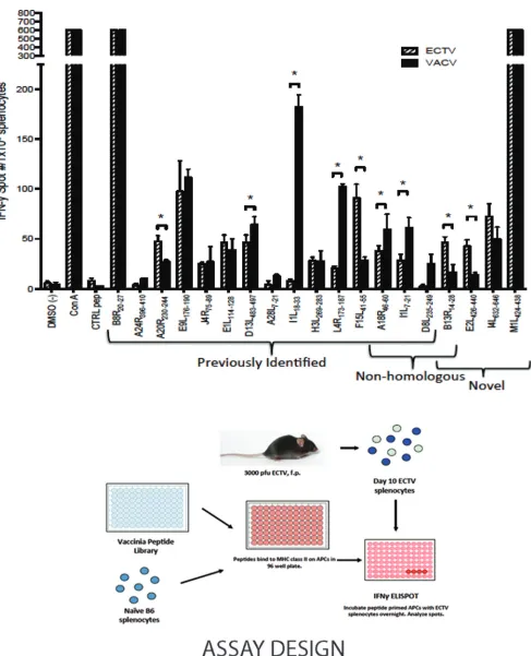
Epitope mapping.The 1,022 peptides used for mapping were a subset of a previously described library (34). Briefly, peptides were synthesized as crude material by Pepscan Systems and mimotopes ranging from 12 to 15 amino acids in length were used previously to identify VACV epitopes (34). The peptides were screened for reactivity against splenocytes from ECTV-primed mice at various time points postinfection. Splenocytes from naive mice were used as antigen-presenting cells. Naive splenocytes were incubated with peptide (final concentration, 2g/ml) at 37°C and 5% CO2. Peptide-primed splenocytes were then coincubated overnight
with either ECTV-specific whole splenocytes or TCD4⫹isolated from
splenocytes (Dynal mouse CD4 negative isolation kit; Invitrogen).
IFN-␥-positive T cell responses were assayed by IFN-␥ELISpot (BD). Spots were counted using ImmunoSpot software (Cellular Technology Lim-ited). To account for varying signal-to-noise ratios, we also calculated a stimulation index (SI), defined as (SFC [spot-forming cell] experiment results)/(SFC background) (34). Peptides with average spot numbers of ⱖ20, means of 1⫻106effector T cells,Pvalues of⬍0.05, and SI values of ⬎2 in three independent experiments were considered positive.
ICS assay.ICS assays were performed as previously described (15). Briefly, bone marrow-derived dendritic cells (1⫻106) generated using
previously published methods (35) were either pulsed with peptides (3g/ml) for 1 h in a 96-well plate or were infected with VACV WR (multiplicity of infection [MOI] of 5) for between 10 and 18 h before the addition of 1⫻106to 2⫻106splenocytes (pooled from two to five mice that were immunized with VACV WR for 10 days). Two hours later, brefeldin A (10g/ml) was added, and cells were cultured for another 6 h before staining according to the protocol of the BD Fix/Perm solution kit (BD Biosciences). At least 1.5⫻106to 2⫻106events per sample were
collected using an LSRII fluorescence-activated cell sorting (FACS) sys-tem (BD Biosciences) and were analyzed with FlowJo software (Tree Star). Background values were determined from samples pulsed with dimethyl sulfoxide (DMSO) only (no peptide) and were subtracted from the exper-imental values. At least three independent experiments were performed for each peptide or peptide pool. A peptide was considered positive if the average of the individual experiments was at least 1 standard deviation above the background.
RESULTS
T
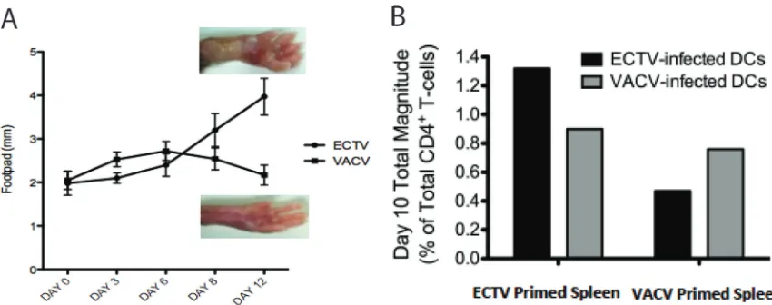
CD4ⴙresponse magnitude after infection with ECTV or VACV.
The distinct pathogenesis of ECTV compared with that of VACV
in C57BL/6 mice, despite
⬎
92% genetic identity (
36
), could be
appreciated following footpad inoculation of these viruses (
Fig.
1A
). VACV-induced inflammation was often detectable by day 3
postinfection, while the onset of ECTV-induced swelling was
typ-ically not appreciable until day 6. This can be ascribed, at least in
part, to greater subversion of early immune responses by ECTV
than by VACV (
14
,
37
). However, while VACV-induced
inflam-mation remained relatively mild and eventually subsided,
ECTV-associated swelling became considerable, ultimately leading to
ne-crosis and loss of the limb within
⬃
21 days of infection in most
cases, consistent with previous reports (
38
). In both infections, we
found day 10 postinfection to be the time point at which
virus-specific T
CD4⫹responses could be discriminately measured,
pro-viding an optimal signal-to-noise ratio (data not shown). The
di-vergent pathogenesis of ECTV and VACV in mice, despite a high
degree of genetic similarity, provides an ideal experimental system
to explore the influence of virulence and infectivity on the
result-ing T
CD4⫹responses following poxvirus infection.
The global T
CD4⫹responses to ECTV and VACV were initially
compared to determine differences in the overall magnitudes of
the responses to these two distinct poxviruses. We primed
C57BL/6 mice with ECTV or VACV, and 7 days later, cocultured
splenocytes with bone marrow-derived dendritic cells (BMDCs)
infected with either ECTV or VACV. Using flow cytometry, we
measured the magnitude of the T
CD4⫹responses by examining
several T cell functional outputs (
Fig. 1B
). Heterologous
stimula-tion produced approximately 62% (VACV T
CD4⫹to ECTV
BMDC) to 67% (ECTV T
CD4⫹to VACV BMDC) of the numbers
of activated T
CD4⫹produced by homologous stimulation. This
was 25 to 30% lower than what would be anticipated based upon
the degree of sequence identity shared by the proteomes of VACV
and ECTV (
36
,
39
). This suggested a disparity in the T
CD4⫹re-sponses to the two poxviruses extending beyond sequence
heter-ogeneity, a possibility that was first investigated by assessing
re-sponses to individual epitopes.
T
CD4ⴙspecificities elicited by VACV and ECTV.
Previous
mapping of the MHC-II-restricted C57BL/6 response to
intraperito-neal VACV infection was accomplished with a library of synthetic
peptides, comprising
⬃
30% of the predicted transcriptome of VACV
on November 7, 2019 by guest
http://jvi.asm.org/
(
34
). That study identified 14 specificities, with late-phase antigens
predominating among the list of parent proteins (
34
). Utilizing 1,022
of those peptides (comprising
⬃
15% of the predicted transcriptome
of VACV), we compared the reactivities elicited by ECTV and VACV
in C57BL/6 mice by ELISpot analysis (
Fig. 2
) (
34
). Since the common
route of entry for ECTV is through abrasions on the skin (
38
,
40–42
),
we performed footpad injection at the standard dose (3,000 PFU) as
the route of infection for this study. Importantly, for the ELISpot
screening, both the route and dose of VACV were matched to allow
for direct comparison with the results obtained following ECTV
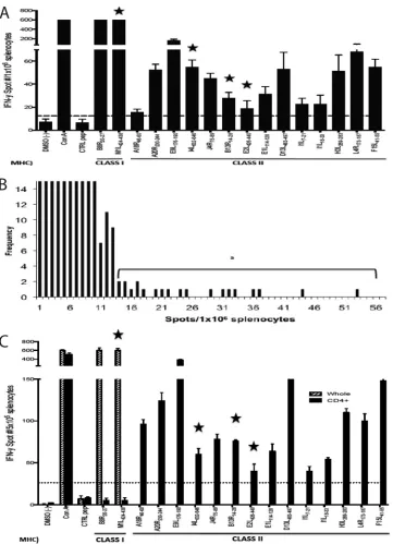
priming. Spleens were harvested 10 days after infection, and the
fre-quency of reproducible ECTV epitopes (
Fig. 3A
) measured was
rela-tively low, on the order of 1 to 2% (
Fig. 3B
), consistent with the
previous VACV screen (
34
).
The majority of peptides screened were fully conserved
be-tween VACV and ECTV, consistent with the high degree of
ho-mology. Fourteen distinct and reproducible MHC-II-restricted
specificities were identified in response to ECTV infection (
Fig. 3A
and
C
and
Table 1
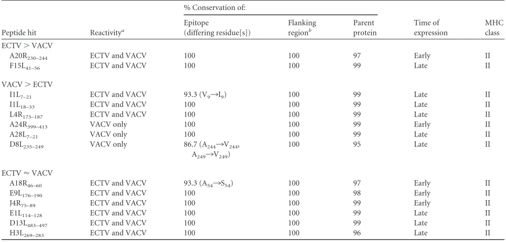
). Eleven of these had been previously identified
in the VACV screen, with two common epitopes (residues 46 to 60
of A18R [A18R
46 – 60] and I1L
7–21) differing by a single, apparently
neutral, amino acid (
Table 2
). Thus, three epitopes (I4L
632– 646,
B13R
14 –28, and E2L
426 – 440) were novel (
Table 3
). Interestingly,
when we screened VACV-specific T
CD4⫹(
Fig. 2
, solid bars), these
three specificities were also elicited, although B13R
14 –28and
E2L
426 – 440were just above the limit of detection. Three other
specificities identified in the original VACV screen (D8L
238 –252,
A28L
10 –24, and A24R
399 – 413) were not elicited by ECTV. This
could not be attributed to sequence heterogeneity for any of the
three because A28L
10 –24and A24R
399 – 413are 100% conserved.
And while the ECTV sequence differs from D8L
238 –252by two
residues (A
244¡
V
244and A
249¡
V
249), the ECTV counterpart was
also nonreactive (
Fig. 4
).
At the same time, we expanded the screen to include CD8
⫹T
cell (T
CD8⫹) specificities and identified a novel and relatively
po-tent MHC class I (MHC-I)-restricted epitope, M1L
424 – 438, that
elicited T
CD8⫹responses after both ECTV and VACV infections
(
Fig. 2
and
3C
). The minimal H2-K
b-restricted epitope within the
M1L
424 –338peptide was defined via a series of truncated peptides
as M1L
426 – 434(IIIPFIAYF) (data not shown).
The sum of the differences in T
CD4⫹specificities depicted in
Fig. 2
,
3
, and
4
do not, on their own, account for the unexpected
deficit in cross-reactivity shown by the results in
Fig. 1
.
Particu-larly striking are the unequal responses to homologous epitopes.
For example, despite the sequence identity, ECTV elicits
signifi-cantly greater responses to the A20R
230 –244, E2L
426 – 440, and
F15L
41–55epitopes than VACV. Conversely, VACV elicits far
greater responses to the I1L
7–21, I1L
18 –33, and L4R
173–187epitopes.
These differences in both directions indicate qualitative
differ-ences in the T
CD4⫹responses to the two viruses that are
indepen-dent of epitope sequence. This led us to assess additional attributes
of the resulting T
CD4⫹responses.
Minimal influence of the ECTV IFN-
␥
binding protein on
assay results.
One factor we needed to address at an early stage
was the IFN-
␥
binding protein expressed by both ECTV and
VACV, since only the ECTV version has specificity for murine
IFN-
␥
(
43
). Thus, the comparative ELISpot assays might have
been compromised by this selective activity. To address this, we
compared the responses of mice to wild-type (WT) ECTV and a
recombinant strain that lacks the soluble IFN-
␥
receptor (B8R in
VACV). The T
CD4⫹responses to WT and
⌬
evm0158
ECTV were
generally quite similar, with only a few significant differences (
Fig.
5
). For example, the E2L
426 – 440and F15L
41–55responses were
re-duced and the I1L
18 –33response elevated in comparison to the
responses of these epitopes to WT ECTV. Collectively, these data
indicate that
⌬
evm0158
ECTV does not significantly affect
virus-specific T
CD4⫹reactivity or IFN-
␥
production. Compatible with
this finding,
⌬
evm0158
ECTV was not appreciably attenuated
in
vivo
in our hands (unpublished data).
Comparative
functional
profiles
of
epitope-specific
T
CD4ⴙfrom ECTV- or VACV-primed mice.
To assess
functional-ity using intracellular cytokine staining (ICS) and polychromatic
flow cytometry, we pooled six peptides (
Table 4
, boldface) that
consistently elicited robust T
CD4⫹responses in ELISpot assays, five
FIG 1Divergent pathogenesis and magnitude of TCD4⫹response to ECTV and VACV. (A) C57BL/6 mice were infected with either VACV or ECTV (3,000 PFU
per footpad). Footpad swelling was measured using a caliper and recorded in millimeters at the indicated days postinfection. Representative photos of footpads at day 12 postinfection with either ECTV or VACV are shown. (B) TCD4⫹response magnitude, represented as the frequency of total responding TCD4⫹from mice
primed with either VACV and ECTV (3,000 PFU per footpad). Responses were measured by intracellular cytokine staining after splenocytes were stimulated with BMDCs infected with ECTV or VACV (MOI⫽1). Total magnitude was calculated after background subtraction by summing across all combinations of cells producing at least one of the following functions: CD107a, IFN-␥, IL-2, and TNF-␣.
on November 7, 2019 by guest
http://jvi.asm.org/
[image:3.585.76.501.65.233.2]FIG 2Comparative epitope specificities and magnitudes of responses to VACV- and ECTV-primed splenocytes. Naive splenocytes were incubated with synthetic poxvirus peptides ranging from 12 to 15 amino acids in length and screened for reactivity against splenocytes from ECTV-primed mice 10 days postinfection. Naive splenocytes were incubated with peptide (final concentration, 2g/ml). Peptide-primed splenocytes were coincubated overnight with whole splenocytes from ECTV-primed mice (3,000 PFU per footpad [f.p.]) 10 days postinfection. IFN-␥-positive T cell responses were assayed by IFN-␥ELISpot. To determine the level of statistical significance, Student’sttest was performed using the mean of triplicate values of the response. #, number; *,P⬍0.05; limits, means⫾standard deviations (SD); APC, antigen-presenting cell. Data are representative of 3 independent experiments.
on November 7, 2019 by guest
http://jvi.asm.org/
[image:4.585.51.538.60.661.2]being completely conserved between ECTV and VACV. Three
cy-tokines were measured: IFN-
␥
, interleukin-2 (IL-2), and tumor
necrosis factor alpha (TNF-
␣
). In general, these molecules play
well-described roles in antiviral immunity (
44–47
), and both
IFN-
␥
and TNF-
␣
have been found to be particularly important
for protection following poxvirus infection (
48
,
49
). In order to
identify T
CD4⫹with cytotoxic potential, we also assessed
degranu-lation by measuring the surface expression of CD107a after
stim-ulation (
50
). In agreement with our prior study (
15
), the average
T
CD4⫹response profile across all six epitopes was comparable
be-tween the two viruses (
Fig. 6A
). However, there were significant
differences in the overall frequency of cytokine production when
FIG 3Determination of ECTV-specific TCD4⫹epitopes in C57BL/6 mice. A synthetic VACV peptide library of 1,022 peptides, ranging from 12 to 15 amino acids
in length, was screened for reactivity against splenocytes from ECTV-primed mice (3,000 PFU per footpad) 10 days postinfection. Naive splenocytes were incubated with peptide (final concentration, 2g/ml). Peptide-primed splenocytes were coincubated overnight with ECTV-specific whole splenocytes. (A) IFN-␥-positive T cell responses were assayed by IFN-␥ELISpot. Stars indicate previously unreported poxvirus epitopes, and MHC class I or MHC class II epitopes are indicated below. (B) Frequency map of the positive peptides identified in panel A. (C) Confirmation that the epitopes identified in panel A are recognized by TCD4⫹. A screen was performed as described in the legend to panel A with splenocytes or purified TCD4⫹from ECTV-primed mice. Limits, means⫾SD.
MHC class I or MHC class II epitopes are indicated below.
on November 7, 2019 by guest
http://jvi.asm.org/
[image:5.585.113.474.63.562.2]individual functions were assessed (
Fig. 6B
), and it became clear
that ECTV-specific T
CD4⫹released significantly more cytotoxic
granules after peptide stimulation.
Next, we examined the functional profiles of the individual
specificities. Despite the functional similarity between
ECTV-and VACV-specific T
CD4⫹at a global (peptide pool) level, we
observed heterogeneity in the individual response profiles (
Fig.
6C
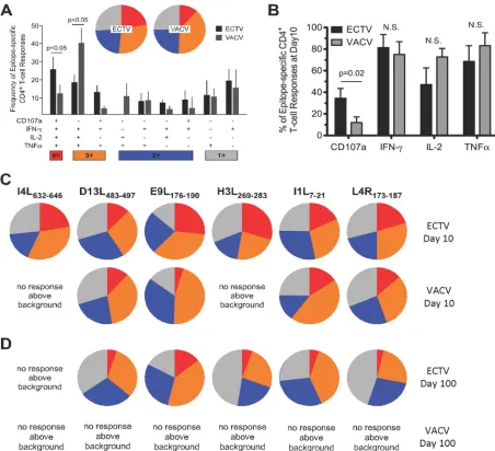
). Of note, VACV-infected mice did not yield a response
above the background for two specificities, I4L
632– 646and
H3L
269 –283(
Fig. 6C
). Of the specificities that could be
com-pared, E9L
176 –190-specific T
CD4⫹displayed the greatest
func-tional divergence.
Additionally, due to the importance and relevance of
long-lasting poxvirus immunization strategies (
51
,
52
,
113
), we
exam-ined epitope-specific T
CD4⫹functionality and persistence into the
memory phase (
ⱖ
100 days postinfection). We found that five of
the six ECTV specificities were detectable (greater than or equal to
0.05% of total T
CD4⫹) at 100 days postinfection (reactivity to
I4L
632– 646was undetectable by ICS), with minor degradation of
functionality over time in each case. In striking contrast, no
indi-vidual VACV-specific epitopes were detectable at 100 days
postin-fection, even though a 1,000-fold high dose of VACV was used for
priming (
Fig. 6D
). In the context of this study, these data suggest
[image:6.585.42.543.80.265.2]that ECTV and VACV infections mediate distinct priming and
TABLE 1Identification of ECTV-specific epitopesa
Peptide hit Sequence VACV protein
Time of
expression Function
MHC class
B8R20–27 TSYKFESV B8R (VACWR190) [A190] Early Virulence I
M1L424–438 KSIIIPFIAYFVLMH M1L (VACWR030) [A030] Early Unknown I
A18R46–60 PKGFYASPSVKTSLV A18R (VACWR138) Early Regulation II
A20R230–244 GDNIFIPSVITKSGK A20R (VACWR141) [A141] Early Regulation II
E9L176–190 PSVFINPISHTSYCY E9L (VACWR065) Early Regulation II
I4L632–646 EFQVVNPHLLRVLTE I4L (VACWR073) [A073] Early Regulation II
J4R75–89 DDDYGEPIIITSYLQ J4R (VACWR096) Early Regulation II
B13R14–28 ENVFISPASISSVLT B13R (VACWR195) [A195] Early Virulence II
E2L426–440 RLMFEYPLTKEASDH E2L (VACWR058) [A058] Early Unknown II
E1L114–128 VLTIKAPNVISSKIS E1L (VACWR057) [A057] Late Regulation II
D13L483–497 PKIFFRPTTITANVS D13L (VACWR118) [A118] Late Structural II
I1L7–21 QLVFNSISARALKAY VACWR070 (I1L) Late Structural II
I1L18–33 LKAYFTAKINEMVDE I1L (VACWR070) Late Structural II
H3L269–283 PGVMYAFTTPLISFF H3L (VACWR101) Late Structural II
L4R173–187 ISKYAGINILNVYSP L4R (VACWR091) [A091] Late Structural II
F15L41–56 TPRYIPSTSISSSNI F15L (VACWR054) Late Unknown II
aBoldface indicates previously unreported epitopes.
TABLE 2TCD4⫹poxvirus-specific epitopes
Peptide hit Reactivitya
% Conservation of:
Time of expression
MHC class Epitope
(differing residue[s])
Flanking regionb
Parent protein
ECTV⬎VACV
A20R230–244 ECTV and VACV 100 100 97 Early II
F15L41–56 ECTV and VACV 100 100 99 Late II
VACV⬎ECTV
I1L7–21 ECTV and VACV 93.3 (V9¡I9) 100 99 Late II
I1L18–33 ECTV and VACV 100 100 99 Late II
L4R173–187 ECTV and VACV 100 100 99 Late II
A24R399–413 VACV only 100 100 99 Early II
A28L7–21 VACV only 100 100 99 Late II
D8L235–249 VACV only 86.7 (A244¡V244,
A249¡V249)
100 95 Late II
ECTV⬇VACV
A18R46–60 ECTV and VACV 93.3 (A54¡S54) 100 97 Early II
E9L176–190 ECTV and VACV 100 100 98 Early II
J4R75–89 ECTV and VACV 100 100 99 Early II
E1L114–128 ECTV and VACV 100 100 99 Late II
D13L483–497 ECTV and VACV 100 100 99 Late II
H3L269–283 ECTV and VACV 100 100 96 Late II
a
Stimulation index (SI) of⬎2.
bIncludes 5 amino acid residues upstream and downstream from the epitope.
on November 7, 2019 by guest
http://jvi.asm.org/
[image:6.585.39.546.465.708.2]functional imprinting of epitope-specific T
CD4⫹that affect
persis-tence into the memory phase.
A greater frequency of effector T
CD4ⴙis elicited by ECTV
dur-ing acute infection.
Having examined CD107a expression among
the poxvirus-specific responses (
Fig. 6
), we looked in more detail
at the effector status of T
CD4⫹responses after infection. Our recent
work points to the importance of cytolytic function by T
CD4⫹dur-ing acute ECTV infection of mice (
9
). Here, we asked whether
cytolytic function is a poxvirus-specific phenomenon or unique to
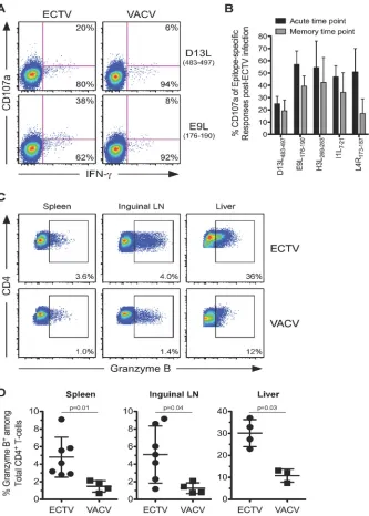
ECTV. Using flow cytometric analysis, we observed significant
differences in the surface mobilization of cytotoxic granules by
T
CD4⫹that were dependent upon both the epitope specificity and
identity of the infecting poxvirus. The T
CD4⫹responses to two
conserved epitopes, D13L
483– 497and E9L
176 –190, serve as an
illus-tration of these points, with ECTV infection inducing E9L
176 –190-specific T
CD4⫹with greater degranulation capacity than T
CD4⫹with other specificities (
Fig. 7A
). Additionally, compared with
VACV, ECTV infection consistently elicited a higher proportion
of responding T
CD4⫹with discernible CD107a expression after
stimulation (
Fig. 7A
). The frequency of degranulation typically
declined over time but was better maintained for some epitope
specificities (
Fig. 7B
).
Granzyme B (gzmB) is a major proapoptotic mediator stored
within cytotoxic granules. As an additional way to assess cytotoxic
potential, we measured the global levels of this molecule within
total T
CD4⫹at acute time points postinfection with both viruses in
the liver, inguinal lymph nodes, and spleen. We found that total
T
CD4⫹in all three locations within ECTV-infected mice expressed
2- to 3-fold-higher levels of grzB than were observed with VACV
(
Fig. 7C
and
D
). The enhanced grzB expression observed from
ECTV T
CD4⫹suggests that the induction of cytolytic T
CD4⫹is
char-acteristic of the murine host response to ECTV and not generally
associated with murine poxvirus infection, as it is absent in
VACV-immunized animals.
To further explore differential effector phenotypes between
ECTV and VACV T
CD4⫹, we examined the expression levels of
macrophage inflammatory protein 1
␣
(MIP1
␣
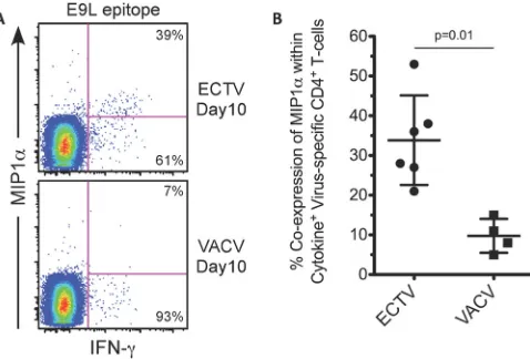
) from ECTV- and
VACV-specific T
CD4⫹. MIP1
␣
is a proinflammatory chemokine
involved in the recruitment of immune cells, and its expression by
T cells is associated with a more robust effector profile (
53–55
).
We found greater expression of MIP1
␣
from ECTV-specific
T
CD4⫹than from VACV T
CD4⫹(
Fig. 8
), offering additional
evi-dence that, in general, ECTV-specific T
CD4⫹display a greater
ef-fector-like profile than their VACV counterparts.
DISCUSSION
This study has revealed several ways in which host responses to the
natural murine poxvirus, ECTV, differ considerably from those to
VACV, the predominant model for examining poxvirus virulence
and immunity in mice (
16
,
56–59
). The initial experiments
re-vealed a level of cross-reactivity,
⬃
62 to 67%, that was far lower
than that expected by the degree of homology (92%). The basis for
this became clear when we examined individual specificities with a
12- to 15-mer peptide library. Eleven of 14 previously identified
VACV-induced specificities were elicited by ECTV (
34
), with the
remaining three specificities (D8L
238 –252, A28L
10 –24, and
A24R
399 – 413) failing to develop in response to ECTV. The screen
also uncovered four novel poxvirus epitopes for both ECTV and
VACV, three of which were MHC class II (I4L
632– 646, B13R
14 –28,
and E2L
426 – 440) and one of which was MHC class I (M1L
426 – 434,
the precise boundaries being subsequently determined by a
trun-cated-peptide series). Several factors may explain why the three
MHC-II epitopes were not discovered in an earlier screen (
33
).
Different routes of infection can affect T
CD4⫹differentiation, since
they determine the initial cell types that interact with the virus
(
60
), and prior screens utilized an intraperitoneal challenge (
34
),
whereas here, we employed a dermal footpad challenge to mimic
the natural infection route of ECTV (
42
). Additionally, prior
VACV epitope screens utilized B cells to present synthetic peptides
to T
CD4⫹(
34
), and differential costimulatory molecule expression
[image:7.585.39.549.79.162.2]by unique antigen-presenting cell types can alter signaling at the
TABLE 3Previously unreported poxvirus-specific epitopes
Peptide hit Reactivitya
% Conservation of:
Time of expression
MHC class Epitope
(differing residue)
Flanking regionb
Parent protein
M1L424–438 ECTV and VACV 100 96 96 Early I
I4L632–646 ECTV and VACV 100 100 97 Early II
B13R14–28 ECTV only 93.3 (P20¡S20) 100 95 Early II
E2L426–440 ECTV only 100 100 98 Early II
aStimulation index (SI) of⬎2.
b
Includes 5 amino acid residues upstream and downstream from the epitope.
FIG 4Comparative epitope reactivities to D8L238 –252between VACV- and
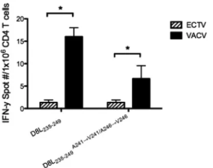
ECTV-primed splenocytes. Naive splenocytes were incubated with synthetic D8L238 –252(VACV sequence) or D8L238 –252A244¡V244/A249¡V249(ECTV
se-quence) at a final concentration of 2g/ml. Peptide-primed splenocytes were then coincubated overnight with whole splenocytes from ECTV-primed mice or VACV-primed mice (3,000 PFU per footpad) 10 days postinfection.
IFN-␥-positive T cell responses were assayed by IFN-␥ELISpot. To determine the level of statistical significance, Student’sttest was performed using the mean of triplicate values of the response. *,P⬍0.05.
on November 7, 2019 by guest
http://jvi.asm.org/
[image:7.585.62.271.472.637.2]immunological synapse, resulting in shifted T
CD4⫹activation
thresholds (
61
).
The novel MHC-I epitope, M1L
426 – 434, induced relatively
equivalent responses from both ECTV and VACV splenocytes
that were nearly as potent as the immunodominant B8R
20 –27for both VACV and ECTV splenocytes (
Fig. 2
), independent of
infection route and dose (data not shown).
In silico
analyses
that utilized algorithm-based predictions for MHC-I binders
within the VACV transcriptome identified a 10-mer H-2D
bclass I epitope (TSNVITDQTV/M1L
291–300) within the M1L
parent protein (
56
) but not at the 426 – 434 location.
The lack of ECTV T
CD4⫹reactivity to the three specificities
identified in the original VACV screen could not be accounted for
by sequence heterogeneity, as A28L
10 –24and A24R
399 – 413are both
100% conserved. And although there were two amino acid
differ-ences in the D8L
238 –252epitope (A
244¡
V
244and A
249¡
V
249), the
possibility that these changes prevent proper processing of the
antigen or prevent binding to the I-A
bmolecule was discounted by
the observation that VACV T
CD4⫹responds to the ECTV
ho-molog. Thus, in all three cases, factors extrinsic to epitope
com-position are at work. One clear difference between the two viruses
is the course of infection. ECTV productively infects a wider range
of murine cell types, including dendritic cells, epidermal T cells,
and keratinocytes (
14
,
41
,
62
). Thus, different sets of
antigpresenting cells with differing processing capabilities will be
en-gaged. Furthermore, ECTV produces species-specific factors that
allow for evasion and subversion of host responses and far greater
replication in mice, as well as a greater antigen load, which will
affect the levels of epitope display. Virulence factors and antigen
load can also affect the cytokine milieu, which is markedly
differ-ent in the two infections (
15
,
63–67
) and which can strongly
in-fluence antigen-processing capabilities (
68
,
69
). Chief among the
cytokines of interest is IFN-
␥
, which, in addition to influencing
the expression of antigen-processing components (
68–71
), drives
upregulation of MHC-II (
72
). This was especially true for these
investigations since ECTV but not VACV encodes a soluble IFN-
␥
receptor (B8R) that binds to murine IFN-
␥
(
25
,
73
,
74
). However,
deletion of B8R did not have a substantial impact on overall T
CD4⫹magnitude or on the individual T
CD4⫹reactivities, consistent with
our observation that
⌬
evm0158
is not attenuated
in vivo
(unpub-lished data).
Differences in participating antigen-presenting cells and
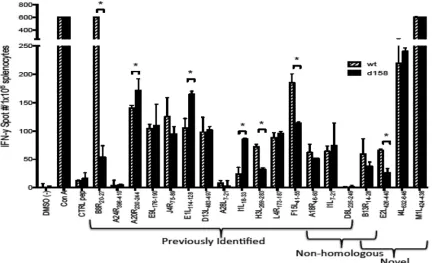
[image:8.585.78.510.67.330.2]anti-FIG 5Comparative epitope specificities and magnitudes of responses between WT ECTV- and⌬evm0158ECTV-primed splenocytes. Naive splenocytes were incubated with synthetic poxvirus peptides ranging from 12 to 15 amino acids in length (final concentration, 2g/ml). Peptide-primed splenocytes were coincubated overnight with whole splenocytes from WT ECTV- or⌬evm0158ECTV-primed mice (3,000 PFU per footpad) 10 days postinfection. IFN-␥-positive T cell responses were assayed by IFN-␥ELISpot. To determine the level of statistical significance, Student’sttest was performed using the mean of triplicate values of the response. *,P⬍0.05; limits, mean⫾SD. Data are representative of 3 independent experiments.
TABLE 4Selected TCD4⫹/MHC class II poxvirus-specific epitopesa
Peptide Amino acid sequence
A20R230–244 GDNIFIPSVITKSGK
I4L632–646 EFQVVNPHLLRVLTE
F15L41–55 TPRYIPSTSISSSNI
E1L114–128 VLTIKAPNVISSKIS
I1L7–21 QLVFNSISARALKAY
I1L18–33 LKAYFTAKINEMVDE
D13L483–497 PKIFFRPTTITANVS
E9L176–190 PSVFINPISHTSYCY
H3L269–283 PGVMYAFTTPLISFF
L4R173–187 ISKYAGINILNVYSP
aBoldface indicates peptides that consistently elicited robust T
CD4⫹responses in
ELISpot assays and were pooled to assess functionality using intracellular cytokine staining (ICS) and polychromatic flow cytometry.
on November 7, 2019 by guest
http://jvi.asm.org/
[image:8.585.40.287.579.697.2]gen load can also affect the functional character of the T
CD4⫹re-sponse (
75–79
). Indeed, we found that for the majority of the
epitopes tested (5 of 6), ECTV-specific T
CD4⫹demonstrated a
higher effector capacity; most notably enhanced were cytolytic
potential and MIP1
␣
expression. Furthermore, VACV T
CD4⫹ex-hibited responses above the background for just 4 of the 6 epitopes
examined at 10 days postinfection, while ECTV-specific T
CD4⫹reacted strongly to all 6. We also identified higher percentages of
grzB-positive cells from the tissues of ECTV-infected mice (
Fig.
7C
and
D
). This was true of all three tissues examined at acute time
points postinfection. This result demonstrates that the generation
of robust cytolytic T
CD4⫹is not a general property of all poxvirus
infections and is consistent with earlier reports that increased
an-tigen loads drive the development of cytolytic T
CD4⫹in both acute
and chronic viral infections (
80
,
81
).
Previous work by Fang et al. demonstrated that cytolytic T
CD4⫹contribute to the suppression and host control of ECTV
replica-tion (
9
), consistent with the well-documented pleiotropic
antivi-ral effects of grzB expression. In addition to triggering apoptosis
during ECTV infection, despite the production of antiapoptotic
proteins such as B13R (
82
,
83
), grzB has been shown to directly
suppress VACV replication via cleavage of eukaryotic initiation
factor 4 gamma 3 (eIF4G3), a protein essential for the initiation of
protein translation (
84
). Increased grzB production may also play
a role in the greater inflammation shown with ECTV at the site of
infection (footpad) (
Fig. 1
) by inducing apoptosis of endothelial
FIG 6Comparative functional profiles of epitope-specific TCD4⫹from ECTV- or VACV-primed mice. (A) TCD4⫹responses to a poxvirus-specific class II peptide
pool (Table 4) at day 10 were divided according to the relative contribution of each functional combination. Permutations that did not contribute significantly to the functional profile are not shown due to space constraints. Responses are grouped according to the degree of positivity and matched to the colors in the pie graphs for responses to the peptide pool at day 10. (B) Proportion of poxvirus-specific TCD4⫹at day 10 postinfection for each specific function measured after
stimulation. (C and D) Functional profiles of TCD4⫹responsive to a selected cohort of poxvirus epitopes at day 10 (C) and day 100 (D) postinfection are shown
for ECTV-infected (3,000 PFU per footpad) and VACV-infected (3,000 PFU per footpad or 3⫻106PFU per footpad) mice. (A to D) All depicted data were from
pooled cells of three mice at each time point and represent the average of two independent experiments. Bars represent the means, and error bars indicate the standard deviations.Pvalues are defined where applicable; N.S., not significant. Data are representative of 3 independent experiments.
on November 7, 2019 by guest
http://jvi.asm.org/
[image:9.585.64.516.65.477.2]cells (anoikis) via granule exocytosis (
85
) and remodeling of
ex-tracellular matrix through cleavage of vitronectin, fibronectin,
and laminin (
86
).
We also assessed the functional profiles of several
peptide-spe-cific T
CD4⫹at memory (day 100) time points using flow cytometry
(
Fig. 6D
). While ECTV infection stimulated robust long-term
T
CD4⫹memory, none of the 6 epitopes examined yielded a
re-sponse above the background at day 100 postinfection with
VACV, even when the input dose was 1,000-fold-greater for
VACV than for ECTV. This was unexpected, since prior studies
have shown persistence of VACV-specific T
CD4⫹in both humans
(
52
) and mice (
15
,
66
). In our own earlier study (
15
), we detected
both T
CD4⫹memory responses at day 75 postinfection, utilizing a
more comprehensive peptide pool than the one examined here,
and VACV-infected presenting cells. Thus, while long-term T
CD4⫹memory to VACV may not be completely absent, it is significantly
compromised.
The considerably greater difference between ECTV- and
FIG 7ECTV-specific TCD4⫹degranulate and express larger amounts of granzyme B during acute infection. (A) Proportions of D13L- or E9L-specific TCD4⫹
splenocytes coexpressing CD107a and IFN-␥from ECTV- or VACV-primed mice (3,000 PFU per footpad) at 10 days postinfection. Percentages represent the fraction of overlaid cells that fall within each quadrant. (B) Percentages of D13L-, E9L-, H3L-, I1L-, or L4R-specific TCD4⫹splenocytes expressing CD107a from
ECTV-primed mice (3,000 PFU per footpad) at acute (10 days postinfection) and memory (75 to 100 days postinfection) time points. (C) Percentages of grzB-positive (grzB⫹) cells from bulk TCD4⫹collected from the spleen, inguinal lymph nodes (LN), or liver of ECTV- or VACV-primed mice (3,000 PFU per
footpad) at 10 days postinfection. (D) Quantification of the average percentages of grzB⫹from bulk TCD4⫹collected from the spleen, inguinal LN, or liver of
ECTV- or VACV-primed mice (3,000 PFU per footpad) at acute time points postinfection. Depicted data are from pooled cells of three mice and represent the average and SD of two independent experiments.Pvalues are defined where applicable. Data are representative of 2 independent experiments.
on November 7, 2019 by guest
http://jvi.asm.org/
[image:10.585.127.460.65.531.2]VACV-specific T
CD4⫹at the memory phase is consistent with the
earlier demonstration that differences in the initial antiviral T
CD4⫹characters can become exaggerated as populations transition to
central and/or effector memory cells (
87
,
88
). Several factors
might contribute to this amplification effect. Early expression of
IL-15 by phagocytes and innate immune cells has been found to
induce both a cytolytic T
CD4⫹character and T
CD4⫹effector
mem-ory cells (
89–91
) and has also been associated with NK cell
recruit-ment, shown to be essential for natural resistance to ECTV (
92
). In
addition, the above-mentioned parameters of antigen exposure
and persistence can greatly affect the initial and long-term
effec-torlike properties of responding T
CD4⫹(
77
,
87
,
93
). At the same
time, excessive antigen exposure can also lead to exhaustion in
some settings (
94
). A greater understanding of the factors that set
the balance between long-lasting T
CD4⫹effector-memory and
ex-haustion has been elusive (
87
,
94
) but will likely be critical for
insight into the profound differences in T
CD4⫹memory to two
such homologous viruses.
The long-term protection against smallpox conferred by
VACV is well known (
52
,
95–105
). In a recent study of smallpox
vaccinees, the levels of VACV-specific antibody, generally held to
be the measure of protection (
52
), remained stable over many
years postimmunization. In contrast and in line with our findings,
VACV-specific T
CD4⫹memory declined over time (
52
). Due to the
complexity of poxviruses, which clearly contain an abundance of
MHC-II-restricted epitopes (this study and reference
34
), this
de-cline may not meaningfully impact protection. Alternatively or in
addition, memory T
CD4⫹may not play a critical role in protection
from smallpox. The importance of robust T
CD4⫹memory
popu-lations for protection from agents such as influenza virus and
hepatitis A and C viruses is more apparent (
106–111
). Whether
this is due to their relative simplicity and the limited number of
T
CD4⫹epitopes they encode or other factors remains to be
deter-mined.
ECTV is proving to be an excellent model both in terms of
providing an opportunity to study a natural host-pathogen
rela-tionship under defined conditions and in being highly relevant to
other poxviruses (
112
), including smallpox and monkeypox (
11
,
12
). The overall results of this study highlight interesting
distinc-tions between the T
CD4⫹epitope reactivity profiles of ECTV and
VACV. A better understanding of the mediators that influence
T
CD4⫹
reactivity and function is critical to designing better
vac-cines and antiviral therapeutics. Factors such as tropism and
rep-lication efficiency are likely major reasons for the functional
dif-ferences observed between ECTV and VACV T
CD4⫹responding to
common epitopes. Moreover, distinct host-pathogen
relation-ships may ultimately play a predominant role in both shaping the
T
CD4⫹repertoire and influencing the functional imprinting and
differentiation of poxvirus-specific T
CD4⫹. The striking cytolytic
character of T
CD4⫹induced by ECTV and the inability of VACV to
drive the development of this protective T
CD4⫹subset (
89
) or a
substantial memory T
CD4⫹population demonstrate the impact of
epitope-extrinsic mediators on T
CD4⫹repertoire, function, and
persistence. Further study in this comparative poxvirus model
may yield additional insights into the design of vaccine strategies
that lead to more robust and long-lived T
CD4⫹responses.
ACKNOWLEDGMENTS
This work was supported by National Institutes of Health grant U19-AI083008.
Research in this publication includes work carried out at the Kimmel Cancer Center Flow Cytometry Facility, which is supported in part by NCI Cancer Center support grant P30CA56036.
REFERENCES
1.Janssen EM, Lemmens EE, Wolfe T, Christen U, von Herrath MG, Schoenberger SP.2003. CD4⫹T cells are required for secondary expan-sion and memory in CD8⫹T lymphocytes. Nature421:852– 856.http: //dx.doi.org/10.1038/nature01441.
2.Nakanishi Y, Lu B, Gerard C, Iwasaki A.2009. CD8(⫹) T lymphocyte mobilization to virus-infected tissue requires CD4(⫹) T-cell help. Na-ture462:510 –513.http://dx.doi.org/10.1038/nature08511.
3.Yin L, Calvo-Calle JM, Cruz J, Newman FK, Frey SE, Ennis FA, Stern LJ.2013. CD4⫹T cells provide intermolecular help to generate robust antibody responses in vaccinia virus-vaccinated humans. J. Immunol. 190:6023– 6033.http://dx.doi.org/10.4049/jimmunol.1202523. 4.Crotty S, Kersh EN, Cannons J, Schwartzberg PL, Ahmed R.2003. SAP
is required for generating long-term humoral immunity. Nature421: 282–287.http://dx.doi.org/10.1038/nature01318.
5.Darrah PA, Patel DT, De Luca PM, Lindsay RW, Davey DF, Flynn BJ, Hoff ST, Andersen P, Reed SG, Morris SL, Roederer M, Seder RA. 2007. Multifunctional TH1 cells define a correlate of vaccine-mediated protection against Leishmania major. Nat. Med.13:843– 850.http://dx .doi.org/10.1038/nm1592.
6.Zhu J, Paul WE.2008. CD4 T cells: fates, functions, and faults. Blood 112:1557–1569.http://dx.doi.org/10.1182/blood-2008-05-078154. 7.Wan YY, Flavell RA.2009. How diverse—CD4 effector T cells and their
functions. J. Mol. Cell Biol. 1:20 –36.http://dx.doi.org/10.1093/jmcb /mjp001.
8.Soghoian DZ, Jessen H, Flanders M, Sierra-Davidson K, Cutler S, Pertel T, Ranasinghe S, Lindqvist M, Davis I, Lane K, Rychert J, Rosenberg ES, Piechocka-Trocha A, Brass AL, Brenchley JM, Walker BD, Streeck H.2012. HIV-specific cytolytic CD4 T cell responses during acute HIV infection predict disease outcome. Sci. Transl. Med. 4:123ra25.http://dx.doi.org/10.1126/scitranslmed.3003165.
9.Fang M, Siciliano NA, Hersperger AR, Roscoe F, Hu A, Ma X, Shamsedeen AR, Eisenlohr LC, Sigal LJ. 2012. Perforin-dependent CD4⫹T-cell cytotoxicity contributes to control a murine poxvirus in-FIG 8ECTV-specific TCD4⫹express larger amounts of MIP1␣during acute
infection. (A) Representative staining showing higher expression of MIP1␣by ECTV-specific TCD4⫹. E9L-specific TCD4⫹responses from ECTV- or
VACV-primed mice (3,000 PFU per footpad) at 10 days postinfection are shown. Percentages represent the fractions of IFN-␥⫹cells that were either positive or negative for MIP1␣after peptide stimulation. (B) Quantification of MIP1␣ expression among several different peptide-specific TCD4⫹responses. Total
percentages of virus-specific TCD4⫹splenocytes coexpressing MIP1␣and at
least one additional cytokine from ECTV- or VACV-primed mice (3,000 PFU per footpad) at 10 days postinfection are shown. Depicted data are represen-tative of four independent experiments.Pvalue was determined using the Mann-Whitney test. Bars and whiskers represent the means and SD.
on November 7, 2019 by guest
http://jvi.asm.org/
[image:11.585.44.283.67.229.2]fection. Proc. Natl. Acad. Sci. U. S. A.109:9983–9988.http://dx.doi.org /10.1073/pnas.1202143109.
10. Mahy BW, Almond JW, Berns KI, Chanock RM, Lvov DK, Pettersson RF, Schatzmayr HG, Fenner F.1993. The remaining stocks of smallpox virus should be destroyed. Science262:1223–1224.http://dx.doi.org/10 .1126/science.8235651.
11. Anderson PD, Bokor G.2012. Bioterrorism: pathogens as weapons. J. Pharm. Pract.25:521–529.http://dx.doi.org/10.1177/0897190012456366. 12. Folio LR, Yao EF.2007. US military smallpox vaccination program:
occupational impact of immunizations on aircrew in Air Mobility Com-mand, US Air Force. J. Am. Osteopath. Assoc.107:547–553.
13. Whitby M, Street AC, Ruff TA, Fenner F.2002. Biological agents as weapons 1: smallpox and botulism. Med. J. Aust.176:431– 433. 14. Esteban DJ, Buller RM.2005. Ectromelia virus: the causative agent of
mousepox. J. Gen. Virol.86:2645–2659.http://dx.doi.org/10.1099/vir.0 .81090-0.
15. Hersperger AR, Siciliano NA, Eisenlohr LC.2012. Comparable poly-functionality of ectromelia virus- and vaccinia virus-specific murine T cells despite markedly different in vivo replication and pathogenicity. J. Virol.86:7298 –7309.http://dx.doi.org/10.1128/JVI.00038-12. 16. Tscharke DC, Smith GL.1999. A model for vaccinia virus pathogenesis
and immunity based on intradermal injection of mouse ear pinnae. J. Gen. Virol.80(Pt 10):2751–2755.
17. Jenkins MK, Khoruts A, Ingulli E, Mueller DL, McSorley SJ, Reinhardt RL, Itano A, Pape KA.2001. In vivo activation of antigen-specific CD4 T cells. Annu. Rev. Immunol. 19:23– 45. http://dx.doi.org/10.1146 /annurev.immunol.19.1.23.
18. Tao X, Constant S, Jorritsma P, Bottomly K.1997. Strength of TCR signal determines the costimulatory requirements for Th1 and Th2 CD4⫹T cell differentiation. J. Immunol.159:5956 –5963.
19. Steinman RM, Hawiger D, Nussenzweig MC.2003. Tolerogenic den-dritic cells. Annu. Rev. Immunol.21:685–711.http://dx.doi.org/10.1146 /annurev.immunol.21.120601.141040.
20. Iwasaki A, Medzhitov R.2004. Toll-like receptor control of the adaptive immune responses. Nat. Immunol.5:987–995.http://dx.doi.org/10.1038 /ni1112.
21. Zhu J, Yamane H, Paul WE.2010. Differentiation of effector CD4 T cell populations. Annu. Rev. Immunol.28:445– 489.http://dx.doi.org/10 .1146/annurev-immunol-030409-101212.
22. Khan S, van den Broek M, Schwarz K, de Giuli R, Diener PA, Groettrup M.2001. Immunoproteasomes largely replace constitutive proteasomes during an antiviral and antibacterial immune response in the liver. J. Immu-nol.167:6859 – 6868.http://dx.doi.org/10.4049/jimmunol.167.12.6859. 23. Smith VP, Alcami A.2000. Expression of secreted cytokine and
chemo-kine inhibitors by ectromelia virus. J. Virol.74:8460 – 8471.http://dx.doi .org/10.1128/JVI.74.18.8460-8471.2000.
24. Alcami A, Smith GL.1992. A soluble receptor for interleukin-1 beta encoded by vaccinia virus: a novel mechanism of virus modulation of the host response to infection. Cell71:153–167.http://dx.doi.org/10.1016 /0092-8674(92)90274-G.
25. Alcami A, Smith GL.1995. Vaccinia, cowpox, and camelpox viruses encode soluble gamma interferon receptors with novel broad species specificity. J. Virol.69:4633– 4639.
26. Alcami A, Symons JA, Smith GL.2000. The vaccinia virus soluble alpha/beta interferon (IFN) receptor binds to the cell surface and pro-tects cells from the antiviral effects of IFN. J. Virol.74:11230 –11239. http://dx.doi.org/10.1128/JVI.74.23.11230-11239.2000.
27. Spriggs MK, Hruby DE, Maliszewski CR, Pickup DJ, Sims JE, Buller RM, VanSlyke J.1992. Vaccinia and cowpox viruses encode a novel secreted interleukin-1-binding protein. Cell71:145–152.http://dx.doi .org/10.1016/0092-8674(92)90273-F.
28. Born TL, Morrison LA, Esteban DJ, VandenBos T, Thebeau LG, Chen N, Spriggs MK, Sims JE, Buller RM.2000. A poxvirus protein that binds to and inactivates IL-18, and inhibits NK cell response. J. Immunol. 164:3246 –3254.http://dx.doi.org/10.4049/jimmunol.164.6.3246. 29. Bowie A, Kiss-Toth E, Symons JA, Smith GL, Dower SK, O’Neill LA.
2000. A46R and A52R from vaccinia virus are antagonists of host IL-1 and toll-like receptor signaling. Proc. Natl. Acad. Sci. U. S. A.97:10162– 10167.http://dx.doi.org/10.1073/pnas.160027697.
30. Loparev VN, Parsons JM, Knight JC, Panus JF, Ray CA, Buller RM, Pickup DJ, Esposito JJ.1998. A third distinct tumor necrosis factor receptor of orthopoxviruses. Proc. Natl. Acad. Sci. U. S. A.95:3786 – 3791.http://dx.doi.org/10.1073/pnas.95.7.3786.
31. Samuelsson C, Hausmann J, Lauterbach H, Schmidt M, Akira S, Wagner H, Chaplin P, Suter M, O’Keeffe M, Hochrein H. 2008. Survival of lethal poxvirus infection in mice depends on TLR9, and ther-apeutic vaccination provides protection. J. Clin. Invest.118:1776 –1784. http://dx.doi.org/10.1172/JCI33940.
32. Xu RH, Cohen M, Tang Y, Lazear E, Whitbeck JC, Eisenberg RJ, Cohen GH, Sigal LJ.2008. The orthopoxvirus type I IFN binding pro-tein is essential for virulence and an effective target for vaccination. J. Exp. Med.205:981–992.http://dx.doi.org/10.1084/jem.20071854. 33. Rubio D, Xu RH, Remakus S, Krouse TE, Truckenmiller ME, Thapa
RJ, Balachandran S, Alcami A, Norbury CC, Sigal LJ.2013. Crosstalk between the type 1 interferon and nuclear factor kappa B pathways con-fers resistance to a lethal virus infection. Cell Host Microbe13:701–710. http://dx.doi.org/10.1016/j.chom.2013.04.015.
34. Moutaftsi M, Bui HH, Peters B, Sidney J, Salek-Ardakani S, Oseroff C, Pasquetto V, Crotty S, Croft M, Lefkowitz EJ, Grey H, Sette A.2007. Vaccinia virus-specific CD4⫹T cell responses target a set of antigens largely distinct from those targeted by CD8⫹T cell responses. J. Immu-nol.178:6814 – 6820.http://dx.doi.org/10.4049/jimmunol.178.11.6814. 35. Siciliano NA, Skinner JA, Yuk MH.2006. Bordetella bronchiseptica
mod-ulates macrophage phenotype leading to the inhibition of CD4⫹T cell pro-liferation and the initiation of a Th17 immune response. J. Immunol.177: 7131–7138.http://dx.doi.org/10.4049/jimmunol.177.10.7131.
36. Chen N, Danila MI, Feng Z, Buller RM, Wang C, Han X, Lefkowitz EJ, Upton C.2003. The genomic sequence of ectromelia virus, the causative agent of mousepox. Virology317:165–186.http://dx.doi.org/10.1016 /S0042-6822(03)00520-8.
37. O’Gorman WE, Sampath P, Simonds EF, Sikorski R, O’Malley M, Krutzik PO, Chen H, Panchanathan V, Chaudhri G, Karupiah G, Lewis DB, Thorne SH, Nolan GP.2010. Alternate mechanisms of initial pattern recognition drive differential immune responses to related pox-viruses. Cell Host Microbe8:174 –185.http://dx.doi.org/10.1016/j.chom .2010.07.008.
38. Marchal J.1930. Infectious ectromelia: a hitherto undescribed virus disease of mice. J. Pathol. Bacteriol.33:713–728.http://dx.doi.org/10 .1002/path.1700330317.
39. Gubser C, Hue S, Kellam P, Smith GL. 2004. Poxvirus genomes: a phylogenetic analysis. J. Gen. Virol.85:105–117.http://dx.doi.org/10 .1099/vir.0.19565-0.
40. Fenner F.1981. Mousepox (infectious ectromelia): past, present, and future. Lab. Anim. Sci.31:553–559.
41. Roberts JA.1962. Histopathogenesis of mousepox. II. Cutaneous infec-tion. Br. J. Exp. Pathol.43:462– 468.
42. Fenner F.1947. Studies in infectious ectromelia in mice; natural trans-mission; the portal of entry of the virus. Aust. J. Exp. Biol. Med. Sci. 25:275–282.http://dx.doi.org/10.1038/icb.1947.39.
43. Sakala IG, Chaudhri G, Buller RM, Nuara AA, Bai H, Chen N, Karupiah G.2007. Poxvirus-encoded gamma interferon binding protein dampens the host immune response to infection. J. Virol.81:3346 –3353. http://dx.doi.org/10.1128/JVI.01927-06.
44. Karupiah G.1998. Type 1 and type 2 cytokines in antiviral defense. Vet. Immunol. Immunopathol. 63:105–109. http://dx.doi.org/10.1016/S0165 -2427(98)00086-5.
45. Isaacs A, Lindenmann J.1957. Virus interference. I. The interferon. Proc. R. Soc. Lond. B Biol. Sci.147:258 –267.http://dx.doi.org/10.1098 /rspb.1957.0048.
46. D’Souza WN, Schluns KS, Masopust D, Lefrancois L.2002. Essential role for IL-2 in the regulation of antiviral extralymphoid CD8 T cell responses. J. Immunol. 168:5566 –5572. http://dx.doi.org/10.4049 /jimmunol.168.11.5566.
47. Seo SH, Webster RG.2002. Tumor necrosis factor alpha exerts powerful anti-influenza virus effects in lung epithelial cells. J. Virol.76:1071–1076. http://dx.doi.org/10.1128/JVI.76.3.1071-1076.2002.
48. Ruby J, Bluethmann H, Peschon JJ.1997. Antiviral activity of tumor necrosis factor (TNF) is mediated via p55 and p75 TNF receptors. J. Exp. Med.186:1591–1596.http://dx.doi.org/10.1084/jem.186.9.1591. 49. Chaudhri G, Panchanathan V, Buller RM, van den Eertwegh AJ,
Claassen E, Zhou J, de Chazal R, Laman JD, Karupiah G. 2004. Polarized type 1 cytokine response and cell-mediated immunity deter-mine genetic resistance to mousepox. Proc. Natl. Acad. Sci. U. S. A. 101:9057–9062.http://dx.doi.org/10.1073/pnas.0402949101.
50. Betts MR, Brenchley JM, Price DA, De Rosa SC, Douek DC, Roederer M, Koup RA.2003. Sensitive and viable identification of antigen-specific
on November 7, 2019 by guest
http://jvi.asm.org/
CD8⫹T cells by a flow cytometric assay for degranulation. J. Immunol. Methods281:65–78.http://dx.doi.org/10.1016/S0022-1759(03)00265-5. 51. Fenner F.1989. Risks and benefits of vaccinia vaccine use in the world-wide smallpox eradication campaign. Res. Virol.140:465– 466.http://dx .doi.org/10.1016/S0923-2516(89)80126-8.
52. Hammarlund E, Lewis MW, Hansen SG, Strelow LI, Nelson JA, Sexton GJ, Hanifin JM, Slifka MK.2003. Duration of antiviral immu-nity after smallpox vaccination. Nat. Med.9:1131–1137.http://dx.doi .org/10.1038/nm917.
53. Casazza JP, Betts MR, Price DA, Precopio ML, Ruff LE, Brenchley JM, Hill BJ, Roederer M, Douek DC, Koup RA.2006. Acquisition of direct antiviral effector functions by CMV-specific CD4⫹T lymphocytes with cellular maturation. J. Exp. Med.203:2865–2877.http://dx.doi.org/10 .1084/jem.20052246.
54. Casazza JP, Brenchley JM, Hill BJ, Ayana R, Ambrozak D, Roederer M, Douek DC, Betts MR, Koup RA.2009. Autocrine production of beta-chemokines protects CMV-Specific CD4 T cells from HIV infec-tion. PLoS Pathog.5:e1000646.http://dx.doi.org/10.1371/journal.ppat .1000646.
55. Hersperger AR, Pereyra F, Nason M, Demers K, Sheth P, Shin LY, Kovacs CM, Rodriguez B, Sieg SF, Teixeira-Johnson L, Gudonis D, Goepfert PA, Lederman MM, Frank I, Makedonas G, Kaul R, Walker BD, Betts MR.2010. Perforin expression directly ex vivo by HIV-specific CD8 T-cells is a correlate of HIV elite control. PLoS Pathog.6:e1000917. http://dx.doi.org/10.1371/journal.ppat.1000917.
56. Moutaftsi M, Peters B, Pasquetto V, Tscharke DC, Sidney J, Bui HH, Grey H, Sette A.2006. A consensus epitope prediction approach iden-tifies the breadth of murine T(CD8⫹)-cell responses to vaccinia virus. Nat. Biotechnol.24:817– 819.http://dx.doi.org/10.1038/nbt1215. 57. Bauer S, Bathke B, Lauterbach H, Patzold J, Kassub R, Luber CA,
Schlatter B, Hamm S, Chaplin P, Suter M, Hochrein H.2010. A major role for TLR8 in the recognition of vaccinia viral DNA by murine pDC? Proc. Natl. Acad. Sci. U. S. A.107:E139.http://dx.doi.org/10.1073/pnas .1008626107. (Reply, 107:E140,http://dx.doi.org/10.1073/pnas.1009858107.) 58. Symons JA, Adams E, Tscharke DC, Reading PC, Waldmann H, Smith
GL.2002. The vaccinia virus C12L protein inhibits mouse IL-18 and promotes virus virulence in the murine intranasal model. J. Gen. Virol. 83:2833–2844.
59. Moutaftsi M, Salek-Ardakani S, Croft M, Peters B, Sidney J, Grey H, Sette A.2009. Correlates of protection efficacy induced by vaccinia vi-rus-specific CD8⫹T-cell epitopes in the murine intranasal challenge model. Eur. J. Immunol. 39:717–722. http://dx.doi.org/10.1002/eji .200838815.
60. Pepper M, Linehan JL, Pagan AJ, Zell T, Dileepan T, Cleary PP, Jenkins MK.2010. Different routes of bacterial infection induce long-lived TH1 memory cells and short-long-lived TH17 cells. Nat. Immunol.11: 83– 89.http://dx.doi.org/10.1038/ni.1826.
61. Kleindienst P, Brocker T. 2005. Concerted antigen presentation by dendritic cells and B cells is necessary for optimal CD4 T-cell immunity in vivo. Immunology 115:556 –564. http://dx.doi.org/10.1111/j.1365 -2567.2005.02196.x.
62. Spohr de Faundez I, Gierynska M, Niemialtowski MG, Malicka E, Popis A. 1995. Ectromelia virus establishes a persistent infection in spleen dendritic cells and macrophages of BALB/c mice following the acute disease. Adv. Exp. Med. Biol.378:257–261.http://dx.doi.org/10 .1007/978-1-4615-1971-3_57.
63. Briody BA.1959. Response of mice to ectromelia and vaccinia viruses. Bacteriol. Rev.23:61–95.
64. Fenner F.1948. The pathogenesis of the acute exanthems; an interpre-tation based on experimental investigations with mousepox; infectious ectromelia of mice. Lancetii:915–920.
65. Fenner F.1949. Mouse-pox; infectious ectromelia of mice; a review. J. Immunol.63:341–373.
66. Harrington LE, van der Most R, Whitton JL, Ahmed R.2002. Recom-binant vaccinia virus-induced T-cell immunity: quantitation of the re-sponse to the virus vector and the foreign epitope. J. Virol.76:3329 – 3337.http://dx.doi.org/10.1128/JVI.76.7.3329-3337.2002.
67. Mahalingam S, Foster PS, Lobigs M, Farber JM, Karupiah G.2000. Interferon-inducible chemokines and immunity to poxvirus infections. Immunol. Rev. 177:127–133. http://dx.doi.org/10.1034/j.1600-065X .2000.17720.x.
68. Delvig AA, Lee JJ, Chrzanowska-Lightowlers ZM, Robinson JH.2002.
TGF-beta1 and IFN-gamma cross-regulate antigen presentation to CD4 T cells by macrophages. J. Leukoc. Biol.72:163–166.
69. Collins T, Korman AJ, Wake CT, Boss JM, Kappes DJ, Fiers W, Ault KA, Gimbrone MA, Jr, Strominger JL, Pober JS.1984. Immune inter-feron activates multiple class II major histocompatibility complex genes and the associated invariant chain gene in human endothelial cells and dermal fibroblasts. Proc. Natl. Acad. Sci. U. S. A.81:4917– 4921.http://dx .doi.org/10.1073/pnas.81.15.4917.
70. Hastings KT.2013. GILT: shaping the MHC class II-restricted pep-tidome and CD4 T cell-mediated immunity. Front. Immunol.4:429. http://dx.doi.org/10.3389/fimmu.2013.00429.
71. Li P, Gregg JL, Wang N, Zhou D, O’Donnell P, Blum JS, Crotzer VL. 2005. Compartmentalization of class II antigen presentation: contribu-tion of cytoplasmic and endosomal processing. Immunol. Rev.207:206 – 217.http://dx.doi.org/10.1111/j.0105-2896.2005.00297.x.
72. Steimle V, Siegrist CA, Mottet A, Lisowska-Grospierre B, Mach B. 1994. Regulation of MHC class II expression by interferon-gamma me-diated by the transactivator gene CIITA. Science265:106 –109.http://dx .doi.org/10.1126/science.8016643.
73. Oseroff C, Kos F, Bui HH, Peters B, Pasquetto V, Glenn J, Palmore T, Sidney J, Tscharke DC, Bennink JR, Southwood S, Grey HM, Yewdell JW, Sette A.2005. HLA class I-restricted responses to vaccinia recognize a broad array of proteins mainly involved in virulence and viral gene regulation. Proc. Natl. Acad. Sci. U. S. A.102:13980 –13985.http://dx.doi .org/10.1073/pnas.0506768102.
74. Symons JA, Tscharke DC, Price N, Smith GL.2002. A study of the vaccinia virus interferon-gamma receptor and its contribution to virus virulence. J. Gen. Virol.83:1953–1964.
75. Igyarto BZ, Kaplan DH.2013. Antigen presentation by Langerhans cells. Curr. Opin. Immunol.25:115–119.http://dx.doi.org/10.1016/j.coi .2012.11.007.
76. Duraes FV, Thelemann C, Sarter K, Acha-Orbea H, Hugues S, Reith W.2013. Role of major histocompatibility complex class II expression by non-hematopoietic cells in autoimmune and inflammatory disorders: facts and fiction. Tissue Antigens82:1–15.http://dx.doi.org/10.1111/tan .12136.
77. Harari A, Vallelian F, Meylan PR, Pantaleo G.2005. Functional het-erogeneity of memory CD4 T cell responses in different conditions of antigen exposure and persistence. J. Immunol.174:1037–1045.http://dx .doi.org/10.4049/jimmunol.174.2.1037.
78. Harari A, Vallelian F, Pantaleo G.2004. Phenotypic heterogeneity of antigen-specific CD4 T cells under different conditions of antigen per-sistence and antigen load. Eur. J. Immunol.34:3525–3533.http://dx.doi .org/10.1002/eji.200425324.
79. Shortman K, Liu YJ.2002. Mouse and human dendritic cell subtypes. Nat. Rev. Immunol.2:151–161.http://dx.doi.org/10.1038/nri746. 80. Appay V.2004. The physiological role of cytotoxic CD4(⫹) T-cells: the
holy grail? Clin. Exp. Immunol.138:10 –13.http://dx.doi.org/10.1111/j .1365-2249.2004.02605.x.
81. Brown DM.2010. Cytolytic CD4 cells: direct mediators in infectious disease and malignancy. Cell. Immunol.262:89 –95.http://dx.doi.org/10 .1016/j.cellimm.2010.02.008.
82. Pardo J, Galvez EM, Koskinen A, Simon MM, Lobigs M, Regner M, Mullbacher A.2009. Caspase-dependent inhibition of mousepox repli-cation by gzmB. PLoS One4:e7512.http://dx.doi.org/10.1371/journal .pone.0007512.
83. Macen JL, Garner RS, Musy PY, Brooks MA, Turner PC, Moyer RW, McFadden G, Bleackley RC.1996. Differential inhibition of the Fas- and granule-mediated cytolysis pathways by the orthopoxvirus cytokine re-sponse modifier A/SPI-2 and SPI-1 protein. Proc. Natl. Acad. Sci. U. S. A. 93:9108 –9113.http://dx.doi.org/10.1073/pnas.93.17.9108.
84. Marcet-Palacios M, Duggan BL, Shostak I, Barry M, Geskes T, Wilkins JA, Yanagiya A, Sonenberg N, Bleackley RC.2011. Granzyme B inhibits vaccinia virus pr