Spherules for Semliki Forest Virus
Katri Kallio,aKirsi Hellström,aGiuseppe Balistreri,a* Pirjo Spuul,a* Eija Jokitalo,aTero Aholaa,b
Institute of Biotechnologyaand Department of Food and Environmental Sciences,bUniversity of Helsinki, Helsinki, Finland
The replication complexes of positive-strand RNA viruses are always associated with cellular membranes. The morphology of the replication-associated membranes is altered in different ways in different viral systems, but many viruses induce small mem-brane invaginations known as spherules as their replication sites. We show here that for Semliki Forest virus (SFV), an alphavi-rus, the size of the spherules is tightly connected with the length of the replicating RNA template. Cells with different model
tem-plates, expressed intransand copied by the viral replicase, were analyzed with correlative light and electron microscopy. It was
demonstrated that the viral-genome-sized template of 11.5 kb induced spherules that were⬃58 nm in diameter, whereas a
tem-plate of 6 kb yielded⬃39-nm spherules. Different sizes of viral templates were replicated efficiently intrans, as assessed by
radio-active labeling and Northern blotting. The replication of two different templates, incisandtrans, yielded two size classes of
spherules in the same cell. These results indicate that RNA plays a crucial determining role in spherule assembly for SFV, in di-rect contrast with results from other positive-strand RNA viruses, in which either the presence of viral RNA or the RNA size do not contribute to spherule formation.
M
ost plant viruses and many important human and animalpathogens are positive-strand RNA viruses. Upon infection, the positive-strand RNA acts both as an mRNA for the viral rep-lication proteins, and subsequently as a template for the synthesis of complementary minus-strand RNA. The minus strands are produced only in small quantities, and are confined to replication sites, where they act as templates for multiple rounds of
positive-strand RNA synthesis (1). The RNA replication of positive-strand
RNA viruses always takes place in association with cytoplasmic membranes, which undergo extensive morphological and
func-tional modification during infection (2). Different virus groups
utilize different membranes for replication. The membranous en-vironment is essential for replication, and has been proposed to act as a concentrating device, a structural scaffold, a protective
environment, as well as an activator of replication proteins (3–5).
The mechanisms leading to the formation of virus-modified membranes remain poorly understood.
Alphaviruses, including Semliki Forest virus (SFV) and Sindbis virus, are positive-strand RNA viruses that are usually transmitted by mosquitoes between mammalian and avian hosts. In humans, several alphaviruses cause febrile disease connected with
enceph-alitis or arthritis (6). The alphavirus genome is⬃11.5 kb in length,
contains a 5=cap structure, a 3=poly(A) tail, and two open reading
frames. The first open reading frame covers almost two-thirds of the genome and encodes the replication proteins of the virus. They are expressed as a polyprotein precursor P1234 that is cleaved to four final components, nonstructural proteins nsP1-nsP4. The second open reading frame, encoding the structural proteins, is expressed via a subgenomic mRNA, which is produced through
internal initiation on the minus-strand template (7). SFV RNA
replication takes place in small membrane invaginations termed
spherules (8,9). The alphavirus spherule has a diameter of⬃60
nm, and its interior is always connected to the cytoplasm by a narrow neck structure. Thousands of spherules first arise at the plasma membrane and are later found also on the surfaces of
endo/lysosomal vacuoles (10,11). Recent electron tomography
analyses have indicated that the replication sites of flaviviruses,
such as Dengue virus and West Nile virus, are also membrane invaginations that resemble the classical spherule invaginations, although they are much larger in size (average diameter almost 90
nm) (12,13).
Many plant viruses produce spherule structures that are strik-ingly similar to those of SFV in size and appearance. The plant virus spherules are most commonly found on the endoplasmic reticulum membranes, and have been extensively characterized
for brome mosaic virus (BMV) (1,14). A third well-studied virus
generating spherules is Flock house virus (FHV), a nodavirus un-related to BMV or SFV. FHV infects insects and induces spherules
on mitochondrial membranes (15). As in the case of alphaviruses,
for BMV and FHV there is also good evidence that RNA synthesis
takes place at the spherules (14,15). BMV encodes only two
rep-lication proteins, 1a and 2apol. It is notable that BMV 1a, a distant
homolog of alphavirus nsP1 and nsP2, gives rise to spherule
struc-tures when expressed alone in the absence of viral RNA (14). In
striking contrast, alphavirus or FHV replicase proteins are inca-pable of producing spherules on their own. FHV and SFV addi-tionally require a viral RNA template and an active viral polymer-ase for spherule formation. This implies that there are at least two
distinct pathways by which spherules are generated (16,17). For
FHV, it has been shown that the size of the spherules remains the same, even if the length of the viral RNA template varies over a
10-fold range (16).
We demonstrate here that for SFV, the length of the replicating
Received7 March 2013Accepted3 June 2013
Published ahead of print12 June 2013
Address correspondence to Tero Ahola, [email protected].
* Present address: Giuseppe Balistreri, Institute of Biochemistry, ETH Zurich, Switzerland; Pirjo Spuul, European Institute of Chemistry and Biology, University of Bordeaux, INSERM 1045, Bordeaux, France.
Copyright © 2013, American Society for Microbiology. All Rights Reserved.
doi:10.1128/JVI.00660-13
on November 7, 2019 by guest
http://jvi.asm.org/
RNA template determines the size of the replication complex spherules. Thus, the assembly mechanism of SFV replication spherules must differ from those of BMV and FHV, and factors other than proteins need to be explicitly considered in the models describing the formation of membranous replication structures.
MATERIALS AND METHODS
Cell culture, viruses, and infection.Baby hamster kidney cells (BHK-21) were cultured in Dulbecco modified Eagle medium supplemented with 10% fetal bovine serum, 2 mML-glutamine, 100 U of penicillin/ml, and 100g of streptomycin/ml. BSR T7/5 cells, a derivative of BHK cells stably expressing T7 RNA polymerase (18), were cultured in the same medium further supplemented with 2% Bacto tryptose phosphate broth, 1% non-essential amino acids, and 1 mg of G418/ml for selection of T7 polymerase expression. Cells for correlative light and electron microscopy (CLEM) experiments were grown on no. 2 glass-bottom P35G-2-14-C-Grid dishes for transfections or on P35G-1.5-14-C dishes for infections (MatTek).
Wild-type SFV (wtSFV) was propagated as described previously (4). The replicon construct pSFV1-ZsG and the luciferase-expressing virus SFV-Rluc have been described (10,19). For EM experiments, BSR T7/5 cells were infected with wtSFV at 50 PFU/cell and fixed at 3 h postinfec-tion.
Plasmid constructs. Plasmids for P123Z4 and templates Tshort, Tmed, and Tlong have been described (17). To construct a short template suitable for CLEM experiments, the marker gene mCherry was PCR am-plified with primers creating BglII and a SalI restriction sites and cloned under the control of the subgenomic promoter of the pUC18 templ⫹ construct (17). This template was designated as TshortCh. A Tmax tem-plate construct was created by cloning human nonmuscle myosin 9 heavy chain (MYH9; human ORFeome library, clone ID 100000287; Open Bio-systems) into the Tlong template. MYH9 was PCR amplified with primers creating Bsu36I and BspDI restriction sites and cloned inside the -galac-tosidase gene.
DNA and RNA transfections.For CLEM experiments, BSR T7/5 cells were transfected with plasmids expressing polyproteins and the templates Tshort, Tmed, or Tlong with jetPRIME transfection reagent (Polyplus transfection) according to the manufacturer’s instructions. One micro-gram each of polyprotein and template plasmid was cotransfected per 35-mm dish in 2 ml of regular BSR medium. To maximize Tmax template replication, more template plasmid (3.75g) was used in cotransfection with polyprotein plasmid (1g) in 2 ml of minimal essential medium (MEM) supplemented with 0.2% bovine serum albumin, 20 mM HEPES, and 2 mML-glutamine using Lipofectamine LTX (Invitrogen) reagent. The cells were fixed at 24 h (Tmax) or 16 h (other templates) posttrans-fection.
For RNA labeling and luciferase measurement experiments BSR T7/5 cells were transfected with Lipofectamine LTX transfection reagent ac-cording to manufacturer’s instructions. For six-well plate, 1g of poly-protein plasmid and 1.25g of template plasmid were cotransfected per well in 2 ml of MEM supplemented as described above. For the luciferase assay with the Tmax template, a 3-fold-higher amount of template DNA was used.Renillaluciferase activity was measured as previously described (17).
To transfect linearized DNA, the Tshort and Tmed template plasmids were cleaved to completion with SacI and Tlong with RsrII, followed by purification with a GeneJET gel extraction kit (Thermo Scientific). Then, 1g of polyprotein plasmid and 1.25g of linearized template were cotransfected with Lipofectamine LTX for all experiments, and analysis was carried out at 16 h posttransfection.
For CLEM experiments with the SFV replicon, the plasmids pSFV1-ZsG and Tmed were first linearized with SpeI and SacI, respectively.In vitrotranscription was performed with SP6 or T7 RNA polymerases for pSFV1-ZsG and Tmed, respectively, in a mixture containing 1 mM (each) ATP, CTP, and UTP, 0.5 mM GTP, 1 mM cap analog m7G(5=)ppp(5=)G (New England BioLabs), 5 mM dithiothreitol, and 50 U of RNasin
(Pro-mega). Finally, BHK cells were transfected with 1g of either replicon RNA alone or together with 1g of Tmed RNA by using Lipofectamine LTX and fixed at 12 h posttransfection.
RNA synthesis intrans-replication system. BSR T7/5 cells were transfected with plasmids expressing polyproteins and templates or in-fected with wtSFV at 1 PFU/cell. Cells transin-fected with Tmed plasmid only, polyprotein plasmid only or mock-transfected cells served as con-trols. Duplicate dishes of each set were treated with 2g of actinomycin D/ml, starting 1 h prior to labeling. Cells were labeled at 3, 5, and 7 h posttransfection with 30Ci of [3H]uridine/dish for 1 h in the presence of
actinomycin D (2g/ml) in 1 ml of medium. Samples were washed five times with cold phosphate-buffered saline (PBS) and were lysed with 2% sodium dodecyl sulfate (SDS) in PBS at 72°C. Labeled RNAs were precip-itated with 10% trichloroacetic acid (TCA) on ice for 30 min. RNA was collected on glass fiber filters (GF/C; Whatman) and washed once with 10% TCA, four times with 5% TCA, and once with 96% ethanol. The amount of incorporated label was determined by liquid scintillation. After subtraction of mock background, the results were divided by the number of uridine residues in each RNA to obtain the relative numbers of labeled RNAs. In control experiments it was verified that T7-driven transcription of template plasmid did not give rise to additional background compared to mock-transfected samples, since the DNA-driven transcription of T7 was also inhibited by actinomycin D.
RNA isolation and Northern blotting.BSR T7/5 cells were trans-fected with plasmids expressing templates alone or cotranstrans-fected with plasmids expressing replicase polyproteins by using Lipofectamine LTX reagent and incubated for 16 h. BHK cells were infected with SFV-Rluc at 10 PFU/cell for 4 h. Cells were lysed and collected with TRIsure reagent (Bioline), followed by RNA isolation according to the manufacturer’s instructions except that an additional phenol (pH 5.0)-chloroform ex-traction was performed prior to precipitation. A 2-g portion of total RNA was fractionated on a denaturing 1% agarose gel and transferred to a positively charged Amersham Hybond-N⫹nylon filter (GE Healthcare) by capillary blotting overnight. RNA was cross-linked to the membrane with Stratalinker (Stratagene).32P-labeled antisense probes were made by in vitrotranscription with T7 RNA polymerase from PCR-amplified DNA fragments containing sequences of the Rluc gene and the T7 promoter. The probe for positive-strand RNA detection corresponded to nucleo-tides 32 to 676, and the probe for negative-strand RNA detection corre-sponded to nucleotides 38 to 623 of the Rluc gene as present on template constructs. Prehybridization and hybridization were performed as de-scribed previously (20), except that 106cpm of the RNA probe was
in-cluded. Hybridization was performed at 60°C overnight. The membrane was washed as described previously (21).
CLEM.All CLEM samples were fixed with 2% glutaraldehyde in 0.1 M sodium-cacodylate buffer for 30 min at room temperature and washed with the buffer. Cells were immediately imaged with a Leica SP2 confocal microscope using HC PL APO 20⫻/0.7 CS (air) and HCX PL APO 63⫻/ 1.2 W Corr/0.17 CS (water) objective lenses or a Leica TCS SP5II HCS A confocal microscope using an HC PL APO 20⫻/0.7 CS (air) objective lens. Fluorescence mode was used to obtain images from RNA-replicating cells and reflection or differential interference contrast mode to image the grid of the dish. Samples were then prepared for transmission electron micros-copy. Briefly, samples were stained with reduced buffered osmium tetrox-ide and uranyl acetate and processed for flat embedding and ultrathin sectioning as previously described (22). Positive cells were relocated on electron microscopy based on previously taken fluorescence and reflec-tion images and imaged with JEOL 1200 EX II transmission electron mi-croscope operated at 80 kV. Images were acquired with Gatan Erlangshen ES5000 W, model 782 (Gatan, Inc.). Alternatively, a newer microscope model, JEOL JEM-1400 (80 kV), and bottom-mounted camera, Gatan Orius SC 1000B, were used for EM imaging.
Spherule measurements.The spherules were analyzed from electron micrographs with Image Pro Plus software (Media Cybernetics). Refer-ence calibrations were made for every image. The average diameter for
on November 7, 2019 by guest
http://jvi.asm.org/
each individual spherule was calculated from two values measured or-thogonally. The average was rounded to the nearest full nm, and the values were plotted as histograms to visualize the distribution of diameters.
Western blotting.BSR T7/5 cells were transfected with plasmids as described above by using Lipofectamine LTX reagent and incubated for 16 h. BHK cells were infected with SFV-Rluc at 10 PFU/cell for 4 h. Total cell lysates were fractionated on 10% SDS-polyacrylamide gels, followed by transfer to Hybond-ECL (Amersham Biosciences). Filters were blocked against nonspecific binding using 5% nonfat dry milk powder and probed with specific antibodies against SFV nsP3 and nsP4 (9). Equal loading was confirmed by probing the same filter with an antibody for-actin (Sigma-Aldrich). Signals were obtained by incubating the filters with secondary antibodies IRDye800CW donkey anti-rabbit IgG (Li-Cor Biosciences) and Alexa Fluor 680 anti-mouse IgG (Invitrogen) and scanning the filters with Odyssey system (Li-Cor).
RESULTS
The length of replicating RNA affects the size of spherules.We
have recently established an efficient plasmid-based replication
system for SFV (17). In this system, replication proteins expressed
from a separate plasmid can replicate RNA templates expressed in
trans. Membranous replication structures arising during replica-tion were studied by CLEM, showing that SFV replicareplica-tion proteins alone cannot induce the formation of spherules, but an RNA
tem-plate and functional polymerase are both required (17). Since the
trans-replication system permits the use of different kinds of tem-plate RNAs, we wanted to assess the role of a temtem-plate in the assembly of replication complexes. First, a short RNA template suitable for CLEM experiments was constructed. TshortCh ex-presses the red fluorescent protein mCherry from the subgenomic
promoter of SFV and, to minimize template size, it lacks the
Re-nilla luciferase (Rluc) marker present in the other templates (Fig. 1A).
When the TshortCh template-producing plasmid was trans-fected to cells alone, no fluorescence was detected. As expected, when TshortCh was cotransfected with replicase polyprotein ex-pressing plasmid, red fluorescence was detected in some of the
cells, indicating active RNA replication (Fig. 1B). After the CLEM
procedure, EM analysis revealed abundant spherule-like struc-tures at the plasma membrane in all of the replication-positive
cells (Fig. 1CtoG) but not in negative cells. It was evident that the
spherules produced during TshortCh replication were much smaller than those observed during SFV infection. Therefore, a comprehensive analysis of spherule size was undertaken for dif-ferent sizes of RNA templates using thin-section EM images
de-rived from CLEM samples, as shown schematically inFig. 1HtoJ.
Cells replicating short (⬃1.3-kb), medium-sized (⬃3.0-kb),
and long (⬃6.0-kb) templates intranswere analyzed with CLEM
and compared to SFV-infected cells. The diameters of altogether 4,229 spherules derived from multiple cells in three to four inde-pendent experiments for each construct/virus were measured. The size distribution was plotted in a histogram as explained in
Materials and Methods (Fig. 2A). In SFV infected-cells (genome,
⬃11.5 kb), the average spherule diameter was 58 nm with a
stan-dard deviation of⫾4.5 nm (n⫽1807) (Fig. 2B), whereas the long
template yielded an average of 45⫾5.2 nm (n⫽1067) (Fig. 2D),
the medium-sized template yielded an average of 39⫾3.6 nm
(n⫽898) (Fig. 2E), and short template yielded an average of 40⫾
6.4 nm (n⫽457) (Fig. 2F). The spherule size distribution for the
virus-infected sample and the medium-sized template was
essen-tially nonoverlapping (Fig. 2A). The spherule-like structures
pro-duced during short template replication were more difficult to identify and analyze. The size distribution was wider, and it mostly
overlapped with that of the medium-sized template (Fig. 2A).
However, there were also smaller structures, which may be under-represented, since they were difficult to find and definitely identify
as “spherules” (Fig. 2F).
After the detection of spherules remarkably different in size
between thetrans-replication system and wtSFV, it was necessary
to study whether the difference was caused by the transfection-based system. The template construct Tmax (11.2 kb) was created to address this question. Similarly to the other template con-structs, Tmax had a fluorescent marker to identify the replicating
cells and Rluc marker at the 5=end of the genome (Fig. 1A).
Al-though the replication efficiency of Tmax was relatively low (see below), cells with efficient replication were found for CLEM anal-ysis, and spherules were measured in two independent
experi-ments. The average size was found to be 57⫾4.9 nm (n⫽950),
which is very close to that observed for wtSFV (Fig. 2AandC).
Therefore, the smaller spherules induced by thetrans-replication
system cannot be due to a specific property of the system itself. Efficiency of replication for different sizes of RNA templates. We have previously characterized luciferase activity after Tshort, Tmed, and Tlong transfection and shown that the shorter tem-plates produced the highest activities, which appeared relatively
early after transfection (17). The replication efficiency of Tmax
was also first established with luciferase assays. The luciferase ac-tivity after Tmax plus replicase polyprotein transfection was much
lower than that observed for the efficient Tmed template (Fig. 3A).
Furthermore, for Tmax the difference between the template alone versus template plus replicase was only observed at late time
points of⬎12 h after transfection (Fig. 3A). In accordance with
these findings, the red fluorescent signal indicating replication also appeared very late and in only a few cells for Tmax, compared
to the other templates, giving only⬃3% of replication-positive
cells (Fig. 3B). This may be due to the low transfection efficiency of
the large plasmid and/or to the lower stability of the large RNA template. However, these few cells represent true replication, since transfection of a polymerase mutant with the templates did not yield any positive cells. Due to the low efficiency at the cell popu-lation level, Tmax was omitted from the following experiments.
To measure the RNA replication activity of the other templates directly, cells replicating different RNAs were labeled with 1-h
pulses of [3H]uridine in the presence of actinomycin D, and
in-corporation into virus-specific template RNA was measured. When compared to wtSFV, the numbers of RNA molecules
syn-thesized intrans-replication complexes were high with the Tshort,
Tmed, and Tlong templates (Fig. 3C). The RNA synthesis
in-creased slightly from 6-h to 10-h time points with all templates and with wtSFV, indicating that new replication complexes were being formed at these time points. The high efficiency of RNA replication demonstrated that the different sizes of spherules were fully functional.
Northern blotting showed that both the positive-strand and negative-strand RNAs generated from Tshort, Tmed, and Tlong
templates were of the expected sizes (Fig. 3D). It appears that the
template constructs alone also produce some larger RNA species, which may be due to the well-known poor efficiency of T7
poly-merase termination signals (23). However, these longer
positive-sense RNAs did not give rise to equivalent quantities of negative-strand products during replication. In the case of Tshort, the
on November 7, 2019 by guest
http://jvi.asm.org/
longer negative strands were most prominent but amounted
to⬍10% of the total negative strands. Overall, in this Northern
blot analysis of RNAs within the cell population, the accumulated levels of Tshort and Tmed RNAs in the presence of the polyprotein
were higher than that of Tlong, a finding which is in agreement with the result that fewer cells were replication positive after Tlong
transfection (Fig. 3B). Tlong was reproducibly seen in lower
quan-tities in Northern blotting than in RNA labeling experiments. The
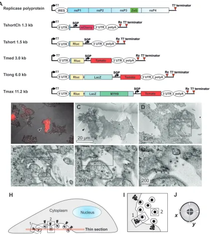
FIG 1Visualization of the replication of RNA templates by CLEM and analysis of spherule size. (A) Schematic of the SFV replicase polyprotein and template constructs used in the present study. The size of the templates, excluding poly(A), is indicated. Both the polyprotein mRNA (containing an internal ribosome entry site [IRES]) and the template RNAs (containing viral 5=and 3=untranslated regions [UTRs]) are expressed from T7 promoters. ZsG indicates the fluorescent protein ZsGreen fused with replicase protein nsP3, and SGP indicates SFV subgenomic promoter, which directs the expression of red fluorescent markers mCherry or Tomato from the template RNA. Rluc,Renillaluciferase; LacZ,-galactosidase; MYH9, myosin 9 heavy chain; Rz, hepatitis delta virus antigenomic ribozyme. (B to G) After cotransfection of polyprotein and TshortCh template constructs, CLEM technique was used to visualize the identified replication-positive cells (red in panel B, marked with an arrowhead in panels B and C) at the EM level (C to G). Boxes indicate the area shown in the subsequent higher-magnification image. (H to J) Schematic of the sectioning of cells and EM analysis of spherules. Thin sections were made horizontally starting from the bottom (H). Plentiful spherules were commonly detected on the bottom plasma membrane that was facing the cell culture dish. In these sections, most spherules are cut as indicated for spherule number 2 and therefore appear to be dissociated from the plasma membrane (I), although they are always connected to it by a neck structure above the plane of the section. In the bottom sections spherules that are cut “sideways,” like spherule number 1, and thus showing the neck, are more rare. We have not observed differences in size when spherule sizes have been measured in the two different cutting orientations. The diameter of each spherule was defined as the average of two orthogonally measured distances (J).
on November 7, 2019 by guest
http://jvi.asm.org/
[image:4.585.85.503.65.530.2]reasons for this are not understood but could be related to the lower stability of longer RNAs, since labeling was used to measure RNAs synthesized during 1 h at earlier time points, whereas Northern blotting detected the total accumulated RNAs at 16 h. Finally, it was also verified that the expression level of the replicase
remained constant when different templates were used (Fig. 3E),
indicating that the amount of the replicase proteins present did not influence the results of RNA replication levels or spherule size.
Transfection of linearized DNA.To avoid the synthesis of
ex-tended positive-strand templates by the T7 polymerase (Fig. 3D),
the template plasmids were linearized after the virus-specific se-quences, as described in Materials and Methods. Although linear DNA is not commonly used for transfection, in this particular case it worked well. Replication initiated by linearized template to-gether with circular replicase expression plasmid yielded 100- to
1,000-fold more luciferase activity than the template alone (Fig.
4A). This result is comparable to that obtained with circular
plas-mids (17) and also the overall level of activity for the most efficient
template, Tshort, was only slightly lower for linear DNA than for
circular DNA (Fig. 4A).
In Northern blotting, transfection of linear DNA only yielded a single correct-size band of T7 transcript in the absence of viral
replicase for each of the templates (Fig. 4B). However, in the
pres-ence of viral replicase, larger negative-strand bands were still
vis-ible, essentially as before (⬃10% of for Tshort) (Fig. 4B). This
suggests that the viral replicase may synthesize small amounts of concatemeric RNAs or covalently linked negative and positive strands, since similar larger RNAs were present for the
positive-strand RNAs in the presence of the replicase (Fig. 4B). Thus, the
larger RNAs appear in the system independent of the original T7 transcripts and could not be completely eliminated.
In CLEM experiments with this transfection setup, Tshort
yielded spherules of 32⫾5.4 nm (n⫽545), Tmed 42⫾4.6 nm
(n⫽654), and Tlong 48⫾7.3 nm (n⫽839). Therefore, the,
spherules of Tmed and Tlong were unchanged compared to the
previous results (Fig. 2), whereas the spherules of Tshort
ap-peared slightly smaller than before, and smaller than those
pro-duced by Tmed (Fig. 4C). Thus, these results confirmed that
template RNA length is a crucial determinant of spherule size. They also suggested further experimental approaches to exam-ine the types of RNAs incorporated into a forming spherule (see Discussion).
Generation of two sizes of spherules in the same cell.The
influence of RNA size was further corroborated by analyzing cells transfected with SFV replicons. These are RNAs
replicat-ing incisthat lack the structural open reading frame and are
thus intermediate in size between SFV genome and the long
trans-replication template (Fig. 5A). For the replicon alone, 2,890 spherules were analyzed, and their average size was found
to be 56 ⫾ 8 nm. This value was larger than expected and
relatively close to that produced by the full-length genome of SFV. However, the replicon experiment was carried out in BHK cells in contrast to all of the previous experiments in BSR cells. We therefore measured the replicon spherules again in BSR
cells and obtained a value of 49.5⫾2.9 nm (n⫽749), which
was intermediate between Tlong and Tmax, as expected. Sim-ilarly, SFV itself produced slightly larger spherules in BHK cells than in BSR cells (data not shown). This difference between the
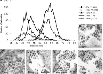
FIG 2Spherule sizes induced by different lengths of replicating RNA. (A) Distribution of spherule diameters for different lengths of RNA templates. The number of spherules detected having a particular diameter is plotted as a histogram for each of the indicated templates. (B to E) Representative images of spherules during SFV infection (B), and during Tmax (C), Tlong (D), Tmed (E), and Tshort (F) replication. All of the images are in the same scale; the template is indicated for each panel.
on November 7, 2019 by guest
http://jvi.asm.org/
[image:5.585.111.474.71.331.2]cell lines appeared consistently but, due to the small magnitude of the effect, it was not pursued further. It could be due to biological differences (e.g., lipid composition) causing a differ-ence in actual spherule size or to different behaviors of the structures under EM fixation and staining conditions in the two cell lines.
In three independent cotransfection experiments of SFV-based replicon RNA, together with Tmed template RNA in BHK cells,
4,746 spherules were studied. In this system, bothcis-replication
of replicon andtrans-replication of medium size template take
place in many of the transfected cells (Fig. 5BandC). The
corep-lication gave rise to two clearly distinct populations of spherules with different diameters, indicating that two sizes of spherules
could be produced in the cells at the same time (Fig. 5DandE).
The diameters of the two populations closely corresponded to
those observed for each of the templates independently (Fig. 2E
and5F). This experiment also supported the high efficiency of
trans-replication for the Tmed template, since it produced
ap-proximately as many spherules as thecis-replication of the
repli-con RNA in cells replicating both RNAs. A further interesting
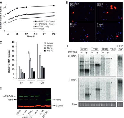
FIG 3Replication of different RNA templates. (A) Replication efficiency of genome-size construct (Tmax) was studied with Rluc assay. Triplicate samples were analyzed at the indicated time points. Mock transfection, Tmax alone and Tmed replication served as controls. (B) Efficiency of replication as analyzed by fluorescence. BSR T7/5 cells transfected with the indicated template together with the polyprotein were analyzed for replication, as indicated by Cherry/Tomato signal at 16 h (other templates) or 24 h (Tmax) posttransfection. The percentage of positive cells is indicated. The data are from one of multiple, reproducible experiments. Nuclei have been stained blue with DAPI (4=,6=-diamidino-2-phenylindole). (C) RNA synthesis levels were analyzed by labeling infected (wtSFV), control (mock), or transfected cells for 1 h, ending at the time points indicated with3[H]uridine in the presence of actinomycin D. Tshort, Tmed, or Tlong
templates were transfected with the viral polyprotein. After subtraction of mock background, the radioactive counts in precipitated RNA were divided by the number of uridine residues in each RNA to visualize the relative numbers of labeled RNAs. (D) Northern blot of positive-sense and negative-sense RNAs in the presence or absence of replicase polyprotein as indicated. RNA from SFV-Rluc-infected cells (in the same blot) is shown as a control. The negative-strand blot was exposed eight times longer than the positive-strand blot. The bands corresponding to the sizes of the three templates are marked with asterisks, and ethidium bromide staining of rRNAs is shown below as a loading control. (E) Western blot of the replicase proteins when the different templates were replicated. SFV-Rluc infection is shown as a control. The arrows indicate nsP4, nsP3 (during virus infection), nsP3-ZsG fusion (produced duringtrans-replication), and-actin loading control.
on November 7, 2019 by guest
http://jvi.asm.org/
[image:6.585.83.504.64.466.2]observation was that the two sizes of spherules were intermixed
with each other (Fig. 5E) and not concentrated to separate regions
of the cell.
DISCUSSION
We have shown here that the size of the membranous replication complex, usually called a spherule, induced by SFV strongly cor-relates with the length of the template RNA. The viral RNA of 11.5
kb replicating normally incisand atrans-replication template of
similar size both yielded spherules of⬃58 nm in diameter (Fig.
2A). It is currently unknown whether an even longer RNA would
yield larger spherules, since thetrans-replication of Tmax was only
observed in few cells, probably due to limitations of transfection efficiency and/or RNA stability, and thus experiments with longer RNAs would require further technical advances. At the other
ex-treme, we consistently observed spherules of⬃39 nm in diameter
for Tmed template of 3 kb (Fig. 2A). The smallest spherules thus
far were observed with the linearized Tshort template, giving a
diameter of⬃32 nm (Fig. 4C). The definite recognition of very
small spherules is challenging, which may limit our observations at the smaller end of scale.
The current results are in direct contrast to what has been ob-served for two other positive-strand RNA viruses, FHV and BMV
(14,16), indicating that the formation of SFV spherules is a
flexi-ble process that utilizes the replicating RNA in a different manner than the two other viruses. FHV and SFV are similar in that for both of them spherule formation requires the replicase proteins,
RNA, and an active polymerase (16,17). However, for FHV it has
been demonstrated that the spherule size is independent of the
length of the RNA template varying over a 10-fold range (16), and
thus far only one size class of spherules has been observed.
In BMV the replicase protein 1a alone can form spherules (14),
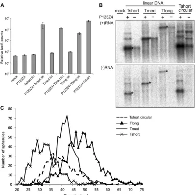
FIG 4trans-Replication initiated by transfection of linear DNA. (A) The indicated templates, with or without replicase expression plasmids, were transfected to BSR cells, and triplicate samples were analyzed at 16 h posttransfection for luciferase activity. Lin, linearized plasmid. (B) Northern blot of positive-sense and negative-sense RNAs in the presence or absence of replicase polyprotein as indicated. The negative-strand blot was exposed four times longer than the positive-strand blot. The bands corresponding to the sizes of the three templates are marked with asterisks. Tshort circular is included as a control. (C) Distribution of spherule diameters for different lengths of RNA templates. The number of spherules detected having a particular diameter is plotted as a histogram for each of the indicated templates. Tshort circular data fromFig. 2Ais included for comparison and marked with a dotted line.
on November 7, 2019 by guest
http://jvi.asm.org/
[image:7.585.93.493.66.464.2]and therefore it is reasonable that the properties of this protein determine the main features of the process, and the viral RNA appears to have no effect. Interestingly, smaller spherules have been observed in the BMV system with some 1a protein mutants, most having alterations in the membrane binding alpha helix of 1a
(24,25). Deletion of the genes for host reticulon proteins that are
components of the BMV spherules, and reduction in the amounts of cellular unsaturated fatty acids also gave rise to smaller
spher-ules (26,27). In several cases, the small spherule phenotype also
dramatically reduced viral RNA synthesis, suggesting that altered spherule assembly could lead to functional defects. In the case of the SFV spherules observed here, the replication of small RNAs in
smaller spherules proceeded very efficiently (Fig. 3), indicating
that they were fully competent for RNA replication.
It should be noted that both BMV and FHV have segmented genomes, with the segment sizes varying between 2.1 to 3.2 kb and 1.4 to 3.1 kb, respectively. During virus multiplication, each of the segments has to be replicated efficiently and packed to progeny particles. Based on volume calculations of the spherules, it has been speculated that several small segments might be
incorpo-rated into a single spherule simultaneously (15). In contrast, the
SFV genome is ca. 11.5 kb in length and, based on similar consid-erations, we hypothesize that SFV is likely to have only one copy of the negative strand within a spherule. Since SFV spherules arise
during active RNA synthesis (17) (seeFig. 6for models), the
form-ing spherule will contain the initial positive-strand template and the nascent/completed negative strand. Therefore, it could be the length or the positive strand or the combined length of the positive and negative strands that determines the spherule size. Interest-ingly, our results tentatively suggest a different size distribution for Tshort spherules when the initial mix of positive strands
con-tained also 3=-extended RNA species produced by T7 polymerase
(slightly larger spherule sizes overall) versus when only proper
Tshort-sized RNAs were initially present (linearized Tshort
tem-plates produced slightly smaller spherules) (Fig. 4C). To confirm
and extend this result regarding the size of the initial template, we are currently studying the replication competence, negative strands and spherules produced by “short” positive-strand tem-plates that do not contain proper Tshort-sized RNAs but consist
only of 3=-extended RNA species.
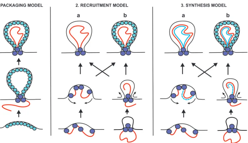
At least three different models can be proposed for the
forma-tion of spherules in relaforma-tion to the template RNA (Fig. 6): (i) in the
packaging model, RNA template is packaged to spherules, which have been preformed by viral protein components; (ii) in the re-cruitment model, spherules are formed by RNA and protein to-gether at the stage of RNA recruitment preceding replication; and (iii) in the synthesis model, the formation of spherules takes place concomitantly with RNA replication. Further variation in these models can be introduced by the presence or absence of an inner protein shell and by the nature of the intermediate stages during
formation (Fig. 6). For BMV, the packaging model has been
pro-posed as a possibility (16), whereas both FHV and SFV need an
active polymerase and appear to represent variants of the synthesis model. In the recruitment and synthesis models, RNA could the-oretically determine the size of the spherule, if the mechanism for spherule formation is flexible in this regard. For FHV, the size of
the spherules remains fixed (16) and might be determined, for
instance, by the packing of the replication proteins within the spherules.
It has been proposed, with some good evidence from EM la-beling and from calculations based on purified membrane
prepa-rations, that BMV and FHV spherules contain⬎100 copies of the
major virus-encoded replicase component, which could form an
inner shell that stabilizes the spherule (14,15). This shell could
also include host proteins, such as reticulons for BMV or
am-phiphysins for SFV (26,28). In the case of SFV, there is no
infor-FIG 5Two sizes of spherules detected in the same cell. (A) Schematic of the SFV replicon and Tmed RNAs prepared byin vitrotranscription. (B and C) The two RNAs were cotransfected, and cells expressing both red and green fluorescence (filled arrowheads), indicating the replication of both Tmed and the replicon, were chosen for spherule measurements after CLEM. The cell population also contains cells expressing green fluorescence only (open arrowhead) derived from the replicon but lacking Tmed replication. (D) The distribution of spherule diameters in cells replicating both RNAs was plotted as a histogram. (E) Larger (white arrowhead) and smaller (black arrowhead) spherules visualized in the same EM field from a cell replicating the two RNAs. (F) Spherules produced by the transfection of replicon RNA alone were uniform in size and corresponded to the larger spherules.
on November 7, 2019 by guest
http://jvi.asm.org/
[image:8.585.72.517.69.269.2]mation regarding the presence of a protein shell. Interestingly, the formation of membrane invaginations without an inner coating is catalyzed by the endosomal sorting complex required for trans-port (ESCRT) involved in the formation of multivesicular bodies
(MVBs) (29). The intermediates in the formation of the inner
vesicles in MVBs have a strong resemblance to spherules, and their sizes in mammalian cells vary between 40 and 100 nm, with an
average of 56 nm (30). There is evidence from tomato bushy stunt
virus, another spherule-generating plant virus (31), that ESCRT
proteins are crucial for the formation of viral replication
com-plexes (32). The ESCRT proteins are also involved in retrovirus
budding during particle production (33), which therefore might
share some common features with spherule formation, with the crucial difference that the spherules remain attached to mem-branes via the neck structure and do not pinch off.
The intermediate stages of spherule formation (Fig. 6) have
thus far not been detected (1), which might be due to their
tran-sient nature. Currently, we are addressing the question of inter-mediates by analyzing early time points from a very
high-multi-plicity SFV infection (10) using electron tomography. An
interesting hypothesis for further study is that the SFV RNA could act as a structural component within the spherule in such a way that the inclusion of the entire RNA within the forming spherule would contribute to the determination of its size. The size deter-mination could be effected through the binding of multiple copies of proteins to the RNA or, given the requirement for an active polymerase, it could be connected to the completed replication of the entire RNA.
ACKNOWLEDGMENTS
We thank Mervi Lindman and Arja Strandell for excellent technical assis-tance in EM.
This study was supported by the Academy of Finland (grant 127214)
and the Sigrid Jusélius Foundation. K.K. was supported in part by a fel-lowship from the Helsinki Graduate Program in Biotechnology and Mo-lecular Biology, and K.H. was supported by an Academy of Finland post-doctoral fellowship.
REFERENCES
1.den Boon JA, Ahlquist P.2010. Organelle-like membrane compartmen-talization of positive-strand RNA virus replication factories. Annu. Rev. Microbiol.64:241–256.
2.Belov G, van Kuppeveld FJM.2012. (⫹)RNA viruses rewire cellular pathways to build replication organelles. Curr. Opin. Virol.2:740 –747. 3.Salonen A, Ahola T, Kääriäinen L.2005. Viral RNA replication in
asso-ciation with cellular membranes. Curr. Top. Microbiol. Immunol.285:
139 –173.
4.Spuul P Salonen A, Merits A, Jokitalo E, Kääriäinen L, Ahola T.2007. Role of the amphipathic peptide of Semliki Forest virus replicase protein nsP1 in membrane association and virus replication. J. Virol.81:872– 883. 5.Miller S, Krijnse-Locker J.2008. Modification of intracellular membrane
structures for virus replication. Nat. Rev. Microbiol.6:363–374. 6.Griffin DE.2007. Alphaviruses, p 1023–1067.InKnipe DM, Howley PM
(ed), Fields virology, 5th ed, vol 1. Lippincott/The Williams & Wilkins Co, Philadelphia, PA.
7.Kääriäinen L, Ahola T.2002. Functions of alphavirus nonstructural pro-teins in RNA replication. Prog. Nucleic Acids Res. Mol. Biol.71:187–222. 8.Grimley PM, Berezesky IK, Friedman RM.1968. Cytoplasmic structures associated with an arbovirus infection: loci of viral ribonucleic acid syn-thesis. J. Virol.2:1326 –1338.
9.Kujala P Ikäheimonen A, Ehsani N, Vihinen H, Auvinen P, Kääriäinen L.2001. Biogenesis of the Semliki Forest virus RNA replication complex. J. Virol.75:3873–3884.
10. Spuul P, Balistreri G, Kääriäinen L, Ahola T.2010. Phosphatidylinositol 3-kinase-, actin-, and microtubule-dependent transport of Semliki Forest virus replication complexes from the plasma membrane to modified lyso-somes. J. Virol.84:7543–7557.
11. Frolova EI, Gorchakov R, Pereboeva L, Atasheva S, Frolov I. 2010. Functional Sindbis virus replicative complexes are formed at the plasma membrane. J. Virol.84:11679 –11695.
12. Welsch S, Miller S, Romero-Brey I, Merz A, Bleck CK, Walther P, Fuller SD, Antony C, Krijnse-Locker J, Bartenschlager R.2009. Composition
FIG 6Three classes of models for spherule formation: packaging, recruitment, and synthesis models. The mature spherules, depicted at the top, could either contain an inner shell (variant b) or be devoid of the shell (variant a). Intermediates in spherule formation could potentially include “open” dome-like structures (variant a, middle row) or “closed” structures already containing a neck-like constriction (variant b). Positive-strand RNA is shown in red, and negative-strand RNA is shown in blue. Spheres represent proteins that could be located at the neck and/or form an inner shell. See the text for further discussion.
on November 7, 2019 by guest
http://jvi.asm.org/
[image:9.585.89.503.62.301.2]and three-dimensional architecture of the dengue virus replication and assembly sites. Cell Host Microbe5:365–375.
13. Gillespie LK, Hoenen A, Morgan G, Mackenzie JM.2010. The endo-plasmic reticulum provides the membrane platform for biogenesis of the flavivirus replication complex. J. Virol.84:10438 –10447.
14. Schwartz M Chen J, Janda M, Sullivan M, den Boon J, Ahlquist P.2002. A positive-strand RNA virus replication complex parallels form and func-tion of retrovirus capsids. Mol. Cell9:505–514.
15. Kopek BG, Perkins G, Miller DJ, Ellisman MH, Ahlquist P. 2007. Three-dimensional analysis of a viral RNA replication complex reveals a virus-induced mini-organelle. PLoS Biol. 5:e220. doi:10.1371/journal .pbio.0050220.
16. Kopek BG, Settles EW, Friesen PD, Ahlquist P. 2010. Nodavirus-induced membrane rearrangement in replication complex assembly re-quires replicase protein A, RNA templates, and polymerase activity. J. Virol.84:12492–12503.
17. Spuul P Balistreri G, Hellström K, Golubtsov AV, Jokitalo E, Ahola T.
2011. Assembly of alphavirus replication complexes from RNA and pro-tein components in a noveltrans-replication system in mammalian cells. J. Virol.85:4739 – 4751.
18. Buchholz UJ, Finke S, Conzelmann KK. 1999. Generation of bovine respiratory syncytial virus (BRSV) from cDNA: BRSV NS2 is not essential for virus replication in tissue culture, and the human RSV leader region acts as a functional BRSV genome promoter. J. Virol.73:251–259. 19. Pohjala L, Barai V, Azhayev A, Lapinjoki S, Ahola T.2008. A
luciferase-based screening method for inhibitors of alphavirus replication applied to nucleoside analogues. Antivir. Res.78:215–222.
20. Tarn WY, Yario TA, Steitz JA.1995. U12 snRNA in vertebrates: evolu-tionary conservation of 5=sequences implicated in splicing of pre-mRNAs containing a minor class of introns. RNA1:644 – 656.
21. Tarn WY, Steitz JA.1996. A novel spliceosome containing U11, U12, and U5 snRNPs excises a minor class (AT-AC) intron in vitro. Cell84:801– 811.
22. Puhka M, Vihinen H, Joensuu M, Jokitalo E.2007. Endoplasmic retic-ulum remains continuous and undergoes sheet-to-tubule transformation during cell division in mammalian cells. J. Cell Biol.179:895–909.
23. Macdonald LE, Zhou Y, McAllister WT.1993. Termination and slippage by bacteriophage T7 RNA polymerase. J. Mol. Biol.232:1030 –1047. 24. Wang X, Lee WM, Watanabe T, Schwartz M, Janda M, Ahlquist P.
2005. Brome mosaic virus 1a nucleoside triphosphatase/helicase domain plays crucial roles in recruiting RNA replication templates. J. Virol.79:
13747–13758.
25. Liu L, Westler WM, den Boon JA, Wang X, Diaz A, Steinberg HA, Ahlquist P.2009. An amphipathic alpha-helix controls multiple roles of brome mosaic virus protein 1a in RNA replication complex assembly and function. PLoS Pathog.5:e1000351. doi:10.1371/journal.ppat.1000351. 26. Diaz A, Wang X, Ahlquist P.2010. Membrane-shaping host reticulon
proteins play crucial roles in viral RNA replication compartment for-mation and function. Proc. Natl. Acad. Sci. U. S. A.107:16291–16296. 27. Zhang J, Diaz A, Mao L, Ahlquist P, Wang X.2012. Host acyl coenzyme A binding protein regulates replication complex assembly and activity of a positive-strand RNA virus. J. Virol.86:5110 –5121.
28. Neuvonen M, Kazlauskas A, Martikainen M, Hinkkanen A, Ahola T, Saksela K. 2011. SH3 domain-mediated recruitment of host cell am-phiphysins by alphavirus nsP3 promotes viral RNA replication. PLoS Pathog.7:e1002383. doi:10.1371/journal.ppat.1002383.
29. Wollert T, Hurley JH.2010. Molecular mechanism of multivesicular body biogenesis by ESCRT complexes. Nature464:864 – 869.
30. Murk JLAN, Humbel BM, Ziese U, Griffith JM, Posthuma G, Slot JW, Koster AJ, Verkleij AJ, Geuze HJ, Kleijmeer MJ. 2003. Endosomal compartmentalization in three dimensions: implications for membrane fusion. Proc. Natl. Acad. Sci. U. S. A.100:13332–13337.
31. McCartney AW, Greenwood JS, Fabian MR, White KA, Mullen RT.
2005. Localization of the tomato bushy stunt virus replication protein p33 reveals a peroxisome-to-endoplasmic reticulum sorting pathway. Plant Cell17:3513–3531.
32. Barajas D, Jiang Y, Nagy PD.2009. A unique role for the host ESCRT proteins in replication of tomato bushy stunt virus. PLoS Pathog.
5:e1000705. doi:10.1371/journal.ppat.1000705.
33. Carlton JG, Martin-Serrano J.2009. The ESCRT machinery: new func-tions in viral and cellular biology. Biochem. Soc. Trans.37:195–199.