Role in Determining the Preferential CD4
ⴙT Cell Immortalization/
Transformation Tropism
Priya Kannian,a,bSoledad Fernandez,d,eKathryn S. Jones,fPatrick L. Greena,b,c,d
Center for Retrovirus Research, The Ohio State University, Columbus, Ohio, USAa
; Department of Veterinary Biosciences, The Ohio State University, Columbus, Ohio, USAb ; Department of Molecular Virology, Immunology, and Medical Genetics, The Ohio State University, Columbus, Ohio, USAc
; Comprehensive Cancer Center and Solove Research Institute, The Ohio State University, Columbus, Ohio, USAd
; Center for Biostatistics, The Arthur James Cancer Hospital and Research Institute, The Ohio State University, Columbus, Ohio, USAe
; Basic Science Program, SAIC-Frederick, Inc., Frederick National Laboratory for Cancer Research, Frederick, Maryland, USAf
Human T lymphotropic virus type 1 (HTLV-1) mainly causes adult T cell leukemia and predominantly immortalizes/transforms
CD4ⴙT cells in culture. HTLV-2 is aleukemic and predominantly immortalizes/transforms CD8ⴙT cells in culture. We have
shown previously that the viral envelope is the genetic determinant of the differential T cell tropism in culture. The surface com-ponent (SU) of the HTLV-1 envelope is responsible for binding to the cellular receptors for entry. Here, we dissect the HTLV-1 SU further to identify key domains that are involved in determining the immortalization tropism. We generated HTLV-1 enve-lope recombinant virus containing the HTLV-2 SU domain. HTLV-1/SU2 was capable of infecting and immortalizing freshly
isolated peripheral blood mononuclear cells in culture. HTLV-1/SU2 shifted the CD4ⴙT cell immortalization tropism of
wild-type HTLV-1 (wtHTLV-1) to a CD8ⴙT cell preference. Furthermore, a single amino acid substitution, N195D, in HTLV-1 SU
(Ach.195) resulted in a shift to a CD8ⴙT cell immortalization tropism preference. Longitudinal phenotyping analyses of thein
vitrotransformation process revealed that CD4ⴙT cells emerged as the predominant population by week 5 in wtHTLV-1
cul-tures, while CD8ⴙT cells emerged as the predominant population by weeks 4 and 7 in wtHTLV-2 and Ach.195 cultures,
respec-tively. Our results indicate that SU domain independently influences the preferential T cell immortalization tropism irrespective of the envelope counterpart transmembrane (TM) domain. We further showed that asparagine at position 195 in HTLV-1 SU is
involved in determining this CD4ⴙT cell immortalization tropism. The slower emergence of the CD8ⴙT cell predominance in
Ach.195-infected cultures suggests that other residues/domains contribute to this tropism preference.
H
uman T lymphotropic virus type 1 (HTLV-1) and type 2 (HTLV-2) are complex retroviruses that share a genome structure (1). In addition to the structural proteins (Gag, Pol, Pro, and Env), they encode regulatory proteins (Tax and Rex) and accessory proteins, including an antisense protein, HBZ (HTLV-1) or APH-2 (HTLV-2) (2–5). Despite their closely re-lated genomic structures, HTLV-1 and HTLV-2 display distinct pathogenic properties. HTLV-1 causes adult T cell leukemia (ATL), HTLV-1-associated myelopathy/tropical spastic parapare-sis (HAM/TSP), and some noninflammatory disorders (6–9). HTLV-2 does not cause leukemia and has been associated with a HAM/TSP-like neurological disease only infrequently (10–12). Another feature that differentiates HTLV-1 and HTLV-2 is the ability to predominantly immortalize (interleukin-2 [IL-2]-de-pendent growth) or transform (IL-2-inde[IL-2]-de-pendent growth) CD4⫹ and CD8⫹T cells, respectively, in culture (13–15). Thein vitroimmortalization/transformation preference for CD4⫹T cells by HTLV-1 is recapitulatedin vivo, as ATL is a CD4⫹T cell leukemia. However, due to its nonleukemogenic nature, HTLV-2-induced CD8⫹T cell immortalization/transformation is primarily anin vitrophenomenon.
We have previously shown that, although the viral Tax protein is indispensable for viral replication and cellular transformation, the preferential immortalization or transformation tropism of HTLV-1 and HTLV-2 is determined by the viral envelope (14,15). Since the primary function of the viral envelope is to facilitate entry into new target cells, it was hypothesized that the cellular receptor complex requirements for HTLV-1 and HTLV-2 could
be different. Subsequently, a number of studies reported that HTLV-1 and HTLV-2 slightly differ in their requirement of host cellular receptors. HTLV-1 requires heparan sulfate proteoglycans (HSPGs) and neuropilin-1 (NRP-1) for initial binding and glu-cose transporter-1 (GLUT-1) for subsequent membrane fusion and entry. Although HTLV-2 shares NRP-1 and GLUT-1 with HTLV-1 for both binding and entry, HSPGs interfere with HTLV-2 binding (16–19). Therefore, together these findings sug-gested a potential role for the viral envelope in mediating prefer-ential T cell transformation, probably at the stage of virus binding to the host cell receptor.
The viral envelope is generated as a polyprecursor protein (gp61) comprised of 488 amino acids which is cleaved into the surface domain (SU-gp46) and transmembrane domain (TM-gp21) (20,21). SU binds to the cellular receptor(s), and then SU and TM undergo significant conformational remodeling, thereby exposing TM to facilitate membrane fusion and subsequent entry into the cell. Functional mapping analysis of the HTLV-1 SU using soluble SU fusion proteins andin vitrobinding assays revealed that the C terminus of the HTLV-1 SU (SU1) binds to the CD4⫹T cells
Received19 April 2013Accepted13 June 2013
Published ahead of print19 June 2013
Address correspondence to Patrick L. Green, [email protected].
Copyright © 2013, American Society for Microbiology. All Rights Reserved.
doi:10.1128/JVI.01079-13
on November 7, 2019 by guest
http://jvi.asm.org/
(22); it resulted in active infection and immortalization of freshly isolated peripheral blood mononuclear cells (PBMCs) in vitro
(23); and the virus efficiently infected and persisted in rabbits (24). However, rabbits infected with the N195D mutant virus ex-hibited a weaker humoral response to SU1 antigen than the rab-bits infected with wild-type HTLV-1 (wtHTLV-1). Additionally, one rabbit infected with the N195D mutant virus generated a strong antibody response to the SU2 antigen. Taken together, these results suggest that the N195D mutation in SU1 could ex-hibit certain biological propertiesin vivothat are similar to those of the HTLV-2 envelope.
In this study, we further dissected the role of HTLV-1 SU in the distinctin vitroimmortalization/transformation tropism charac-teristics. We first generated and characterized recombinant HTLV-1 provirus containing the SU region of wtHTLV-2 enve-lope (HTLV-1/SU2). HTLV-1/SU2 actively replicated in freshly isolated PBMCs, and additionally, HTLV-1/SU2 predominantly immortalized CD8⫹T cells similarly to wtHTLV-2. This suggests that the viral envelope domain SU contributes to the preferential immortalization tropism. We further showed that the N195D substitution in the immunodominant epitope of HTLV-1 SU (Ach.195) also shifted the immortalization tropism toward a CD8⫹T cell preference. This finding indicates that HTLV-1 me-diates the preferential CD4⫹ T cell immortalization tropism through the envelope SU protein and that residue 195 plays a critical role in this preference.
MATERIALS AND METHODS
Cells.729Achneo and 729pH6neo are stable wtHTLV-1 and wtHTLV-2 producer cell lines that were generated and characterized previously and designated 729.HTLV-1 and 729.HTLV-2 (14). The parental 729 B cell line was used as the negative control. All 729 parental and derivative cell lines were maintained in Iscove’s Dulbecco’s minimum essential medium (Cellgro; Mediatech, Manassas, VA). Ach.95 and Ach.195 (a generous gift from Lee Ratner) are human T cell lines that stably express HTLV-1 con-taining an asparagine-to-aspartic acid mutation at residues 95 and 195 of the Env coding sequence, respectively. Ach.95-, Ach.195-, wtHTLV-1-, and wtHTLV-2-producing T cell lines were maintained in RPMI 1640 (Gibco-Invitrogen, Grand Island, NY) and 10 U/ml recombinant human interleukin-2 (rhIL-2; Roche Applied Biosciences, Indianapolis, IN). All media were supplemented to contain 10% fetal bovine serum (Gemini Bio-Products, Sacramento, CA), 2 mM glutamine, penicillin (100 U/ml), and streptomycin (100g/ml; Gibco-Invitrogen, Grand Island, NY). Hu-man peripheral blood mononuclear cells (PBMCs) were isolated from the blood of normal donors by centrifugation over Ficoll-Paque (GE Life Sciences, Piscataway, NY) and were cocultured with irradiated virus pro-ducer cell lines in RPMI 1640 medium supplemented with 20% fetal bo-vine serum (FBS), 2 mM glutamine, antibiotics, and 10 U/ml rhIL-2.
729 B cells using a nucleofection V kit (Amaxa; Lonza, AG, Cologne, Germany) according to the manufacturer’s instructions. Stable transfec-tants containing the desired proviral clones were isolated following incu-bation in 24-well culture plates in medium containing 1 mg/ml of Gene-ticin. After 4 to 5 weeks of selection, viable cells were expanded and maintained in culture for further analysis. The clones were screened for p19 Gag expression in the cell supernatants by an enzyme-linked immu-nosorbent assay (ELISA) (Zeptometrix; Buffalo, NY) per the manufactur-er’s instructions.
PCR-based restriction enzyme digestion.Genomic DNA was ex-tracted from the stable virus producer cell lines using a DNeasy kit (Qia-gen, Valencia, CA) per the manufacturer’s instructions. The primers de-signed to amplify a 1.8-kb product containing the envelope region are Achneo4974(S) (49745=-CATTGGTATTATTTCAAGCTTC4995-3=) and
Achneo6827(AS) (68275=-AGGAAAGAAAAAATGCAGGAGT6848-3=) for wtHTLV-1 and pH6neo4974(S) (49745=-CAAATGGTTCTATTATAAAC
TC4995-3=) and pH6neo6827(AS) (68275=-TTCAGGGTTATGTGGATTT
C6846-3=) for wtHTLV-2. DNA was subjected to PCR performed with a
50-l mixture containing a 160 nM concentration of each of the corre-sponding primer pairs. The cycling profile was a single cycle of 94°C for 10 min followed by 40 cycles of 94°C for 1 min, annealing at 50°C for Achneo primers or 47°C for pH6neo primers for 1 min and 72°C for 1 min, and a single-cycle final extension at 72°C for 5 min. The amplified product was digested with NcoI, and the fragments were resolved on a 1% agarose gel containing ethidium bromide.
Immortalization assay.Approximately 1 million gamma-irradiated (10,000 rads) 729 (negative control), 729.HTLV-1, 729.HTLV-2, 729.HTLV-1/SU2, Ach.95, or Ach.195 cells (the number of virus producer cells was normalized based on p19 Gag output as measured by ELISA) were cocultured with 2⫻106freshly isolated normal human PBMCs in
24-well plates for 8 weeks with weekly changes of media. The cultures were monitored by measuring viability using trypan blue exclusion and p19 Gag production using ELISA on a weekly basis. After 8 weeks, the cultures were harvested and stained using fluorescein isothiocya-nate (FITC)-conjugated anti-human CD3 (clone UCHT1), allocyanin (APC)-conjugated anti-human CD4 (clone RPA-T4), and phyco-erythrin (PE)-conjugated anti-human CD8 (clone HIT8a) antibodies (BD Biosciences, San Jose, CA) and analyzed by flow cytometry (14).
Internalization assay.Studies to examine the internalization of HTLV virions were performed essentially as described previously (27) with the following modifications. Following incubation with virus-containing su-pernatants from HTLV-1-producing cells or uninfected controls for 2 h at 37°C, CD4⫹T cells were fixed and permeabilized using commercially available reagents (BD Cytofix/Cytoperm fixation/permeabilization kit; BD Biosciences). The amount of internalized virions was determined us-ing an antibody directed against the HTLV-1 p19 protein (TP-7; Zep-tometrix) as previously described (27).
Statistical analysis. The means of the normalized percentages of CD4⫹T cells and CD8⫹T cells from the wt1- versus the HTLV-1/SU2-immortalized cultures, as well as wtHTLV-1- versus
on November 7, 2019 by guest
mortalized cultures, were compared for significant differences using analysis of variance (ANOVA). The normalized percentages of the proliferating T cells at each week in the wtHTLV-1-, Ach.95-, and Ach.195-immortalized cultures were compared using a generalized linear model. This model included the following factors: virus group (wtHTLV-1 or Ach.95/Ach.195), time, and the interaction of the two main effects. TheP values of the pairwise comparisons versus the control (wtHTLV-1) at each week were adjusted by Dunnett’s method.
RESULTS
Generation and characterization of the recombinant HTLV-1/
SU2 provirus.Previously, using recombinant viruses in which the
env sequences were exchanged between HTLV-1 and HTLV-2, we observed that the viral envelope confers the distinctin vitro im-mortalization/transformation tropism (14). Subsequently, Jones et al. (18) reported that HTLV-1 and HTLV-2 differ slightly in their host cell receptor requirement for initial binding by SU, al-though they share a receptor to facilitate fusion and entry. Fur-thermore, the same group also showed that the C terminus of the HTLV-1 SU (SU1) is essential for efficient binding to CD4⫹T cells (18). The amino acid homology between HTLV-1 Env and
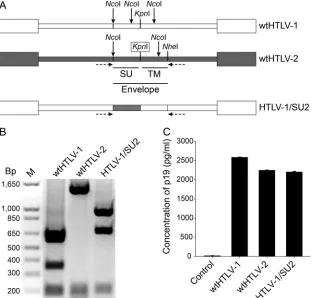
HTLV-2 Env is approximately 63% for the SU glycoprotein. To further dissect the role of the SU region in the distinctin vitroT cell immortalization tropism, we constructed an HTLV-1 recombi-nant that contains SU2 from wtHTLV-2 (HTLV-1/SU2;Fig. 1A). In HTLV-1, since the open reading frames ofenvandHbzdo not overlap, the recombinations in the envelope region do not affect HBZ. Additionally, the poly(A) signal and site are in similar loca-tions forHbzandAph-2, at the 5=end of SU1 and SU2, respec-tively. To determine the ability of recombinant proviral clones to replicate, synthesize viral proteins, and induce cellular immortal-ization, permanent 729 B cell transfectants expressing wt and re-combinant proviral clones were generated and further character-ized. Each of the stable transfectants contained complete copies of the provirus (data not shown), and the presence of the expected
envsequences was confirmed by DNA PCR followed by restriction digestion (Fig. 1B). To monitor the production of viral protein in these stable transfectants, the concentration of p19 Gag in the culture supernatants was quantified by ELISA. As shown inFig. 1C, repre-sentative stable cell clones selected for this study had p19 Gag expres-sion levels similar to those of the wt virus producer cell lines. FIG 1Generation and characterization of the recombinant HTLV-1/SU2 provirus. (A) Genomic organization of the wild-type and recombinant proviral clones used in this study. White and gray boxes indicate wtHTLV-1 and wtHTLV-2 origin, respectively. The envelope region, along with the individual SU domain as well as the specific restriction enzymes used to construct the recombinants, is shown. The missing restriction enzyme site that was generated by site-directed mutagenesis is boxed. The multiple NcoI sites throughout the envelope region are shown by arrows. The horizontal arrows below the proviruses denote PCR primer positions. (B) Recombinant HTLV-1/SU2 provirus contained the exchanged envelope sequences. Approximately 1.8 kb of the viral genome encompass-ing the envelope region was amplified usencompass-ing a PCR oligonucleotide primer pair (shown in panel A) and the genomic DNA extracted from 729 cell lines producencompass-ing wtHTLV-1, wtHTLV-2, or HTLV-1/SU2 viruses. The amplified products were digested with NcoI, and the fragments were resolved by 1% agarose gel electro-phoresis and visualized with ethidium bromide. The expected fragments were observed as follows: for wtHTLV-1, 693 bp, 614 bp, 361 bp, and 205 bp; for wtHTLV-2, 1,491 bp, 205 bp, and 157 bp; and for HTLV-1/SU2, 963 bp, 693 bp, and 204 bp. (C) Viral protein production from the wild-type and recombinant virus producer cell lines. One million cells per virus producer cell line or the negative 729B cell line were cultured in 24-well plates for 72 h in triplicate. The culture supernatant from each cell line was collected and tested for p19 Gag output using a commercial ELISA kit. The bars depict the average values of the results from three independent experiments, and the error bars depict the standard deviations.
on November 7, 2019 by guest
http://jvi.asm.org/
[image:3.585.137.450.68.366.2]HTLV-1 recombinants containing HTLV-2 SU exhibited a
shift in the immortalization tropism to CD8ⴙT cells.We next
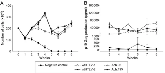
determined whether the recombinant viruses had the ability to immortalize human PBMCs using a well-established cocultiva-tion immortalizacocultiva-tion assay (14). Cell number and viability were examined at weekly intervals to monitor the cellular transforma-tion process and the characteristic expansion of cells from the mixed PBMC population. A growth curve from a representative experiment is depicted inFig. 2A. When cocultured with irradi-ated uninfected 729 cells, PBMCs showed a progressive loss of viable cells over time and eventually died off approximately 5 to 6 weeks postplating. This observation is consistent with previously published reports (14, 15). In contrast, the immortalization of PBMCs was apparent in coculture wells containing producer cells of 729.HTLV-1 (wtHTLV-1), 729.HTLV-2 (wtHTLV-2), or 729.HTLV-1/SU2. This initial drop in cell numbers at week 1 in these cocultures could be attributed to HTLV-1 infection-induced apoptosis/senescence and normal cell death due to lack of anti-genic stimuli. Cell numbers and viabilities were similar for all virus-producing cells throughout the experiment. Viral replica-tion was assessed by quantitareplica-tion of p19 Gag producreplica-tion in the culture supernatant starting at 3 weeks postcocultivation. At that time point, HTLV-infected PBMCs typically produce viral parti-cles and the virion production from the residual irradiated viral producer cells becomes negligible (Fig. 2B). All cultures contain-ing wt or recombinant HTLV showed p19 Gag production. How-ever, note that this assay is principally used as a surrogate marker to ensure that the increasing proliferation of T cells (Fig. 2A) is induced by virus infection (Fig. 2B) rather than as a quantitative measure of differences in virus replication between the recombi-nant and wt viruses.
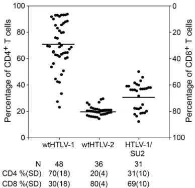
To determine if the exchange of SU sequence altered the im-mortalization tropism, we evaluated phenotypes of cells immor-talized by wtHTLV-1, wtHTLV-2, or HTLV-1/SU2. Individual wells of cells at 8 weeks postcoculture were stained with anti-CD3, anti-CD4, and anti-CD8 antibodies and analyzed by flow cytom-etry. The data from multiple wells in two independent experi-ments are depicted inFig. 3. Since HTLV-1 and HTLV-2 mainly immortalize/transform CD4⫹and CD8⫹T cells, respectively, the actively proliferating cells at the end of 8 weeks in culture most
likely represent a combination of these two T cell types. Therefore, we normalized the actual percentages of CD3⫹CD4⫹and CD3⫹ CD8⫹T cells to 100 in each of the wells analyzed. Our results revealed that wtHTLV-1 immortalized 70% CD4⫹ T cells and 30% CD8⫹T cells, while wtHTLV-2 immortalized 20% CD4⫹T
⫻
PBMCs in 24-well plates with 10 U/ml of rhIL-2. These cultures were fed with fresh media on a weekly basis. Data represent the results from a representative of two independent experiments. (A) Cell viability was determined by trypan blue exclusion using three random wells from each of the cocultures on a weekly basis. The symbols denote the averages and the error bars denote the standard deviations at each time point for each of the corresponding cocultures. (B) The active replication of HTLV in each of these cocultures was determined by measuring p19 Gag output in the supernatant using ELISA. The symbols denote the averages and the error bars denote the standard deviations in the p19 Gag levels from three random wells of the corresponding cocultures.
FIG 3Recombinant HTLV-1/SU2 shifted the CD4⫹T cell immortalization preference. The immortalization cocultures set up as described in theFig. 2
legend were harvested after 8 weeks, stained with anti-CD3, anti-CD4, and anti-CD8 antibodies, and analyzed by flow cytometry. The average actual per-centages of CD4⫹and CD8⫹T cells in the two freshly isolated PBMCs were 58% and 30%, respectively. The percentages of CD3⫹CD4⫹and CD3⫹CD8⫹ T cells in each of the outgrowing wells were determined. The percentages in each well were normalized to 100, and the percentages of CD4⫹T cells are plotted in the graph. The cultures from two independent experiments were combined and plotted. The graph shows the percentages of CD4⫹T cells in ascending order in the leftyaxis and the percentages of CD8⫹T cells in descending order in the rightyaxis for each well denoted by a symbol. The horizontal bars denote the average percentage of CD4⫹T cells immortalized by each of the viruses. The average percentages along with standard deviations (SD) and the total number of wells analyzed for each of the virus-immortalized cultures are listed below the graph. The average CD4⫹T cell percentages of HTLV-1/SU2-immortalized cultures were significantly different from that of the wtHTLV-1-immortalized cultures (P⬍0.0001; Holm-Sidak method).
on November 7, 2019 by guest
[image:4.585.145.445.65.196.2] [image:4.585.324.518.370.557.2]cells and 80% CD8⫹T cells. This finding is consistent with our previously published results (14,15). HTLV-1/SU2 immortalized 31% CD4⫹T cells and 69% CD8⫹T cells. This indicates that the SU2 component shifted the immortalization tropism of HTLV-1 from predominantly CD4⫹T cells toward a predominantly CD8⫹ T cell population, which is similar to the wtHTLV-2 data. The differential tropism results seen in comparisons of wtHTLV-1 and HTLV-1/SU2 were statistically significant (P⬍0.0001; Holm-Sidak method).
Thus, taken together, the data indicate that recombinant HTLV-1/SU2, like the parental viruses, was replication competent and was capable of inducing sustained proliferation and immor-talization of newly isolated PBMCs. However, the recombinant HTLV-1/SU2 virus shifted the immortalization tropism prefer-ence from CD4⫹to CD8⫹T cells. Therefore, SU of the viral en-velope contributes to the preferential immortalization/transfor-mation tropism of HTLV-1. Moreover, this shift in tropism also helps explain the dramatic difference in p19 Gag production levels between HTLV-1- and HTLV-1/SU2- or HTLV-2-infected PBMCs shown inFig. 2B. HTLV expression in CD8⫹T cells is generally lower than in CD4⫹T cells. Thus, the levels of expres-sion of p19 are similar for the viruses present in the expanding CD8⫹cells, and those levels are much lower than that seen with wtHTLV-1, which favors immortalization of CD4⫹T cells.
N195D substitution in the HTLV-1 SU immunodominant
domain alters T cell immortalization tropism.At position 195 of
the HTLV-1 SU domain, a conservative amino acid substitution (N195D) did not alter the function of the envelope, but a noncon-servative amino acid substitution was not tolerated. In previous studies, an HTLV-1 provirus carrying the N195D substitution (Ach.195) was able to efficiently infect and immortalize fresh PBMCsin vitro(23) and replicatein vivoin newly infected rabbits (24). However, 2 of 4 Ach.195-infected rabbits mounted an al-tered humoral response; one rabbit had no antibody response, and the other rabbit had a stronger antibody response to SU2 than to SU1 antigen. Although the altered immune response was demon-strated in a very small number of animals, it is interesting that a
single amino acid change can trigger such an altered response, suggesting an important role of this particular amino acid and position in the conformation of SU1. A similar N-to-D substitu-tion at posisubstitu-tion 95 in HTLV-1 (Ach.95) elicited a humoral re-sponse similar to that seen with the wtHTLV-1, in addition to other similarities in the biologic properties. Interestingly, the SU2 domain carries a Q at position 91 and a D at position 191, corre-sponding to positions 95 and 195 in the SU1 domain, respectively, suggesting that the HTLV-1 envelope with N195D might be more like the HTLV-2 envelope functionally.
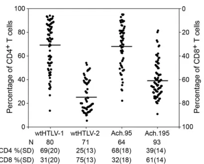
Therefore, we assessed thein vitroT cell immortalization abil-ity of Ach.95 and Ach.195. The weekly growth curves indicated that Ach.95 and Ach.195, in similarity to the parental wtHTLV-1 and the wtHTLV-2 strains, were capable of immortalizing freshly isolated PBMCs in culture (Fig. 4A). The virus-immortalized cul-tures also revealed constant p19 Gag production, confirming ac-tive viral replication in these proliferating cells (Fig. 4B). PBMCs cultured with uninfected 729 cells died by week 4 to 5 (Fig. 4A), and the supernatants did not have any detectable p19 Gag levels throughout the 8-week time course (Fig. 4B). We note in this assay that the levels of p19 Gag production in cultures infected with the wtHTLV-1 (Ach) and the Ach.95 and Ach.195 were similar, whereas HTLV-2-infected cultures expressed p19 Gag at levels that were much lower but above the level seen with the negative control. Differences in protein production levels between HTLV-1 and HTLV-2 have been reported by us and others previ-ously and are primarily attributed to Tax-1 and Tax-2 differential transactivation functions (5,28,29). The cultures were analyzed by flow cytometry to determine the CD4/CD8 phenotype of the predominantly proliferating CD3⫹T cell type (Fig. 5). Our results showed that wtHTLV-1-immortalized population was composed of 69% CD4⫹T cells and 31% CD8⫹T cells, while the wtHTLV-2-immortalized population was composed of 25% CD4⫹T cells and 75% CD8⫹T cells. Again, these results were consistent with the results presented inFig. 3and our previously published re-ports (14,15). The cell population immortalized by Ach.95, like that immortalized by wtHTLV-1, was composed of 68% CD4⫹T FIG 4HTLV-1 envelope SU domain mutant viruses Ach.95 and Ach.195 were capable of infecting and immortalizing fresh PBMCs in culture. Approximately 106virus producer cells (the number of cells was normalized to the equivalent p19 Gag output) or 729B negative-control cells were irradiated (10,000 rads) and
cocultured with 2⫻106freshly isolated human PBMCs in 24-well plates for 8 weeks with 10 U/ml of rhIL-2. These cultures were fed with fresh media on a weekly
basis. Data are representative of three independent experiments. (A) Cell viability was determined by trypan blue exclusion using three random wells from each of the cocultures on a weekly basis. The symbols denote the averages and the error bars denote the standard deviations at each time point for each of the corresponding cocultures. (B) The active replication of HTLV in each of these cocultures was determined by measuring p19 Gag output in the supernatant using ELISA. The symbols denote the averages and the error bars denote the standard deviations in the p19 Gag levels from three random wells of the corresponding cocultures.
on November 7, 2019 by guest
http://jvi.asm.org/
[image:5.585.124.463.66.217.2]cells and 32% CD8⫹T cells. In contrast, the cells immortalized by Ach.195 consisted of 39% CD4⫹T cells and 61% CD8⫹T cells; thus, there was a shift from a CD4⫹T cell to a CD8⫹T cell im-mortalization preference. The difference corresponding to this shift between wtHTLV-1 and Ach.195 was statistically significant (P⬍0.0001; Dunn’s method). Therefore, asparagine at position 195 in the SU domain of the HTLV-1 envelope is involved in dictating this CD4⫹T cell immortalization tropism in culture.
N195D substitution in the SU domain of the HTLV-1 enve-lope dictates the shift in immortalization tropism during the
selective clonal expansion process.We next evaluated the T cell
clonal expansion process using a 9-weekin vitroimmortalization assay for the preferential T cell tropism exhibited by the N-to-D substitution at residues 95 and 195 of the SU domain. For this, we analyzed the percentages of proliferating CD4⫹and CD8⫹T cells from eight wells of the wtHTLV-1-, wtHTLV-2-, Ach.95-, or Ach.195-immortalized cultures on a weekly basis for 9 weeks. The actual percentages of CD4⫹and CD8⫹T cells in the freshly iso-lated PBMCs were 57% and 31%, respectively. Cell viability counts and a p19 Gag ELISA performed at each week indicated efficient virus-induced immortalization by all four viruses (data not shown). As expected, the cultures with the negative-control 729 cells died within 4 to 5 weeks (data not shown). Due to the large dead cell population from week 1, the negative cultures were not phenotyped. The weekly phenotype analysis revealed that Ach.95-immortalized cultures, in similarity to the wtHTLV-1-im-mortalized cultures, had selectively expanded CD4⫹T cells as the predominant population from week 5 onward (Fig. 6). The predominant CD8⫹ T cell population emerged by week 4 in wtHTLV-2-immortalized cultures. However, predominantly CD8⫹T cell clonal selection in the Ach.195-immortalized cul-tures, which represented a shift from the predominant CD4⫹T cell population in the wtHTLV-1-immortalized cultures, emerged
at week 7. The differences at weeks 7 to 9 between the wtHTLV-1-and Ach.195-immortalized predominant T cell phenotypes were statistically significant (P⬍0.0001).
Taken together, these results indicate that the viral envelope dictates the preferential immortalization/transformation of the predominant T cell type and that residue 195 in SU is within a critical domain that contributes to the protein interactions that potentially mediate this preferential immortalization tropism in HTLV-1-infected cells. Our results suggest that preferential im-mortalization tropism occurs during the clonal expansion pro-cess, which likely corresponds to the clinical latency period in infected individuals, rather than during the initial virus entry stage.
DISCUSSION
Although HTLV-1 and HTLV-2 share a genomic structure, they exhibit distinct pathogeneses. HTLV-1 predominantly immortal-izes/transforms CD4⫹T cells in culture and mainly causes ATL, which is a CD4⫹T cell leukemia. HTLV-2 predominantly immor-talizes/transforms CD8⫹T cells in culture (13–15) but is not as-sociated with leukemia. Although the Tax protein is critical for transformation by HTLV-1 and HTLV-2, we have previously shown that the envelope dictates which T cell type (CD4⫹or CD8⫹) is preferentially immortalized or transformed in culture (14). Other retroviral envelopes, including the envelopes of Jaagsiekte sheep retrovirus and Friend spleen focus-forming virus, have the capacity to transform cells or play a key role in oncogen-esis (30,31). Nevertheless, the observation was paradigm shifting for HTLV, since at that time it was generally believed that HTLV-1 and HTLV-2, along with their simian counterparts, shared com-mon host cellular receptors for binding and entry. Subsequent studies on host cell receptor requirements provided evidence that FIG 5Ach.195 shifted the CD4⫹T cell immortalization preference. The
im-mortalization cocultures were set up as described in the legend toFig. 2. The average actual percentages of CD4⫹and CD8⫹T cells in the freshly isolated PBMCs were 56% and 30%, respectively. The graph shows the percentages of the CD4⫹T cells in ascending order in the leftyaxis and the percentages of CD8⫹T cells in descending order in the rightyaxis for each well denoted by a symbol. The bar denotes the average percentage of CD4⫹T cells transformed by each of the viruses. The average percentages along with standard deviations and the total number of wells analyzed for each of the virus-immortalized cultures are denoted below the graph. The average CD4⫹T cell percentage in Ach.195-immortalized cultures was significantly different from that of the wtHTLV-1-immortalized cultures (P⬍0.0001; Dunn’s method).
FIG 6Longitudinal phenotype analysis of the T cells immortalized by wtHTLV-1, wtHTLV-2, Ach.95, or Ach.195. The immortalization cocultures were set up as described in the legend toFig. 2. Eight wells from the cocultures with each of the four viruses were analyzed for the percentage of CD3⫹CD4⫹ and CD3⫹CD8⫹T cells at each time point. The black circle symbols represent wtHTLV-1-, the black diamond symbols represent wtHTLV-2-, the gray x symbols represent Ach.95-, and the gray circle symbols represent Ach.195-immortalized cultures. The corresponding vertical bars represent the 95% confidence interval for each of the viruses at the given time point. The trend lines or fitted lines represent the overall or average trends across time points. These fitted lines were obtained by fitting a generalized linear model. The model adjusted mean values were compared at each time point. The differ-ences in the mean values between wtHTLV-1- and Ach.195-immortalized cul-tures were statistically significant from week 7 onward (P⬍0.0001; general-ized linear model with Dunnet’s method for multiplicity adjustment).
on November 7, 2019 by guest
[image:6.585.63.268.63.228.2] [image:6.585.318.522.64.211.2]HTLV-1 and HTLV-2 displayed slightly different receptor re-quirements for initial binding, which is mediated by the SU do-main of the viral envelope. The do-main difference is the requirement of HSPGs for initial binding—HSPGs are essential for efficient HTLV-1 binding, while their presence is detrimental to HTLV-2 binding. However, it was shown that the two viruses utilized the same GLUT-1 receptor followed by subsequent fusion of the membranes and entry into the cell (16–19,32). In this study, we characterized the viral envelope SU component to determine its independent contribution to the preferential CD4⫹T cell immor-talization tropism. In the context of the entire HTLV-1 provirus, the exchange of SU2 domain from wtHTLV-2 shifted the CD4⫹T cell immortalization preference to a CD8⫹T cell preference. This indicates that the SU domain independently influences the pref-erential immortalization/transformation of T cells, irrespective of the TM counterpart. This result is consistent with our recentin vivostudy, which clearly confirms that HTLV-1 and HTLV-2 do not exhibit any CD4⫹or CD8⫹T cell preference at the infection stage. Those studies also showed that the T cell predominance emerged only after 4 to 5 weeks of coculture, providing strong evidence that the observed preferential tropism is not determined by the initial infection of the cell but by later interactions of the envelope with the infected cell (33).
The oncogenicity of the feline leukemia virus (FeLV) has been shown to be determined by the SU of that virus, along with the long terminal repeat (34). It has been shown that the exchange of the SU domain from an FeLV variant (FeLV-945) into FeLV-A altered the disease outcome entirely from a thymic lymphoma of T cell origin to a multicentric (nonthymic) lymphoma of B cell ori-gin (34). This result indicated that the SU domain determined the tumorigenic outcome. However, in HTLV, although Tax is the key oncoprotein, the envelope mainly mediates the T cell immortal-ization/transformation tropism. Our previous longitudinal phe-notype analysis of HTLV-immortalizedin vitrocultures revealed that HTLV-1 and HTLV-2 induced the proliferation of both CD4⫹and CD8⫹T cells in the initial weeks postcoculture (33). However, after 4 to 5 weeks, the preferred T cell population emerged via selection and clonal expansion (33). These results, taken together with results in this study, led us to hypothesize that the SU1 protein expressed in HTLV-infected cells might interact with host cellular factors to mediate the preferential clonal expan-sion of CD4⫹T cells. Future studies will focus on the identifica-tion of these potential factors.
Delamarre et al. (22) examined the effect of single conservative and nonconservative amino acid substitutions in the SU1 protein on intracellular maturation and function through syncytium for-mation. They showed that 19/23 nonconservative single amino acid substitutions impaired envelope function but 5/7 conserva-tive single amino acid changes resulted in normal envelope func-tion, indicating that the SU protein does not tolerate amino acid changes that are not conserved between HTLV-1, HTLV-2, and STLV-1. Two of these five amino acid residues (SU195and SU1195)
are within the immunodominant epitopes of SU1 (SU188 –98and
SU1175–199) that trigger the neutralizing antibody response in
HTLV-1-infected patients and asymptomatic carriers (35–38). In-terestingly, both these residues, SU195and SU1195, carry an
aspar-agine and their species-conservative amino acid substitution was aspartic acid (N95D and N195D). Note that in HTLV-2 SU, the corresponding residue at position 91 is a glutamic acid and not aspartic acid, unlike the corresponding residue at position 191.
Proviral clones of HTLV-1 carrying N95D and N195D substitu-tions (Ach.95 and Ach.195) were capable of infecting and immor-talizing freshly isolated PBMCs (23).In vivostudies showed that rabbits inoculated with Ach.95 or Ach.195 virus-producing cells had proviral loads similar to those of rabbits infected with wtHTLV-1, indicating similar levels of infection by the wild-type and the mutant viruses. However, 2/4 Ach.195-inoculated rabbits exhibited an altered humoral response to the envelope protein compared to wtHTLV-1- or Ach.95-inoculated rabbits (differ-ences included no humoral response or a stronger humoral re-sponse to SU2 antigen). Additionally, all four Ach.195-inoculated rabbits had an antibody response to SU1 antigen that was lower than that seen with wtHTLV-1- or Ach.95-inoculated rabbits (24). In this study, we showed that N195D shifted the CD4⫹T cell immortalization tropism of wtHTLV-1 using anin vitro immor-talization assay.
Experiments to directly compare the abilities of wtHTLV-1 virus and virus carrying the N195D mutation (Ach.195) to bind and enter cells revealed that these two viruses bound to and en-tered CD4⫹T cells at similar levels (Fig. 7). Our longitudinal phenotyping results showed that Ach.95 and Ach.195, like wtHTLV-1 and wtHTLV-2, initially induced the proliferation of both CD4⫹and CD8⫹T cells. However, the selection of the cor-responding predominant CD4⫹ or CD8⫹ T cell population emerged at week 5 and week 7 in Ach.95- and Ach.195-immortal-ized cultures, respectively. These results indicate that the aspara-gine at residue 195 of the SU1 domain is actively involved in po-tential cellular interactions that result in the predominant selection of a particular T cell clone postinfection. It is important that the percentage of CD4⫹and CD8⫹T cells is determined by the total number of live cells (infected and uninfected). It is pos-sible that in the initial weeks, a small number of uninfected cells could be proliferating along with the infected cells. However, after FIG 7ACH.195 binds and enters primary CD4⫹T cells at levels similar to wtHTLV-1 (ACH) levels. Primary CD4⫹T cells were incubated with virus supernatant from T cell producer cell lines (ACH, ACH.195, and MT-2) or uninfected control as indicated. Three hours later, the cells were washed, per-meabilized, and stained with antibody to the core protein MA and the levels of internalized virions determined by flow cytometry analyses as described in Materials and Methods. Dark lines, staining with anti-HTLV p19 antibody; light lines, staining with mouse IgG1 (isotype control).
on November 7, 2019 by guest
http://jvi.asm.org/
[image:7.585.317.527.67.256.2]181 and 197 in the immunodominant epitope, are important for envelope function. This opens new avenues for exploration of the protein interactions with the immunodominant epitope of the HTLV-1 envelope that might result in the preferential transfor-mation of CD4⫹T cells. In rabbits, the first cytotoxic T cell (CTL) response is to the viral envelope, followed closely by Tax (39). Robek and Ratner (28) have shown that the transcriptional func-tion of Tax in the CREB/ATF pathway is important for the pref-erential transformation of CD4⫹T cells by HTLV-1. Moreover, Sibon et al. (29) showed that clonal expansion of HTLV-1-in-fected cells is the result of proliferation and acquisition of genetic rearrangements in CD4⫹T cells versus protected accumulation of CD8⫹T cells. Taking these results together, we hypothesize that the immunodominant epitope SU175–199could play a dual role in
the pathogenesis of HTLV-1. This epitope is initially expressed in large quantities and induces humoral and cytotoxic immune re-sponses to the cells that express this protein. However, once Tax is downregulated and CTLs begin to actively remove HTLV-1 pro-tein-expressing cells, the virus downregulates overall protein ex-pression. The low levels of SU1175–199probably trigger a slow
pro-gressive selection of the transformed T cell type for clonal expansion, in concert with Tax and potentially other viral pro-teins. Thus, our results provide novel insights into HTLV-1 patho-genesis and open avenues to explore the potential of low-level periodic booster vaccines against this SU175–199epitope in
HTLV-1-infected carriers to suppress disease progression. In addition, it might be possible to use this epitope as bait for the elucidation of the underlying mechanism for the preferential transformation tropism in HTLV-1 and HTLV-2.
ACKNOWLEDGMENTS
We thank Kate Hayes-Ozello for editorial comments on the manuscript and Tim Vogt for figure preparation.
This work was supported by grants from the National Institutes of Health (CA100730 and CA077556) to P.L.G. and in part with federal funds from the National Cancer Institute, National Institutes of Health, under contract HHSN26120080001E (K.S.J.). This research was sup-ported in part by the Intramural Research Program of the NIH, National Cancer Institute, Center for Cancer Research.
The content of this publication does not necessarily reflect the views or policies of the Department of Health and Human Services, nor does mtion of trade names, commercial products, or organizamtions imply en-dorsement by the U.S. Government.
REFERENCES
1.Lairmore M, Franchini G.2007. Human T-cell leukemia virus types 1 and 2, p 2071–2106.InFields B, Knipe D, Howley P, Chanock R, Monath T,
7.Hinuma Y, Nagata K, Hanaoka M, Nakai M, Matsumoto T, Kinoshita K-I, Shirakawa S, Miyoshi I.1981. Adult T-cell leukemia: Antigen in an ATL cell line and detection of antibodies to the antigen in human sera. Proc. Natl. Acad. Sci. U. S. A.78:6476 – 6480.
8.Osame M, Usuku K, Izumo S, Ijichi N, Amitani H, Igata A, Matsumoto M, Tara M.1986. HTLV-I associated myelopathy, a new clinical entity. Lanceti:1031–1032.
9.Yoshida M, Miyoshi I, Hinuma Y.1982. Isolation and characterization of retrovirus from cell lines of human adult T-cell leukemia and its implica-tion in the disease. Proc. Natl. Acad. Sci. U. S. A.79:2031–2035. 10. Araujo A, Hall WW.2004. Human T-lymphotropic virus type II and
neurological disease. Ann. Neurol.56:10 –19.
11. Bartman MT, Kaidarova Z, Hirschkorn D, Sacher RA, Fridey J, Gar-ratty G, Gibble J, Smith JW, Newman B, Yeo AE, Murphy EL.2008. Long-term increases in lymphocytes and platelets in human T-lymphotropic virus type II infection. Blood112:3995– 4002.
12. Orland JR, Engstrom J, Fridey J, Sacher RA, Smith JW, Nass C, Garratty G, Newman B, Smith D, Wang B, Loughlin K, Murphy EL. 2003. Prevalence and clinical features of HTLV neurologic disease in the HTLV Outcomes Study. Neurology61:1588 –1594.
13. Wang T-G, Ye J, Lairmore M, Green PL.2000. In vitro cellular tropism of human T-cell leukemia virus type 2. AIDS Res. Hum. Retroviruses 16:1661–1668.
14. Xie L, Green PL.2005. Envelope is a major viral determinant of the distinct in vitro cellular transformation tropism of human T-cell leukemia virus type 1 (HTLV-1) and HTLV-2. J. Virol.79:14536 –14545. 15. Ye J, Xie L, Green PL.2003. Tax and overlapping Rex sequences do not
confer the distinct transformation tropisms of HTLV-1 and HTLV-2. J. Virol.77:7728 –7735.
16. Ghez D, Lepelletier Y, Jones KS, Pique C, Hermine O.2010. Current concepts regarding the HTLV-1 receptor complex. Retrovirology.7:99. 17. Ghez D, Lepelletier Y, Lambert S, Fourneau JM, Blot V, Janvier S,
Arnulf B, van Endert PM, Heveker N, Pique C, Hermine O. 2006. Neuropilin-1 is involved in human T-cell lymphotropic virus type 1 entry. J. Virol.80:6844 – 6854.
18. Jones KS, Fugo K, Petrow-Sadowski C, Huang Y, Bertolette DC, Lisin-ski I, Cushman SW, Jacobson S, Ruscetti FW. 2006. Human T-cell leukemia virus type 1 (HTLV-1) and HTLV-2 use different receptor com-plexes to enter T cells. J. Virol.80:8291– 8302.
19. Manel N, Kim FJ, Kinet S, Taylor N, Sitbon M, Battini JL.2003. The ubiquitous glucose transporter GLUT-1 is a receptor for HTLV. Cell115: 449 – 459.
20. Hattori S, Kiyokawa T, Imagawa K-I, Shimizu F, Hashimura E, Seiki M, Yoshida M.1984. Identification ofgagandenvgene products of human T-cell leukemia virus (HTLV). Virology136:338 –347.
21. Lee TH, Coligan JE, McLane MF, Sodroski JG, Popovic M, Wong-Staal F, Gallo RC, Haseltine W, Essex M.1984. Serological cross-reactivity between envelope gene products of type I and type II human T-cell leuke-mia virus. Proc. Natl. Acad. Sci. U. S. A.81:7579 –7583.
22. Delamarre L, Pique C, Pham D, Tursz T, Dokhelar MC.1994. Identi-fication of functional regions in the human T-cell leukemia virus type I SU glycoprotein. J. Virol.68:3544 –3549.
23. Tsukahara T, Wielgosz MM, Ratner L.2001. Characterization of enve-lope glycoprotein mutants for human T-cell leukemia virus type 1 infec-tivity and immortalization. J. Virol.75:9553–9559.
24. Silverman LR, Phipps AJ, Montgomery A, Fernandez S, Tsukahara T,
on November 7, 2019 by guest
Ratner L, Lairmore MD.2005. In vivo analysis of replication and immu-nogenicity of proviral clones of human T-lymphotropic virus type 1 with selective envelope surface-unit mutations. Blood106:3602–3608. 25. Kimata JT, Wong FH, Wang JJ, Ratner L. 1994. Construction and
characterization of infectious human T-cell leukemia virus type I molec-ular clones. Virology204:656 – 664.
26. Chen IY, McLaughlin J, Gasson JC, Clark SC, Golde DW.1983. Mo-lecular characterization of genome of a novel human T-cell leukaemia virus. Nature305:502–505.
27. Jones KS, Petrow-Sadowski C, Bertolette DC, Huang Y, Ruscetti FW. 2005. Heparan sulfate proteoglycans mediate attachment and entry of human T-cell leukemia virus type 1 virions into CD4⫹T cells. J. Virol. 79:12692–12702.
28. Robek MD, Ratner L.1999. Immortalization of CD4⫹and CD8⫹ T-lymphocytes by human T-cell leukemia virus type 1 Tax mutants ex-pressed in a functional molecular clone. J. Virol.73:4856 – 4865. 29. Sibon D, Gabet AS, Zandecki M, Pinatel C, Thete J, Delfau-Larue MH,
Rabaaoui S, Gessain A, Gout O, Jacobson S, Mortreux F, Wattel E. 2006. HTLV-1 propels untransformed CD4 lymphocytes into the cell cy-cle while protecting CD8 cells from death. J. Clin. Invest.116:974 –983. 30. Maeda N, Palmarini M, Murgia C, Fan H.2001. Direct transformation
of rodent fibroblasts by jaagsiekte sheep retrovirus DNA. Proc. Natl. Acad. Sci. U. S. A.98:4449 – 4454.
31. Silver JE, Fredrickson TN.1983. Susceptibility to Friend helper virus leukemias in CXB recombinant inbred mice. J. Exp. Med.158:1693–1702. 32. Pinon JD, Klasse PJ, Jassal SR, Welson S, Weber J, Brighty DW, Sattentau QJ.2003. Human T-cell leukemia virus type 1 envelope glyco-protein gp46 interacts with cell surface heparan sulfate proteoglycans. J. Virol.77:9922–9930.
33. Kannian P, Yin H, Doueiri R, Lairmore MD, Fernandez S, Green PL. 2012. Distinct transformation tropism exhibited by human T lymphotro-pic virus type 1 (HTLV-1) and HTLV-2 is the result of postinfection T cell clonal expansion. J. Virol.86:3757–3766.
34. Chandhasin C, Coan PN, Levy LS.2005. Subtle mutational changes in the SU protein of a natural feline leukemia virus subgroup A isolate alter disease spectrum. J. Virol.79:1351–1360.
35. Baba E, Nakamura M, Tanaka Y, Kuroki M, Itoyama Y, Nakano S, Niho Y.1993. Multiple neutralizing B-cell epitopes of human T-cell leukemia virus type 1 (HTLV-1) identified by human monoclonal antibodies. A basis for the design of an HTLV-1 peptide vaccine. J. Immunol.151:1013– 1024.
36. Kuroki M, Nakamura M, Itoyama Y, Tanaka Y, Shiraki H, Baba E, Esaki T, Tatsumoto T, Nagafuchi S, Nakano S, et al.1992. Identification of new epitopes recognized by human monoclonal antibodies with neu-tralizing and antibody-dependent cellular cytotoxicity activities specific for human T cell leukemia virus type 1. J. Immunol.149:940 –948. 37. Palker TJ, Riggs ER, Spragion DE, Muir AJ, Scearce RM, Randall RR,
McAdams MW, McKnight A, Clapham PR, Weiss RA, et al. 1992. Mapping of homologous, amino-terminal neutralizing regions of human T-cell lymphotropic virus type I and II gp46 envelope glycoproteins. J. Virol.66:5879 –5889.
38. Tanaka Y, Tanaka R, Hoshino H.1994. Identification of a novel neutral-ization epitope on envelope gp46 antigen of human T-cell-leukemia virus-type-II (HTLV-II). Int. J. Cancer59:655– 660.
39. Haynes RA, 2nd, Phipps AJ, Yamamoto B, Green P, Lairmore MD. 2009. Development of a cytotoxic T-cell assay in rabbits to evaluate early immune response to human T-lymphotropic virus type 1 infection. Viral Immunol.22:397– 405.