Fiber Knobs and Desmoglein 2
Hongjie Wang,aRoma Yumul,aHua Cao,aLiang Ran,cXiaolong Fan,cMaximilian Richter,aForrest Epstein,aJulie Gralow,d Chloe Zubieta,ePascal Fender,fAndré Liebera,b
University of Washington, Division of Medical Genetics, Seattle, Washington, USAa
; University of Washington, Department of Pathology, Seattle, Washington, USAb
; Beijing Normal University, Beijing, Chinac
; Fred Hutchinson Cancer Research Center, Seattle, Washington, USAd
; European Synchrotron Radiation Facility, Grenoble, Francee
; Unit of Virus Host Cell Interactions, UMI3265, CNRS/EMBL/UJF, Grenoble, Francef
Human adenovirus (Ad) serotypes Ad3, Ad7, Ad11, and Ad14, as well as a recently emerged strain of Ad14 (Ad14p1), use the
epi-thelial junction protein desmoglein 2 (DSG2) as a receptor for infection. Unlike Ad interaction with CAR and CD46, structural
details for Ad binding to DSG2 are still elusive. Using an approach based on
Escherichia coli
expression libraries of random Ad3
and Ad14p1 fiber knob mutants, we identified amino acid residues that, when mutated individually, ablated or reduced Ad knob
binding to DSG2. These residues formed three clusters inside one groove at the extreme distal end of the fiber knob. The Ad3
fiber knob mutant library was also used to identify variants with increased affinity to DSG2. We found a number of mutations
within or near the EF loop of the Ad3 knob that resulted in affinities to DSG2 that were several orders of magnitude higher than
those to the wild-type Ad3 knob. Crystal structure analysis of one of the mutants showed that the introduced mutations make
the EF loop more flexible, which might facilitate the interaction with DSG2. Our findings have practical relevance for cancer
therapy. We have recently reported that an Ad3 fiber knob-containing recombinant protein (JO-1) is able to trigger opening of
junctions between epithelial cancer cells which, in turn, greatly improved the intratumoral penetration and efficacy of
therapeu-tic agents (I. Beyer, et al., Clin. Cancer Res. 18:3340 –3351, 2012; I. Beyer, et al., Cancer Res. 71:7080 –7090, 2011). Here, we show
that affinity-enhanced versions of JO-1 are therapeutically more potent than the parental protein in a series of cancer models.
W
e recently identified DSG2 as the main receptor for a group
of species B adenoviruses, including adenovirus serotype 3
(Ad3), a serotype which is widely distributed in the human
pop-ulation (
1
). We found that the DSG2-interacting domain(s)
within Ad3 is formed by several fiber knobs (
2
). This specific mode
of Ad3 fiber knob-DSG2 interaction provides a high avidity and is
functionally relevant for opening of epithelial junctions (
1
,
2
). The
latter involves clustering of DSG2 and activation of pathways that
are reminiscent of an epithelial-to-mesenchymal transition,
in-cluding the phosphorylation of mitogen-activated protein kinase
(MAPK) and the downregulation of junction protein expression
(
1
,
3
,
4
). The ability to open epithelial junctions appears to be
important for Ad3 penetration into and spread within airway
ep-ithelial cells (
1
,
2
,
4
). In a recent study, we attempted to find the
minimal moiety within the Ad3 capsid that confers efficient
bind-ing to DSG2 (
2
). We generated a small recombinant protein which
contains the Ad3 fiber knob and a domain that allows for the
self-dimerization of trimeric Ad3 fiber knobs (JO-1) (
2
). JO-1 can
be readily produced in
Escherichia coli
and purified by affinity
chromatography. In polarized epithelial cell cultures, JO-1
trig-gered the opening of intercellular junctions, while intravenous
injection of JO-1 into mice with epithelial tumors allowed for
better penetration of anti-cancer drugs (
3
,
5
).
The first goal of the present study was to further delineate
structural features of the Ad3 fiber knob-DSG2 interaction. This
included identifying the amino acid residues within the Ad3 fiber
knob that are involved in binding to DSG2 and creating JO-1
mutants with reduced and ablated binding to DSG2. The second
goal of this study, which has translational relevance, was to further
improve JO-1 by enhancing its affinity to DSG2, thereby
increas-ing its therapeutic effect. This was done by identifyincreas-ing mutants
with increased binding to DSG2.
Both goals were achieved using an
E. coli
expression library of
Ad3 fiber knob mutants. We have identified residues in three
dif-ferent clusters within the Ad3 fiber knob that are critically
in-volved in binding to DSG2. All residues are localized within one
groove at the distal end of the fiber knob facing the receptor. We
then assessed the effect of these mutations on the fiber knob’s
ability to open epithelial junctions by measuring the
transepithe-lial electrical resistance in polarized epithetransepithe-lial cells
in vitro
and the
ability to enhance the efficacy of a chemotherapy drug in mice
with epithelial xenograft tumors. As expected, when mutations
with reduced affinity to DSG2 were introduced into JO-1, the
resulting proteins were less capable of opening epithelial
junc-tions. On the other hand, a number of mutations that increased
the affinity of JO-1 to DSG2 displayed a stronger activity in
open-ing of epithelial junctions. Overall, these studies indicate a
corre-lation between the affinity of Ad3 fiber knobs to DSG2 and
subse-quent effects on epithelial junctions.
The third goal of this study was to delineate the
DSG2-inter-acting fiber knob residues of another DSG2-targeting Ad serotype,
the newly emerged strain Ad14p1 (
6
), which is considered to be
more pathogenic/virulent than the parental strain (Ad14-deWit)
(
7–9
). The beta sheet distribution of Ad14p1 differs from that of
Ad3, which could result in differences in the mode of DSG2
bind-Received8 July 2013 Accepted7 August 2013 Published ahead of print14 August 2013
Address correspondence to André Lieber, [email protected], or Pascal Fender, [email protected].
Copyright © 2013, American Society for Microbiology. All Rights Reserved.
doi:10.1128/JVI.01825-13
on November 7, 2019 by guest
http://jvi.asm.org/
ing. Therefore, we generated an
E. coli
expression library of
Ad14p1 fiber knob mutants to identify the DSG2-interacting
res-idues of Ad14p1.
MATERIALS AND METHODS
Proteins.Recombinant human DSG2 (hDSG2) protein was from Leinco Technologies, Inc. (St. Louis, MO). The Ad3 fiber knob was derived from Ad3 virus, GB strain, obtained from the ATCC. The Ad14p1 fiber knob is derived from Ad14p1 virus, strain Portland 2971/2007, provided by the Centers for Disease Control and Prevention (Atlanta, GA) (6). The fiber knobs were produced inE. coliwith N-terminal 6-His tags using the pQE30 expression vector (Qiagen, Valencia, CA) and purified by nickel-nitrilotriacetic acid (Ni-NTA) agarose chromatography as described else-where (10).
Cell lines.293, HeLa, and A549 cells were maintained in Dulbecco’s modified Eagle medium (DMEM) supplemented with 10% fetal bovine serum (FBS), a mix of 100 U/ml penicillin and 100g/ml streptomycin (P/S), 2 mM glutamine (Glu), and 1⫻MEM nonessential amino acid solution (Invitrogen, Carlsbad, CA). Colon cancer T84 cells (ATCC CCL-248) were cultured in a 1:1 mixture of Ham’s F12 medium and DMEM, 10% FBS, Glu, and P/S. Ovc316 cells are Her2/neu-positive epithelial tumor cells derived from an ovarian cancer biopsy specimen (11). Ovc316 cells were cultured in mammary epithelial cell growth medium (MEGM) containing 3g/liter human epidermal growth factor (hEGF), 5g/liter insulin, 5 mg/liter hydrocortisone, 26 mg/liter bovine pituitary extract, 25 mg/liter amphotericin B (Lonza, Mapleton, IL), 1% FBS, 100 IU penicil-lin, 100g/liter streptomycin, and 10 mg/liter ciprofloxacin. MDA-MB-231 cells, a triple-negative breast cancer cell line (ATCC-HTB-26), were cultured in Leibovitz’s L-15 medium supplemented with 10% FBS, 100 IU penicillin, and 100g/liter streptomycin. TC1-DSG2 cells were derived from TC1 cells, a C57BL/6 lung cancer cell line that expresses HPV16 E6 and E7 (12). TC1 cells were transduced with a vesicular stomatitis virus protein G (VSV-G)-pseudotyped lentivirus vector expressing human DSG2 (1). A clone that expressed human DSG2 at a level seen in human tumors was selected forin vivostudies.
Adenoviruses.Propagation, [methyl-3H]thymidine labeling,
purifi-cation, and titer determination for wild-type Ad3 (wt Ad3) was performed as described elsewhere (13). Ad3-GFP is a wild-type Ad3-based vector containing a cytomegalovirus-green fluorescent protein (CMV-GFP) ex-pression cassette inserted into the E3 region (1). Viral particle (VP) con-centrations were determined spectrophotometrically by measuring the optical density at 260 nm (OD260). Titers of PFU were performed using
293 cells as described elsewhere (14). The VP/PFU ratio was 20:1 for all virus preparations.
Ad3 knob library.The coding sequence of the Ad3 knob (amino acids [aa] 108 to 319) containing the last two shaft repeats was obtained by PCR from Ad3 DNA using primers P1 (5=ATCACGGATCCGGTGGCGGTTC TGGCGGTGGCTCCGGTGGCGGTTCTAACAAACTTTGCAGTAAA CTC 3=) and P2 (5=CTCAGCTAATTAAGCTTAGTCATCTTCTCTAAT ATAGGA3=) and cloned into pQE30 (Qiagen, Valencia, CA) for expres-sion inE. coli. The resulting plasmid was called pQE-Ad3knob. Random mutagenic PCR was performed based on a protocol published elsewhere (15,16). Briefly, 20 fmol pQE-Ad3knob DNA template; 30 pmol (each) PCR primers (Pmut1, 5=-CCAATTCTATTGCACTTAAGAATAACACT TTATGGACAGGT-3=; Pmut2, 5=-GTCCAAGCTCAGCTAATTAAGCT TAGTCATCTTC-3=); 2.5l, 3.5, 5, or 10l of 10⫻mutagenic buffer (70 mM MgCl2, 500 mM KCl, 100 mM Tris [pH 8.3 at 25°C], 0.1% [wt/vol]
gelatin); 10l 5 mM MnCl2; 10l deoxynucleoside triphosphate (dNTP)
mix (2 mM dGTP, 2 mM dATP, 10 mM dCTP, 10 mM dTTP); and 5 U of Taqpolymerase (Promega, Madison, WI) were mixed in a final volume of 100l. PCR conditions were 94°C for 1 min, 45°C for 1 min, and 72°C for 1 min (30 cycles). The mutant PCR products (615 bp in length containing mutations only in the reading frame of the fiber knob head) were purified, digested with appropriate enzymes, and cloned into the plasmid pQE-Ad3knob. For quality control of the random mutagenic library, the
liga-tion product was transformed intoE. coliM15 (Qiagen, Valencia, CA) and plated on kanamycin and ampicillin plates, and 50 colonies were ran-domly picked for sequencing.
Ad14 library.The coding sequence of the Ad14p1 knob (aa 108 to 323) containing the last two shaft repeats was obtained by PCR from Ad14p1 DNA using primers P1 (5=CATCACGGATCCGGTGGCGGTTC TGGCGGTGGCTCCGGTGGCGGTTCTAATAAACTTTGTACCAAAT TGGGAGAAGG 3=) and P2 (5=GCTAATTAAGCTTAGTCGTCTTCTC TGATGTAGTAAAAGG 3=) and cloned into pQE30 (Qiagen, Valencia, CA) for expression inE. coli. The resulting plasmid was called pQE-Ad14p1knob. Random mutagenic PCR was performed by using PCR primers (Pmut1, 5=-AACACCCTGTGGACAGGAGTTAACCC⫺3=; Pmut2, 5=-CTCAGCTAATTAAGCTTAGTCGTC-3=). The mutant PCR products (594 bp in length, containing mutations only in the reading frame of the fiber knob head) were purified, digested with appropriate enzymes, and cloned into the plasmid pQE-Ad14p1knob. For quality control of the random mutagenic library, the ligation product was transformed intoE. coliM15 (Qiagen, Valencia, CA) and plated on kanamycin and ampicillin plates, and 50 colonies were randomly picked for sequencing.
Colony assays.The Ad3 or Ad14p1 knob mutant plasmid library was transformed into XL-1 Blue or M15E. colihost strains and plated on LB plates with appropriate antibiotics, i.e., ampicillin or ampicillin and kanamycin, respectively. After overnight growth, a 0.45-m Durapore filter membrane (Millipore, Billerica, MA) was placed on top of the colo-nies. The membrane was peeled off and placed carefully, with the colonies facing upwards, on two sheets of Whatman 3MM paper soaked in LB medium supplemented with antibiotics and 1 mM isopropyl--D -thioga-lactopyranoside (IPTG). Protein expression of the colonies was induced for 6 h at 30°C, after which the filter with the colonies was placed on top of a nitrocellulose filter and Whatman 3MM paper soaked in native lysis buffer (20 mM Tris-Cl [pH 8], 300 mM NaCl, 50 mM MgCl2, 0.1 mg/ml
lysozyme, 0.75 mg/ml DNase I, and half of a Complete EDTA-free pro-tease inhibitor cocktail tablet/10 ml [Roche, Palo Alto, CA]). The filter sandwich was incubated at room temperature for 10 min and then freeze-thawed 4 times for 10 min at⫺80°C and 10 min at 30°C. The nitrocellu-lose membrane was removed from the sandwich and blocked with 3% bovine serum albumin (BSA) in Tris-buffered saline-Tween 20 (TBST) at 4°C overnight. The blot was then incubated with 0.1 ng/ml of recombi-nant DSG2 protein (Leinco, St. Louis, MO) in TBST-BSA, followed by the addition of mouse anti-DSG2 monoclonal antibodies (MAbs) (clone 6D8; SeroTec Ltd., Oxford, United Kingdom) and anti-mouse IgG horse-radish peroxidase (HRP) conjugate. Colonies without DSG2 binding were picked and cultured in 3 ml LB medium overnight. Protein expression was induced with 1 mM IPTG for 5 h, and the bacteria were then pelleted, resuspended in SDS loading buffer, and freeze-thawed 3 times. After elec-trophoresis, proteins were transferred to nitrocellulose and incubated with anti-His antibodies (MCA1396; Sertec) to assess Ad knob trimeriza-tion. To screen for mutants with stronger binding to DSG2, the Ad3 knob mutant library was transformed into the M15E. colihost strain. Protein expression of the colonies was induced for only 20 min at room temper-ature. The colonies that showed the most intense DSG2 binding signal were picked.
Western blotting.Mini-Protean precast gels (Bio-Rad, Hercules, CA) with 4 to 15% gradient polyacrylamide were used. A total of 1g protein mixed with 2⫻loading buffer (10 mM Tris-HCl, pH 6.8, 200 mM dithio-threitol [DTT], 4% SDS, 20% glycerol, 0.2% bromophenol blue) was loaded per lane. Samples were either boiled (B) for 5 min or loaded un-boiled (UB). The following running buffer was used: 25 mM Tris, pH 8.3, 0.192 M glycine, 0.1% SDS. After electrophoresis, proteins were trans-ferred to nitrocellulose and incubated with recombinant human DSG2 protein and anti-DSG2 antibodies as described previously (1). The West-ern blots were scanned and quantified using ImageJ 1.32 software (Na-tional Institutes of Health, Bethesda, MD). JO-1 band intensity was set as 100%. For analysis of MAPK activity, polarized T84 cultures were lysed in
Adenovirus Type 3 and 14p1 Fiber Knob Binding to DSG2
on November 7, 2019 by guest
http://jvi.asm.org/
20 mM HEPES (pH 7.5), 2 mM EGTA, 10% glycerol, 1% Triton X-100, 1 mM phenylmethylsulfonyl fluoride (PMSF), 200M Na3VO4, and
pro-tease inhibitors on ice. After sonication, samples were pelleted and protein containing supernatant stored at⫺80°C. Fifteeng of total protein was used for Western blotting with MAb against phospho-p44/42 MAPK (Erk1/2; Thr202/Tyr204) (Cell Signaling Danvers, MA) or MAb against mouse anti-Erk1/2 (Cell Signaling).
Competition assays.HeLa cells were detached from culture dishes by incubation with Versene and washed with phosphate-buffered saline (PBS). A total of 105cells per tube were resuspended in 50l of ice-cold
adhesion buffer (DMEM supplemented with 2 mM MgCl2, 1% FBS, and
20 mM HEPES) containing different concentrations of Ad3 fiber knob protein and incubated on ice for 1 h.3H-labeled wild-type Ad3 virus then
was added in adhesion buffer at a multiplicity of infection (MOI) of 8,000 VP per cell to a final volume of 100l. After 1 h of incubation on ice, cells were pelleted and washed twice with 0.5 ml of ice-cold PBS. After the last wash, the supernatant was removed and the cell-associated radioactivity was determined by a scintillation counter. The number of VP bound per cell was calculated by using the virion-specific radioactivity and the num-ber of cells.
Surface plasmon resonance.Acquisitions were done on a Biacore 3000 instrument. HBS-N (GE Healthcare, Pittsburgh, PA) supplemented with 2 mM CaCl2was used as the running buffer in all experiments at a flow rate of 5l/min. Immobilization on a CM4 sensorchip (Biacore) was performed using DSG2 at 10g/ml diluted in 10 mM acetate buffer, pH 4.5, injected for 10 min on an ethyl(dimethylaminopropyl) carbodiimide-N-hydroxysuccinimide (EDC-NHS)-activated flow cell. A control flow cell was activated by EDC-NHS and inactivated by ethanolamine. Differ-ent concDiffer-entrations of Ad3 fiber knob proteins were injected for a 3-min association time followed by a 2.5-min dissociation time, and the signal was automatically subtracted from the background of the ethanolamine-deactivated EDC-NHS flow cell. Kinetic and affinity constants were cal-culated using BIAeval software.
Crystallography.Crystallization conditions for wt Ad3 and K217E/ F224S knob mutants were from the service of the High-Throughput Screening Laboratory at Hauptman Woodward Medical Research Insti-tute. For diffraction studies, wt Ad3 and the K217E/F224S knob mutant were crystallized using the hanging-drop method. Crystals were grown using a reservoir solution of 1.65 M MgSO4(7H2O) in 0.1 M TAPS buffer,
pH 9.0, and a protein solution of 15 mg/ml. Crystals were frozen using a cryoprotectant composed of 85% reservoir and 15% glycerol (vol/vol). Data collection was performed at 100 K on ID14-4 of the ESRF using the EDNA pipeline (17). Data were indexed and scaled using XDS/XSCALE (18,19), and the structure was solved by molecular replacement (Protein Data Bank [PDB] code1H7Z) with the program PHASER (20). The model was built and refined using COOT (21) and PHENIX (22), respec-tively (Table 1). The structure of the adenovirus 3 knob domain K217E and F224S mutant has been assigned the RCSB ID code rcsb080687 and PDB code4LIY.
Three-dimensional (3D) structure.Pymol software was used to ana-lyze the structure. Mutations in the Ad3 knob domain (PDB code1H7Z) were stained using different colors on the purple isosurface. Monomers of Ad3 knob mutant K217E/F224S were drawn in colored cartoons with mutations in sticks and overlaid on the gray cartoon view of the wild-type Ad3 fiber knob.
Negative-stain electron microscopy.Recombinant JO-2 protein was visualized by negative-stain EM to assess its assembly status. The standard mica-carbon preparation was used with protein at 0.1 mg/ml. Sample was stained using 1% (wt/vol) sodium silicotungstate (pH 7.0) and visualized on a JEOL-1200 electron microscope at 100 kV.
Permeability assay.A total of 5⫻105T84 cells were seeded in 12-mm
transwell inserts (polyethylene terephthalate [PET] membrane with 0.4-m pore size; Corning, NY) and cultured for⬎14 days until transep-ithelial electrical resistance (TEER) was stable. Culture medium was
changed every 2 to 3 days. The cells were exposed to DSG2 ligands (20 g/ml) in adhesion medium (DMEM, 1% FBS, 2 mM MgCl2, 20 mM
HEPES) for 15 min at room temperature, and TEER was measured and calculated as described elsewhere (23).
Animal studies.All experiments involving animals were conducted in accordance with the institutional guidelines set forth by the University of Washington. Mice were housed in specific-pathogen-free facilities. Im-munodeficient (CB17) mice (strain NOD.CB17-Prkdcscid/J) were
ob-tained from the Jackson Laboratory. Human DSG2 transgenic mice con-tain 90 kb of the hDSG2 locus and express hDSG2 at a level and in a pattern similar to that of humans (4).
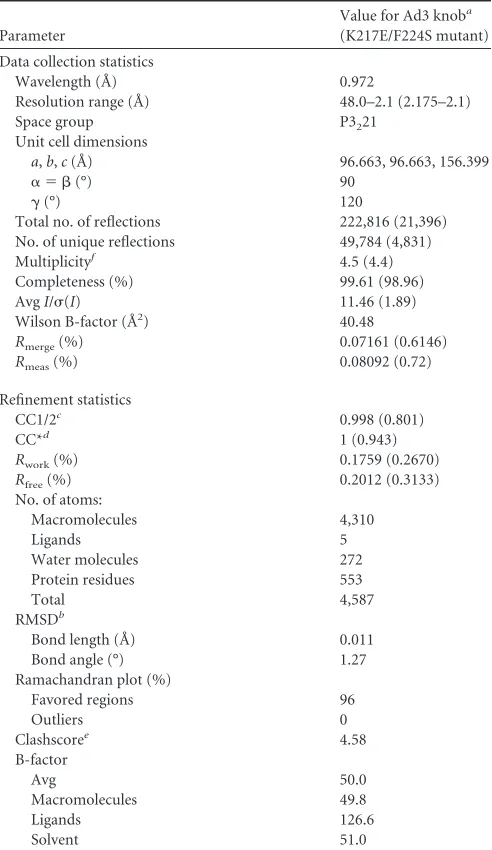
[image:3.585.299.545.77.502.2]A549, MDA-MB-231, and Ovc316 xenograft tumors were established by injection of the corresponding tumor cells into the mammary fat pad TABLE 1Data collection and refinement statistics
Parameter
Value for Ad3 knoba
(K217E/F224S mutant)
Data collection statistics
Wavelength (Å) 0.972
Resolution range (Å) 48.0–2.1 (2.175–2.1)
Space group P3221
Unit cell dimensions
a,b,c(Å) 96.663, 96.663, 156.399
␣ ⫽ (°) 90
␥(°) 120
Total no. of reflections 222,816 (21,396)
No. of unique reflections 49,784 (4,831)
Multiplicityf 4.5 (4.4)
Completeness (%) 99.61 (98.96)
AvgI/(I) 11.46 (1.89)
Wilson B-factor (Å2) 40.48
Rmerge(%) 0.07161 (0.6146)
Rmeas(%) 0.08092 (0.72)
Refinement statistics
CC1/2c 0.998 (0.801)
CC*d 1 (0.943)
Rwork(%) 0.1759 (0.2670)
Rfree(%) 0.2012 (0.3133)
No. of atoms:
Macromolecules 4,310
Ligands 5
Water molecules 272
Protein residues 553
Total 4,587
RMSDb
Bond length (Å) 0.011
Bond angle (°) 1.27
Ramachandran plot (%)
Favored regions 96
Outliers 0
Clashscoree 4.58
B-factor Avg 50.0 Macromolecules 49.8 Ligands 126.6 Solvent 51.0 a
Statistics for the highest-resolution shell are shown in parentheses.
bRMSD, root mean square deviations.
c
CC(1/2) is the percentage of correlation between intensities from random half data sets.
d
CC* is the estimate of the correlation coefficient of the data to the true intensities.
eThe clashscore is the number of overlaps greater than 0.4 Å per 1,000 atoms.
f
Multiplicity is the average number of observations for each reflection.
on November 7, 2019 by guest
http://jvi.asm.org/
(1:1 with Matrigel) of CB17 mice. TC1-DSG2 tumors were established by subcutaneous injection of TC1-DSG2 cells into DSG2 transgenic mice. JO-0, JO-1, JO-2, or JO-4 was intravenously injected 1 h before the appli-cation of chemotherapeutic drugs: irinotecan (Camptosar; Pfizer Inc., Groton, CT), pegylated liposomal doxorubicin (Lipodox; Sun Pharmaceuti-cals IN, India), cetuximab (Erbitux; ImClone, Somerville, NJ), and nanopar-ticle albumin-conjugated paclitaxel (nab-paclitaxel; Abraxane; Abraxis Bio-sciences, Summit, NJ). Tumor volumes were measured three times a week. Each treatment group consisted of a minimum of 5 mice. Animals were sac-rificed and the experiment terminated when tumors in one of the groups reached a volume of 800 mm3or tumors displayed ulceration.
Anti-JO-4 antibodies.Anti-JO-4 antibody concentrations in human serum samples were measured by enzyme-linked immunosorbent assay (ELISA). Plates were coated with rabbit polyclonal Ad3 fiber anti-bodies (1), followed by recombinant JO-4, human serum samples (1:2 to 1:1,000 dilution), and anti-human IgG-HRP. Serum samples from ovar-ian cancer patients were provided by the Pacific Ovarovar-ian Cancer Research Consortium.
3D structure.Pymol software was used to visualize the 3D structure of the Ad3 fiber knob (MMDB code 16945, PDB code1H7Z) (24).
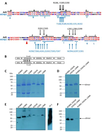
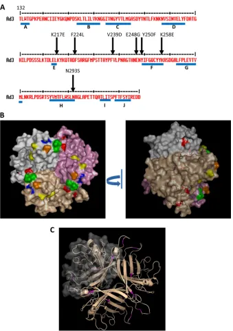
Statistical analysis.All results are expressed as means ⫾standard deviations (SD). Two-way analysis of variance (ANOVA) for multiple FIG 1Residues found to be critically involved in binding to DSG2. (A) Shown are the amino acid sequences of the Ad3 and Ad14p1 fiber knob. Beta sheets present in the Ad3 knob (PDB code1H7Z_A) and Ad14 knob (PDB code3F0Y_A) are indicated by blue lines. Black arrows indicate residues within the Ad3 fiber knob which, when mutated individually, ablate or reduce binding to DSG2. Compared to the parental strain of Ad14 (deWit), Ad14p1 had a deletion of two amino acid residues within the FG loop of the fiber protein knob (45), indicated by a red triangle. (B) Schematic structure of dimeric Ad3 fiber knob mutants. The fiber knob domain and one shaft motif was fused through a flexible linker to a homodimerizing K-coil domain (2). The proteins are self-dimerizing and can be purified by His-Ni-NTA affinity chromatography. (C to F) Analysis of binding of dimeric Ad3 fiber knob mutants to soluble DSG2. (C and D) Coomassie staining. Teng of purified Ad3 fiber knob (unboiled) was loaded per lane. Trimeric forms of the fiber knobs are indicated by an arrow. The gel contained SDS and the loading buffer containing DTT, which caused the disassembly of dimers of trimeric fiber knobs, as previously reported (2). (E and F) Western blot using soluble recombinant DSG2 as a probe, followed by anti-DSG2-MAb and anti-mouse IgG-HRP. For comparison, JO-1 (0.5g/lane) is shown. The Western blots were scanned and signals were quantified.
Adenovirus Type 3 and 14p1 Fiber Knob Binding to DSG2
on November 7, 2019 by guest
http://jvi.asm.org/
[image:4.585.126.463.62.499.2]testing was applied. Animal numbers andPvalues are indicated in the figure legends.
PDB accession number.The Ad3 fiber knob domain of the K217E and F224S mutant was deposited in PDB under code4LIY.
RESULTS
Residues critical for DSG2 binding.
We first focused our work on
Ad3. High-affinity binding to DSG2 and subsequent epithelial
junction opening requires several trimeric fiber knobs in a spatial
constellation present in the virion, PtDd, or dimerized (trimeric)
Ad3 fiber knob (e.g., JO-1) (
2
). The recombinant (trimeric) fiber
knob with two shaft motifs but without the dimerization domain
[image:5.585.39.287.77.199.2](Ad3 knob monomer) binds to DSG2 with an affinity that is
or-ders of magnitude less than that of JO-1, is not able to block Ad3
infection, and does not trigger junction opening (
1
,
2
,
13
).
How-ever, the affinity of Ad3 knob monomer is high enough to detect
binding in Western blot analyses in which soluble DSG2 is used as
a probe. Therefore, we used an
E. coli
expression library of
His-tagged Ad3 knob monomer mutants to identify the amino acid
residues within the Ad3 fiber knob that are critical for DSG2
bind-ing. To generate this library, we employed mutagenic PCR (
15
,
16
)
in a protocol that generated, on average, one to two amino acid
substitutions per knob. The Ad3 fiber knob library in
E. coli
XL-1
Blue was plated on agar plates, knob expression was induced by
IPTG, and colonies were screened for DSG2 binding using
recom-binant DSG2 and anti-DSG2 antibodies. A first screening round
of
⬃
10,000 colonies for variants that did not bind to DSG2
re-vealed 240 candidate colonies. When analyzed by Western
blot-ting for the 6
⫻
His tag, 40 of the 240 colonies showed expression of
trimeric fiber knob, indicating the absence of major
conforma-tional changes. The remaining variants had truncated fiber knobs
or did not form trimers. The corresponding 40 plasmids were
sequenced. The vast majority of colonies had single-amino-acid
substitutions within the fiber knob. If multiple amino acid
substi-tutions per knob were encountered, new Ad3 knob genomes
con-taining the corresponding mutations individually were
synthe-sized. Further rounds of colony screening did not uncover other
regions, indicating that all of the DSG2-interacting residues had
been found. A total of 8 independent mutants then were used for
subsequent studies (
Fig. 1A
). For all subsequent studies, we
gen-erated self-dimerizing forms of the Ad3 fiber mutants (
Fig. 1B
)
and purified them by affinity chromatography using Ni-NTA
col-umns. The purified dimerized knob mutants were analyzed for
TABLE 2Analysis of Ad3 fiber mutants
Mutant
% Residual DSG2 binding by Western blottinga
% Inhibition of:
Infection in the presence of dimeric knobb
Attachment in the presence of dimeric knobc
N186D 5.3 32.7 56.5
V189G 14 54 81
S190P 7.1 30.9 73.4
D261N 0 5.3 45
F265L 0 17.6 55.6
L296R 3.6 5.1 73.7
E299V 20 50.7 97.3
N186D, D261N 7.0 5.2 23.5
D261, L296R 7.0 1.5 20.1
N186D, D261N, L296R 0 0 18.5
aQuantitative analysis of Western blot bands corresponding to Ad3 knob trimers. The
intensity of the wt Ad3 fiber knob was taken as 100%.
bThe data reflect the ability of dimeric Ad3 fiber knob mutants to inhibit Ad3-GFP
infection (Fig. 3B). The higher the percentage, the stronger the inhibition. Inhibition by JO-1 (dimeric wt Ad3 knob) is taken as 100%.
c
Corresponding data for Ad3 virus attachment.n⫽3. Averages are shown. The standard deviations were less than 10%.
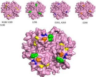
FIG 23D model of the Ad3 fiber knob. The structure is based on that of PDB accession number1H7Z_A. (Upper) Four critical areas involved in DSG2 binding. The critical residues are shown on the pink isosurface of the trimeric fiber knob. The view is from the top (apical side) facing the receptor. (Lower) All critical residues combined. On the right is an enlargement of the groove after a slight side rotation.
on November 7, 2019 by guest
http://jvi.asm.org/
[image:5.585.136.449.441.691.2]DSG2 binding by Western blotting (
Fig. 1C
to
F
). All mutants
were severely reduced in binding compared to JO-1 (i.e., the
dimerized form containing the wt Ad3 fiber knob). Mutants
D261N and F265L were almost completely ablated for DSG2
binding, while the other mutants had different levels of residual
DSG2 binding (3.6 to 20% of the wt Ad3 knob level) (
Table 2
). The
residues identified as being critical for Ad3 knob binding to DSG2
were in three different areas of the Ad3 fiber knob within the
CD-loop/D-beta sheet (N186D, V189G, and S190P), the
FG-loop/G-beta sheets (D261N and F265L), and the H-beta
sheet/HI-loop (L292A, L296R, and E299V) (
Fig. 1A
). In a 3D model of the
Ad3 fiber knob (PDB accession number
1H7Z_A
), all identified
FIG 3Competition of Ad3 virus by dimerized Ad3 knob mutants. (A) Relative attachment of3H-labeled Ad3 virus in the presence of dimeric fiber knob mutants. A total of 1.8⫻105HeLa cells were incubated with Ad3 knob mutants at concentrations of 2.5 and 100g/ml on ice for 1 h. Four hundred PFU/cell of3H-Ad3 virus was added on ice for another hour. Unbound virus particles were washed away. Attachment of virus particles incubated with PBS was taken as 100%.n⫽ 3. (B) Competition of Ad3-GFP virus infection on HeLa cells. A total of 1.5⫻105HeLa cells were seeded into 24-well plates. Cells were incubated with the Ad3 knob mutants at increasing concentrations for 1 h at room temperature. One hundred PFU/cell of Ad3-GFP virus was added, and GFP expression was analyzed 18 h later by flow cytometry. (Left) Percentage of GFP-positive cells. (Right) Mean fluorescence intensity (MFI).n⫽3. The standard deviations were less than 10%. (C) Relative attachment of3H-labeled Ad3 virus in the presence of dimeric fiber knob mutants with multiple mutations. The study was performed as described for panel B. The standard deviations were less than 10%. (D) Competition of Ad3-GFP virus infection on HeLa cells. Geo MFI, geometric mean fluorescence intensity. The study was performed as described for panel C. The standard deviations were less than 10%.
Adenovirus Type 3 and 14p1 Fiber Knob Binding to DSG2
on November 7, 2019 by guest
http://jvi.asm.org/
residues were located at the apical side of the fiber knob and
fol-lowed one specific groove in the knob (
Fig. 2
). It is noteworthy
that, except for F265L, the substitutions resulted in a change of
charge in the corresponding residue. Differences in migrations
patterns in polyacrylamide gels (for example, that for D261N)
indicate that the substitutions have caused conformational
changes. We are currently attempting to crystallize these mutants
to analyze their 3D structure.
To create an Ad3 fiber knob and eventually an Ad3 virus with
maximum ablation of DSG2 binding, we introduced multiple
mu-tations in the three identified areas, specifically a combination of
N186D and D261N, a combination of D261N and L296R, and a
combination of N186D, D261N, and L296R. As expected, the
combination of mutations in all three critical regions conferred
the highest level of ablation (
Table 2
and
Fig. 1F
).
Because of its relevance as a recently emerged pathogen, we
also generated a library of (monomeric) Ad14p1 fiber knob
mu-tants. A first screening revealed
⬃
300 candidate colonies for
vari-ants that did not bind to DSG2. When analyzed by Western
blot-ting for the 6
⫻
His tag, 45 of the 300 colonies showed expression of
trimeric fiber knob. Sequencing of these variants revealed 15
in-dependent mutants with reduced binding to DSG2 (
Fig. 1A
).
In-terestingly, in spite of a different beta sheet distribution, the amino
acid residues that were critical for Ad14p1 knob binding were in
the same three regions that were identified for the Ad3 fiber knob.
Because of these similarities, we performed further studies only
with selected Ad3 fiber knob mutants.
Functional validation.
Competition studies were performed
on HeLa cells, which express DSG2 (
1
). We first studied the
at-tachment of
3H-labeled Ad3 virus after preincubation of cells with
dimeric Ad3 fiber knobs (
Fig. 3A
). Reduction in Ad3 virus binding
was compared to that at preincubation with JO-1, i.e., the dimeric
protein that contained the wild-type Ad3 fiber knob. Inhibition of
binding by JO-1 was taken as 100%. The mutants L296R, D261N,
and F265L blocked Ad3 virus binding the least (5.1, 5.3, and
17.6%), followed by mutants S190P, N186D, and E299V (30.9,
32.7, and 50.7% reduced binding, respectively) (
Table 2
). A
sim-ilar assay setup was used to measure the ability of dimeric Ad3
knob mutants to block transduction of HeLa cells by an Ad3-GFP
vector. Transduction was measured based on GFP expression
(
Fig. 3B
). Similar to what we observed in the attachment study,
Ad3-GFP infection was least reduced by preincubation with
mu-tants D261N and F265L, followed by mumu-tants N186D, S190P,
L296R, and V189G. Taking the DSG2 binding (Western blotting),
attachment, and infection competition data together, we
con-cluded that the area containing residues 261 to 265 is the most
critical area in DSG2 binding. The region around residues 186 to
190 also contributes to binding, while the region containing
resi-due 299 appears to be only marginally involved in binding.
Dimeric Ad3 knob mutants with combined mutations were also
analyzed for their ability to compete with Ad3 virus for
attach-ment (
Fig. 3C
) and infection (
Fig. 3D
). The mutant with
muta-tions in all three areas (N185, D261, and L296) did not block Ad3
binding or infection even at concentrations of 200
g/ml,
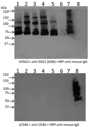
indicat-ing that it is nearly ablated for DSG2 bindindicat-ing. Notably, when usindicat-ing
soluble CD46 (sCD46) as a probe in the Western blot analysis of
wild-type Ad3 fibers, no specific binding was observed (
Fig. 4
).
This indicates that Ad3 only inefficiently binds to CD46.
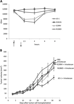
Correlation of reduced DSG2 binding and weaker ability to
open epithelial junctions.
A straightforward assay for the
junc-tion-opening function of dimeric Ad3 knob mutants is based on
measuring the transepithelial electrical resistance (TEER) in
transwell cell cultures. Epithelial cancer cells are cultured until the
TEER is constant, when major intercellular junctions are formed.
Addition of JO-1 for 1 h to the apical side of the transwell cultures
resulted in a rapid decrease in the TEER, indicating opening of
junctions (
Fig. 5A
). Incubation with mutant D261N had no effect
on the TEER. N186D and E299V had intermediate effects that
correlated with the residual binding of the corresponding fiber
knobs to DSG2. In previous studies, we have also established that
JO-1 triggered changes in epithelial junctions of xenograft tumors
and increased the anti-tumor efficacy of chemotherapeutics with
high molecular masses, for example, irinotecan. (Irinotecan has a
molecular mass of 586.7 Da and is used to treat colon and lung
cancer). We used this effect to assess the function of dimeric Ad3
knob mutants
in vivo
(
Fig. 5B
). Similar to what we observed
in
vitro
, JO-1 enhanced irinotecan therapy while mutants with
re-duced DSG2 binding had no significant effect on irinotecan
effi-cacy.
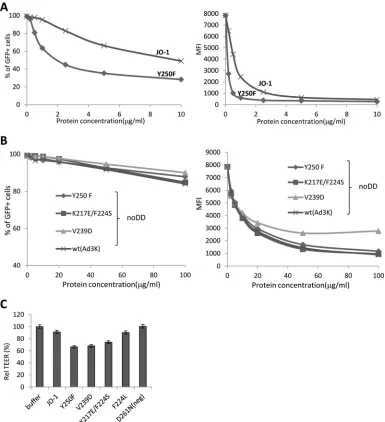
Affinity-enhanced dimeric Ad3 fiber knobs.
As outlined
above, JO-1 is relevant for cancer therapy. Therefore it is
impor-tant to better understand structural details of its interaction with
DSG2 and create JO-1 mutants with increased affinity to DSG2.
Affinity enhancement of biologics is used to (i) decrease their
effective dose, (ii) increase their half-lives, (iii) potentially increase
their therapeutic effects, and (iv) circumvent the adverse effects of
antibodies generated by patients against the biologic (e.g.,
neutral-ization or changes to the pharmokinetics). To make JO-1
ana-FIG 4Analysis of Ad3 fiber knob binding to soluble CD46. Ad3 fiber knobs containing different numbers of shaft motifs and either the wild-type Ad3 fiber knob (lane 1, S6/Kn; lane 2, S5/Kn; lane 3, S4/Kn; lane 4, Ad3-S3/Kn; lane 5, Ad3-S2/Kn; lane 6, Ad3-S/Kn), JO-1 (lane 7) or the CD46-binding Ad35 fiber knob (lane 8) were blotted and hybridized with soluble DSG2 (upper) or soluble CD46 (lower). Binding was detected by anti-DSG2 MAb or anti-CD46 MAb.
on November 7, 2019 by guest
http://jvi.asm.org/
[image:7.585.329.512.64.326.2]logues with increased affinity, we screened the
E. coli
expression
library with random mutations within JO-1 for variants with
in-creased binding to DSG2. Out of 10,000 colonies plated, 20
colo-nies with the most intense DSG2 signals were picked, and plasmid
DNA was sequenced. Seven different mutants with one or two
amino acid substitutions were identified: Y250F, K217E
⫹
F224S,
N293S, V239D, F224L, E248G
⫹
K258E, and L277R
⫹
N293D. The
localizations of the residues in the primary and 3D structures of
the Ad3 fiber knob are shown in
Fig. 6
. Notably, most of the
mutations were localized within the EF loop, indicating that this
loop is involved in stabilizing the interaction between Ad3 and
DSG2. V239 and Y250 are not exposed at the knob surface,
sug-gesting a structural change in the knob rather than an involvement
in direct binding to DSG2 (
Fig. 6B
, right). Recombinant mutant
dimeric Ad3 knob proteins then were purified. To measure the
affinity of the mutants to DSG2, we performed surface plasmon
resonance studies. The outcome of studies with knobs containing
the dimerization domain was complex, most likely due to the fact
that these mutants formed multimeric complexes. Therefore, we
performed studies with knob proteins lacking the dimerization
domain (noDD). The association rate constant (
K
aor
K
on) and
the dissociation rate constant (
K
dor k
off), as well as the
K
D (equi-FIG 5Correlation of reduced DSG2 binding with the ability to open epithelial junctions. (A) Transepithelial electrical resistance (TEER) measured on polarized colon cancer T84 cells. Cells were cultured in transwell chambers until the TEER was constant, i.e., tight junctions had formed. A total of 5g of dimeric Ad3 fiber knobs in PBS was then added for 1 h to the apical chamber. TEER was measured at the indicated time points.n⫽6. For time points 1.5 and 4 h, the difference between JO-1 and the D261N and N186D mutants was significant (P⬍0.01). The arrows indicate the addition and removal of Ad3 fiber knobs. (B) Enhancement of irinotecan therapy. A total of 4⫻106A549 cells were injected subcutaneously into CB17-SCID/beige mice. Once the tumor reached a volume of⬃100 mm3 (day 15 after implantation), the mice were injected intravenously with 2 mg/kg JO-1, E299V, N186D, or PBS, followed by an intravenous injection of irinotecan (37.5 mg/kg) 1 h later. The treatment was repeated on day 25.n⫽5. The differences between the irinotecan versus E299V plus irinotecan groups or irinotecan versus N186 plus irinotecan groups were not significant. The difference between irinotecan and JO-1 plus irinotecan was significant (P⬍0.01) from day 20 on.Adenovirus Type 3 and 14p1 Fiber Knob Binding to DSG2
on November 7, 2019 by guest
http://jvi.asm.org/
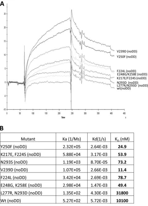
[image:8.585.135.449.68.504.2]librium dissociation constant) of wt Ad3 knob and all knob
mu-tants, are shown in
Fig. 7
. In agreement with previous studies (
2
),
we found that the wt Ad3 knob without the dimerization domain
bound to DSG2 with only relatively low affinity (
K
D⫽
10
M).
With the exception of mutant L227R/noDD plus N293D/noDD,
all mutants identified in the colony blot screen had higher
affini-ties to DSG2. Notably, the affiniaffini-ties of mutant Y250F/noDD or
V239D/noDD were 885- or 405-fold higher than those of wt
Ad3knob/noDD. The high affinity of the different mutants was
mainly due to a faster association with DSG2 rather than a change
in the dissociation rate. The only exception to this trend was
mu-tant N293S/noDD, for which the association rate was the lowest of
the mutants. However, this was partially compensated for by a
slower dissociation rate. Together, these results indicate that wt
Ad3 knob (noDD) binding to DSG2 is mostly limited by a slow
association rate that can be improved by a panel of mutations.
FIG 6Amino acid substitutions that increase the binding to DSG2. (A) Shown is the amino acid sequence of the Ad3 fiber knob. Beta sheets are indicated by blue lines. Arrows indicate residues within the Ad3 fiber knob which, when mutated, yielded stronger signals in colony blot assays, indicating stronger binding to DSG2. (B) The isosurface of the three knob monomers is colored in gray, pink, and light brown. Mutants involved in DSG2 recognition are shown in the same color asFig. 1B(yellow, green, blue, and orange), and mutations enhancing the binding are shown in red. (Left) Top view; (right) side view. V239 and Y250 are not exposed at the top, suggesting a structural change in the knob rather than an involvement in direct binding to DSG2. (C) Localization of all mutations that enhance the binding to DSG2. Residues are show in magenta in two knob monomers. The isosurface of one monomer is shown in gray transparency.
on November 7, 2019 by guest
http://jvi.asm.org/
[image:9.585.124.463.62.543.2]These mutations do not appear to modify the stability of this
in-teraction but the balance of association versus dissociation,
result-ing in higher affinities of ligands to the receptor.
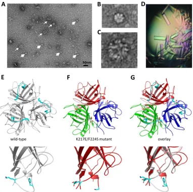
To better understand structural elements that enhance binding
to DSG2, we performed a more detailed analysis with mutant
K217E/F224S. Transmission electron microscopy with uranyl
acetate-stained K217E/F224S fiber knobs containing the
dimerization domain showed particles with 6 knobs representing
dimerized trimeric fibers (
Fig. 8A
, thick arrows, and B).
Interest-ingly, under these conditions, fiber knobs also formed regularly
shaped arrogates (with
⬃
30-nm diameter) resembling collapsed
PtDd (
Fig. 8A
, thin arrows, and C). We then performed X-ray
crystallography studies to resolve the structure of the K217E/
F224S mutant at the atomic level (
Fig. 8D
to
H
). As expected, the
K217E/F224S mutant formed a monotrimer of fiber knobs (
Fig.
8E
). The 3D structure of the mutant was overlaid with that of the
wild-type Ad3 fiber knob (
Fig. 8F
to
H
). This revealed that the EF
loop in the K217E/F224S mutant was completely disordered. This
loop is at the base of the knob domain at the junction with the fiber
shaft. Therefore, the K217E/F224S mutations may allow for easier
binding by increasing the flexibility of this loop region.
Correlation of increased affinity with stronger ability to
open epithelial junctions.
For the following studies, we used Ad3
fiber knob forms containing the dimerization domain. To analyze
the selected high-affinity mutants, we performed competition
in-fection studies with Ad3-GFP on HeLa cells and the dimeric forms
of the affinity-enhanced Ad3 fiber mutants (
Fig. 9A
). Based on
GFP expression, all dimeric mutants except mutant L277R
⫹
N293D inhibited Ad3-GFP infection significantly more than JO-1.
Notably, the nondimerized forms of Ad3 fiber knobs with increased
affinity to DSG2 were unable to act as competitors in transduction
studies (
Fig. 9B
). Higher affinity to DSG2 resulted in an increased
capability to open epithelial junctions in transwell cultures (
Fig. 9C
).
Compared to JO-1, the TEER in cultures incubated with the mutants
V250F, V239D, and K217E
⫹
F224S was significantly higher.
Two of the affinity-enhanced versions of JO-1, V293D and
Y250F, and V250F were analyzed in an
in vivo
assay. We called
these mutants JO-2 and JO-4, respectively. The first study was
performed in a xenograft model derived from A549 cells, similar
to the study described for
Fig. 5B
.
Figure 10A
shows that the
af-finity-enhanced mutants JO-2 and JO-4 increased irinotecan
ther-apy significantly more than JO-1 (
P
⬍
0.05 starting from day 27).
Furthermore, JO-4 (
K
D, 11.4 nM) was significantly more efficient
than JO-2 (
K
D, 24.9 nM), indicating a correlation between affinity
to DSG2 and therapeutic effect. Additional studies were
per-formed in xenograft tumors derived from Ovc316 cells (
11
,
25
).
Ovc316 cells are Her2/neu-positive epithelial tumor cells derived
from an ovarian cancer biopsy specimen. These cells can undergo
epithelial-to-mesenchymal transition (EMT) and the reverse
pro-cess, mesenchymal-to-epithelial-transition (MET), under specific
conditions
in vitro
and
in vivo
. A subfraction of Ovc316 cells that is
positive for Nanog, CD133, and E-cadherin is enriched for cancer
stem cells, i.e., self-renewing cells with pluripotent potential and
tumor-forming ability (
25
). Therefore, Ovc316 cells closely model
the heterogeneity and plasticity seen in tumors
in situ
. Intravenous
injection of JO-1 at a dose of 2 mg/kg of body weight 1 h before
injection of pegylated liposomal doxorubicin (PLD), a drug that is
widely used for chemotherapy of ovarian cancer, significantly
in-creased the treatment efficiency (
Fig. 10B
). Importantly, at a dose
of 0.5 mg/kg, JO-4 had an even greater stimulating effect on PLD
therapy. Finally, we tested JO-4 in a model for triple-negative
breast cancer (TNBC). TNBC is characterized by a lack or
mini-mal expression of estrogen receptor (ER) and progesterone
recep-tor (PR) and the absence of Her2/neu overexpression. TNBC
ac-counts for 15% of all breast cancers. Overall survival is poor
compared to that of patients who have other phenotypes. A
char-acteristic feature of TNBC is high levels of DSG2 and epithelial
junctions. Promising clinical results in the treatment of TNBC
have been achieved with nanoparticle albumin-conjugated
pacli-taxel (nab-paclipacli-taxel) alone or in combination with the EGF
re-ceptor (EGFR)-targeting mAb cetuximab (
26
). Our study showed
that JO-4 significantly increased nab-paclitaxel/cetuximab
effi-cacy in a mouse model with orthotopic TNBC tumors (
Fig. 10C
).
Because of its therapeutic relevance, we further studied JO-4 in an
adequate mouse tumor model. Because Ad3 virus and Ad3 fiber
knob derivatives do not bind to mouse cells and tissues, we used
human transgenic mice that expressed human DSG2 in a pattern
and at a level similar to that of humans (
4
). These mice were
subcutaneously implanted with syngeneic TC1-hDSG tumors.
When tumors reached a volume of
⬃
600 mm
3, JO-1 or JO-4 was
intravenously injected for safety and efficacy studies. Both JO-1
FIG 7SPR analysis of nondimerized Ad3 fiber knob interactions with DSG2. (A) DSG2 was immobilized on sensorchips, and background was automati-cally subtracted from the control flow cell. The Ad3 fiber knobs (without a dimerization domain, termed noDD) were injected for 3 min at 2.5g/ml, followed by a 2.5-min dissociation period. (B) Summary of SPR data. A con-centration range from 2.5 to 10g/ml of the knobs was injected, and kinetics and affinity parameters were evaluated using BIAeval software. The extracted data are shown in the table. Wt, Ad3 fiber knob without mutations.
Adenovirus Type 3 and 14p1 Fiber Knob Binding to DSG2
on November 7, 2019 by guest
http://jvi.asm.org/
[image:10.585.43.283.66.399.2]and JO-4 serum concentrations declined more than one order of
magnitude within an hour after injection, and the decline was
significantly greater for JO-4 (
P
⬍
0.01 for 1 h postinjection) (
Fig.
11A
). After 1 h postinjection, JO-1 and JO-4 concentrations
reached a plateau with
⬃
100 ng/ml. We also analyzed
hematolog-ical parameters after intravenous JO-1 and JO-4 injection. Blood
chemistry did not show abnormal changes. Blood cell counts were
normal except for lymphocyte and platelet numbers, which
de-creased early after injection (
Fig. 11B
). Lymphocyte and platelet
counts reached a nadir at 24 h postinfection with significantly
lower numbers for JO-4 (
P
⬍
0.01). Interestingly, lymphocyte and
platelet counts returned to normal levels faster in JO-4-injected
mice than in JO-1-treated animals.
JO-1 and JO-4 are virus-derived proteins and are
immuno-genic. In immunocompetent mice, serum IgG antibodies against
these proteins can be detected by ELISA 2 weeks after injection (
5
).
One theoretical premise for affinity enhancement of therapeutic
pro-teins is that it circumvents neutralizing serum antibodies. To test this,
we performed repeated injections of JO-1 and JO-4 in an
immuno-competent hDSG2 mouse tumor model with TC1-hDSG2 tumors
(
Fig. 11C
). After two treatment cycles of JO-1 and PLD, treatment
was stopped and tumors were allowed to regrow. The 3rd and 4th
treatment cycles were started on days 28 and 35, respectively. At the
time of the 3rd cycle, serum anti-JO-1 antibodies were detectable by
ELISA. Importantly, in both the 3rd and 4th treatment cycles, JO-1
and JO-4 had an enhancing effect on PLD therapy, whereby the
en-hancing effect was significantly stronger for JO-4.
Overall, our functional studies with affinity-enhanced dimeric
Ad3 fiber mutants demonstrate a correlation between DSG2
af-finity and epithelial junction opening/therapeutic effects.
DISCUSSION
Residues involved in Ad3 knob binding to DSG2.
Unlike Ad
in-teraction with CAR and CD46 (
27
,
28
), structural details of Ad
interaction with DSG2 are still elusive. Although the crystal
struc-ture of the Ad3 fiber knob has been resolved, for DSG2, the 3D
FIG 8Electron microscopy and 3D structure of Ad3 fiber knob mutant JO-2. (A to C) Negative staining of JO-2 with SST. Dimeric forms can be seen, but higher organizations are also visible. A heterogeneous complex of around 50 nm is depicted by thin arrows, and a smaller, regular dodecahedral-like particle is depicted by thick arrows. Closeup views are presented in panels B and C. (D to G) Crystallographic structure of the nondimerized form of the K217E/F224S mutant. (D) Protein crystals. (E) The wild-type Ad3 knob is colored in gray, and the EF loop 217-224 is in cyan. This is the loop which becomes disordered in the mutant. There is no density for these residues in the mutant structure. (F) The mutant is displayed as a cartoon, and each monomer is colored red, blue, or green. (G) Overlay of these two structures shows that the EG loop is completely disordered in the K217E/F224S mutant. The bottom panels show closeup views of one monomer. K217 and F224 appear as blue sticks.
on November 7, 2019 by guest
http://jvi.asm.org/
[image:11.585.103.485.65.445.2]structure of only the most distal of the four extracellular domains
(ECD) is available (MMDB code 59843). However, our previous
competition studies with monoclonal antibodies against different
DSG2 domains indicated that ECDs 3 and 4 are involved in
bind-ing to Ad3 (
1
). In this study, we used mutagenesis-based analyses
to identify the amino acid residues within the Ad3 fiber knob that
are critical for binding to DSG2. Mutagenic analysis of DSG2 was
not possible, because, when expressed in
E. coli
, the protein did
not bind to Ad3, indicating that posttranslational processing is
required to create active Ad3 binding sites within DSG2 (data not
shown). The identified residues, critical for Ad3 knob binding to
DSG2, were in three different areas of the Ad3 fiber knob and
formed a potential binding pocket localized in a groove at the
distal end of the fiber knob facing the receptor. Notably, binding
of other Ad serotypes to CAR or CD46 primarily involves regions
at the lateral or basal side of the corresponding fiber knobs (
10
,
29
). Our data indicate that Ad3 uses a different binding strategy.
We are currently performing crystallography studies with dimeric
Ad3 fiber knobs and DSG2. Considering that multimeric Ad3 fiber
knobs cluster several DSG2 molecules (
2
), it is expected that the 3D
structure of this complex will be complicated. It remains to be studied
whether the residues critical for Ad3 fiber knob binding to DSG2 will
also be involved in binding of other species B Ads to DSG2. Notably,
while D261, F265, and E299 are conserved in all four
DSG2-interact-ing Ads (Ad3, Ad7, Ad11, and Ad14), other critical residues (N186,
V189, and L296) differ between these serotypes (
Fig. 12
).
FIG 9Analysis of dimeric Ad3 fiber knob mutants with increased affinity to DSG2. (A) Competition of Ad3-GFP virus infection on HeLa cells with dimeric affinity-enhanced mutant Y250F and JO-1 (dimeric wt Ad3 fiber knob). The experimental settings are the same as those described forFig. 3C. (Left) Percentage of GFP-positive cells. (Right) Mean fluorescence intensity.n⫽3. The standard deviations were less than 10%. (B) Competition of Ad3-GFP virus infection on HeLa cells by Ad3 knob mutants with enhanced DSG2 binding but without a dimerization domain. A total of 1.5⫻105HeLa cells were seeded into a 24-well plate. Cells were incubated with the Ad3 knob mutants at increasing concentrations for 1 h at room temperature. One hundred PFU/cell of Ad3GFP virus was then added, and GFP expression was analyzed 18 h later. (C) TEER on colon cancer T84 cells. The experimental settings were the same as those described forFig. 5A. The TEER at 4 h is shown.n⫽3.
Adenovirus Type 3 and 14p1 Fiber Knob Binding to DSG2
on November 7, 2019 by guest
http://jvi.asm.org/
[image:12.585.100.485.66.488.2]Ad14 is an important research object because of the recent
appearance of a new strain (Ad14p1). Having never been
previ-ously documented in the United States, Ad14p1 was reported in
March and April 2006 during routine surveillance at several U.S.
military recruit training centers (
30
). During March to June of the
following year, a total of 140 additional cases of confirmed
HAdV-B14p1 respiratory illness were reported in patients in Oregon,
Washington, and Texas (
31
). Thirty-eight percent of these
pa-tients were hospitalized, including 17% who were admitted to
intensive care units; 5% of patients died. Outbreaks of
HAdV-B14p1 were subsequently detected in the other 5 bases and in
civilian populations in Washington, Oregon (
32
), Alaska (
33
),
Wisconsin, and Pennsylvania (
7
,
9
), as well as in Canada (
8
),
China (
34
), and South Korea (
35
). At this point, the molecular
basis for the high pathogenicity and/or virulence of Ad14p1 is
unclear. We attempted to delineate the structural components for
Ad14p1 binding to DSG2. The beta sheet distribution of Ad14p1
differs from that of Ad3 (
Fig. 1A
).Therefore, similar to
CD46-interacting serotypes (
36
,
37
), it is possible that DSG2-interacting
Ads vary in their binding strategy to DSG2, which could result in
different DSG2 binding areas. However, the screening of an
Ad14p1 fiber knob mutant library did not support this hypothesis.
The areas involved in DSG2 binding were essentially the same for
Ad3 and Ad14p1 fiber knobs. Nevertheless, our finding are
rele-vant for the treatment of Ad14p1 viremia, specifically for the
pro-duction of Ad14p1 inhibitors or high-affinity decoys that can
trig-ger the opsonization of virus present in the blood circulation or
airway.
It has been reported that, in addition to DSG2, Ad3 can use
CD46 as a receptor to infect cells if DSG2 is absent (
38
).
Previ-ously, we found that in polarized normal epithelial cells, DSG2 is
trapped in tight junctions and is not accessible from the apical
side, while CD46 is present on both membrane sides (
1
).
There-fore, we speculate that CD46 can serve as a relatively inefficient
entry receptor for Ad3, while
de novo
-produced Ad3 and Ad3
penton-dodecahedra interact with DSG2, open epithelial junctions,
and allow for efficient lateral spread of Ad3 or penetration into deeper
tissue layers and blood circulation. The ability to individually ablate
the Ad3 knob residues that are critical for DSG2 and CD46 binding
should make it possible to prove this hypothesis.
Affinity-enhanced fiber knobs.
Most of the mutations that
increased the affinity to DSG2 were localized within the EF loop,
FIG 10Combination of affinity-enhanced JO-1 versions with chemotherapy. (A) Enhancement of irinotecan (I) therapy. The experimental settings were the same as those described forFig. 5B. The differences in the groups JO-1 plus I versus JO-2 plus I and JO-2 plus I versus JO-4 plus I were significant from day 20 on.n⫽5. (B) JO-4 enhances PLD therapy in an ovarian cancer model at a lower dose than JO-1. Mammary fat pad tumors were established from primary ovarian cancer Ovc316 cells. Treatment was started when tumors reached a volume of 100 mm3. Mice were injected intravenously with 2 mg/kg JO-1 or with 0.5 mg/kg JO-4, followed by an intravenous injection of PLD (1 mg/kg) 1 h later. Treatment was repeated weekly. (C) JO-4 enhances therapy in poor-prognosis triple-negative breast cancer (TNBC). A total of 4⫻106TNBC MDA-MB-231 cells were injected into the mammary fat pad of CB17 SCID/beige mice. JO-4 (2 mg/kg) was intravenously injected 1 h before the application of cetuximab (C) (10 mg/kg, intraperitoneal) and nab-paclitaxel (nab-P) (5 mg/kg, intravenous). Treatment was given weekly.n⫽10.P⬍0.01 at day 25 for nab-P plus C versus JO-4 plus nab-P plus C.
on November 7, 2019 by guest
http://jvi.asm.org/
[image:13.585.116.473.66.384.2]indicating that this loop is involved in stabilizing the interaction
between Ad3 and DSG2. Interestingly, unlike Ad7, Ad11, and
Ad14, the Ad3 fiber knob has two additional residues (VL)
fol-lowed by a proline in this area. Therefore, this loop could be
ex-tended further and the proline could orient it in a way that might
allow for better interaction with the receptor. The analysis of the
3D structure of one of these mutants at the atomic level supports
this conclusion. These studies indicate that the introduced
muta-tions make the loop more flexible, which might facilitate the
in-teraction with DSG2.
The identification of Ad3 knobs with higher affinity than the wt
Ad3 knob has implications for Ad3-mediated gene therapy.
Re-FIG 11Pharmacokinetics, toxicity, and immunogenicity of JO-4. (A) Serum clearance of JO-1 and JO-4. hDSG2 transgenic mice with subcutaneous TC1-hDSG2 tumors (⬃600 mm3) were intravenously injected with JO-1 or JO-4 (2 mg/kg), and serum samples were analyzed by ELISA.n⫽3. Note that theyaxis has a log scale. (B) Lymphocyte and platelet counts in hDSG2/TC1-hDSG2 transgenic mice after JO-1 or JO-4 injection.n⫽3. (C) Therapy studies in immunocompetent hDSG2 transgenic mice with TC1-hDSG2 tumors. When tumors reached a volume of⬃80 mm3, JO-1 or JO-4 (2 mg/kg) or PBS was injected intravenously, followed 1 h later by PLD (Doxil; 1.5 mg/kg, intravenous). Treatment was repeated as indicated by arrows. Tumors were then allowed to regrow for about 2 weeks. From day 15 on, serum anti-JO-1/JO-4 antibodies were detectable by ELISA. Two more treatment cycles were performed at day 28 and day 35. JO-1 and JO-4 continued to be effective after multiple treatment cycles, even in the presence of detectable antibodies. The difference between JO-1/PLD and JO-4/PLD is significant from day 31 on.n⫽10.
Adenovirus Type 3 and 14p1 Fiber Knob Binding to DSG2
on November 7, 2019 by guest
http://jvi.asm.org/
[image:14.585.95.490.65.558.2]cently, gene transfer vectors based on Ad3 have shown promise for
cancer therapy in clinical trials (
39
). Theoretically,
affinity-en-hanced Ad3 vectors could be used at lower doses and outcompete
neutralizing antibodies. Recently, attempts were undertaken to
incorporate high-affinity ligands into measles virus (
40
) and
Ad5-based vectors (
31
,
41
,
42
) in order to increase efficacy and
speci-ficity of target cell infection
in vivo
. Based on our findings in this
study, a similar strategy can now be pursued for Ad3 vectors.
In addition to improving Ad3 vectors, affinity-enhanced
ver-sions of JO-1 have translational relevance. Most solid tumors are
of epithelial origin, and although malignant cells are
dedifferenti-ated, they maintain intercellular junctions, a key feature of
epithe-lial cells, both in the primary tumor as well as in metastatic lesions
(
5
,
43
). These intercellular junctions represent a protection
mech-anism against attacks by the host immune system and pose
phys-ical barriers that prevent intratumoral penetration and
dissemi-nation of cancer therapeutics, including monoclonal antibodies
and chemotherapy drugs (
5
,
43
). When injected intravenously
into mice with xenograft or syngeneic DSG2 transgenic tumors,
JO-1 markedly enhanced therapeutic effects with a variety of
che-motherapy drugs as well as monoclonal antibodies (
3
,
5
). In this
study, we have shown that new affinity-enhanced versions of JO-1
(e.g., JO-4) increased the efficacy of cancer therapeutics
signifi-cantly more than JO-1 (irinotecan, nab-paclitaxel, pegylated
lipo-FIG 12Alignment of fiber knob sequences. The residues that ablate/reduce Ad3 knob binding to DSG2 are indicated.
FIG 13Sera from humans and hypervaccinated mice do not inhibit activity of JO-4. Analysis of human serum for binding with JO-4 by ELISA is shown. Rabbit polyclonal antibodies against the Ad3 fiber knob were used for capture, followed by recombinant JO-1 protein, human serum (dilutions 1:20 to 1:1,000), and anti-human IgG-HRP. Commercial human Ab serum depleted for IgG was used as a negative control (blue horizontal line). Serum from a scientist who routinely works with Ad3 virus was used as a positive control. P1 to P38 are serum samples from ovarian cancer patients obtained from the Pacific Ovarian Cancer Research Consortium.
on November 7, 2019 by guest
http://jvi.asm.org/
[image:15.585.121.463.65.334.2] [image:15.585.100.488.518.673.2]somal doxorubicin, and cetuximab) in four tumor models (A549,
Ovc316, MDA-MB231, and TC1-DSG2). Studies of DSG2
trans-genic mice with syngeneic tumors showed that serum JO-4 levels
rapidly decreased, most likely due to binding to DSG2 on tissues.
Previous studies showed that, in addition to tumors, lymphocytes
and platelets of transgenic hDSG2 mice express hDSG2 (similar to
human and monkeys) (
1
,
4
). Along these lines, we found that JO-4
injection resulted in a transient reduction of lymphocyte and
platelet counts.
Despite the fact that approximately one-third of humans have
neutralizing antibodies against Ad3 (
1
), in a recent study with
serum from ovarian cancer patients we found detectable
(bind-ing) antibodies against JO-4 in only 10% of patients (
n
⫽
38) (
Fig.
13
). However, it is certain that adaptive immune responses against
intravenously administered JO-4 will develop in humans,
partic-ularly after repeated injection. In this context, it is noteworthy that
anti-JO-4 antibodies generated after injection into
immunocom-petent mice appeared not to critically inhibit the function of JO-4.
The data shown in
Fig. 11C
demonstrate that JO-1 and JO-4
con-tinue to be effective after multiple treatment cycles, even in the
presence of detectable antibodies. Because the therapeutic effect
after repeated injection was significantly greater for JO-4, we
spec-ulate that JO-4 is more potent not only in junction opening but
also in disrupting complexes between the junction opener and
serum antibodies.
In summary, our studies uncover important structural details
of Ad3 and Ad14p1 fiber knob binding to DSG2. Furthermore, it
shows a correlation between the affinity of Ad3 fiber knobs to
DSG2 and subsequent effects on epithelial junctions. Finally, the
generation of affinity-enhanced recombinant dimeric Ad3 fiber
knobs has implications for cancer therapy.
ACKNOWLEDGMENTS
The study was supported by NIH grants R01 CA080192 and R01 HLA078836.
We thank the Pacific Ovarian Cancer Research Consortium for pro-viding patient serum samples.
REFERENCES
1.Wang H, Li ZY, Liu Y, Persson J, Beyer I, Moller T, Koyuncu D, Drescher MR, Strauss R, Zhang XB, Wahl JK, III, Urban N, Drescher C, Hemminki A, Fender P, Lieber A.2011. Desmoglein 2 is a receptor for adenovirus serotypes 3, 7, 11 and 14. Nat. Med.17:96 –104.
2.Wang H, Li Z, Yumul R, Lara S, Hemminki A, Fender P, Lieber A.2011. Multimerization of adenovirus serotype 3 fiber knob domains is required for efficient binding of virus to desmoglein 2 and subsequent opening of epithelial junctions. J. Virol.85:6390 – 6402.
3.Beyer I, van Rensburg R, Strauss R, Li Z, Wang H, Persson J, Yumul R, Feng Q, Song H, Bartek J, Fender P, Lieber A.2011. Epithelial junction opener JO-1 improves monoclonal antibody therapy of cancer. Cancer Res.71:7080 –7090.
4.Wang H, Beyer I, Persson J, Song H, Li Z, Richter M, Cao H, van Rensburg R, Yao X, Hudkins K, Yumul R, Zhang XB, Yu M, Fender P, Hemminki A, Lieber A.2012. A new human DSG2-transgenic mouse model for studying the tropism and pathology of human adenoviruses. J. Virol.86:6286 – 6302.
5.Beyer I, Cao H, Persson J, Song H, Richter M, Feng Q, Yumul R, van Rensburg R, Li Z, Berenson R, Carter D, Roffler S, Drescher C, Lieber A.2012. Coadministration of epithelial junction opener JO-1 improves the efficacy and safety of chemotherapeutic drugs. Clin. Cancer Res.18: 3340 –3351.
6.Wang H, Tuve S, Erdman DD, Lieber A.2009. Receptor usage of a newly emergent adenovirus type 14. Virology387:436 – 441.
7.Carr MJ, Kajon AE, Lu X, Dunford L, O’Reilly P, Holder P, De Gascun CF, Coughlan S, Connell J, Erdman DD, Hall WW. 2011. Deaths
associated with human adenovirus-14p1 infections, Europe, 2009 –2010. Emerg. Infect. Dis.17:1402–1408.
8.Girouard G, Garceau R, Thibault L, Oussedik Y, Bastien N, Li Y.2013. Adenovirus serotype 14 infection, New Brunswick, Canada, 2011. Emerg. Infect. Dis.19:119 –122.
9.Kajon AE, Lu X, Erdman DD, Louie J, Schnurr D, George KS, Koop-mans MP, Allibhai T, Metzgar D.2010. Molecular epidemiology and brief history of emerging adenovirus 14-associated respiratory disease in the United States. J. Infect. Dis.202:93–103.
10. Wang H, Liaw YC, Stone D, Kalyuzhniy O, Amiraslanov I, Tuve S, Verlinde CL, Shayakhmetov D, Stehle T, Roffler S, Lieber A. 2007. Identification of CD46 binding sites within the adenovirus serotype 35 fiber knob. J. Virol.81:12785–12792.
11. Strauss R, Sova P, Liu Y, Li Z-Y, Tuve S, Pritchard D, Brinkkoetter P, Moller T, Wildner O, Pesonen S, Hemminki A, Urban N, Drescher C, Lieber A.2009. Epithelial phenotype of ovarian cancer mediates resis-tance to oncolytic adenoviruses. Cancer Res.15:5115–5125.
12. Tuve S, Chen BM, Liu Y, Cheng TL, Toure P, Sow PS, Feng Q, Kiviat N, Strauss R, Ni S, Li ZY, Roffler SR, Lieber A.2007. Combination of tumor site-located CTL-associated antigen-4 blockade and systemic reg-ulatory T-cell depletion induces tumor-destructive immune responses. Cancer Res.67:5929 –5939.
13. Tuve S, Wang H, Ware C, Liu Y, Gaggar A, Bernt K, Shayakhmetov D, Li Z, Strauss R, Stone D, Lieber A.2006. A new group B adenovirus receptor is expressed at high levels on human stem and tumor cells. J. Virol.80:12109 –12120.
14. Shayakhmetov DM, Papayannopoulou T, Stamatoyannopoulos G, Lieber A.2000. Efficient gene transfer into human CD34(⫹) cells by a retargeted adenovirus vector. J. Virol.74:2567–2583.
15.Cadwell RC, Joyce GF. 1994. Mutagenic PCR. PCR Methods Appl. 3:S136 –S140.
16. Cadwell RC, Joyce GF.1992. Randomization of genes by PCR mutagen-esis. PCR Methods Appl.2:28 –33.
17. Incardona MF, Bourenkov GP, Levik K, Pieritz RA, Popov AN, Svens-son O.2009. EDNA: a framework for plugin-based applications applied to X-ray experiment online data analysis. J. Synchrotron Radiat.16:872– 879. 18. Kabsch W.2010. Integration, scaling, space-group assignment and
post-refinement. Acta Crystallogr. D Biol. Crystallogr.66:133–144.
19. Kabsch W.2010. Xds. Acta Crystallogr. D Biol. Crystallogr.66:125–132. 20. McCoy AJ, Grosse-Kunstleve RW, Adams PD, Winn MD, Storoni LC, Read RJ.2007. Phaser crystallographic software. J. Appl. Crystallogr.40: 658 – 674.
21. Emsley P, Lohkamp B, Scott WG, Cowtan K.2010. Features and devel-opment of Coot. Acta Crystallogr. D Biol. Crystallogr.66:486 –501. 22. Adams PD, Afonine PV, Bunkoczi G, Chen VB, Davis IW, Echols N,
Headd JJ, Hung LW, Kapral GJ, Grosse-Kunstleve RW, McCoy AJ, Moriarty NW, Oeffner R, Read RJ, Richardson DC, Richardson JS, Terwilliger TC, Zwart PH. 2010. PHENIX: a comprehensive Python-based system for macromolecular structure solution. Acta Crystallogr. D Biol. Crystallogr.66:213–221.
23. Walters RW, Freimuth P, Moninger TO, Ganske I, Zabner J, Welsh MJ. 2002. Adenovirus fiber disrupts CAR-mediated intercellular adhesion al-lowing virus escape. Cell110:789 –799.
24. Durmort C, Stehlin C, Schoehn G, Mitraki A, Drouet E, Cusack S, Burmeister WP.2001. Structure of the fiber head of Ad3, a non-CAR-binding serotype of adenovirus. Virology285:302–312.
25. Strauss R, Li ZY, Liu Y, Beyer I, Persson J, Sova P, Moller T, Pesonen S, Hemminki A, Hamerlik P, Drescher C, Urban N, Bartek J, Lieber A. 2011. Analysis of epithelial and mesenchymal markers in ovarian cancer reveals phenotypic heterogeneity and plasticity. PLoS One6:e16186. doi:
10.1371/journal.pone.0016186.
26. Ueno NT, Zhang D. 2011. Targeting EGFR in triple negative breast cancer. J. Cancer2:324 –328.
27. Bewley MC, Springer K, Zhang YB, Freimuth P, Flanagan JM.1999. Stru