Green
fl
uorescent protein-based assays for
high-throughput functional characterization and
ligand-binding studies of biotin protein ligase
†
Samuel P. Askin, Thomas E. H. Bond and Patrick M. Schaeffer*
InE. coliand other prokaryotes such asStaphylococcus aureus, andMycobacterium tuberculosis, biotin protein ligase (BirA) is an emerging drug target as it is the sole enzyme capable of biotin transfer onto the BCCP subunit of ACC. There is currently a gap in simple yet efficient assays for rapidly identifying and characterising inhibitors targeting BirA. We present for thefirst time the development and validation of a simple and reliable DSF-GTP assay for the high-throughput screening of BirA:ligand interactions using a new GFP-tagged BirA ofE. coli. In addition, we developed a new GFP-based biotinylation activity assay taking advantage of a GFP tethered with an AviTag. The data obtained with these assays revealed new insights into how the binding of individual or combinations of ligands affect the overall thermal stability and affinity of BirA. The DSF-GTP assay has aZ0value of 0.785 that makes it an excellent tool for future high-throughput screening of inhibitory compounds.
Introduction
Drug discovery is a rapidly expanding and diversifying eld. This is in part due to the need to identify alternatives to current therapeutics which are failing as a result of emerging global antibiotic resistance by prevalent bacteria. Strategies to combat antibiotic resistance centre upon the identication of novel compounds to inhibit already established drug targets, identi-cation of new drug targets or target pathways, or a combina-tion of the two. In identifying novel targets in a disease, there is a necessity to comprehensively characterize the target. This allows screening in optimal conditions for monitoring activity or stability of the target as well as a prior, in-depth under-standing of specic ligand interactions of the target.
A critical pathway to survival of both eukaryotes and prokaryotes that is targeted in new therapies is fatty acid biosynthesis.1 InE. coli, fatty acid biosynthesis reactions are processed by individual enzymes, following the type II fatty acid synthase pathway. The fatty acid biosynthesis pathway inE. coli
is well characterized and begins via the catalysis of the conversion of acetyl-CoA to malonyl-CoA by the acetyl-coA carboxylase (ACC). Critical to this process is the transfer of biotin to the biotin carboxyl carrier protein (BCCP) subunit of ACC. ACC, and hence the entire fatty acid biosynthesis pathway,
is therefore dependent on the synthesis, concentration, and downstream processing of biotin.2,3
InE. coliand other prokaryotes such asStaphylococcus aureus, andMycobacterium tuberculosis, biotin protein ligase (BirA) is an emerging drug target as it is the sole enzyme capable of biotin transfer onto the BCCP subunit of ACC.4–6 Further to this, the BirA ofE. colialso regulates expression of bio genes and subse-quently biotin synthesis through binding to the biotin operator (bioO), a 40 bp inverted repeat that overlaps and controls both biotin operon promoters.7,8The apoform of BirA (apoBirA) binds to biotin and ATP which are converted to the adenylated inter-mediate biotinyl-50-AMP (bio-50-AMP) with the release of PPi.9The bio-50-AMP:BirA complex has been described as the holoform of the protein (holoBirA). Biotin is subsequently transferred by holoBirA onto BCCP. The formation of an amide bond between the bio-50-AMP and a single lysine residue of the BCCP subunit allows for downstream processing of fatty acid chain extension to then take place. The holoBirA also acts as a co-repressor through its binding tobioO.7,10–16
The interactions between BirA and its ligands have been
extensively studied via numerous methods. However, the
development of high-throughput (HT) assays capable of screening large libraries of inhibitors in a drug screening context has been limited.17The emergence of technologies such as differential scanning uorimetry (DSF), led by the widely adopted Thermouor assay,18has been enterprizing in theeld of drug discovery and protein characterization for their ability to prole thousands of conditions simultaneously in small volumes. Recently, a new DSF platform based on the screening of GFP-tagged proteins (DSF-GTP) was developed to prole protein thermal stability in high-throughput (HT).19–21This new Comparative Genomics Centre, James Cook University, DB21, James Cook Drive,
Townsville, QLD 4811, Australia. E-mail: patrick.schaeff[email protected]; Fax: +61 7 4781 6078; Tel: +61 7 4781 4448
†Electronic supplementary information (ESI) available. See DOI: 10.1039/c5ay03064a
Cite this:Anal. Methods, 2016,8, 418
Received 24th November 2015 Accepted 25th November 2015
DOI: 10.1039/c5ay03064a
www.rsc.org/methods
Methods
PAPER
Open Access Article. Published on 02 December 2015. Downloaded on 16/05/2017 00:56:22.
This article is licensed under a
Creative Commons Attribution 3.0 Unported Licence.
platform does not require the addition of a solvatochromic dye to the reaction thus eliminating the possibility of interferences with protein–ligand interactions. DSF-GTP has also the added advantage that it can be performed in the presence of additional proteins.
Here, we describe for the rst time the development and validation of a DSF-GTP assay for the HT screening of Bir-A:ligand interactions using a new GFP-tagged BirA ofE. coli. In addition, taking advantage of GFP and the GFP-tagged BirA we developed a suite of new sensitiveuorescence-based secondary screening and activity assays for BirA in preparation for future screening of inhibitory compounds and to decipher their mechanisms of action.
Materials and methods
Cloning, expression and purication of proteins
The GFP-AviTag vector (pSA241) was cloned by digesting the
GFP-HA coding vector pIM05722 and the AviTag vector
pSA23623 with KpnI, and dephosphorylation of pSA236 over-hangs to prevent vector reclosing. The GFP sequence from pIM057 was then ligated into the digested pSA236 to produce pSA241. The direction of insert ligation was checked by colony PCR following transformation intoE. coliDH12S cells.
BirA and BirA-GFP expression vectors were derived from
pIM022 (pETc-GK-GFP).19 The BirA coding sequence was
amplied fromE. coliDH12S genomic DNA using the primers
psJCU319: 50-AAAAAACTTAAGAAGGATAACACCGTGCCACTG-30
and psJCU320: 50
-AAAAAAGCTAGCTTTTTCTGCACTACG-CAGGG-30. The PCR product was digested with NheI and ligated into an EcoRV and NheI digested pIM022 to produce p6HIS-BirA-GFP (pSA239). The plasmid pSA239 was digested with NheI and an end-lling reaction performed to introduce a stop codon between the BirA and GFP coding regions to form p6HIS-BirA (pSA267) using a previously described method.19
BirA was expressed and puried as previously described.23 GFP-AviTag was expressed and puried similarly to a previously described procedure.24BirA-GFP was expressed inE. coliSingle Step KRX competent cells (Promega). E. coliSingle Step KRX cells were transformed with pSA239 and grown in LB media supplemented with ampicillin (100mg ml1) overnight at 37C, shaken at 200 rpm. 100 ml cultures of LB supplemented with ampicillin (100mg ml1) were then inoculated and incubated at 37C with shaking at 200 rpm until the optical density (OD600)
reached1, at which time expression of BirA-GFP was induced with 0.1% (nal) rhamnose. Temperature was then reduced to 16C and incubation with shaking continued until OD reached 3–5. Cells were harvested, lysed and puried by Ni-affinity chromatography as previously described,24however cell pellets were resuspended in BirA buffer (25 mM Tris (pH 8), 100 mM NaCl, 5% v/v glycerol). BirA-GFP and BirA purity was assessed by SDS-PAGE and quantication performed by Bradford assay.
GFP-based biotinylation activity assay
The biotinylation activity of BirA-GFP was determined analo-gously to a recently published method.23Biotinylation of
GFP-AviTag was performed in 50ml reactions containing 0.3mM BirA-GFP, 0.5 mM biotin, 2.5 mM ATP, 1 mM MgCl2and 30mM
GFP-AviTag in 25 mM Tris (pH 8.0). Control reactions without BirA-GFP were performed in parallel. Reactions were incubated at room temperature for 1 h. Following incubation, biotin was removed from the reactions by Ni-affinity purication using Pronity IMAC nickel resin (Biorad) and 1.5 ml spin columns. Reactions were bound to 50 ml pre-equilibrated nickel resin slurry for 5 min at 4C, then passed through the resin twice by centrifugation at 1000 g for 1 min at 4 C. The resin was washed twice with 500ml of 25 mM Tris (pH 8.0) supplemented with 5% glycerol and 150 mM NaCl by centrifugation at 1000g
for 1 min at 4C. The proteins were eluted from the spin columns with 50 ml of the same buffer supplemented with 200 mM imidazole and centrifugation at 1000 g for 1 min at 4 C following 5 min incubation at 4C. The eluate was then passed a second time through the same spin column as forrst elution. Following purication, 7.5ml of protein elution from spin column (see above) with or without BirA-GFP were incubated with 0.75ml Stv (streptavidin, 5 mg ml1, Invitrogen) at room
temperature for 15 min in duplicate. Reactions were then sub-jected to SDS-PAGE and the uorescence of GFP-AviTag ana-lysed by integration of protein bands (ImageJ, NIH, USA) to determine the extent of biotinylation through binding to Stv. Control reactions were performed similarly by omitting BirA-GFP or Stv as indicated (cf. Fig. 1A). An additional control reaction was performed including the addition of competing biotin (0.3 mM) to Stv prior to addition to the reacted GFP-AviTag. All control reactions were processed in identical fashion
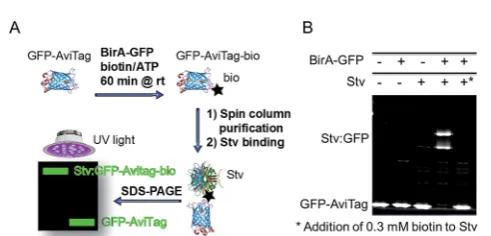
Fig. 1 GFP-based biotinylation activity assay. (A) Schematic repre-sentation of the GFP-based biotinylation activity assay workflow. Recombinant GFP-AviTag (30mM) is reacted at room temperature for 1 h with recombinant BirA-GFP (0.3mM) in the presence of ATP (2.5 mM), biotin (0.5 mM) and MgCl2(1 mM) for biotinylation of the AviTag in 25 mM Tris–HCl (pH 8). The biotinylated GFP-AviTag (GFP-AviTag-bio) is then bound to a 1.5 ml spin column containing Ni-affinity resin to eliminate excess biotin. The bound GFP-AviTag-bio is eluted in buffer containing 200 mM imidazole by centrifuging at 1000g. Following elution, GFP-AviTag-bio is incubated with excess streptavidin (Stv) for 15 min at room temperature and subjected to SDS-PAGE. Samples are loaded without a heat-denaturation step to allow for direct in-gelfluorescence detection of GFP. Fluorescent proteins are analysed by direct UV-irradiation of the gel and integration of GFPfluorescence. (B) Detection of GFP-AviTag biotinylation by SDS-PAGE and Stv-induced band-shift analysis. Bio-tinylation reactions were performed and processed as described in Fig. 1A. Control reactions were performed in the absence of BirA-GFP and Stv, or by saturating Stv with excess biotin (*) as indicated.
Open Access Article. Published on 02 December 2015. Downloaded on 16/05/2017 00:56:22.
This article is licensed under a
[image:2.595.306.552.421.539.2]for comparison to account for protein losses occurred during purication and reaction steps and for normalization. Three independent experiments were performed.
Electrophoretic mobility shiassay
Oligonucleotides psJCU349 50
-TAATCGACTTGTAAACCAAATT-GAAAAGATTTAGGTTTACAAGTCTACAC-30 and psJCU350 50
- GTGTAGACTTGTAAACCTAAATCTTTTCAATTTGGTTTACAAGTC-GATTA-30(400mM in 25 mM Tris, 50 mM NaCl) comprising the
bioO40 bp sequence with 5 bpanking sequences (underlined) were mixed together and annealed by heating at 80C for 2 min followed by slow cooling to room temperature (rt).
All reactions contained 1mM BirA-GFP, 1mMbioO, 10 mM Tris (pH 8), 0.83% glycerol and 50 mM NaCl. In addition to these components some reactions were spiked with 0.83 mM biotin, 0.83 mM ATP and MgCl2, and combinations thereof as
indicated in Fig. 2A. All reactions were incubated at rt for 15 min, then analysed by agarose gel electrophoresis followed by staining with GelRed and integration of uorescent bands (ImageJ, NIH, USA).
Size exclusion chromatography
For SEC analysis of apo- and holoBirA-GFP, BirA-GFP (100ml at 20mM in BirA buffer) was combined with 100ml of either : 25 mM Tris (pH 8) or 25 mM Tris (pH 8) supplemented with 2 mM of biotin, ATP and MgCl2. For SEC in the presence ofbioO,
BirA-GFP wasrst mixed with bioO(100ml at 20mM each in BirA buffer) and incubated for 10 min at rt prior to mixing with 100ml
of 25 mM Tris (pH 8) supplemented with 2 mM of biotin, ATP and MgCl2. All reactions were incubated for 10 min at rt. The
reactions were then injected in a Superdex 200 10/300 GL column (GE Lifesciences) equilibrated in BirA buffer using
a Biologic Duoow Chromatography System (Biorad) with
aowrate of 0.25 ml min1. Following fractionation of
BirA-GFP, 200ml of each fraction was analysed using auorescence plate reader (Victor 3V Wallac 1420, Perkin-Elmer) set to 355 nm excitation/535 nm emission.
SEC analysis of BirA was performed as described above, however the concentrations of BirA andbioOwere increased 10-fold in all cases. UV absorbance was used to determine the peaks and quaternary structure of BirA in each of the condi-tions. Fractions containing the peak expected to be the bio-O:BirA dimer (10ml) were also analysed by SDS-PAGE followed by Coomassie Blue staining (cf.ESI Fig. S2†).
DSF-GTP assay
All reactions (50ml) containing 3mM BirA-GFP were performed in a reaction buffer consisting of 15 mM Tris–HCl (pH 8), 10 mM NaCl and 0.5% (v/v) glycerol. Ligands and additives were added to the reaction buffer as indicated in the following sections. All reactions were equilibrated for 10 min at rt prior being subjected to melt-curve analysis using a real-time thermal cycler (IQ5 iCycler, Bio-Rad). Temperature range was set from 35–85C, increasing in 0.5C increments every 30 s. Data was analysed as previously described.20All reactions were performed at least in duplicates.
Biotin and ATP dependence. To determine the effect of biotin on theTmof BirA-GFP, reactions were performed with
increasing concentrations of biotin ranging from 500 nM to 1 mM as indicated in Fig. 3. To determine the combined effects of ATP and biotin, reactions were performed with increasing concentrations of both species ranging from 500 nM to 1 mM and 1 mM MgCl2. To determine the effect of ATP on theTmof
biotin-bound BirA-GFP, reactions were performed with
increasing concentrations of ATP ranging from 500 nM to 1 mM, 1 mM biotin and 1 mM MgCl2.
Nucleotide specicity. To determine the effect of different nucleotides on theTmof BirA-GFP, reactions were performed in
the presence of 1 mM of either ATP, CTP, UTP or GTP and 1 mM MgCl2. To determine the effect of different nucleotides on the
Tm of biotin-bound BirA-GFP, reactions were performed as
above in the presence of 1 mM biotin. All nucleotide stocks were buffered (pH 8).
Results and Discussion
Biotin transfer activity of BirA and BirA-GFPThe production of a functional GFP-tagged BirA (BirA-GFP) was required for the DSF-GTP assay. First, the biotinylation activity of BirA-GFP was determined with a new GFP-based biotinylation activity assay to assess whether GFP-tagging of BirA was not detrimental to its activity. The new assay relies on complex formation between a biotinylated GFP containing the AviTag
biotinylation sequence (GLNDIFEAQKIEWHE)25–28 and
Fig. 2 DNA-binding activity and dimerization of BirA-GFP and BirA. (A) Determination ofbioO-binding to BirA-GFP by EMSA. BirA-GFP (1mM) was reacted withbioO(0.5mM) in the presence of 1 mM biotin, 1 mM ATP or 1 mM biotin, 1 mM ATP and 1 mM MgCl2for 10 min at rt. Reactions were then analysed by agarose gel electrophoresis and GelRed staining. The positions of freebioOprobe (C) and shifted bands are indicated. Identical experiments were also performed with BirA (cf.ESI Fig. S1†). (B) Top: SEC analysis of BirA-GFP. BirA-GFP (10mM) was injected either alone, in the presence of 1 mM biotin, 1 mM ATP and 1 mM MgCl2, or in the presence ofbioO(10mM), 1 mM biotin, 1 mM ATP and 1 mM MgCl2. The proteins were detected through thefl uo-rescence of the GFP domain of BirA-GFP. Bottom: SEC analysis of BirA. Reactions were as described for BirA-GFP, however BirA andbioO were present at 100mM each and protein peaks were determined by UV absorption.
Open Access Article. Published on 02 December 2015. Downloaded on 16/05/2017 00:56:22.
This article is licensed under a
[image:3.595.49.291.411.571.2]a streptavidin (Stv) probe23 resulting in an electrophoretic mobility shiof the complex detectable by SDS-PAGE (Fig. 1A). The GFP-based biotinylation activity assay is identical in prin-ciple to our recent ‘in-gel biotin–protein conjugate detection assay’.23 Here, a new AviTag-labelled GFP (GFP-AviTag) was developed and used as substrate for BirA-GFP. GFP-AviTag (30mM) was biotinylated by BirA-GFP at room temperature (rt) for 1 h. Aer removal of excess biotin the biotinylated GFP-AviTag was bound to Stv and analysed by SDS-PAGE. For this assay it is important to not heat-denature the samples before loading onto the polyacrylamide gel to avoid loss of the GFP uorophore. The biotinylation activity assay is not intended for measuring enzyme kinetics but it is a useful rapid end-point assay and has the potential to be applied for the identication of biotinylation inhibitors.
In-geluorescence quantitation of the reacted GFP-AviTag showed that BirA-GFP was able to biotinylate 96% of the protein in 1 h at rt (Fig. 1B). Abolition of biotinylated GFP-Avi-Tag:Stv complex formation by the addition of 0.3 mM biotin to Stv prior addition of GFP-AviTag (Fig. 1B, far right lane) conrmed that the GFP-shi observed was the result of Stv
binding. Faint higher order bands were visible in those wells that Stv was included. These were due to a protease contami-nation present in the commercial Stv sample (data not shown). However the level of proteolysis was negligible in the context of determining the activity of BirA-GFP and did not affect quanti-tation. The level of biotinylation obtained with BirA-GFP and the GFP-AviTag corresponds well toin vitrobiotinylation activity levels previously reported by us and others for a variety of tagged and untagged BirA.23,29 Indeed, BirA and BirA-GFP yielded almost identical levels of biotinylated AviTag probes in the same conditions23demonstrating that the tagging of BirA does not signicantly affect its activity.
Dimerization and DNA-binding activity of BirA and BirA-GFP
A 50 bp double strand oligonucleotide consisting of the 40 bp
bioOsequenceanked at either end by 5 bp of natural chro-mosomal sequences was used to test the DNA-binding activities of BirA (cf.ESI Fig. S1†) and BirA-GFP (Fig. 2A). The binding of
BirA and BirA-GFP to bioO was analysed by EMSA
demon-strating that the presence of the GFP did not signicantly affect Fig. 3 Thermal stability of BirA-GFP by DSF-GTP. (A) DSF-GTP melt-curves (d(RFU)/dT) demonstrating the effect of increasing biotin concentrations on BirA-GFP thermal stability. BirA-GFP (3mM) was reacted with increasing concentrations of biotin ranging from 500 nM to 1 mM for 10 min at rt prior to analysis by DSF-GTP. (B) The concentration dependence of biotin onTmof BirA-GFP.DTmvalues were calculated by subtractingTmof apoBirA-GFP from theTmobtained in presence of increasing [biotin]. (C) Nucleotide specificity of BirA-GFP. Effect of different nucleotides (1 mM) as indicated on theTmof BirA-GFP in the presence or absence of 1 mM biotin and MgCl2. BirA-GFP was reacted with each nucleotide (with or without biotin/MgCl2). (D) Comparison of the effect of simultaneous increase of [biotin], [ATP] and [MgCl2] (red squares)vs. independent increase of [ATP] in the presence of 1 mM biotin and MgCl2(open triangles) on BirA-GFPTm. Reactions of BirA-GFP with increasing [biotin] (black circles) were also performed for comparison. For all experiments, error bars represent standard deviations (n¼3).
Open Access Article. Published on 02 December 2015. Downloaded on 16/05/2017 00:56:22.
This article is licensed under a
[image:4.595.114.483.50.364.2]BirA DNA-binding activity. Previous studies have shown that holoBirA binds with the greatest affinity to bioO– i.e. in the presence of the co-repressor bio-50-AMP.7,30,31The biotin-bound BirA and apoBirA bind cooperatively tobioOwith10-fold and
50-fold lower affinity respectively than holoBirA,31 and this was also evident with our GFP-tagged species in our high-salt conditions (Fig. 2A). The same difference in affinity was also conrmed with the BirA species (cf.ESI Fig. S1†).
The quaternary structure of BirA and BirA-GFP was then analysed by size exclusion chromatography (SEC). BirA (injected at 100mM initial concentration) was detected by UV absorption whereas BirA-GFP could be analysed byuorimetry at a ten-fold lower concentration (i.e.10 mM initial concen-tration). Both proteins remained in a monomeric form in their apoforms. Previous data have shown that apoBirA and the biotin-bound form can dimerize in low salt conditions at mM concentrations.31 These complexes would therefore not be observed in our high-salt SEC conditions. A slight shi was observed for both holoBirA and holoBirA-GFP (Fig. 2B). These shis in elution suggest a dramatic change in conformation of the proteins or a fast exchanging dimer form in our SEC conditions. Previous studies indicate that dimerization of holoBirA is inuenced by ionic strength with reported KD
values for dimerization of1 and 10mM in low- and high-salt respectively.30,31 As expected, a dramatic shi in elution was observed for both holoBirA and holoBirA-GFP upon addition of bioO reecting their dimer formation and bioO binding (Fig. 2B and C; ESI Fig. S2†). The high sensitivity of the uo-rimetric SEC assay compared to UV detection, makes it an attractive secondary assay for the identication of molecules capable of interfering with dimer formation and DNA-binding activity of BirA-GFP.
HT characterization of BirA-GFP by DSF-GTP
The structural and functional characterization of BirA-GFP in the previous sections demonstrated that the tethering of GFP has no effect on BirA activity. The use of DSF-GTP requires that a transition in the melting curve can be observed corresponding to the BirA domain unfolding before complete loss of GFP uorescence at80C (Fig. 3A). We identied a clear transition in the melting curve of BirA-GFP and validated the assay with increasing concentrations of biotin for which increasing tran-sition midpoint (Tm) values could be recorded as illustrated in
Fig. 3A. For this, BirA-GFP (3mM) was reacted with increasing concentrations of biotin ranging from 488 nM–1 mM for 10 min at rt, prior to determination ofTmby DSF-GTP. Fig. 3B
illus-trates the perfect correlation between BirA-GFP thermal stability and biotin concentration obtained from data presented in Fig. 3A. The thermal stability of BirA-GFP (41C) increased progressively by up to12.5C in the presence of 1 mM biotin (Fig. 3A and B). A best-t trendline was obtained with aR-square value > 0.99 reecting the excellent reproducibility of the assay. The quality of the new HT DSF-GTP assay for BirA was further assessed byZ0-factor32determination which yielded an excellent
Z0value of 0.785 (n¼96).
BirA-GFP was then proled for nucleotide specicity both alone and in combination with biotin and MgCl2. When the
effect of nucleotides alone was investigated, theTmof BirA-GFP
increased only by2C with 1 mM ATP. GTP was the most
stabilizing2.9C, and CTP and UTP the least stabilizing <2C. Interestingly, theTmof BirA-GFP was increased by12C in the
presence of 1 mM biotin, and by20.5C when reacted with both 1 mM biotin and ATP (i.e.leading to formation of bio-50 -AMP; Fig. 3C). We did not observe any cumulative stabilizing effects for UTP, CTP or GTP that could suggest the formation of higher affinity intermediates with these species in the presence of biotin. The large increase inTmof BirA-GFP upon binding of
both ATP and biotin suggesting the formation of bio-50-AMP is well supported by the recently reportedKD of 50 pM for the
holocomplex.33 Our data correlates well with the sequential binding of biotin and ATP by BirA, with biotin bindingrst leading to the formation of a specic ATP binding site.13–15,34–36 This site is largely non-existent and nonspecic prior to biotin binding. A disordered loop of the apoBirA structure (i.e. resi-dues 212–233) known as the adenylate binding loop (ABL) is involved in ATP binding.36 The ABL becomes ordered upon binding of ATP by BirA and folds over it.15
BirA has previously been shown to have very low in vitro
biotinylation activity with CTP (10%) and GTP (5%).17Our data suggests that GTP binds marginally better to the non-specic binding pocket of apoBirA-GFP in the absence of biotin. Nevertheless, considering the very low affinity of BirA for all nucleotides in the absence of biotin (Fig. 3C) and the total absence of cumulative stabilisation with GTP, CTP and UTP in the presence of biotin, it is unlikely that any of these nucleo-tides could lead to formation of the ABL in the absence of biotin. The low biotinylation activity of BirA observed in the presence of high concentrations of CTP or GTP17could therefore only be explained by reaction of BirA-bound biotin with GTP or CTP without ABL formation. To our knowledge, this is therst study of the sequential binding of different nucleotides and their specicity to the ATP binding site using DSF.
We have previously been able to estimate the apparent binding parameters (Kobs) of glycerol kinase and Tus for their
respective ligands with DSF-GTP in conditions of [ligand] > [protein] using a simple graphical method. In both cases, theKobs
were in agreement with the dissociation constants (KD) obtained
with alternative quantitative methods.20,21K
obsis dened as the
concentration of ligand at which the titration curve trendline for a given ligand (Fig. 3D) intercepts with log[ligand]-axis at theTm
of the unliganded protein. The thermal slopes are identical within a protein for different ligands since they are determined by the unfolding enthalpy.37It is therefore important to note that
Kobsis not directly comparable toKDalthough changes inKobs
will be proportional to changes inKD. Using our simple graphical
method we obtained aKobsof 231 nM (ESI Table S1†) which was 5-fold higher than the previously reported KD of 45 nM38 –
similar to the range of endogenous biotin concentration inE. coli.
The difference observed between ourKobsand the previously
re-portedKDmay potentially be due to the different conditions and
pH in our study as well as temperature effects and possible steric hindrances from the GFP.
Open Access Article. Published on 02 December 2015. Downloaded on 16/05/2017 00:56:22.
This article is licensed under a
Next, we investigated the combined effect of ATP and biotin leading to bio-50-AMP production on BirA-GFP's thermal stability (Fig. 3D). Based on theKDof 50 pM that has recently
been reported for the holoBirA complex, we expected that a sharp increase in BirA-GFP thermal stability would occur upon bio-50-AMP production resulting in a sigmoidal titration curve around ligand concentrations corresponding to the BirA-GFP concentration used in the reaction as well as the presence of two peaks corresponding to the unbound and bound protein form. For this, two different experimental setups were performed: (a) the effects of serial dilutions of an equimolar solution of ATP and biotin were analysed on BirA-GFPTm; and (b) the
concen-tration of biotin was kept constant and the effects of serial dilutions of ATP were analysed. When [biotin] was kept constant (1 mM) and [ATP] was varied, the lowest concentration of ATP tested (500 nM) was not stabilizing BirA-GFP beyond the effect of 1 mM biotin (cf.Fig. 3D; black circles). A very small sigmoi-dicity was observed at2mM ATP followed by a steady increase in BirA-GFPTmwith higher ATP concentrations (Fig. 3D). When
both [biotin] and [ATP] were increased simultaneously (Fig. 3D; red squares), two distinct BirA-GFP Tm peaks were observed
between 1.9–7.8mM immediately suggesting aKD< 10 nM as
expected. OneTmpeak corresponded to the effect of biotin only
(Fig. 3D; black circles) and the otherTmpeak that was sharply
increased–i.e.converging towards the curve obtained forxed [biotin] with increasing [ATP] (Fig. 3D; open triangles)–most likely corresponded to the holoBirA-GFP complex. When the concentration of ligands was >8mM, only the higherTmpeaks
were observed reecting the curve obtained for xed [biotin] with increasing [ATP] (Fig. 3D; cf. open triangles with red squares). As the increase in BirA-GFPTmis very sharp >1mM
biotin and ATP– i.e.much more than would be expected for merely additive effects of biotin and ATP–we can conclude that the very sharp increase in BirA-GFPTmreects formation of the
tighter bio-50-AMP:BirA-GFP complex. It also suggests that binding of biotin signicantly increases the affinity for ATP by several orders of magnitude through formation of the biotin-induced ATP binding site and ABL.
The titration curve obtained with biotin (Fig. 3D; black circles) has a much steeper slope than the ones obtained in the presence of both ATP and biotin (Fig. 3D; open triangles and red squares) revealing that the biotin-bound BirA-GFP form might have a signicantly smaller enthalpy of unfolding compared to the ATP and biotin form. This argument has to be taken with caution due to the complexity of the system. Indeed, formation and dissociation of the bio-50-AMP adduct which binds much tighter than biotin and ATP to BirA-GFP, might be limiting steps inuencing the slope of these titration curves. Nevertheless, due to the linearity of the titration curves suggesting 100% conver-sion of ATP and biotin into bio-50-AMP (Fig. 3D and ESI Table S1†) we decided to determine the Kobs values from the two
different experimental setups. Here, a Kobsof 3.3 pM was
ob-tained in increasing [biotin] and [ATP] which was similar to the
Kobsof 7.4 pM in constant [biotin] and increasing [ATP]. These
Kobsvalues reected quite well the tight affinity andKDof BirA
with the bio-50-AMP adduct (i.e.50 pM in 200 mM KCl and pH 8) reported by Eginton et al.33 although our reactions were
performed with the natural substrates ATP and biotin, in low salt and slightly higher pH conditions.
To our knowledge this is the rst time that the biotin-induced conformational changes leading to ATP binding and bio-50-AMP:BirA-GFP complex could be analysed by DSF in such depth. Overall the combined analyses of nucleotide specicity and ligand concentration dependence demonstrate the versa-tility and breadth of information that can be easily and rapidly gained using DSF-GTP for BirA-GFP characterization.
Conclusions
In this study, we demonstrate the utility and high quality of a new HT DSF-GTP assay for the high-throughput character-ization of BirA (Z0¼0.785), an emerging drug target for anti-biotic development. BirA could be fused to a C-terminal GFP without the requirement for any further solubility tags for high-level production of soluble and functional proteins. BirA-GFP was fully functional when compared to BirA and was able to biotinylate 96% of GFP-AviTag in 1 h at rt. Our results ob-tained with the different natural BirA ligands reected well the previously known properties of BirA as well as revealed new insights into how its binding to individual or combinations of ligands affects the overall thermal stability of the protein. With the validation of E. coli BirA-GFP in the new DSF-GTP, SEC, EMSA and GFP-based biotinylation activity assays now at hand, the subject of future work will be to apply this suite of assays for HT screening and identication of BirA inhibitors and to deci-pher their mechanisms of action and structure activity rela-tionships. It is expected that our workow will be adaptable to other bacterial BirA orthologs as well as the human hol-ocarboxylase synthetase and that it will signicantly streamline their target characterization to screening processes by reducing time and cost.
Con
fl
ict of interest
The authors declare that they have no conict of interest.
References
1 J. W. Campbell and J. E. Cronan,Annu. Rev. Microbiol., 2001,
55, 305–332.
2 J. W. Campbell and J. E. Cronan, J. Bacteriol., 2001, 183, 5982–5990.
3 K. Magnuson, S. Jackowski, C. O. Rock and J. E. Cronan,
Microbiol. Rev., 1993,57, 522–542.
4 N. R. Pendini, M. Y. Yap, S. W. Polyak, N. P. Cowieson, A. Abell, G. W. Booker, J. C. Wallace, J. A. Wilce and M. C. Wilce,Protein Sci., 2013,22, 762–773.
5 B. P. Duckworth, K. M. Nelson and C. C. Aldrich,Curr. Top. Med. Chem., 2012,12, 766–796.
6 T. P. Soares da Costa, W. Tieu, M. Y. Yap, N. R. Pendini, S. W. Polyak, D. Sejer Pedersen, R. Morona, J. D. Turnidge, J. C. Wallace, M. C. Wilce, G. W. Booker and A. D. Abell,
J. Biol. Chem., 2012,287, 17823–17832.
Open Access Article. Published on 02 December 2015. Downloaded on 16/05/2017 00:56:22.
This article is licensed under a
7 E. D. Streaker and D. Beckett,J. Mol. Biol., 2003,325, 937– 948.
8 J. Abbott and D. Beckett,Biochemistry, 1993,32, 9649–9656. 9 O. Prakash and M. A. Eisenberg,Proc. Natl. Acad. Sci. U. S. A.,
1979,76, 5592–5595.
10 P. R. Adikaram and D. Beckett,J. Mol. Biol., 2012,419, 223– 233.
11 E. D. Streaker and D. Beckett,Protein Sci., 2006, 15, 1928– 1935.
12 E. D. Streaker and D. Beckett,Biochemistry, 2006,45, 6417– 6425.
13 L. H. Weaver, K. Kwon, D. Beckett and B. W. Matthews,
Protein Sci., 2001,10, 2618–2622.
14 L. H. Weaver, K. Kwon, D. Beckett and B. W. Matthews,Proc. Natl. Acad. Sci. U. S. A., 2001,98, 6045–6050.
15 Z. A. Wood, L. H. Weaver, P. H. Brown, D. Beckett and B. W. Matthews,J. Mol. Biol., 2006,357, 509–523.
16 H. Zhao and D. Beckett,J. Mol. Biol., 2008,380, 223–236. 17 B. Ng, S. W. Polyak, D. Bird, L. Bailey, J. C. Wallace and
G. W. Booker,Anal. Biochem., 2008,376, 131–136.
18 M. W. Pantoliano, E. C. Petrella, J. D. Kwasnoski, V. S. Lobanov, J. Myslik, E. Graf, T. Carver, E. Asel, B. A. Springer, P. Lane and F. R. Salemme, J. Biomol. Screening, 2001,6, 429–440.
19 M. J. Moreau, I. Morin and P. M. Schaeffer, Mol. BioSyst., 2010,6, 1285–1292.
20 M. J. J. Moreau, I. Morin, S. P. Askin, A. Cooper, N. J. Moreland, S. G. Vasudevan and P. M. Schaeffer,RSC Adv., 2012,2, 11892–11900.
21 M. J. Moreau and P. M. Schaeffer, Mol. BioSyst., 2013, 9, 3146–3154.
22 I. Morin, S. P. Askin and P. M. Schaeffer,Analyst, 2011,136, 4815–4821.
23 A. E. Sorenson, S. P. Askin and P. M. Schaeffer,Anal.Methods, 2015,7, 2087–2092.
24 D. B. Dahdah, I. Morin, M. J. Moreau, N. E. Dixon and P. M. Schaeffer, Chem. Commun., 2009, 3050–3052, DOI: 10.1039/b900905a.
25 P. J. Schatz,Bio-Technol., 1993,11, 1138–1143.
26 D. Beckett, E. Kovaleva and P. J. Schatz,Protein Sci., 1999,8, 921–929.
27 M. G. Cull and P. J. Schatz,Methods Enzymol., 2000,326, 430– 440.
28 M. Howarth and A. Y. Ting,Nat. Protoc., 2008,3, 534–545. 29 Y. Li and R. Sousa,Protein Expression Purif., 2012,82, 162–
167.
30 E. Eisenstein and D. Beckett,Biochemistry, 1999,38, 13077– 13084.
31 E. D. Streaker, A. Gupta and D. Beckett,Biochemistry, 2002,
41, 14263–14271.
32 J. H. Zhang, T. D. Y. Chung and K. R. Oldenburg,J. Biomol. Screening, 1999,4, 67–73.
33 C. Eginton, W. J. Cressman, S. Bachas, H. Wade and D. Beckett,J. Mol. Biol., 2015,427, 1695–1704.
34 K. Kwon, E. D. Streaker, S. Ruparelia and D. Beckett,J. Mol. Biol., 2000,304, 821–833.
35 K. Kwon and D. Beckett,Protein Sci., 2000,9, 1530–1539. 36 S. Naganathan and D. Beckett,J. Mol. Biol., 2007,373, 96–
111.
37 D. Matulis, J. K. Kranz, F. R. Salemme and M. J. Todd,
Biochemistry, 2005,44, 5258–5266.
38 K. Kwon, E. D. Streaker and D. Beckett,Protein Sci., 2002,11, 558–570.
Open Access Article. Published on 02 December 2015. Downloaded on 16/05/2017 00:56:22.
This article is licensed under a

![Fig. 3Thermal stability of BirA-GFP by DSF-GTP. (A) DSF-GTP melt-curves (independent increase of [ATP] in the presence of 1 mM biotin and MgClnucleotide (with or without biotin/MgClsubtracting[biotin] (black circles) were also performed for comparison](https://thumb-us.123doks.com/thumbv2/123dok_us/148638.25782/4.595.114.483.50.364/thermal-stability-independent-presence-mgclnucleotide-mgclsubtracting-performed-comparison.webp)