Identifies a CD8
ⴙT Cell Subset Associated with Viral Control during
Chronic Human Immunodeficiency Virus Infection
Federico Simonetta,a,b,cStéphane Hua,a,bCamille Lécuroux,a,bSandie Gérard,a,bFaroudy Boufassa,dAsier Sáez-Cirión,e Gianfranco Pancino,eCécile Goujard,b,fOlivier Lambotte,a,b,fAlain Venet,a,bChristine Bourgeoisa,b
INSERM, U1012, Le Kremlin-Bicêtre, Francea; Université Paris-Sud, UMR-S1012, Le Kremlin-Bicêtre, Franceb; Division of Hematology, Department of Medical Specialties, Geneva University Hospitals, Geneva, Switzerlandc; INSERM UMR-S 1018, Centre d’études en santé publique, Le Kremlin-Bicêtre, Franced; Institut Pasteur, Unité de Régulation des Infections Rétrovirales, Paris, Francee; AP-HP, Hôpital Bicêtre, Service de Médecine Interne et Immunologie clinique, Le Kremlin-Bicêtre, Francef
ABSTRACT
During HIV infection, increased CD57 expression among CD8ⴙT cells has been associated with immune senescence and defec-tive immune responses. Interestingly, CD57-expressing CD8ⴙT cells exhibit a dual profile, being simultaneously highly cyto-toxic (terminally differentiated effectors) and poorly proliferative (replicative senescent). Recent publications point toward a positive role of CD57-expressing CD8ⴙT cell subsets, presumably due to their high cytolytic activity. We further investigated the phenotype of CD57-expressing CD8ⴙT cells in healthy donors and during HIV infection combining CD57 expression to Eome-sodermin (EOMES), a T box transcription factor which determines, coordinately with T-bet, effector and memory CD8ⴙT cell differentiation. We defined in healthy donors two functionally distinct CD57-expressing CD8ⴙT cell subsets exhibiting different levels of EOMES expression: EOMEShiCD57ⴙand EOMESintCD57ⴙCD8ⴙT cells. EOMEShiCD57ⴙcells exhibited low cyto-toxic activity but preserved proliferative capacity and interleukin 7 (IL-7) receptor expression, whereas EOMESintCD57ⴙcells exhibited obvious cytotoxic functions and a more terminally differentiated phenotype. We next performed a similar analysis in different contexts of HIV infection: primary infected patients, long-term viremic patients, aviremic patients treated with antiret-roviral therapy, and HIV controllers; we demonstrated a higher percentage of CD57-expressing cells in all HIV-infected patients regardless of virological status. When heterogeneity in EOMES expression among CD57 cells was taken into account, we de-tected significantly higher proportions of EOMEShiCD57ⴙcells among HIV-specific and nonspecific CD8ⴙT cells from HIV controllers than in aviremic antiretroviral-treated patients and viremic patients. Importantly, such a peculiar non-terminally differentiated EOMEShiCD57ⴙphenotypic profile was associated with viral control.
IMPORTANCE
This study demonstrates that functional heterogeneity exists among CD57-expressing CD8 T cells, which include both termi-nally differentiated, highly cytotoxic EOMESintCD57ⴙCD8ⴙT cells and less differentiated EOMEShiCD57ⴙCD8 T cells, which do not exhibit immediate cytotoxic functions but present high proliferative capacity. Interestingly, HIV controllers present a high proportion of EOMEShiCD57 cells among CD57-expressing HIV-specific CD8 T cells compared to both long-term viremic
and aviremic antiretroviral therapy (ART)-treated patients, suggesting a beneficial role for this cell subset in viral control.
D
uring chronic HIV infection, virus-specific CD8⫹ T cells functionally decline, progressively losing their proliferative capacity and cytotoxic potential and progressing to exhaustion and/or senescence (1,2) except in rare individuals: the HIV con-trollers (HIC). These patients exhibit persistently undetectable HIV RNA in the absence of antiretroviral therapy (ART) (3) and maintain polyfunctional HIV-specific CD8⫹T cells which retain proliferative potential (4–6) as well as the ability to produce effec-tor cytokines and cytotoxic molecules (5–8). Such a peculiar, nonexhausted profile has been related to the presence of longer telomeres and higher levels of constitutive telomerase activity in HIV-specific CD8⫹T cells from HIC (2). CD57 expression iden-tifies senescent human T cells displaying a terminally differenti-ated phenotype (1,10–12) and increases during HIV infection, probably as a result of chronic immune activation (11,13). Inter-estingly, CD57-expressing CD8⫹T cells exhibit a dual profile, being simultaneously highly efficient cytotoxic cells (terminally differentiated effectors) (14) and poor proliferative (replicative senescence) subsets (1).However, recent publications provided new insights on the
role of CD57-expressing cells during HIV infection. Lee et al. demonstrated that HIV and cytomegalovirus (CMV) differently regulate CD57 expression on CD8⫹T cells, inducing terminal differentiation in CMV infection but accumulation of less differ-entiated cells in HIV infection, as assessed by a decreased propor-tion of CD57-expressing cells among CD28⫺CD8⫹T cells (15). The same group also demonstrated that proportions of CD57-expressing CD28⫺CD8⫹T cells were increased following ART treatment (16). Additionally, low proportions of CD28⫺CD8⫹T
Received11 July 2014 Accepted28 July 2014
Published ahead of print6 August 2014
Editor:G. Silvestri
Address correspondence to Christine Bourgeois, [email protected]. F.S. and S.H. contributed equally to this article.
Copyright © 2014, American Society for Microbiology. All Rights Reserved.
doi:10.1128/JVI.02013-14
on November 7, 2019 by guest
http://jvi.asm.org/
cells expressing CD57 were a predictive marker of mortality among ART-treated HIV-infected patients with advanced disease (16). These recent data point toward a positive role for CD57-expressing CD8⫹T cell subsets, presumably due to their high cy-tolytic activity, in contrast to the deleterious impact of immune senescence, usually associated with the CD57-expressing subsets. We further investigated the phenotype of CD57-expressing CD8⫹ T cells combining CD57 expression to Eomesodermin (EOMES), a T box transcription factor which determines, coordinately with T-bet, effector CD8⫹ T cell differentiation, regulating interferon gamma (IFN-␥), perforin, and granzyme B expression (17–19), as well as memory CD8⫹T cell transition and maintenance (20–22). EOMES expression has been reported to be upregulated in early effectors and to further increase during memory differentiation (20). During murine chronic viral infections, maintained high T-bet expression has been associated with terminal effector differ-entiation (23, 24), whereas high EOMES expression correlates with the long-term memory fraction (25) and characterizes cells exhibiting increased proliferative potential, granzyme B produc-tion, and cytotoxicity (26). At present, the precise role performed by EOMES during HIV infection remains unclear: a recent report showed that EOMES expression was increased in viremic HIV patients (27), whereasEomesmRNA has been shown to signifi-cantly decrease in HIV-specific CD8⫹T cells from primary HIV infection to chronic phase (28), suggesting that loss of EOMES expression could be associated with CD8⫹T cell functional de-cline.
In this report, we demonstrate functional heterogeneity among CD57-expressing CD8⫹T cells. By combining CD57 with EOMES expression, we were able to identify in healthy donors (HD) two functionally distinct subsets: EOMEShiCD57⫹CD8⫹T cells
dis-playing a memory phenotype and EOMESintCD57⫹CD8⫹T cells
exhibiting high cytotoxic functions and a terminally differentiated phenotype. We performed a cross-sectional analysis of EOMES and CD57 expression in CD8⫹T cells isolated from different groups of HIV-infected patients: primary infected patients, ART-treated aviremic patients, chronically viremic patients, and HIV controllers. We showed that CD57 expression is increased in all HIV-infected patient groups studied, including HIV controllers. Taking into account heterogeneity in EOMES expression among CD57-expressing CD8⫹T cells, we showed that HIV controllers maintained significantly lower proportions of EOMESintCD57⫹
cells and higher fractions of EOMEShiCD57⫹cells among both
HIV-specific and nonspecific CD8⫹T cells than both untreated viremic and ART-treated aviremic patients and that such a pecu-liar phenotype was associated with viral control.
These data suggest that CD57 expressionper seis not a reliable marker of terminal differentiation, whereas the combination of EOMES and CD57 provided a more accurate insight on the tight balance between proliferating memory and cytotoxic terminally differentiated cells during HIV infection.
MATERIALS AND METHODS
Study participants.We collected samples from 147 HIV-infected individ-uals with their informed consent. Thirty-two primary infected untreated patients were enrolled in the French ANRS multicenter PRIMO cohort (Agence Nationale de Recherche sur le SIDA, CO06). Primary infection was defined by HIV RNA positivity and by a negative or emerging anti-body response. We also studied 30 untreated chronically infected patients who are referred to as viremic patients. Thirty-one ART-treated aviremic
individuals presenting plasma HIV RNA levels of⬍50 copies/ml and treated for at least 12 months were included in the study. Fifty-four pa-tients were enrolled in the French HIV controller cohort (ANRS, CO21 CODEX) (inclusion criteria: no ART, HIV infection for⬎5 years, five last consecutive plasma HIV RNA values of⬍400 copies/ml). HIV RNA de-tection assay with a dede-tection limit reaching⬍40 copies/ml was per-formed for all samples from HIV controllers. Clinical and biologic char-acteristics of participants are shown inTable 1. All HIV-infected patient groups were age matched with the exception of HIV controllers, which by definition are long-term HIV-infected patients and are older than others. Peripheral blood samples from 21 non-HIV-infected blood donors were obtained from the Etablissement Français du Sang (Saint Louis Hospital, Paris, France). These donors were selected to be age matched with HIV controllers.
Laboratory studies. (i) Cell preparation.Peripheral blood mononu-clear cells (PBMCs) were isolated from anticoagulated blood by Ficoll density gradient centrifugation. Human leukocyte antigen (HLA) typing was done with the complement-dependent microlymphocytotoxic tech-nique (One Lambda, Montpellier, France). Cells were cryopreserved in liquid nitrogen for subsequent analysis.
(ii) Flow cytometry.PBMCs were analyzed by 10-color flow cytom-etry. Cryopreserved cells were thawed, and after incubation with purified Fc receptor binding inhibitor (e-Bioscience), samples were stained with labeled antibodies against surface markers for 15 min at 4°C. Conjugated antibodies against the following were used: CD4 (fluorescein isothiocya-nate [FITC]), CD57 (phycoerythrin [PE]-CF594), CD27 (Alexa Fluor 700), CD45RA (allophycocyanin [APC]-H7), CD8 (V450), and CD3 (V500) from BD Biosciences (San Jose, CA) and CD57 (FITC or PE) from Miltenyi. Intracellular staining for cytotoxic molecules was performed using anti-granzyme B (FITC or Alexa Fluor 700; clone GB11 [BD Biosci-ences]) and antiperforin (FITC; clone B-D48 [Diaclone]). Intranuclear detection of EOMES (anti-EOMES Alexa Fluor 647; clone WD1928 [e-Bioscience]) and T-bet (anti-T-bet peridinin chlorophyll protein [PerCP]-Cy5.5; clone 4B10 [e-Bioscience]) was performed on fixed and permeabilized cells by following the manufacturer’s instructions (e-Bio-science). Detection of perforin and granzyme B was performedex vivo
without any stimulation or brefeldin/monensin incubation. Samples were acquired on an LSRFortessa cell analyzer (BD Biosciences), and data files were analyzed using FlowJo software (Tree Star Inc.).
[image:2.585.301.544.79.166.2]Peptide-HLA class 1 pentamers.HIV-specific CD8⫹T cells were identified by employing soluble PE-labeled peptide-HLA class 1 pentam-ers (Proimmune, Oxford, United Kingdom) derived from the HIV Gag, Nef, Pol, and Env proteins. The following epitopes were used: the HLA-A*0201-restricted peptide ligands SLYNTVATL (Gag residues 77 to 85) and ILKEPVHGV (Pol residues 476 to 484), the A*0301-restricted pep-tide ligands RLRPGGKKK (Gag residues 20 to 28) and QVPLRPMTYK (Nef residues 73 to 82), the A*1101-restricted ligand AVDLSHFLK (Nef residues 84 to 92), the A*2402-restricted peptide ligand RYPLTFGWCY TABLE 1Characteristics of HIV-infected patients and healthy controlsa
Group (no. of individuals) Age (yrs)
Viral load (log10 copies/ml)
CD4 count (cells/l)
Healthy donors (21) 49 (31–58)
Primary infected patients (32) 35 (30–40) 5.2 (4.7–5.8) 469 (363–614) ART untreated patients (30) 37 (29–42) 4.4 (3.9–4.8) 488 (361–732) ART treated patients (31) 40 (33–48) ⬍1.7 771 (544–893)
HIV controllers (54) 47 (40–53) ⬍2.6 771 (596–931)
aA total of 168 subjects were included in this study, in the 5 groups listed: healthy
donors, HIV-infected patients studied during primary HIV infection, non-ART-treated viremic patients, aviremic (viral load⬍50 copies/ml) ART-treated patients, and HIV controllers, defined as showing spontaneous viral control for more than 5 years (viral load⬍400 copies/ml). Median values and 25th and 75th percentiles are presented (in parentheses) for age, viral load, and CD4 count for each group.
on November 7, 2019 by guest
http://jvi.asm.org/
(Nef residues 134 to 143), the B*0702-restricted peptide ligand IPRRI RQGL (Env residues 848 to 856), the B*0801-restricted peptide ligands GEIYKRWII (Gag residues 259 to 267) and FLKEKGGL (Nef residues 90 to 97), and the B*2705-restricted peptide ligand KRWIILGLNK (Gag res-idues 263 to 272).
Statistical methods.Statistical analysis was performed using Graph-Pad prism software. Nonparametric Mann-Whitney test was employed to compare cell subsets or patient groups. Spearman’s rank test was used to determine correlations.Pvalues above 0.05 were considered not statisti-cally significant.
RESULTS
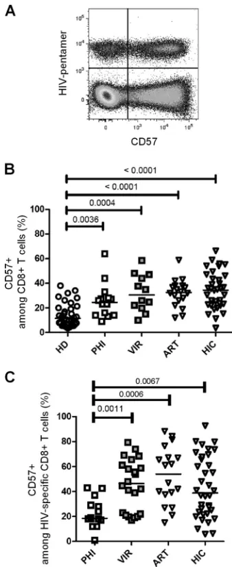
CD57 expression is increased on both total and HIV-specific CD8ⴙT cells in chronically HIV-infected patients indepen-dently of viral load.We first compared degrees of CD57 surface expression in CD8⫹T cells isolated from HIV-negative subjects and from different HIV-infected patient groups, namely, subjects with primary HIV infection (PHI), long-term viremic (VIR) pa-tients, ART-treated aviremic papa-tients, and HIV controllers (HIC). For HIV-infected patients, analysis was performed on both total and HIV-specific CD8⫹ T cells identified using HIV epitope-HLA-I pentamers (Fig. 1A). In healthy donors, 11.5% (median; interquartile range [IQR], 3.5% to 38.0%) of CD8⫹T cells ex-pressed CD57 (Fig. 1B). Analysis of CD57 expression in HIV-infected individuals revealed a significant increase in CD57-ex-pressing CD8⫹ T cell proportions in primary HIV-infected patients (24.5%; IQR, 9.0% to 64.0%;P⫽ 0.0036), untreated viremic chronically infected patients (30.5%; IQR, 10.0% to 58.6%;P⫽0.0004), and aviremic ART-treated patients (32.7%; IQR, 12.0% to 58.9%;P⬍0.0001) compared with healthy donors (Fig. 1B). Interestingly, CD8⫹T cells isolated from HIV control-lers also expressed increased levels of CD57 (34.6%; IQR, 3.7% to 66.4%;P⬍0.0001) compared to those expressed by healthy do-nors (Fig. 1B). These results indicate that CD57 expression in CD8⫹T cells is increased during early and more advanced phases of HIV infection, independently of active viral replication.
We next assessed CD57 expression at the surface of HIV-spe-cific CD8⫹T cells. HIV-specific CD8⫹T cells from PHI patients displayed only low proportions of CD57-expressing cells (18.50%; IQR, 1.0% to 43.0%) (Fig. 1C). Conversely, untreated viremic chronically infected patients displayed significantly higher pro-portions of CD57-positive HIV-specific CD8⫹ T cells (46.3%; IQR, 17.0% to 79.4%) than PHI patients (P⫽0.0011) (Fig. 1C). Among aviremic patients, HIV-specific CD8⫹T cells from ART-treated patients and from HIC contained high proportions of CD57-positive cells (ART-treated patients, 54.0% [IQR, 15.0% to 88.3%]; HIC, 38.8% [IQR, 5.9% to 92.7%]) (Fig. 1C). All chron-ically infected groups exhibited significantly higher proportions of CD57-expressing HIV-specific CD8⫹T cells than did PHI pa-tients (P⫽0.0011, P⫽0.0006, and P⫽0.0067 for VIR and ART-treated patients and HIC, respectively). These results indi-cate that chronically HIV-infected patients, including HIV con-trollers, display high proportions of CD57-expressing HIV-spe-cific and nonspeHIV-spe-cific CD8⫹T cells.
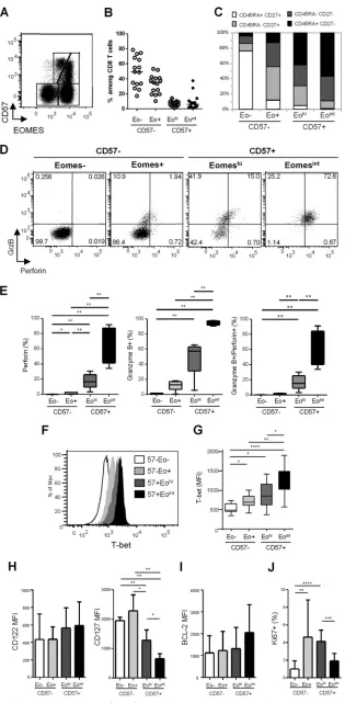
Identification of two distinct CD57-expressing CD8ⴙT cell subsets based on EOMES expression.Expression of the T box transcription factor EOMES has been recently associated with ter-minal differentiation, CD57 being coexpressed with EOMES (29). In an analysis of EOMES and CD57 coexpression in CD8⫹T cells, four cell subsets were identified (Fig. 2AandB): (i) a double neg-ative subset, EOMES⫺CD57⫺, represented approximately half of
CD8⫹T cells in healthy donors (median, 49.8% [IQR, 34.8% to 66.1%]); (ii) an EOMES⫹CD57⫺population represented the sec-ond most represented subset (median, 34.7% [22.7% to 42.3%]). Finally, EOMES⫹CD57⫹cells showed a heterogeneous level of EOMES expression, allowing us to identify two smaller subsets: (iii) an EOMEShigh CD57⫹ (EOMEShi CD57⫹) and (iv) an
EOMESintermediateCD57⫹subset (EOMESintCD57⫹), represent-ing in healthy donors 7.6% (3.7% to 11.4%) and 4.0% (1.4% to 8.7%) of total CD8⫹T cells, respectively. When surface expression of CD45RA and CD27 was analyzed to characterize cellular differ-entiation of the four CD8⫹T cell subsets identified on the basis of
FIG 1CD57 expression in CD8⫹T cells during HIV infection. (A) Represen-tative dot plot of CD57 expression on CD8⫹T cells costained with pentamers folded with HIV-derived peptides recovered from one HIV-infected patient. (B) CD57-expressing cells among CD8⫹T cells in peripheral blood mononu-clear cells (PBMCs) from healthy donors (HD; open circles), primary HIV-infected patients (PHI; open squares), untreated viremic patients (VIR; filled gray squares), aviremic patients treated with ART (ART; open triangles), and HIV controllers (HIC; filled gray triangles). (C) CD57-expressing cells among HIV-specific CD8⫹T cells, identified by pentamer staining as shown in panel A. Median percentages are represented for each group, andPvalues are indi-cated when significant.
on November 7, 2019 by guest
http://jvi.asm.org/
[image:3.585.334.503.65.478.2]FIG 2Phenotypic and functional characterization of EOMES⫹CD8⫹T cells. (A) Representative dot plot of EOMES and CD57 expression in CD8⫹T cells from one healthy donor (HD). (B) Proportions of CD8⫹T cell subsets defined by EOMES and CD57 expression in HD: EOMES⫺CD57⫺(Eo⫺CD57⫺), EOMES⫹
on November 7, 2019 by guest
http://jvi.asm.org/
[image:4.585.136.451.67.708.2]EOMES and CD57 expression, we found that among CD57⫺ CD8⫹T cells, EOMES⫺cells were mainly composed of CD45RA⫹ CD27⫹naive T cells (Fig. 2C). Conversely, EOMES⫹CD57⫺cells contained the highest proportions of CD45RA⫺CD27⫹memory CD8⫹T cells and an important proportion of CD45RA⫺CD27⫺ effector/memory CD8⫹ T cells (Fig. 2C). CD57⫹ cells, either EOMEShior EOMESint, were mainly represented by CD45RA⫺
CD27⫺effector/memory and CD45RA⫹CD27⫺terminally dif-ferentiated CD8⫹ T cells (TEMRA) (Fig. 2C). The EOMESint
CD57⫹CD8⫹T cell subset contained the highest proportions of CD45RA⫹CD27⫺TEMRA CD8⫹T cells, suggesting higher dif-ferentiation of this cell subset. However, we could not associate clear phenotypic discrimination between EOMEShiand EOMESint
CD57-expressing CD8⫹T cell fractions.
The EOMESintCD57ⴙphenotype is associated with higher cytotoxic potential and T-bet coexpression in CD8ⴙT cells.We next studied the cytotoxic potential of the four CD8⫹T cell sub-sets defined by EOMES and CD57 expression based on granzyme B and perforin expression. CD57⫺CD8⫹T cells, either EOMES negative or positive, were almost completely deprived in cytotoxic molecule-producing cells (Fig. 2DandE). EOMEShiCD57⫹cells
contained significantly higher proportions of cells expressing per-forin (16.6% [9.5% to 25.9%]), granzyme B (57.4% [31.1% to 63.0%]), or both (15.3% [9.0% to 25.4%]) than did EOMES⫺ CD57⫺or EOMES⫹CD57⫺cell subsets (Fig. 2DandE). Finally, EOMESintCD57⫹cells presented the highest proportions of per-forin-expressing (73.1% [41.4% to 87.2%]) and granzyme B-ex-pressing (93.8% [91.9% to 97.5%]) cells, the great majority of EOMESintCD57⫹CD8⫹T cells (72.3% [40.1% to 84.5%])
coex-pressing both granzyme B and perforin (Fig. 2DandE). We thus demonstrated that CD57-expressing CD8⫹T cells include two fractions exhibiting highly different cytotoxic potentials that can be segregated using EOMES expression.
As the T box transcription factor T-bet also modulates CD8⫹ T-cell differentiation and granzyme B and perforin production in mice, we assessed T-bet expression in CD8⫹T cell subsets identi-fied based on EOMES and CD57 expression. The lowest levels of T-bet were expressed by EOMES-negative cells (median fluores-cence intensity [MFI], 489 [440 to 651]), in accordance with their less differentiated resting state (Fig. 2FandG). A progressive in-crease in T-bet expression was identified from EOMES⫹CD57⫺ cells (MFI, 696 [597 to 846]) to EOMEShiCD57⫹cells (MFI, 859 [613 to 1,160]) and EOMESintCD57⫹cells (MFI, 1,328 [1,013 to
1,490]) (Fig. 2FandG). Interestingly, the EOMESintCD57⫹ sub-set, which presented the most terminally differentiated effector phenotype and the highest granzyme B and perforin production, expressed the highest levels of T-bet. Collectively, these data indi-cate that EOMESintCD57⫹CD8⫹T cells represent a terminally
differentiated effector cell subset characterized by the highest cy-totoxic potential and highest levels of T-bet expression.
The EOMEShiCD57ⴙphenotype is associated with higher CD127 expression and higher proliferation ability in CD57ⴙ CD8ⴙT cells.We next characterized CD8⫹T cell subsets for surface expression of CD122 and CD127, which are necessary for transduc-tion of interleukin 15 (IL-15) and IL-7 signaling, respectively, and therefore essential for CD8⫹T cell homeostasis. As shown inFig. 2H, we failed to detect any significant difference in CD122 expression at the surface of the four CD8⫹T cell subsets identified based on CD57 and EOMES expression. Conversely, we found that EOMEShiand
EOMESintCD57⫹cells displayed significantly lower levels of CD127 than CD57-negative subsets (Fig. 2H). However, EOMEShiCD57⫹
cells retained higher levels of CD127 expression (mean MFI, 1,282⫾ 351) than did the highly cytotoxic EOMESint CD57⫹ cells
(MFI, 657⫾166) (Fig. 2H).
To determine whether such a difference in CD127 was associ-ated with any homeostatic advantage, we studied the expression of the antiapoptotic molecule BCL-2 as a marker of survival and the expression of Ki-67 as a proliferation marker. As shown inFig. 2I, we failed to detect any significant difference in BCL-2 expression among the four subsets. Conversely, we found lower proportions of Ki-67-expressing cells in EOMES⫺CD57⫺cells (1.0%⫾0.9%) compared to both EOMES⫹CD57⫺(4.6%⫾4.2%) and EOMEShi CD57⫹ CD8⫹ (4.1% ⫾ 1.3%) T cells (Fig. 2J). Interestingly, EOMEShiCD57⫹cell subsets presented significantly higher pro-portions of Ki-67⫹cells than did EOMESintCD57⫹CD8⫹T cells
(1.9%⫾0.8%) (Fig. 2J). Collectively, these data suggest a higher homeostatic potential for EOMEShiCD57⫹CD8⫹T cells, which
exhibited higher expression of IL-7 receptor and proliferative ca-pacity compared with the high immediate cytotoxicity provided by the EOMESintCD57⫹subset.
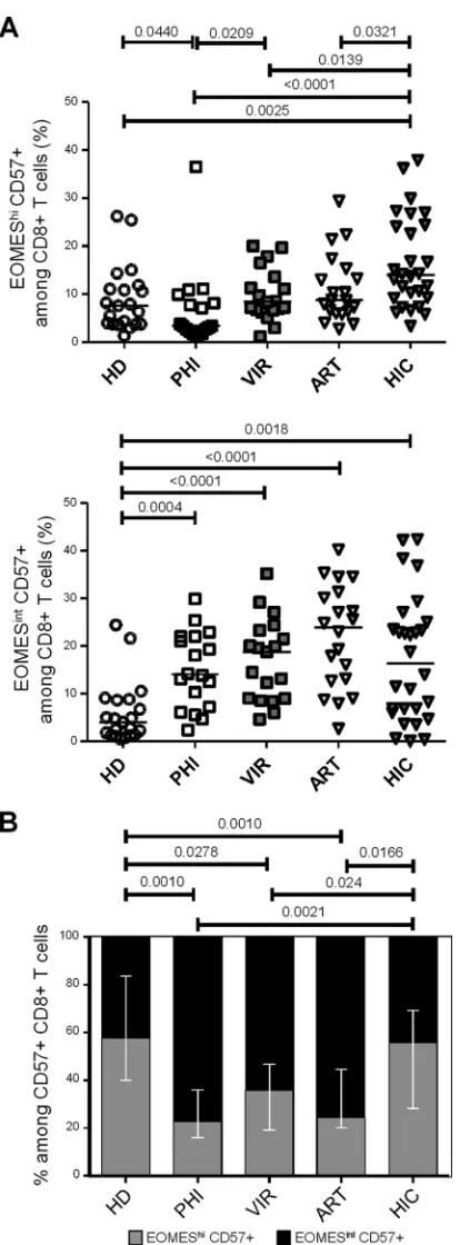
Higher levels of EOMEShiCD57ⴙCD8ⴙT cells in HIV con-trollers.We then investigated CD57 expression during HIV infec-tion introducing EOMES expression. Primary HIV-infected pa-tients displayed significantly reduced proportions of EOMEShi CD57⫹cells (3.4% [1.1% to 36.5%]) compared to those displayed by healthy donors (7.6% [1.3% to 26.2];P⫽0.0440) and viremic individuals (8.4% [1.1% to 20.0%];P⫽0.0209) (Fig. 3A, upper graph). Conversely, proportions of EOMEShi CD57⫹CD8⫹T cells in chronically HIV-infected patients, either viremic or avire-mic ART treated (8.8% [2.8% to 29.4%]), were not different from those observed in healthy individuals (Fig. 3A, upper graph). In-terestingly, CD8⫹T cells from HIV controllers displayed signifi-cantly higher proportions of EOMEShiCD57⫹cells (13.9% [3.3%
to 37.9%]) than did healthy donors (P⫽0.0025) and primary HIV-infected (P⬍0.0001), chronically viremic (P⫽0.0139), and aviremic ART-treated (P ⫽ 0.0321) patients (Fig. 3A, upper
CD57⫺(Eo⫹CD57⫺), EOMEShiCD57⫹(EohiCD57⫹), and EOMESintCD57⫹(EointCD57⫹). (C) Phenotypic characterization of EOMES⫺CD57⫺, EOMES⫹ CD57⫺, EOMEShiCD57⫹, and EOMESintCD57⫹cells based on CD45RA and CD27 expression. Graphs represent proportions of CD45RA⫹CD27⫹(white bars), CD45RA⫺CD27⫹(light gray bars), CD45RA⫺CD27⫺(dark gray bars), and CD45RA⫹CD27⫺(black bars) CD8⫹T cells. (D) Representative dot plots of granzyme B and perforin expression in CD57⫺and CD57⫹CD8⫹T cell subsets defined based on EOMES expression from one healthy donor. (E) Percentages of perforin-positive (left graph), granzyme B-positive (middle graph), and granzyme B- and perforin-coexpressing (right graph) cells among EOMES⫺CD57⫺ (white bars), EOMES⫹CD57⫺(light gray bars), EOMEShiCD57⫹(dark gray bars), and EOMESintCD57⫹(black bars) CD8⫹T cells from HD (n⫽10). (F) T-bet expression in CD8⫹T cell subsets. Flow cytometry histograms show representative results from one individual. (G) Histograms represent the median T-bet median fluorescence intensity (MFI)⫾SD in EOMES⫺CD57⫺(white bars), EOMES⫹CD57⫺(light gray bars), EOMEShiCD57⫹(dark gray bars), and EOMESintCD57⫹(black bars) CD8⫹T cells from 10 HD. (H to J) Mean MFI of CD122 and CD127 (H) and BCL-2 (I) and mean percentages of Ki-67⫹cells (J) in EOMES-CD57⫺(white bars), EOMES⫹CD57⫺(light gray bars), EOMEShiCD57⫹(dark gray bars), and EOMESintCD57⫹(black bars) CD8 T cells from 10 HD. *,P⬍0.05; **,P⬍0.01; ***,P⬍0.001; ****,P⬍0.0001.
on November 7, 2019 by guest
http://jvi.asm.org/
graph). When we focused on the EOMESintCD57⫹CD8⫹T cell
subset, i.e., the fraction exhibiting high cytolytic contentex vivo, we observed higher proportions in HIV-infected patient groups, including primary infected individuals (14.1% [2.4% to 29.9%]; P ⫽ 0.0004), chronically infected patients (18.8% [4.6% to 35.2%]; P ⬍ 0.0001), aviremic ART-treated patients (23.9% [2.7% to 40.2%];P⬍0.0001), and HIV controllers (16.4% [0.2% to 42.3%];P⫽0.0018) than in healthy donors (4.0% [0.5% to 24.4%]), while we failed to detect any significant difference among HIV-infected patient groups (Fig. 3A, lower graph). Because such increases may be directly driven by the higher percentage of CD57-expressing cells among CD8⫹T cells observed in HIV-in-fected patients (as shown inFig. 1B), we analyzed the proportions of EOMEShi CD57⫹ and EOMESint CD57⫹ fractions among CD57-expressing CD8⫹T cells. We observed that primary HIV-infected, chronically viremic, and aviremic ART-treated patients presented a significant reduction in the proportions of EOMEShi
cells among CD57⫹CD8⫹T cells associated with an increase on the EOMESintfraction (Fig. 3B). Interestingly, HIV controllers
represented the only patient group retaining a balance of these two subsets similar to that observed in healthy donors (Fig. 3C). Col-lectively, these results show that while displaying proportions of CD57⫹CD8⫹T cells similar to those of other groups (Fig. 1B), HIV controllers retained higher proportions of EOMEShi cells among CD57⫹cells.
HIV-specific CD8ⴙT cells from HIV controllers present an
EOMEShi CD57ⴙ phenotype. We next assessed EOMES and
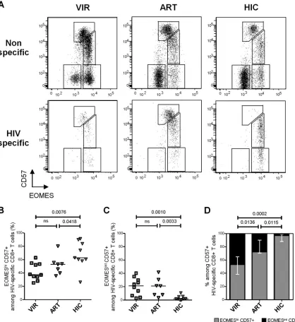
CD57 expression in HIV-specific CD8⫹T cells from chronically viremic patients (n⫽10), aviremic ART-treated individuals (n⫽ 7), and HIV controllers (n⫽9) identified using HIV epitope-HLA-I pentamers (Fig. 4A). We detected significantly higher pro-portions of EOMEShi CD57⫹cells (i.e., the fraction exhibiting higher survival and proliferation profiles) among HIV-specific CD8⫹T cells from HIV controllers (62.8% [26.3% to 91.2%]) than in cells from both chronic viremic (37.7% [25.2% to 62.9%]; P⫽0.0076) and ART-treated (53.1% [35.7% to 80.2%]; P⫽ 0.0418) patients (Fig. 4B). Conversely, lower proportions of EOMESintCD57⫹cells (i.e., the fraction exhibiting higher cyto-lytic molecule contentsex vivo) among HIV-specific CD8⫹T cells were observed in HIV controllers (1.9% [0.2% to 10.7%]) than in both chronic viremic (21.4% [2.7% to 40.1%];P⫽0.0010) and ART-treated (21.1% [3.5% to 41.7%];P⫽0.0418) patients (Fig. 4C). Restricting the analysis to CD57⫹HIV-specific CD8⫹T cells, we observed the lowest proportions of EOMEShi cells among CD57-expressing cells in chronical viremic HIV-infected individ-uals (53.1% [30.9% to 90.3%]) (Fig. 4D). In contrast, CD57⫹ HIV-specific CD8⫹ T cells from HIV controllers were almost completely constituted by EOMEShiCD57⫹cells (96.7% [85.5% to 99.7%]), EOMESintCD57⫹cells being barely detectable (Fig.
4D). These data confirm in HIV-specific CD8⫹T cells results ob-tained in analyses performed on global CD8⫹T cells and indicate that CD57-expressing HIV-specific CD8⫹T cells from HIV con-trollers presented a skewed phenotype toward an EOMEShi
CD57⫹phenotype compared with both those from viremic and aviremic ART-treated patients.
Higher EOMEShi CD57ⴙ proportions among both global
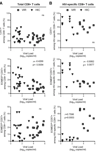
and HIV-specific CD8ⴙT cells are associated with lower viral loads.Finally, we studied whether CD57-expressing CD8⫹T cells subsets correlated with viral load in chronically HIV-infected un-treated patients. HIV load did not correlate with CD57⫹proportions
FIG 3Effects of different phases of HIV infection on CD57- and EOMES-expressing CD8⫹T cell subsets. (A and B) Graphs present EOMEShiCD57⫹ (A) and EOMESintCD57⫹(B) percentages among CD8⫹T cells in PBMCs from HD, primary HIV-infected patients (PHI; open squares), untreated vire-mic patients (VIR; filled gray squares), ART-treated avirevire-mic patients (ART; open triangles), and HIV controllers (HIC; filled gray triangles). (C) Propor-tions of EOMEShiCD57⫹(gray bars) and EOMESintCD57⫹(black bars) among CD57⫹CD8⫹T cells from HD and PHI, VIR, ART, and HIC patients.
Pvalues are indicated.
on November 7, 2019 by guest
http://jvi.asm.org/
[image:6.585.62.269.61.621.2]of either total (Fig. 5A, upper graph) or HIV-specific (Fig. 5B, upper graph) CD8⫹T cells. However, when we took into account CD57⫹ CD8⫹T cell heterogeneity in terms of EOMES expression, we de-tected a significant inverse correlation between viral load and EOMEShiCD57⫹proportions among both total (r⫽ ⫺0.4269;P⫽ 0.0054) and HIV-specific (r⫽ ⫺0.6062;P⫽0.0077) CD8⫹T cells (Fig. 5, middle graphs). In contrast, while no association with viral load was observed when considering the immediately cytotoxic EOMESintCD57⫹subset among total CD8 T cells (Fig. 5A, lower graph), we found a significant, positive correlation between viral lev-els and the proportion of EOMESintCD57⫹cells among HIV-specific
CD8⫹T cells (r⫽ ⫺0.7596,P⫽0.0003) (Fig. 5B, lower graph). Collectively, these results revealed differential impact of CD57-ex-pressing subsets on viral load and supported the notion that the EOMEShiCD57⫹subset, which exhibits a higher proliferation
capac-ity and homeostatic potential but lower immediate cytotoxic poten-tial, could play a role in viral control.
DISCUSSION
The CD57 antigen, a member of the N-CAM family initially de-scribed as a natural killer cell marker, has been reported to identify human CD8⫹T cells displaying high cytotoxic activity and poor
FIG 4HIV-specific CD8⫹T cells from HIV controllers present a peculiar EOMEShiCD57⫹phenotype. (A) Representative dot plots of EOMES and CD57 expression in total (upper graphs) and HIV-specific (lower graphs) CD8⫹T cells from VIR, ART, and HIC patients. (B and C) Proportions of EOMEShiCD57⫹ (B) and EOMESintCD57⫹(C) cells among HIV-specific CD8⫹T cells from VIR (gray filled squares), ART (open triangles), and HIC (gray filled triangles) patients. (D) Proportions of EOMEShiCD57⫹(gray bars) and EOMESintCD57⫹(black bars) among HIV-specific CD57⫹CD8⫹T cells from VIR, ART, and HIC patients.Pvalues are indicated.
on November 7, 2019 by guest
http://jvi.asm.org/
[image:7.585.79.497.67.523.2]proliferative activity leading to the coexistence of terminally dif-ferentiated phenotype and senescence.In vitrostudies revealed functional impairment of HIV-specific CD57⫹CD8⫹T cells as indicated by their high susceptibility to apoptosis and activation-induced cell death associated with a reduced capacity to prolifer-ate in response to appropriprolifer-ate stimulation (1,10,11). Interest-ingly, Lee et al. (16) recently demonstrated that the proportions of CD57-expressing cells among CD28⫺ CD8⫹ T cells were in-creased following ART treatment and represented a favorable prognostic factor during HIV infection, pointing toward a
posi-tive role for the CD57-expressing fraction. We hypothesized that such a discrepancy relied on functional heterogeneity among CD57-expressing CD8⫹T cells. We thus aimed to further charac-terize CD57-expressing CD8⫹T cells during HIV infection by analyzing the expression of Eomesodermin (EOMES) and T-bet, two transcription factors determining coordinately memory (20, 21) and effector (30) CD8⫹T cell fate.
In healthy subjects, we distinguished two functionally different subsets among CD57-expressing cells; the EOMESintCD57⫹
frac-tion but not the EOMEShiCD57⫹fraction exhibited high
gran-FIG 5Relationship between CD57-expressing CD8⫹T cell subsets and viral load. Correlation between viral load and CD57⫹(upper graphs), EOMEShiCD57⫹ (middle graphs), or EOMESintCD57⫹(lower graphs) cells among total (A) and HIV-specific (B) CD8⫹T cells from untreated chronically HIV-infected patients. Different symbols identify patients groups: viremic patients (gray filled squares) and HIV controllers (filled gray triangles). Correlations were evaluated using a Spearman rank correlation coefficient test. Spearmanrindex andPvalue are indicated.
on November 7, 2019 by guest
http://jvi.asm.org/
[image:8.585.137.453.63.556.2]zyme B and/or perforin contents. Our results show that highly cytotoxic, terminally differentiated EOMESintCD57⫹CD8 T cells presented the highest levels of T-bet expression, in accordance with data from Hersperger et al. (31). Conversely, EOMEShi CD57⫹cell subsets presented lower levels of T-bet expression as-sociated with a less differentiated memory phenotype and retain high homeostatic and proliferative potential, as revealed by high levels of CD127 expression and a high proportion of proliferating cells ex vivo, respectively. Such an EOMES/T-bet coexpression pattern supports in human CD8⫹T cells a model in which the relative expression levels of T-bet and EOMES in CD8⫹T cells reciprocally promote a terminal effector differentiation versus a memory fate, as previously suggested for mice (20–22). Such het-erogeneity may provide a rationale for discrepancies concerning functional characteristics of CD57- or EOMES-expressing cells (1, 14,26,29).
CD57⫹CD8⫹T cell proportions increase physiologically dur-ing agdur-ing (32), presumably mostly driven by chronic infections such as with CMV. An increase in CD57 expression during chronic HIV infection has also been documented, occurring prob-ably as a result of chronic immune activation (11,13). Taking advantage of the study of several French cohorts of patients at different phases of HIV infection, we confirmed in this work that CD57 expression in both total and HIV-specific CD8⫹T cells is increased during the chronic phase of infection. Interestingly, equally high percentages of CD57-expressing CD8⫹T cells were observed at the surface of CD8⫹T cells isolated from viremic and aviremic ART-treated patients and HIV controllers. Primary HIV-infected patients exhibited a significantly reduced propor-tion of CD57-expressing HIV-specific CD8⫹T cells compared to those exhibited by all chronically infected groups. Such a low pro-portion of CD57-expressing cells, which include potent cytotoxic CD8⫹T cells, is in accordance with an important role for chronic immune activation on CD57 expression and may participate in the insufficient viral control during primary infection. We thus further focused on the chronic stages of HIV infection. By inte-grating EOMES expression in the analysis, we demonstrated that HIV controllers exhibited a significantly higher proportion of EOMEShiCD57⫹CD8⫹T cells than did both viremic and
avire-mic ART-treated patients. Extending our analysis to HIV-specific responses, we observed that HIV-specific CD8⫹T cells from HIV controllers retain an EOMEShiCD57⫹phenotype, in contrast to cells from viremic and ART-treated aviremic patients, which mostly exhibited terminally differentiated EOMESintCD57⫹cells. Our study suggests that maintenance of high EOMES expression in CD57⫹cells could contribute to the higher efficiency of CD8⫹ T cell responses in HIC (5–8). Indeed, among untreated chroni-cally infected patients, proportions of EOMEShi CD57⫹ cells among total and HIV-specific CD8⫹T cells inversely correlate with viral loads, suggesting a beneficial role for this cell population in viral control. Conversely, the proportion of EOMESintCD57⫹
cells among HIV-specific CD8⫹T cells was positively associated with viral load, suggesting that viral persistence contributed to terminal differentiation. However, viral load is probably not the only factor responsible for the expansion of the EOMESintCD57⫹
fraction, as HIV-specific CD8⫹T cells from HIV controllers still exhibited significantly lower proportions of the EOMESintCD57⫹
fraction and higher proportions of the EOMEShiCD57⫹fraction than did ART-treated aviremic patients (i.e., a patient group with an equivalent viral load).
Previous reports have suggested that HIV-specific CD8⫹T cells from HIV controllers have a greater capacity to produce cy-totoxic molecules (5,8). Our results, demonstrating higher fre-quencies of non-terminally differentiated, actively proliferating EOMEShiCD57⫹CD8⫹T cells in HIV controllers, are only in
apparent contrast with previous reports. Migueles and coworkers reported that high perforin production by CD8⫹T cells from HIV long-term nonprogressors (LNTPs) was a consequence of their ability to more readily proliferate and undergo greater numbers of divisions upon antigen stimulation (5). Our results using EOMES/CD57 identification led to a similar observation: the EOMEShiCD57⫹fraction, which exhibited a higher proliferative
capacity, was associated with better viral control. It is tempting to speculate that the large and rapid expansion of HIV-specific CD8⫹T cells from LNTPs partly reflects the higher fraction of EOMEShiCD57⫹fraction among CD57-expressing HIV-specific
CD8⫹T cells. The use of the EOMES/CD57 combination requires intracellular staining and thus does not allow us to directly dem-onstrate the cytotoxic potential of the EOMEShiCD57⫹fraction
upon in vitro simulation. CD57 expression was higher in the EOMESintCD57⫹fraction than in the EOMEShiCD57⫹fraction,
which leads us to question whether a higher level of CD57 expres-sion may reflect higher terminal differentiation. We may therefore speculate that EOMEShiCD57⫹CD8⫹T cells could represent a
progenitor subset ready to rapidly proliferate and further differ-entiate into fully functional, perforin-producing EOMESint CD57⫹cells upon antigenic encounter. Accordingly, Buckheit et al. have previously demonstrated that HIV-specific CD8⫹T cell subsets exhibit differentin vitrosuppressive activities depending on the time point considered: terminally differentiated cells were more suppressive at an early stage, whereas a less differentiated fraction exhibited an equally suppressive function at later time points (33). Polyfunctionality, a marker of efficient T cell re-sponses, may thus also rely on the diversity of CD8⫹T cell differ-entiation ensuring a large timescale of cytotoxic responses balanc-ing from immediate to late induced cytotoxic activity.
In conclusion, our results demonstrate functional heterogene-ity among CD57-expressing CD8⫹ T cells. CD57-expressing CD8⫹T cells include both terminally differentiated, highly cyto-toxic CD8⫹T cells and less differentiated cells that may act as progenitors capable of rapidly proliferating and further differen-tiating into fully functional, perforin-producing EOMESint
CD57⫹cells upon antigenic encounter. We identified a skewed balance between EOMEShiCD57⫹and EOMESintCD57⫹CD8⫹ T cells in HIV controllers, who exhibited a more preserved EOMEShiCD57⫹fraction. Importantly, such a less differentiated
profile was associated with viral control. Our study suggests that maintenance of high EOMES expression could contribute to the higher efficiency of cytotoxic responses by developing an adequate balance between less differentiated and terminally differentiated fractions.
ACKNOWLEDGMENTS
We thank Marc Tardieu and Jean-François Delfraissy for their support. We declare no competitive financial interests.
This work was supported by the Agence Nationale de la recherche contre le SIDA et les hépatites virales (ANRS) and Fondation de France. Federico Simonetta was also supported by the Fondation pour la recher-che médicale (FRM).
on November 7, 2019 by guest
http://jvi.asm.org/
REFERENCES
1.Brenchley JM, Karandikar NJ, Betts MR, Ambrozak DR, Hill BJ, Crotty LE, Casazza JP, Kuruppu J, Migueles SA, Connors M, Roederer M, Douek DC, Koup RA.2003. Expression of CD57 defines replicative se-nescence and antigen-induced apoptotic death of CD8⫹T cells. Blood 101:2711–2720.http://dx.doi.org/10.1182/blood-2002-07-2103. 2.Lichterfeld M, Mou D, Cung TDH, Williams KL, Waring MT, Huang
J, Pereyra F, Trocha A, Freeman GJ, Rosenberg ES, Walker BD, Yu XG. 2008. Telomerase activity of HIV-1-specific CD8⫹T cells: constitutive up-regulation in controllers and selective increase by blockade of PD li-gand 1 in progressors. Blood112:3679 –3687.http://dx.doi.org/10.1182 /blood-2008-01-135442.
3.Lambotte O, Boufassa F, Madec Y, Nguyen A, Goujard C, Meyer L, Rouzioux C, Venet A, Delfraissy JF.2005. HIV controllers: a homoge-neous group of HIV-1-infected patients with spontahomoge-neous control of viral replication. Clin. Infect. Dis. 41:1053–1056.http://dx.doi.org/10.1086 /433188.
4.Card CM, Keynan Y, Lajoie J, Bell CP, Dawood M, Becker M, Kasper K, Fowke KR.2012. HIV controllers are distinguished by chemokine expression profile and HIV-specific T-cell proliferative potential. J. Ac-quir. Immune Defic. Syndr.59:427– 437.http://dx.doi.org/10.1097/QAI .0b013e3182454fcd.
5.Migueles SA, Laborico AC, Shupert WL, Sabbaghian MS, Rabin R, Hal-lahan CW, Van Baarle D, Kostense S, Miedema F, McLaughlin M, Ehler L, Metcalf J, Liu S, Connors M.2002. HIV-specific CD8⫹T cell proliferation is coupled to perforin expression and is maintained in nonprogressors. Nat. Immunol.3:1061–1068.http://dx.doi.org/10.1038/ni845.
6.Zimmerli SC, Harari A, Cellerai C, Vallelian F, Bart PA, Pantaleo G.2005. HIV-1-specific IFN-gamma/IL-2-secreting CD8 T cells support CD4-independent proliferation of HIV-1-specific CD8 T cells. Proc. Natl. Acad. Sci. U. S. A.102:7239 –7244.http://dx.doi.org/10.1073/pnas.0502393102. 7.Betts MR, Nason MC, West SM, De Rosa SC, Migueles SA, Abraham J,
Lederman MM, Benito JM, Goepfert PA, Connors M, Roederer M, Koup RA.2006. HIV nonprogressors preferentially maintain highly func-tional HIV-specific CD8⫹T cells. Blood107:4781– 4789.http://dx.doi .org/10.1182/blood-2005-12-4818.
8.Hersperger AR, Pereyra F, Nason M, Demers K, Sheth P, Shin LY, Kovacs CM, Rodriguez B, Sieg SF, Teixeira-Johnson L, Gudonis D, Goepfert PA, Lederman MM, Frank I, Makedonas G, Kaul R, Walker BD, Betts MR.2010. Perforin expression directly ex vivo by HIV-specific CD8 T-cells is a correlate of HIV elite control. PLoS Pathog.6:e1000917. http://dx.doi.org/10.1371/journal.ppat.1000917.
9. Reference deleted.
10. Le Priol Y, Puthier D, Lecureuil C, Combadiere C, Debre P, Nguyen C, Combadiere B, Lécureuil C, Combadière C, Debré P, Combadière B. 2006. High cytotoxic and specific migratory potencies of senescent CD8⫹ CD57⫹cells in HIV-infected and uninfected individuals. J. Immunol. 177:5145–5154.http://dx.doi.org/10.4049/jimmunol.177.8.5145. 11. Papagno L, Spina CA, Marchant A, Salio M, Rufer N, Little S, Dong T,
Chesney G, Waters A, Easterbrook P, Dunbar PR, Shepherd D, Cerun-dolo V, Emery V, Griffiths P, Conlon C, McMichael AJ, Richman DD, Rowland-Jones SL, Appay V.2004. Immune activation and CD8⫹T-cell differentiation towards senescence in HIV-1 infection. PLoS Biol.2:E20. http://dx.doi.org/10.1371/journal.pbio.0020020.
12. Petrovas C, Chaon B, Ambrozak DR, Price DA, Melenhorst JJ, Hill BJ, Geldmacher C, Casazza JP, Chattopadhyay PK, Roederer M, Douek DC, Mueller YM, Jacobson JM, Kulkarni V, Felber BK, Pavlakis GN, Katsikis PD, Koup RA.2009. Differential association of programmed death-1 and CD57 with ex vivo survival of CD8⫹T cells in HIV infection. J. Immunol. 183:1120 –1132.http://dx.doi.org/10 .4049/jimmunol.0900182.
13. Lewis DE, Puck JM, Babcock GF, Rich RR.1985. Disproportionate expansion of a minor T cell subset in patients with lymphadenopathy syndrome and acquired immunodeficiency syndrome. J. Infect. Dis.151: 555–559.http://dx.doi.org/10.1093/infdis/151.3.555.
14. Chattopadhyay PK, Betts MR, Price DA, Gostick E, Horton H, Roede-rer M, De Rosa SC.2009. The cytolytic enzymes granyzme A, granzyme B, and perforin: expression patterns, cell distribution, and their relation-ship to cell maturity and bright CD57 expression. J. Leukoc. Biol.85:88 – 97.http://dx.doi.org/10.1189/jlb.0208107.
15. Lee SA, Sinclair E, Hatano H, Hsue PY, Epling L, Hecht FM, Bangsberg DR, Martin JN, McCune JM, Deeks SG, Hunt PW.2014. Impact of HIV
on CD8⫹T cell CD57 expression is distinct from that of CMV and aging. PLoS One9:e89444.http://dx.doi.org/10.1371/journal.pone.0089444. 16. Lee SA, Sinclair E, Jain V, Huang Y, Epling L, Van Natta M, Meinert
CL, Martin JN, McCune JM, Deeks SG, Lederman MM, Hecht FM, Hunt PW.2014. Low proportions of CD28⫺CD8⫹T cells expressing CD57 can be reversed by early ART initiation and predict mortality in treated HIV infection. J. Infect. Dis.210:372–382.http://dx.doi.org/10 .1093/infdis/jiu109.
17. Cruz-Guilloty F, Pipkin ME, Djuretic IM, Levanon D, Lotem J, Lich-tenheld MG, Groner Y, Rao A.2009. Runx3 and T-box proteins coop-erate to establish the transcriptional program of effector CTLs. J. Exp. Med.206:51–59.http://dx.doi.org/10.1084/jem.20081242.
18. Intlekofer AM, Banerjee A, Takemoto N, Gordon SM, Dejong CS,
Shin H, Hunter CA, Wherry EJ, Lindsten T, Reiner SL. 2008.
Anomalous type 17 response to viral infection by CD8⫹T cells lacking T-bet and eomesodermin. Science321:408 – 411.http://dx.doi.org/10 .1126/science.1159806.
19. Pearce EL, Mullen AC, Martins GA, Krawczyk CM, Hutchins AS, Zediak VP, Banica M, DiCioccio CB, Gross DA, Mao CA, Shen H, Cereb N, Yang SY, Lindsten T, Rossant J, Hunter CA, Reiner SL. 2003. Control of effector CD8⫹T cell function by the transcription factor Eomesodermin. Science 302:1041–1043. http://dx.doi.org/10 .1126/science.1090148.
20. Banerjee A, Gordon SM, Intlekofer AM, Paley MA, Mooney EC, Lind-sten T, Wherry EJ, Reiner SL.2010. Cutting edge: the transcription factor eomesodermin enables CD8⫹T cells to compete for the memory cell niche. J. Immunol.185:4988 – 4992.http://dx.doi.org/10.4049/jimmunol .1002042.
21. Intlekofer AM, Takemoto N, Wherry EJ, Longworth SA, Northrup JT, Palanivel VR, Mullen AC, Gasink CR, Kaech SM, Miller JD, Gapin L, Ryan K, Russ AP, Lindsten T, Orange JS, Goldrath AW, Ahmed R, Reiner SL.2005. Effector and memory CD8⫹T cell fate coupled by T-bet and eomesodermin. Nat. Immunol.6:1236 –1244.http://dx.doi.org/10 .1038/ni1268.
22. Rao RR, Li Q, Odunsi K, Shrikant PA.2010. The mTOR kinase deter-mines effector versus memory CD8⫹T cell fate by regulating the expres-sion of transcription factors T-bet and Eomesodermin. Immunity32:67– 78.http://dx.doi.org/10.1016/j.immuni.2009.10.010.
23. Intlekofer AM, Takemoto N, Kao C, Banerjee A, Schambach F, Nor-throp JK, Shen H, Wherry EJ, Reiner SL.2007. Requirement for T-bet in the aberrant differentiation of unhelped memory CD8⫹T cells. J. Exp. Med.204:2015–2021.http://dx.doi.org/10.1084/jem.20070841. 24. Joshi NS, Cui W, Chandele A, Lee HK, Urso DR, Hagman J, Gapin L,
Kaech SM.2007. Inflammation directs memory precursor and short-lived effector CD8(⫹) T cell fates via the graded expression of T-bet transcrip-tion factor. Immunity27:281–295.http://dx.doi.org/10.1016/j.immuni .2007.07.010.
25. McLane LM, Banerjee PP, Cosma GL, Makedonas G, Wherry EJ,
Orange JS, Betts MR.2013. Differential localization of T-bet and Eomes in CD8 T cell memory populations. J. Immunol.190:3207–3215.http://dx .doi.org/10.4049/jimmunol.1201556.
26. Paley MA, Kroy DC, Odorizzi PM, Johnnidis JB, Dolfi DV, Barnett BE, Bikoff EK, Robertson EJ, Lauer GM, Reiner SL, Wherry EJ. 2012. Progenitor and terminal subsets of CD8⫹T cells cooperate to contain chronic viral infection. Science338:1220 –1225.http://dx.doi.org/10.1126 /science.1229620.
27. Hasley RB, Hong C, Li W, Friesen T, Nakamura Y, Kim GY, Park J-H, Hixon JA, Durum S, Hu Z, Sneller MC, Oguariri R, Imamichi T, Lane HC, Catalfamo M.2013. HIV immune activation drives increased Eomes expression in memory CD8 T cells in association with transcriptional downregulation of CD127. AIDS 27:1867–1877. http://dx.doi.org/10 .1097/QAD.0b013e3283618487.
28. Ribeiro-dos-Santos P, Turnbull EL, Monteiro M, Legrand A, Conrod K, Baalwa J, Pellegrino P, Shaw GM, Williams I, Borrow P, Rocha B.2012. Chronic HIV infection affects the expression of the 2 transcription factors required for CD8 T-cell differentiation into cytolytic effectors. Blood119: 4928 – 4938.http://dx.doi.org/10.1182/blood-2011-12-395186. 29. Dolfi DV, Mansfield KD, Polley AM, Doyle SA, Freeman GJ, Pircher H,
Schmader KE, Wherry EJ.2013. Increased T-bet is associated with senes-cence of influenza virus-specific CD8 T cells in aged humans. J. Leukoc. Biol.93:825– 836.http://dx.doi.org/10.1189/jlb.0912438.
on November 7, 2019 by guest
http://jvi.asm.org/
30. Sullivan BM, Juedes A, Szabo SJ, von Herrath M, Glimcher LH.2003. Antigen-driven effector CD8 T cell function regulated by T-bet. Proc. Natl. Acad. Sci. U. S. A.100:15818 –15823.http://dx.doi.org/10.1073/pnas .2636938100.
31. Hersperger AR, Martin JN, Shin LY, Sheth PM, Kovacs CM, Cosma GL, Makedonas G, Pereyra F, Walker BD, Kaul R, Deeks SG, Betts MR. 2011. Increased HIV-specific CD8⫹T-cell cytotoxic potential in HIV elite controllers is associated with T-bet expression. Blood117:3799 –3808. http://dx.doi.org/10.1182/blood-2010-12-322727.
32. Tarazona R, DelaRosa O, Alonso C, Ostos B, Espejo J, Peña J, Solana R.2000. Increased expression of NK cell markers on T lymphocytes in aging and chronic activation of the immune system reflects the accumu-lation of effector/senescent T cells. Mech. Ageing Dev.121:77– 88.http: //dx.doi.org/10.1016/S0047-6374(00)00199-8.
33. Buckheit RW, Salgado M, Silciano RF, Blankson JN.2012. Inhibitory potential of subpopulations of CD8⫹T cells in HIV-1-infected elite suppressors. J. Virol.86:13679 –13688.http://dx.doi.org/10.1128/JVI .02439-12.