Phospholipase A
2GXIIB Regulate Production of Infectious Hepatitis
C Virus
Xinlei Li,aHanfang Jiang,aLinbing Qu,bWenxia Yao,aHua Cai,aLing Chen,bTao Penga,b
‹Laboratory of Viral Immunologya
and State Key Laboratory of Respiratory Disease,b
Guangzhou Institutes of Biomedicine and Health, Chinese Academy of Sciences, Guangzhou, China
ABSTRACT
Hepatitis C virus (HCV) is a major cause of chronic hepatitis, liver cirrhosis, and hepatocellular carcinoma in humans. The life cycle of HCV is closely associated with the metabolism of lipids, especially very-low-density lipoprotein (VLDL) in hepatocytes. Hepatocyte nuclear factor 4␣(HNF4␣), the most abundant transcription factor in the liver, regulates the VLDL secretory path-way. However, the effects of HNF4␣on the HCV life cycle are unclear. In this study, we investigated the regulatory effects of HNF4␣on HCV assembly and secretion. HCV in HNF4␣-deficient hepatocytes showed reduced assembly and secretion but un-changed entry and RNA replication. Bezafibrate, a chemical inhibitor of HNF4␣, suppressed HCV assembly and secretion. HNF4␣downregulation resulted in rearrangement of cytosolic lipid droplets (LDs), as evidenced by the aggregation of large LDs and distorted cytosolic distribution. Phospholipase A2GXIIB (PLA2GXIIB), an HNF4␣-regulated factor involved in VLDL secre-tion, was found to be crucial in HCV secretion. PLA2GXIIB expression was upregulated in hepatocytes harboring HCV sub-genomic replicons or in HCV-infected hepatocytes. This upregulation was transcriptionally controlled in an HNF4␣-dependent manner after HCV infection. Furthermore, PLA2GXIIB combined with microsomal triglyceride transfer protein was found to be responsible for the regulation of HNF4␣-induced HCV infectivity. These results suggest that HNF4␣and its downstream PLA2GXIIB are important factors affecting the late stage of the HCV life cycle and may serve as potential drug targets for the treatment of HCV infection.
IMPORTANCE
The assembly and secretion of hepatitis C virus (HCV) are closely correlated with the very-low-density lipoprotein (VLDL) secre-tory pathway. However, the molecular mechanism by which HCV cooperates with VLDL to facilitate assembly and release is un-clear. In this study, we report the function of hepatocyte nuclear factor 4␣(HNF4␣), the most abundant liver-enriched tran-scription factor, in HCV assembly and release. HNF4␣is crucial in VLDL-mediated lipid transport. We found that HNF4␣ abundance was increased upon HCV infection and that HNF4␣downregulation impaired the assembly and secretion of HCV. We also determined that PLA2GXIIB, a direct target factor of HNF4␣involved in VLDL secretion, was upregulated upon HCV infection. These results suggest that PLA2GXIIB is required in the late stage of the HCV life cycle and that the VLDL secretory pathway is important for HCV assembly and secretion. This study provides insights into the mechanism by which HCV cooper-ates with HNF4␣to be assembled into and released together with VLDL particles.
H
epatitis C virus (HCV) is a major pathogen of liver diseases such as hepatic steatosis, cirrhosis, and hepatocellular carci-noma (1). The current standard treatment for such diseases in-volves alpha interferon plus ribavirin. However, this treatment is limited by its low efficiency in some HCV genotypes, severe side effects, and high cost (2). The direct antiviral agents recently avail-able have shown excellent efficacy against a broad range of HCV genotypes (3). However, these compounds target viral proteins and induce the emergence of drug-resistant viruses. Thus, novel treatment regimens for HCV infection are needed.The host lipid metabolism is known to regulate the life cycle of HCV. HCV particles circulating in the blood exist as HCV lipovi-roparticles that are associated with very-low-density lipoproteins (VLDLs)/low-density lipoproteins (LDLs) (4). Cellular factors, including LDL receptor (LDLR), scavenger receptor class B type I (SR-BI), and Niemann-Pick C1-like 1 (NPC1L1), are required for both HCV entry and lipid uptake (5). Chemical compounds that block these factors show potent anti-HCV activities (6–8). HCV RNA replication occurs in specialized cellular membranes and
shares cellular factors, such as microRNA miR-122, with lipid me-tabolism (9,10). HCV assembly involves nucleocapsid formation in an endoplasmic reticulum (ER)-related compartment in close vicinity to cytosolic lipid droplets (LDs) (11). HCV maturation requires the addition of lipid and/or apolipoproteins, possibly in a post-ER compartment (12,13). Finally, similar to VLDL particles, HCV particles are secreted out of cells through exocytosis. The VLDL secretory pathway is hijacked to facilitate HCV assembly and secretion (14,15). Cellular factors, such as microsomal
tri-Received25 July 2013 Accepted22 October 2013
Published ahead of print30 October 2013
Address correspondence to Tao Peng, [email protected].
X.L. and H.J. are co-first authors.
Copyright © 2014, American Society for Microbiology. All Rights Reserved.
doi:10.1128/JVI.02068-13
on November 7, 2019 by guest
http://jvi.asm.org/
glyceride transfer protein (MTP) and apolipoprotein E (ApoE), are required by both HCV and VLDL (12,16).
Hepatocyte nuclear factor 4␣(HNF4␣) is a hepatocyte-en-riched transcription factor belonging to the nuclear receptor su-perfamily. HNF4␣has important functions in the regulation of lipid metabolism, especially lipid transport. Mice with conditional knockout of the liver HNF4␣gene (HNF4␣Liv KO mice) show disrupted lipid metabolism and transport because of the defi-ciency in VLDL assembly and secretion (17). Many HNF4␣ -reg-ulated factors, including NPC1L1 (18), miR-122 (19), MTP (20), and ApoE (21), modulate the life cycle of HCV. In addition, HNF4␣expression and activity are upregulated in hepatocytes that harbor genotype 1b subgenomic replicon (22). These findings suggest that HNF4␣is involved in the HCV life cycle. However, direct evidence to prove these results remains elusive.
The expression of phospholipase A2GXIIB (PLA2GXIIB), an
atypical member of the phospholipase A2family, is regulated by
HNF4␣. We have previously established a PLA2GXIIB-null
mouse model that developed severe hepatic steatosis and hypo-lipidemia, indicating that PLA2GXIIB is involved in VLDL
secre-tion (23). Other studies have also reported that PLA2GXIIB
ex-pression is remarkably elevated in HCV-related liver tumor tissues (24). However, whether or not PLA2GXIIB is involved in
the life cycle and the mechanism of HCV remains unknown. This study is the first to investigate the regulatory effects of HNF4␣and PLA2GXIIB on the HCV life cycle. Our results
dem-onstrated that HNF4␣downregulation by small interfering RNA (siRNA) or bezafibrate reduced HCV assembly and secretion. PLA2GXIIB was upregulated by HCV infection; PLA2GXIIB
knockdown impaired HCV secretion. These observations provide direct evidence that HNF4␣and its downstream PLA2GXIIB are
required in the assembly and secretion of HCV.
MATERIALS AND METHODS
Cells, viruses, and plasmids.Huh7 and derivative Huh7.5.1 cells were cultured in Dulbecco’s modified Eagle’s medium (DMEM) supplemented with 10% fetal bovine serum (FBS; HyClone, Beijing, China) and 1% penicillin-streptomycin. Huh7-sgHCV1b (genotype 1b [GT1b]) and Huh7.5.1-sgJFH (GT2a) cells harboring genotype 1b and 2a subgenomic (sg) replicons were maintained in the aforementioned medium supple-mented with 400g/ml G418 and 2g/ml puromycin, respectively. Huh7.5.1 cured cells were obtained by treating Huh7.5.1-sgJFH cells with 270 ng/ml of pegylated alpha interferon 2a (Pegasys) for 2 weeks. The cured cells were confirmed by quantifying intracellular HCV RNA copies via quantitative reverse transcription-PCR (qRT-PCR) and by detecting NS3 protein via Western blotting. Huh7.5.1 cells infected with HCV-Jc1EGFP (where EGFP is enhanced green fluorescent protein) at a multi-plicity of infection (MOI) of 0.02 were maintained for more than 2 weeks and used as persistently infected cells in assembly and secretion experi-ments.
The plasmid pFL-J6/JFH/Jc1 used to generate infectious HCV-Jc1 was provided by Apath, LLC. HCV-Jc1 virus was generated by transfecting Huh7.5.1 cells with 10g of Jc1 RNA by electroporation. The viral super-natants were collected at 8 days to 10 days posttransfection. The EGFP gene was inserted into the C terminus of NS5A-encoding sequence within the Jc1 genome to generate an HCV reporter virus termed HCV-Jc1EGFP. Huh7.5.1 cells were transfected with HCV-Jc1EGFP RNA, and the viral supernatants were serially passaged to obtain a cell culture-adapted virus with robust propagation efficiency. HCV-Jc1EGFP virus stocks were produced by infecting Huh7.5.1 cells with the adapted HCV-Jc1EGFP at an MOI of 0.02. At 8 days postinfection, supernatants were collected for the following experiments. An adapted HCV-JFH1 virus
(designated HCV-mutJFH1) from ChinaWave Co., Ltd., grows more rap-idly in a cell culture system and exhibits higher specific infectivity than HCV-Jc1.
Plasmids that encode human HNF4␣ (NCBI accession number NP_000448), MTP (tagged with Flag;NM_000253), ApoE (tagged with HA;NP_000032), and PLA2GXIIB (tagged with V5;NP_115951) proteins were generated by inserting genes into pcDNA3.1(⫹), pcDNA3.1(⫹), lentiviral vector pRlenti, and pcDNA4-V5-His C, respectively. siRNA-resistant HNF4␣- and PLA2GXIIB-encoding plasmids were cloned into pcDNA3.1(⫹) and pRlenti, respectively.
Antibodies and reagents.Western blot analysis, immunofluorescence assay, and flow cytometry (FCM) were performed using primary antibod-ies against the following proteins: HCV core (C7-50; Abcam), HCV NS3 (H23; Abcam), HNF4␣(3113S [Cell Signaling] and Sc-8987X [Santa Cruz]), CD81 (MAB4615; R&D Systems), ApoE (10817-RP02; Sino Bio-logical), and PLA2GXIIB (SAB1103079 [Sigma] and ab85069 [Abcam]). The monoclonal antibody against albumin was provided by Miguel A. Esteban. Monoclonal antibodies against-tubulin, glyceraldehyde-3-phosphate dehydrogenase (GAPDH), Flag, V5, and hemagglutinin (HA) tags were used according to the manufacturer’s instructions. siRNA spe-cifically targeting HNF4␣(25), ApoE (16), CD81 (26), HCV (27), and phosphatidylinositol 4-kinase III alpha (PI4KIII␣) (28) were previously reported. The target sequence of the PLA2GXIIB siRNA is AUA UCG CUG UGA UGC AAA A. Primers used for qRT-PCR are available upon request.
Bezafibrate (B7273; Sigma) was reconstituted in dimethyl sulfoxide (DMSO). Cytotoxicity was examined using alamarBlue reagent (AbD Serotec, Oxford, United Kingdom) in accordance with the manufactur-er’s instructions.
HCV life cycle experiments.The effect of HNF4␣or PLA2GXIIB on HCV entry was determined using an siRNA-mediated knockdown assay according to previously described methods (29). Briefly, Huh7.5.1 cells were plated into a six-well plate and then transfected with HNF4␣or PLA2GXIIB siRNA. HCV-Jc1 was inoculated at an MOI of 0.05 at 48 h posttransfection. Subsequently, the cells were collected to quantify intra-cellular HCV RNA copy numbers at 24 h postinfection. Nonspecific siRNA and CD81 siRNA were used as negative and positive controls, respectively.
GT2a- or GT1b-harboring cells were transfected with specific siRNAs to investigate the effect of HNF4␣or PLA2GXIIB on HCV RNA replica-tion. HCV RNA copy numbers and NS3 expression were quantified at the time points postinfection indicated onFig. 2DtoG. HCV- and PI4KIII␣ -specific siRNAs were used as positive controls.
Huh7.5.1 cells or persistently infected Huh7.5.1 cells were used for HCV assembly and secretion assays. Huh7.5.1 cells were plated on six-well plates at 3⫻105cells/well. After 24 h, the cells were transfected with specific siRNAs at a final concentration of 100 nM or treated with different doses of bezafibrate. At 3 h posttransfection, the transfected Huh7.5.1 cells were infected with HCV-Jc1 or HCV-Jc1EGFP at an MOI of 0.02. Viral supernatants and cell pellets were collected at 72 h postinfection for virus titration, Western blotting, and qRT-PCR. Huh7.5.1 cells that were per-sistently infected with HCV-Jc1EGFP were alternatively used for HCV assembly and secretion assays. The cells were transfected with specific siRNAs or treated with 300g/ml of bezafibrate, followed by the quanti-fication of HCV infectivity in the supernatants or cell lysates at 72 h post-treatment.
Virus titration assay.HCV titers in the cell lysates or viral superna-tants were quantified as previously described (29). Two assays were used for virus titration. For the titration of supernatant HCV-Jc1 titers, viral supernatants were clarified and inoculated on Huh7.5.1 cells by endpoint dilution. At 72 h postinfection, Huh7.5.1 cells were fixed with 4% para-formaldehyde and then immunostained for HCV core antigen. Cell lysates with DMEM containing 10% FBS were subjected to four cycles of freezing and thawing to determine intracellular HCV-Jc1 infectivity. Cell debris was removed by low-speed centrifugation. The supernatants were HNF4␣and PLA2GXIIB Regulate Infectious HCV Production
on November 7, 2019 by guest
http://jvi.asm.org/
collected to determine infectivity as described above. An FCM assay was also used for HCV-Jc1EGFP titration. Approximately 50l to 200l of clarified supernatant or cell lysate was inoculated on Huh7.5.1 cells. The infected cells were collected for FCM at 48 h postinfection. The relative infectivity was then normalized and calculated as a percentage.
Promoter reporter analysis.The PLA2GXIIB promoter reporter plas-mid was previously reported (23). Huh7.5.1 cells were plated on 96-well plates with 2⫻104cells/well and then transfected with the PLA
2GXIIB promoter reporter plasmid and an HNF4␣-encoding plasmid to investi-gate the regulatory effect of HNF4␣on the PLA2GXIIB promoter re-porter. A pSV40-Rluc plasmid that encodesRenillaluciferase (Rluc) was cotransfected to monitor transfection efficiency. An HNF4␣ -binding-de-ficient promoter reporter plasmid was used as a negative control. At 24 h posttransfection, the cells were lysed with Dual-Glo luciferase assay re-agent (E2920; Promega) and firefly and Renilla luciferase activities were measured according to the manufacturer’s instructions. Huh7.5.1 cells were transfected with the PLA2GXIIB promoter reporter and pSV40-Rluc plasmid with or without siRNA targeting HNF4␣(siHNF4␣) to evaluate the regulatory effect of PLA2GXIIB promoter reporter on HCV infection. At 3 h posttransfection, the cells were infected with HCV-Jc1EGFP at an MOI of 0.2. The promoter activities were analyzed at the time points postinfection indicated in the legends toFig. 6GandHand onFig. 6F.
Cytosolic LDs and supernatant ApoE quantification.Huh7.5.1 cells were transfected with HNF4␣or PLA2GXIIB siRNA or treated with beza-fibrate and then inoculated with HCV-mutJFH1 for 72 h. These cells were then trypsinized and collected for Nile Red (lot 22190; AAT Bioquest, Inc., CA) staining and FCM. To measure the rearrangement of LDs, HCV-mutJFH1-infected cells were fixed for LD staining with Nile Red. HCV core protein was monitored by specific antibody and examined with a Zeiss LSM 710 NLO confocal microscope. Supernatants were collected for virus titration. Twenty-five microliters of clarified supernatant was re-solved by 10% SDS-PAGE, and the levels of secreted ApoE and albumin were quantified through Western blotting.
Statistical analysis.Data are presented as means⫾standard errors of the means (SEM) and analyzed byttest or two-way analysis of variance (ANOVA).
RESULTS
HNF4␣regulates HCV assembly and secretion. (i) HNF4␣is increased by HCV replication.HNF4␣is a key mediator of cel-lular lipid metabolism, particularly VLDL assembly and secretion. HNF4␣ expression and activity were increased in HCV
sub-genomic replicon-replicating Huh.8 cells (genotype 1b) (22). To validate this observation, transcripts of HNF4␣and its down-stream factors MTP, PLA2GXIIB, ApoB, and ApoE were
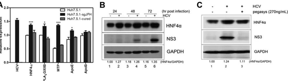
quanti-fied in Huh7.5.1-sgJFH cells (Huh7.5.1 cells with a replicating genotype 2a subgenomic replicon) and Huh7.5.1 cured cells (pro-duced by treating Huh7.5.1-sgJFH cells with Pegasys for 2 weeks to eliminate the replicon). As shown inFig. 1A, the transcripts of HNF4␣, PLA2GXIIB, MTP, and ApoE increased in
Huh7.5.1-sgJFH cells compared with those in Huh7.5.1 cells. When the rep-licon was eliminated, transcripts of these factors returned to levels comparable to those of Huh7.5.1 cells (Fig. 1A). A time course analysis was performed to monitor the expression of HNF4␣ pro-tein in HCV infection. Huh7.5.1 cells were infected with HCV-Jc1EGFP at an MOI of 0.02. The expression of HNF4␣protein was detected at the time points postinfection indicated onFig. 1B. Compared with the noninfected cells, the HCV-infected Huh7.5.1 cells showed increased HNF4␣protein expression (Fig. 1B, com-pare lanes 2, 4, and 6 with lanes 1, 3, and 5, respectively). When treated with Pegasys, the HCV-Jc1EGFP-infected Huh7.5.1 cells showed reduced HNF4␣abundance (Fig. 1C, compare lanes 2 and 3). These results suggest that the upregulated HNF4␣expression is dependent on HCV replication.
These observations suggest that HNF4␣is crucial in HCV life cycle. To test this hypothesis, we evaluated the effect of HNF4␣on different stages of HCV life cycle.
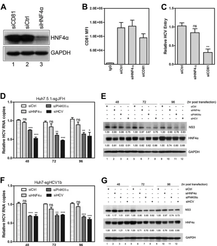
(ii) HNF4␣is not required for HCV entry or RNA replica-tion.To determine whether HNF4␣is required for HCV entry, Huh7.5.1 cells were transfected with HNF4␣-specific siRNA (siHNF4␣) and then incubated for 48 h for efficient knockdown. These cells were then infected with HCV-Jc1, followed by the quantification of intracellular HCV RNA copy numbers at 24 h postinfection. siHNF4␣efficiently decreased HNF4␣protein ex-pression (Fig. 2A, compare lanes 1 and 3) with no considerable effect on cell viability (data not shown). As a positive control, a CD81-specific siRNA (siCD81) reduced CD81 expression by ⬃55% and⬃30% at the transcript (data not shown) and protein (Fig. 2B) levels, respectively. No significant decrease in HCV entry efficacy was observed upon HNF4␣silencing, whereas siCD81 FIG 1HNF4␣expression is induced by HCV infection. (A) Transcripts of HNF4␣and its downstream factors were quantified in Huh7.5.1-sgJFH cells (Huh7.5.1 cells with replicating genotype 2a subgenomic replicon) and Huh7.5.1 cured cells after normalization by GAPDH. Huh7.5.1 cured cells were obtained by treating Huh7.5.1-sgJFH cells with pegylated interferon␣-2a (Pegasys) for 2 weeks. The data are presented as means⫾SEM (n⫽3; *,P⬍0.05; ***,P⬍ 0.001). (B) HNF4␣and HCV NS3 proteins were detected in HCV-Jc1EGFP-infected Huh7.5.1 cells (MOI of 0.02) using specific antibodies and compared with mock-infected cells in a time course analysis. The ratio for the relative band intensity of HNF4␣normalized with GAPDH was calculated. (C) Huh7.5.1 cells infected with HCV-Jc1EGFP (MOI of 0.02) were treated with or without Pegasys at 270 ng/ml and then cultured for 72 h. Abundances of HNF4␣and NS3 proteins were detected by Western blotting using specific antibodies.
on November 7, 2019 by guest
http://jvi.asm.org/
[image:3.585.48.541.65.205.2]FIG 2HNF4␣is not required for HCV entry or RNA replication. (A) Huh7.5.1 cells were transfected with HNF4␣siRNA (siHNF4␣), CD81 siRNA (siCD81), or a scrambled siRNA (siCtrl), followed by inoculation of HCV-Jc1 (MOI of 0.05) at 48 h posttransfection. HNF4␣expression at 48 h posttransfection was detected via Western blot analysis. (B) Cellular membrane CD81 expression was measured by flow cytometry in Huh7.5.1 cells at 48 h posttransfection, and the mean fluorescence intensity of CD81 was calculated. (C) The infected Huh7.5.1 cells were collected at 24 h postinfection, and relative quantification of the change in intracellular HCV RNA copy numbers was calculated after normalization by 18S RNA. (D) Huh7.5.1-sgJFH cells were transfected with siRNA-targeted HNF4␣, phosphatidylinositol 4-kinase III alpha (PI4KIII␣), or HCV. Cell lysates were harvested at the indicated time points posttransfection. Relative quantification of the change in HCV RNA copies was calculated by normalization with 18S RNA. The data are presented as means⫾SEM (n⫽3; *,P⬍0.05;**,P⬍0.01; ***,P⬍0.001). (E) Huh7.5.1-sgJFH cell lysates were harvested at the indicated time points posttransfection to detect HNF4␣and NS3 protein expression via Western blot analysis. The ratios for the relative band intensities of each target protein normalized with GAPDH were calculated. (F) Huh7-sgHCV1b cells were transfected with siHNF4␣and collected to quantify HCV RNA copy numbers. siPI4KIII␣and siHCV were used as positive controls. Relative quantification of the change in HCV RNA copy numbers was calculated at the indicated time points posttransfection after normalization with 18S RNA. The results are shown as means⫾SEM (n⫽3; **,P⬍0.01; ***,P⬍0.001). (G) Detection of HNF4␣and NS3 in Huh7-sgHCV1b cells caused by siHNF4␣, siPI4KIII␣, and siHCV transfection via Western blot analysis. The ratios for relative band intensities normalized by GAPDH were calculated.
HNF4␣and PLA2GXIIB Regulate Infectious HCV Production
on November 7, 2019 by guest
http://jvi.asm.org/
[image:4.585.75.510.61.556.2]suppressed HCV entry efficacy by⬃70% (Fig. 2C). Evaluation of the effects of HNF4␣overexpression on HCV entry revealed no significant changes compared with pcDNA-transfected cells (data not shown). CD81, claudin-1, SR-BI, occludin, LDLR, and NPC1L1 are cellular factors required for HCV entry. Transcripts of these factors were indistinguishable when HNF4␣was knocked down or overexpressed (data not shown). Collectively, these re-sults indicate that HNF4␣is not required for HCV entry.
To determine whether HNF4␣is required for HCV RNA rep-lication, a time course analysis was performed on Huh7.5.1-sgJFH cells. After the cells were transfected with siHNF4␣, HCV RNA copy numbers and NS3 protein expression were quantified at the time points posttransfection indicated onFig. 2DtoG. HCV- and PI4KIII␣-specific siRNAs were used as positive controls (27,28). HNF4␣expression was efficiently reduced (Fig. 2E). However, no significant decrease in HCV RNA copy numbers (Fig. 2D) or NS3 expression (Fig. 2E) was observed. In contrast, siPI4KIII␣and siHCV significantly reduced HCV RNA copy numbers and NS3 protein expression. Similar results were observed in Huh7-sgHCV1b cells (Fig. 2F and G). HNF4␣ overexpression in Huh7.5.1-sgJFH or Huh7-sgHCV1b cells did not affect RNA rep-lication (data not shown). Collectively, these results indicate that HNF4␣is not required for HCV RNA replication.
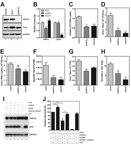
(iii) HNF4␣is required for HCV assembly and secretion.To investigate whether HNF4␣is involved in the late stage of the HCV life cycle, Huh7.5.1 cells were transfected with siHNF4␣ (Fig. 3AandBshow the effect of knockdown) and then infected with HCV-Jc1. ApoE reportedly affects the assembly and secretion of HCV (29). Thus, ApoE siRNA (siApoE) was used as a positive transfection control. The infected cells and culture supernatants were collected at 72 h postinfection. Since HNF4␣was not re-quired for HCV entry or RNA replication, the reduced intracellu-lar HCV RNA copy numbers in the multicycle infection system (Fig. 3C) argued the effect of HNF4␣on the late stage of the HCV life cycle.
Intracellular and supernatant viruses were titrated to deter-mine the effect of siHNF4␣on the late stage of the HCV life cycle. The infectious titers in the viral supernatant from the HNF4␣-silenced cells were reduced by approximately 70% (Fig. 3D). The HCV RNA copy numbers in the supernatant were slightly reduced because of HNF4␣silencing (Fig. 3E). Thus, the specific infectivity (extracellular HCV titer divided by extracellular viral RNA copy number) was significantly reduced by siHNF4␣(Fig. 3F). The infectious titers in the cell lysates were significantly reduced upon siHNF4␣transfection (Fig. 3G). The reduced infectivity in the supernatants and cell lysates indicated that the assembly efficiency of HCV was impaired in the HNF4␣-silenced cells. Moreover, the ratio of the titers in the supernatant to those in the cell lysate was significantly reduced upon siHNF4␣transfection (Fig. 3H), sug-gesting that the secretion of virion particles was also inhibited upon HNF4␣silencing. These results indicate that HNF4␣affects both HCV assembly and secretion, similar to the effect of ApoE. Huh7.5.1 cells were cotransfected with siRNA-resistant HNF4␣ -encoding plasmids and siHNF4␣to rule out any off-target effect. HCV-Jc1EGFP was infected at 3 h posttransfection. Viral super-natants at 72 h postinfection were harvested for FCM assay. HNF4␣protein expression was rescued upon pHNF4␣-resistant transfection (Fig. 3I, compare lanes 3 and 4). Predictably, the re-duced HCV infectivity caused by HNF4␣silencing was recovered upon siHNF4␣-resistant plasmid transfection (Fig. 3J).
Collec-tively, these results show that HNF4␣is required for HCV assem-bly and secretion.
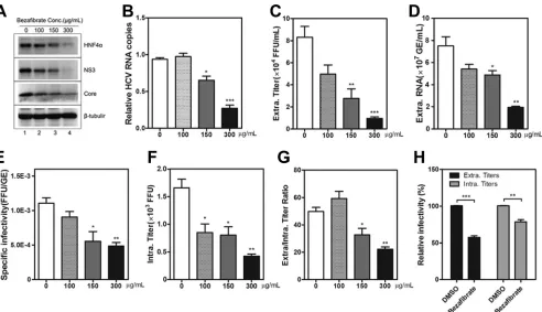
(iv) HNF4␣antagonist bezafibrate suppresses HCV assem-bly and secretion.Bezafibrate is an antagonist of HNF4␣(30). Huh7.5.1 cells were infected with HCV-Jc1EGFP in the presence of bezafibrate to confirm the effect of HNF4␣on the HCV life cycle. At 72 h postinfection, HNF4␣expression was downregu-lated by bezafibrate in a dose-dependent manner (Fig. 4A, com-pare lane 1 with lanes 2 to 4) with no apcom-parent cellular cytotoxicity (data not shown). MTP transcript was also downregulated ac-cordingly (data not shown), indicating that the transcriptional activity of HNF4␣was reduced upon bezafibrate treatment.
To measure the efficiency of HCV RNA replication in bezafi-brate-treated cells, intracellular HCV RNA copies were quantified. A dose-dependent decrease was observed (Fig. 4B). Bezafibrate did not affect HCV entry or RNA replication (data not shown). Thus, the reduction in intracellular HCV RNA copy numbers in-dicated that HCV assembly and secretion were inhibited.
To evaluate the effect of bezafibrate on HCV assembly and secretion, infectious titers in the supernatant and cell lysate were quantified. As shown inFig. 4C, bezafibrate suppressed the infec-tivity of the secreted HCV, with a maximal inhibition level of approximately 90%. HCV RNA copies in the supernatant (Fig. 4D) and specific infectivity (Fig. 4E) were both reduced upon bezafibrate treatment. Intracellular virus titers were quantified. Bezafibrate at a concentration of 300g/ml showed a maximal inhibition level of approximately 75% (Fig. 4F). These results in-dicated that the assembly efficiency of HCV was impaired upon bezafibrate treatment. The ratio of the supernatant infectivity to that in the cell lysate significantly decreased with increasing beza-fibrate concentration (Fig. 4G). High concentrations of bezafi-brate inhibited HCV assembly and secretion, whereas low concen-trations (e.g., 100g/ml) of bezafibrate mainly impaired HCV assembly. Cells persistently infected with HCV were alternatively used for HCV assembly and secretion experiments. Huh7.5.1 cells persistently infected with HCV-Jc1EGFP were treated with 300 g/ml bezafibrate. Viral supernatants and cell lysates were col-lected at 72 h posttreatment. As shown inFig. 4H, intracellular and supernatant infectious titers were reduced upon bezafibrate treatment by approximately 30% and 50%, respectively. Collec-tively, these results reveal that bezafibrate impairs HCV assembly and secretion.
PLA2GXIIB, a downstream factor regulated by HNF4␣, is re-quired for HCV secretion. (i) HNF4␣regulates PLA2GXIIB ex-pression.Given that HCV is assembled into and released together with VLDLs, we hypothesize that certain HNF4␣-regulated VLDL biogenesis-related factors are responsible for the HNF4␣-induced regulation of HCV production. We previously reported that PLA2GXIIB functions in VLDL biogenesis/secretion and that
HNF4␣regulates PLA2GXIIB expression in HepG2 and HeLa cells
(23). Thus, PLA2GXIIB might function in the HCV life cycle. To
test this possibility, we first evaluated whether HNF4␣regulates PLA2GXIIB expression in Huh7.5.1 cells. A PLA2GXIIB promoter
reporter plasmid (pGL3-PLA2GXIIBprom) was cotransfected
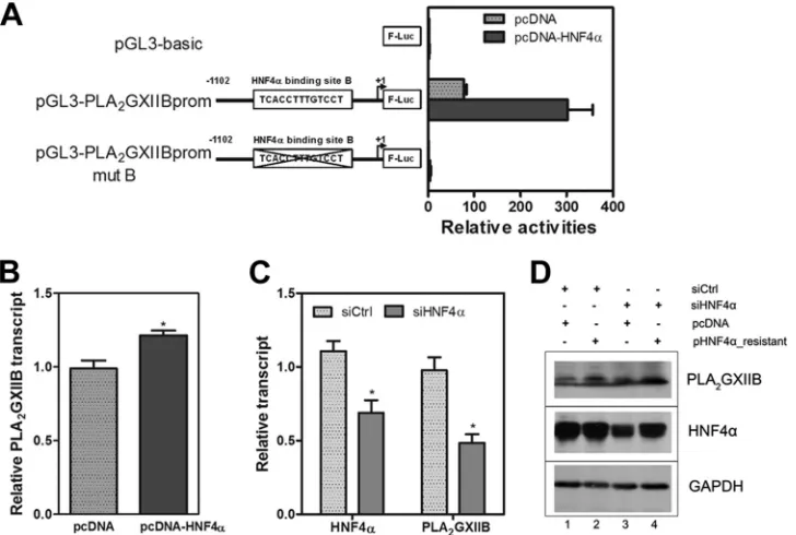
into Huh7.5.1 cells with an HNF4␣-encoding plasmid. HNF4␣ induced the PLA2GXIIB promoter activities by more than 3-fold
compared with the pGL3-basic reporter (Fig. 5A). A mutant PLA2GXIIB promoter reporter plasmid (pGL3-PLA2GXIIBprom
mutB) with a mutated HNF4␣-responsive binding site was used as a negative control, which was not activated by HNF4␣(Fig. 5A).
on November 7, 2019 by guest
http://jvi.asm.org/
Huh7.5.1 cells were transfected with the HNF4␣-encoding plasmid to directly evaluate the regulatory effect of HNF4␣on PLA2GXIIB. The PLA2GXIIB transcript was quantified at 48 h
posttransfection. Ectopic HNF4␣ expression significantly
in-creased PLA2GXIIB transcript (Fig. 5B). PLA2GXIIB protein
abundance was also increased (Fig. 5D, compare lanes 1 and 2). The effect of downregulated HNF4␣expression on PLA2GXIIB
expression in Huh7.5.1 cells was also evaluated. HNF4␣silencing FIG 3HNF4␣is required for HCV assembly and secretion. Huh7.5.1 cells were transfected with siHNF4␣and then inoculated with HCV-Jc1 at an MOI of 0.02 at 3 h posttransfection. The viral supernatants and cells were collected at 72 h postinfection. (A) Detection of HNF4␣caused by siHNF4␣transfection via Western blot analysis. siCtrl and ApoE siRNA (siApoE) were used as negative and positive controls, respectively. (B) Relative change in transcript of HNF4␣and ApoE upon HNF4␣or ApoE silencing in Huh7.5.1 cells at 72 h post-HCV infection. (C) Intracellular HCV RNA copy numbers were quantified after normalization by 18S RNA, induced by siHNF4␣or siApoE. (D) Clarified viral supernatants were titrated by endpoint dilution on Huh7.5.1 cells, and the infectious HCV titers were calculated as the number of focus-forming units (FFU)/ml. (E) Detection of supernatant HCV RNA copies (calculated as genome equivalents [GE]/ml) via qRT-PCR. (F) Specific infectivity of HCV particles after each siRNA treatment was calculated using the extracellular HCV titer divided by the extracellular viral RNA copy number (FFU/GE). (G) Infectious particles in the cell lysates were released from cells by four cycles of freezing and thawing, and the supernatants were clarified for titration (calculated as FFU). (H) The ratios of the infectivity in the supernatants to that in the cell lysates upon siHNF4␣or siApoE treatment were calculated. (I) Huh7.5.1 cells were cotransfected with siHNF4␣and siRNA-resistant HNF4␣DNA, and HCV-Jc1EGFP was inoculated at an MOI of 0.02 at 3 h posttransfection. The cell lysates were collected at 72 h postinfection to detect HNF4␣expression. (J) Viral supernatants upon HNF4␣reconstitution in HNF4␣-silenced cells were collected to quantify HCV infectivity using flow cytometry. Triple independent experiments were performed, and the results are presented as means⫾SEM (n⫽3; *,P⬍0.05;**,P⬍0.01;***,P⬍0.001). Intra, intracellular; extra, extracellular.
HNF4␣and PLA2GXIIB Regulate Infectious HCV Production
on November 7, 2019 by guest
http://jvi.asm.org/
[image:6.585.75.513.62.542.2]downregulated PLA2GXIIB transcript (Fig. 5C) and protein
ex-pression (Fig. 5D, compare lanes 1 and 3). The downregulated PLA2GXIIB expression was recovered when HNF4␣expression
was rescued upon siHNF4␣-resistant plasmid transfection (Fig. 5D, compare lanes 3 and 4). These results indicate that PLA2GXIIB
ex-pression is regulated by HNF4␣in Huh7.5.1 cells.
(ii) PLA2GXIIB expression is upregulated by HCV infection.
A previous study reported that PLA2GXIIB is upregulated in
HCV-related liver tumor tissues (24). Here, three experiments were performed to determine whether or not PLA2GXIIB
expres-sion is induced by HCV infection. First, PLA2GXIIB transcript
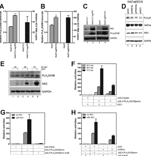
was increased in Huh7.5.1-sgJFH and Huh7-sgHCV1b cells com-pared with those in Huh7.5.1 and Huh7 cells, respectively (Fig. 6A andB). PLA2GXIIB protein expression was also upregulated in
Huh7.5.1-sgJFH and Huh7-sgHCV1b cells (Fig. 6C, compare lanes 1 and 2 and lanes 3 and 4). Huh7-sgHCV1b cells were trans-fected with siPI4KIII␣or siHCV to confirm the regulatory effect of HCV RNA replication on PLA2GXIIB expression. siHNF4␣was
used as a positive control. At 72 h posttransfection, PLA2GXIIB
expression was reduced upon siPI4KIII␣or siHCV silencing com-pared with the level with a scrambled control siRNA (siCtrl), con-comitant with decreased NS3 protein expression (Fig. 6D). This
result suggests that PLA2GXIIB is specifically upregulated in
replicon cells. Second, Huh7.5.1 cells were infected with HCV-Jc1EGFP at an MOI of 0.02. PLA2GXIIB expression was quantified
in a time course analysis. PLA2GXIIB protein expression
com-pared with levels in mock-infected cells was increased concomi-tant with HCV RNA replication (Fig. 6E). Third, the PLA2GXIIB
promoter was activated upon HCV infection (Fig. 6F). These re-sults demonstrate that PLA2GXIIB expression is upregulated by
HCV infection.
(iii) HNF4␣is required for HCV-induced upregulation of PLA2GXIIB.To investigate whether HNF4␣is required for HCV-induced upregulation of PLA2GXIIB, Huh7.5.1 cells were
trans-fected with the wild-type or mutant construct of a PLA2GXIIB
promoter reporter and then infected with HCV-Jc1EGFP. As shown inFig. 6G, HCV infection effectively activated the wild-type PLA2GXIIB promoter reporter but failed to activate the
mu-tant reporter. This result confirmed the importance of HNF4␣in the activation of the PLA2GXIIB promoter. Cotransfection of
siHNF4␣ with the PLA2GXIIB promoter reporter reduced
PLA2GXIIB promoter activity by 2.6-fold (Fig. 6H). A comparable
reduction was also observed in the cells without HCV infection FIG 4HNF4␣antagonist bezafibrate suppresses HCV assembly and secretion. Huh7.5.1 cells were infected with HCV-Jc1EGFP at an MOI of 0.02 in the presence of different concentrations of bezafibrate (0g/ml to 300g/ml). DMSO was used as a vehicle, and the final concentration in the medium was 2‰. (A) At 72 h postinfection, the cell lysates were collected to detect HNF4␣, HCV NS3, and core proteins via Western blotting. (B) Intracellular HCV RNA copies were quantified after normalization by 18S RNA at 72 h postinfection upon different concentrations of bezafibrate treatment. (C) Viral supernatant infectivity (calculated as FFU/ml) upon bezafibrate treatment was determined on Huh7.5.1 cells by endpoint dilution. (D) Measurement of supernatant HCV RNA copies (calculated as GE/ml) upon bezafibrate treatment. (E) Calculation of specific infectivity (described as FFU/GE) upon bezafibrate treatment. (F) Quantification of HCV titers in the cell lysates (calculated as FFU) in Huh7.5.1 cells upon bezafibrate treatment. (G) The ratios of the supernatant infectivity to that in the cell lysates treated with different concentrations of bezafibrate were calculated. (H) Huh7.5.1 cells persistently infected with HCV-Jc1EGFP were inoculated with DMSO or 300g/ml bezafibrate. The relative HCV infectivity (percentage of the value of the DMSO-treated condition) in the supernatants and the cell lysates in persistently infected Huh7.5.1 cells at 72 h postinoculation was determined using flow cytometry. The results are presented as means⫾SEM (n⫽3; *,P⬍0.05; **,P⬍0.01; ***,P⬍0.001).
on November 7, 2019 by guest
http://jvi.asm.org/
[image:7.585.48.540.64.347.2](Fig. 6H). Collectively, these results indicate that HNF4␣is re-quired for HCV-induced upregulation of PLA2GXIIB expression. (iv) PLA2GXIIB is required for HCV secretion.We
investi-gated the susceptibility of HCV entry or HCV RNA replication to PLA2GXIIB silencing to determine whether or not PLA2GXIIB is
required in the HCV life cycle. No inhibition of HCV entry or RNA replication was observed upon PLA2GXIIB knockdown
(data not shown). Thus, we speculated that PLA2GXIIB possibly
functions in the late stage of the HCV life cycle. Huh7.5.1 cells were transfected with siPLA2GXIIB and then infected with
HCV-Jc1EGFP to investigate the effect of PLA2GXIIB on HCV assembly
and secretion. siPLA2GXIIB efficiently reduced PLA2GXIIB
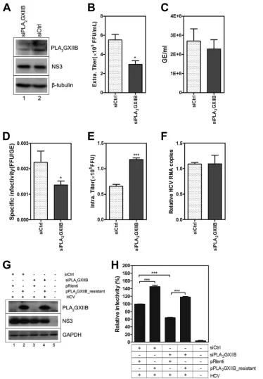
ex-pression (Fig. 7A) with no measurable effect on cell viability (data not shown). Infectious HCV titers in the supernatant were re-duced by approximately 50% upon siPLA2GXIIB transfection
(Fig. 7B), whereas the released HCV RNA copy numbers re-mained unchanged (Fig. 7C). Thus, the specific infectivity (Fig. 7D) was reduced upon PLA2GXIIB silencing. When the HCV
titers in the cell lysates were quantified, a 2-fold accumulation was observed (Fig. 7E). HCV RNA replication remained unchanged, regardless of PLA2GXIIB downregulation (Fig. 7F). These results
indicated that PLA2GXIIB interfered with HCV secretion but not
assembly. A rescue experiment was performed to confirm this conclusion. Huh7.5.1 cells were cotransfected with siPLA2GXIIB
and siRNA-resistant PLA2GXIIB-encoding plasmids (Fig. 7G
shows the knockdown and rescue effects). HCV-Jc1EGFP was then inoculated at 3 h posttransfection. The viral supernatant in-fectivity was determined by FCM. The reduced HCV inin-fectivity by
siPLA2GXIIB was completely recovered by the reconstitution of
PLA2GXIIB expression (Fig. 7H), underscoring the effect of
PLA2GXIIB on infectious HCV production. Collectively, these
findings show that PLA2GXIIB is required for HCV secretion. PLA2GXIIB and MTP reconstitutions recover the reduced ef-fect of HNF4␣silencing on HCV infectivity.To address which factors downstream of HNF4␣are attributed to the HNF4␣ -in-duced regulation of HCV infectivity, the effects of PLA2GXIIB
overexpression were first investigated. Huh7.5.1 cells were trans-fected with plasmids encoding PLA2GXIIB and then were infected
with HCV-Jc1 at an MOI of 0.01 at 24 h posttransfection. As shown inFig. 8A, PLA2GXIIB protein overexpression was readily
detected at all time points postinfection. The infectious titers in the supernatants increased by more than 2-fold compared with the mock-transfected condition at 72 h postinfection (Fig. 8B), coinciding with the result shown inFig. 7H. However, MTP (Fig. 8C) or ApoE (Fig. 8E) overexpression did not increase HCV in-fectivity in the supernatants compared with the mock-transfected condition (Fig. 8DandF). These results show that MTP and ApoE are sufficient in Huh7.5.1 cells for HCV production and that ectopic PLA2GXIIB expression enhances HCV infectivity,
consis-tent with the results under the condition of HNF4␣ overexpres-sion (Fig. 3J).
Reconstitution experiments were performed in HNF4␣-si-lenced Huh7.5.1 cells to identify the factors responsible for the HNF4␣-regulated HCV infectivity. Huh7.5.1 cells were cotrans-fected with siHNF4␣and plasmids encoding PLA2GXIIB, MTP,
or ApoE alone or in combination. HCV-Jc1EGFP was inoculated FIG 5HNF4␣regulates PLA2GXIIB expression in Huh7.5.1 cells. (A) Huh7.5.1 cells were transfected with three plasmids including the wild-type or mutant
PLA2GXIIB promoter reporter plasmid, an HNF4␣-encoding plasmid, and a plasmid encodingRenillaluciferase for transfection efficiency measurement. At 48
h posttransfection, reporter activities were analyzed by a Dual-Glo luciferase kit. The wild-type and the mutant construct of the PLA2GXIIB promoter reporter
lacking the HNF4␣-responsive binding site are described in the schematic. (B) The relative change in PLA2GXIIB transcript in Huh7.5.1 cells transfected with an
HNF4␣-encoding plasmid at 72 h posttransfection was determined compared with the value in mock-transfected cells. The results are presented as means⫾SEM (n⫽3; *,P⬍0.05). (C) The relative change in HNF4␣and PLA2GXIIB transcripts in HNF4␣-silenced Huh7.5.1 cells at 72 h posttransfection was quantified.
The results are presented as means⫾SEM (n⫽3; *,P⬍0.05). (D) Huh7.5.1 cells were cotransfected with siHNF4␣and siRNA-resistant HNF4␣-encoding plasmids. The cell lysates at 72 h posttransfection were collected to detect HNF4␣and PLA2GXIIB protein expression via Western blot analysis.
HNF4␣and PLA2GXIIB Regulate Infectious HCV Production
on November 7, 2019 by guest
http://jvi.asm.org/
[image:8.585.112.476.67.312.2]FIG 6HNF4␣is required for the HCV-induced upregulation of PLA2GXIIB expression. (A) Relative quantification of the change in PLA2GXIIB transcript
normalized to GAPDH in Huh7.5.1-sgJFH cells compared with Huh7.5.1 cells. The data are presented as means⫾SEM (n⫽3; ***,P⬍0.001). (B) Relative quantification of the change in PLA2GXIIB transcript after normalization to GAPDH in Huh7-sgHCV1b cells compared with Huh7 cells. The data are presented
as means⫾SEM (n⫽3; **,P⬍0.01). (C) PLA2GXIIB and HCV NS3 proteins were detected in Huh7.5.1-sgJFH or Huh7-sgHCV1b cells via Western blot
analysis. (D) Huh7-sgHCV1b cells were transfected with siPI4KIII␣or siHCV, with siHNF4␣as a positive control. The abundance of PLA2GXIIB protein was
measured by antibodies against NS3, HNF4␣, and PLA2GXIIB at 72 h posttransfection. (E) PLA2GXIIB protein was measured in HCV-Jc1EGFP-infected
Huh7.5.1 cells (MOI of 0.02) compared with mock-infected cells in a time course analysis. (F) Activities of the PLA2GXIIB promoter reporter in Huh7.5.1 cells
infected with HCV-Jc1EGFP (MOI of 0.2) compared with those of mock-infected cells were quantified in a time course analysis. (G) Huh7.5.1 cells were transfected with the wild-type and mutant PLA2GXIIB promoter reporter plasmids and then inoculated with HCV-Jc1EGFP (MOI of 0.2) at 3 h posttransfection.
Activities of the promoter reporter were quantified at 48 h post-HCV infection. (H) Huh7.5.1 cells were cotransfected with PLA2GXIIB promoter reporter
plasmids and siHNF4␣, and activities of the promoter reporter were analyzed in Huh7.5.1 cells either noninfected or infected with HCV-Jc1EGFP (MOI of 0.2) at 48 h postinfection. psi, postinfection.
on November 7, 2019 by guest
http://jvi.asm.org/
[image:9.585.45.530.67.567.2]FIG 7PLA2GXIIB is required for HCV secretion. (A) Huh7.5.1 cells were transfected with siPLA2GXIIB and then infected with HCV-Jc1EGFP at an MOI of 0.02
at 3 h posttransfection. The cell lysates were collected to detect PLA2GXIIB and HCV NS3 proteins using Western blot analysis at 72 h post-HCV infection. (B)
Infectious HCV titers in the supernatants (FFU/ml) induced by siPLA2GXIIB were quantified compared with values of the siCtrl-treated condition at 72 h
postinfection. (C) Quantification of HCV RNA copies (GE/ml) in the supernatants induced by siPLA2GXIIB at 72 h postinfection. (D) Specific infectivity
(FFU/GE) of the virus in the supernatants upon siPLA2GXIIB treatment was calculated at 72 h postinfection. (E) The intracellular HCV titers induced by
siPLA2GXIIB compared with those induced by siCtrl were quantified at 72 h postinfection. (F) Relative quantification of the change in intracellular HCV RNA
copy numbers in siPLA2GXIIB-transfected Huh7.5.1 cells. (G) Huh7.5.1 cells were transfected with siPLA2GXIIB and siRNA-resistant PLA2GXIIB-encoding
plasmids and then inoculated with HCV-Jc1EGFP at 3 h posttransfection. PLA2GXIIB protein abundance in PLA2GXIIB-silenced Huh7.5.1 cells upon rescue was
detected at 72 h postinfection. (H) The relative infectious HCV titers in the supernatants were quantified by flow cytometry in PLA2GXIIB-rescued Huh7.5.1 cells
compared with the PLA2GXIIB-silenced condition. Experiments were performed at least three times, and the results are shown as means⫾SEM (*,P⬍0.05; **,
P⬍0.01; ***,P⬍0.001).
HNF4␣and PLA2GXIIB Regulate Infectious HCV Production
on November 7, 2019 by guest
http://jvi.asm.org/
[image:10.585.108.482.64.603.2]at 3 h posttransfection at an MOI of 0.02. The infection efficiency was determined by monitoring NS5A-GFP expression (Fig. 8G) in transfected cells at 72 h postinfection. The supernatant infectivity at 72 h postinfection was quantified by FCM assay. In
HNF4␣-silenced Huh7.5.1 cells, HCV infectivity in the supernatants was reduced by⬃50% compared with the level in mock-silenced cells (Fig. 8H). PLA2GXIIB, MTP, or ApoE overexpression alone did
not recover the reduced HCV infectivity upon siHNF4␣ transfec-FIG 8PLA2GXIIB and MTP reconstitutions recover the reduced HCV infectivity by siHNF4␣. Huh7.5.1 cells were transfected with different doses of plasmids
encoding PLA2GXIIB, MTP, or ApoE and then inoculated with HCV-Jc1 (MOI of 0.01) at 24 h posttransfection. Cells and supernatants were collected at the
indicated time points postinfection. (A) Overexpressed PLA2GXIIB and HCV core proteins were detected with antibodies against PLA2GXIIB and core protein
at the indicated time points postinfection. (B) HCV infectivity in the supernatants upon PLA2GXIIB overexpression were titrated and calculated as FFU/ml. (C)
Exogenous MTP-Flag fusion proteins were detected at the indicated time points post-HCV infection with flow cytometry by using intracellular staining with an anti-Flag antibody. (D) The supernatant infectious titers were quantified under the condition of MTP overexpression compared with mock-transfected cells. (E) HCV core and HA-tagged ApoE proteins were detected via Western blot analysis with specific antibodies against core protein and HA tag, respectively. (F) Infectious HCV titers in the supernatants under the condition of ApoE overexpression were quantified. (G) Plasmids encoding PLA2GXIIB, MTP, and ApoE
alone or in combination were cotransfected with siHNF4␣into Huh7.5.1 cells and then inoculated with HCV-Jc1EGFP at an MOI of 0.02 at 3 h posttransfection. The cell lysates were collected to quantify NS5A-GFP frequency (infection efficiency) at 72 h postinfection. (H) The supernatants were collected and inoculated on Huh7.5.1 cells to quantify HCV titers. The relative HCV infectivity in the supernatants after normalization to the siCtrl-transfected condition was calculated as a percentage. The data are presented as means⫾SEM (n⫽3; *,P⬍0.05). “ph” indicates a plasmid that encodes a human protein.
on November 7, 2019 by guest
http://jvi.asm.org/
[image:11.585.45.532.68.548.2]tion (Fig. 8H). The impaired HCV infectivity was completely res-cued when PLA2GXIIB was reconstituted in combination with
MTP (Fig. 8H). Reconstituted PLA2GXIIB and ApoE expression,
as well as reconstituted MTP and ApoE, partially but not signifi-cantly recovered the reduced HCV infectivity by siHNF4␣ trans-fection (Fig. 8H). The reconstituted combination of PLA2GXIIB,
MTP, and ApoE did not completely rescue the impaired HCV infectivity (Fig. 8H), indicating the presence of a complicated in-teraction among these factors. Identical results were also obtained on persistently infected Huh7.5.1 cells (data not shown). These results indicate that PLA2GXIIB and MTP are sufficient to rescue
the reduced HCV infectivity in HNF4␣-deficient cells.
HNF4␣downregulation induces rearrangement of cytosolic LDs.ApoE is the most abundant apolipoprotein on HCV parti-cles; it is crucial for HCV assembly and infectivity (29,31). To determine whether HNF4␣regulates HCV assembly and secre-tion in an ApoE-dependent manner, Huh7.5.1 cells were trans-fected with siHNF4␣or treated with bezafibrate and then infected with HCV-mutJFH1 infection. ApoE abundance and HCV titers in the viral supernatants were quantified at 72 h postinfection. Consistent with earlier results, HNF4␣downregulation remark-ably suppressed the infectious HCV titers in the supernatants (Fig. 9A). However, siHNF4␣or bezafibrate treatment did not remark-ably decrease ApoE secretion (normalized to albumin) (Fig. 9B andC). The abundance of ApoE in the cell lysates upon bezafi-brate treatment was unchanged compared with DMSO treatment (data not shown). These results indicate that HNF4␣affects HCV assembly and secretion in an ApoE-independent manner.
Cytosolic LDs are also crucial for HCV assembly and infectious virus production. The rearrangement of LDs reportedly impairs HCV infectivity (32–34). Huh7.5.1 cells were infected with HCV-mutJFH1 in the presence of bezafibrate and then observed under a confocal microscope at 72 h postinfection to investigate whether or not HNF4␣regulates the rearrangement of LDs. Bezafibrate decreased the number but increased the size of LDs in the HCV-infected cells (Fig. 9DtoF). However, the total fluorescence in-tensity of LDs did not significantly increase (data not shown). Similar to bezafibrate, siHNF4␣transfection yielded large aggre-gates of LDs and distorted cytosolic distributions in the HCV-infected Huh7.5.1 cells (Fig. 9G). This result coincides with the observations in specific HNF4␣-nullDrosophilaor mice (17,35). We then analyzed the location of the HCV core protein on LDs upon bezafibrate treatment. As shown inFig. 9H, bezafibrate re-duced core protein expression and perturbed the typical perinu-clear distribution pattern observed in the HCV-infected cells treated with DMSO. However, the HCV core protein remained localized to the periphery of LDs. Identical results were observed in HNF4␣silencing (data not shown). This result suggests that HNF4␣downregulation affects core protein abundance and cyto-solic distribution but not the location of the core protein on LDs. Therefore, the rearrangement of LDs can be attributed to the effect of HNF4␣on infectious HCV production.
DISCUSSION
Biophysical and ultrastructural studies indicate that the incorpo-ration of HCV particles with VLDLs determines HCV infectivity, aids in HCV entry and release from cells, and helps the virus es-cape from host immune surveillance. HCV hijacks the VLDL se-cretory pathway for its own assembly and secretion (12,15). How-ever, the molecular mechanism remains elusive. In this study, the
importance of HNF4␣, a central nuclear receptor controlling cel-lular lipid transport, in HCV assembly and secretion was illus-trated. We showed that HCV replication induced HNF4␣ -medi-ated upregulation of genes involved in the VLDL pathway. We also demonstrated that HNF4␣downregulation impaired HCV assembly and secretion, whereas HNF4␣ overexpression pro-moted infectious HCV production.
As a key transcription factor for VLDL-associated genes, HNF4␣ affected HCV assembly and secretion by regulating downstream genes, including MTP and PLA2GXIIB. In normal
Huh7.5.1 cells, the overexpression of PLA2GXIIB and not MTP
increased HCV infectivity. In HNF4␣-deficient cells, ectopic PLA2GXIIB or MTP alone did not rescue the reduced HCV
infec-tivity. However, the reduced HCV infectivity was completely re-covered by reconstituted PLA2GXIIB in combination with MTP.
The synergistic effect can be well understood because these two factors function in different stages of the HCV life cycle. Previous studies showed that inhibition of MTP activity reduces HCV as-sembly (12). The current study showed that PLA2GXIIB
knock-down reduced HCV infectivity in the supernatant and accumu-lated intracellular HCV infectivity. These results suggest that PLA2GXIIB is required for HCV secretion. Indeed, VLDL
secre-tion in PLA2GXIIB-null mice is also reduced (23). The unchanged
expression levels of MTP and ApoB suggest that VLDL assembly cannot be distinguished in PLA2GXIIB-null mice. These results
emphasize further the tight interaction between VLDL and HCV assembly and secretion. Although ApoE transcript was upregu-lated by ectopic HNF4␣expression, the abundance of ApoE in the cell lysates and supernatants was not affected by HNF4␣ down-regulation. In normal Huh7.5.1 cells, ApoE silencing reduced both HCV assembly and secretion, supporting previous reports (29). Meanwhile, ectopic ApoE expression did not increase HCV infectivity in the supernatants. This result suggests that ApoE ex-pression is sufficient for HCV life cycle in normal cells. ApoE overexpression alone or in combination with PLA2GXIIB and
MTP did not rescue the reduced HCV infectivity in HNF4␣-defi-cient cells. These results suggest that ApoE may not be a critical downstream factor affecting the regulatory activity of HNF4␣on HCV infectivity. ApoE possibly functioned differently from HNF4␣.
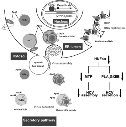
Therefore, we propose a model to illustrate the interaction be-tween HNF4␣and the HCV life cycle (Fig. 10). HCV infection upregulates HNF4␣ expression, which in turn transactivates PLA2GXIIB and MTP. MTP is required in HCV assembly,
whereas PLA2GXIIB facilitates the secretion of HCV
lipoviropar-ticles. Reduced HNF4␣ expression downregulates MTP and PLA2GXIIB, accompanied with cytosolic LD rearrangement,
which in turn impairs HCV assembly and secretion. This model emphasizes the importance of the VLDL secretory pathway for HCV assembly and secretion and suggests a possible mechanism by which HCV cooperates with HNF4␣to be assembled into and released together with VLDL particles.
Lipoprotein components, such as ApoE, triglyceride, and cho-lesterol, associate with HCV particles and therefore determine HCV infectivity (36–38). Among these components, ApoE is re-portedlytrans-activated by HNF4␣and is an abundant compo-nent of HCV particles. However, several lines of evidence suggest that ApoE is not sufficient to endow HCV particles with infectiv-ity. Shimizu et al. reported that the HCV RNA associated with ApoE can be detected in eluted fractions of gel filtration
chroma-HNF4␣and PLA2GXIIB Regulate Infectious HCV Production
on November 7, 2019 by guest
http://jvi.asm.org/
FIG 9HNF4␣downregulation induces rearrangement of cytosolic LDs. (A) Huh7.5.1 cells were transfected with specific siRNAs and then infected with HCV-mutJFH1 at 3 h posttransfection in the absence or presence of 300g/ml bezafibrate. The viral supernatant infectivity was quantified at 72 h postinfection. The data are shown as means⫾SEM (n⫽3;**,P⬍0.01). (B) The viral supernatants at 72 h post-HCV infection were subjected to low-speed centrifugation; 25l of clarified supernatants was resolved by 10% SDS-PAGE, and the levels of secreted ApoE and albumin were quantified through Western blotting. (C) The ratios for the relative band intensity of ApoE after normalization by albumin were calculated (n⫽3). (D) Huh7.5.1 cells were plated on chambered coverglass and then infected the following day with HCV-mutJFH1 (MOI of 2) in the presence of bezafibrate (300g/ml). Cells were fixed at 72 h postinfection to stain lipid droplets (red) with Nile Red (0.1g/ml) and the nucleus (blue) with 4=,6=-diamidino-2-phenylindole (1g/ml). A change in LD number (E) and LD size (F) per cell in bezafibrate or DMSO-treated HCV-mutJFH1-infected Huh7.5.1 cells was quantified. Quantification was performed in approximately 20 individual cell images from bezafibrate- or DMSO-treated conditions using ImageJ software. (G) Huh7.5.1 cells were transfected with siHNF4␣or siPLA2GXIIB, infected with
HCV-mutJFH1 (MOI of 2), and then cultured for an additional 72 h. Cells were fixed and stained for lipid droplets. (H) Huh7.5.1 cells at 72 h post-HCV infection in the presence of bezafibrate (300g/ml) were costained with lipid droplets (red) with Nile Red and HCV core protein (blue) with specific antibody. Colocalization of LDs and HCV core protein in HCV-infected Huh7.5.1 cells upon bezafibrate treatment was detected with confocal analysis. Scale bar, 10m.
on November 7, 2019 by guest
http://jvi.asm.org/
[image:13.585.110.473.64.601.2]tography irrespective of infectivity (39). In addition, ApoE can be detected preferentially in fractions with higher density but not in fractions containing the majority of the total HCV infectivity (40). A recent study has revealed that the abundance of the incorpo-rated ApoE was indistinguishable in HCV particles with different sizes although more than 40% of the total infectivity is contained in a narrow fraction with a density of⬃1.13 g/ml (41). The levels of secreted triglyceride and cholesterol are reduced in HNF4␣Liv KO mice (17). However, whether these two lipids of lipoproteins are responsible for the HNF4␣-induced regulation of HCV spe-cific infectivity must be explored.
Cytosolic LDs are dynamic organelles for cellular lipid storage and transport. In the present study, HNF4␣or PLA2GXIIB
down-regulation induced abnormal aggregation of large LDs and re-duced the number of cytosolic LDs. Such LD rearrangement was accompanied by the reduced expression and distorted cytosolic distribution of HCV proteins, similar to that of
nordihydroguai-aretic acid and cyclophilin inhibitors (33,34). These results echo the notion that cytosolic LDs are important organelles for HCV assembly and secretion (11). As another line of supporting evi-dence, the HNF4␣-specific knockout mice accumulated aggre-gated cytosolic LDs in the hepatocytes (17).
The development of direct-acting antiviral agents that target HCV viral proteins has updated the optimal treatment regimens of chronic HCV infection. However, a high risk of drug resistance has become a major concern. A chemical agent targeting a cellular factor possibly has the potential to overcome the resistance as well as provide broad genotype coverage. In this study, we evaluated the effect of HNF4␣antagonist on HCV treatment. Bezafibrate, an antagonist of HNF4␣, is widely used for treating dyslipidemia. Bezafibrate is also an agonist of peroxisome proliferator-activated receptor ␣ (PPAR␣). However, we found that parallel with HNF4␣downregulation, bezafibrate efficiently inhibited HCV as-sembly and secretion. Such activity was not parallel with the in-FIG 10Proposed model of HNF4␣regulation in the HCV life cycle. HCV infection upregulates HNF4␣expression, which in turn transactivates numerous genes (PLA2GXIIB and MTP) involved in VLDL biogenesis. MTP is required in HCV assembly, whereas PLA2GXIIB facilitates secretion of HCV lipoviroparticles.
Reduced HNF4␣expression downregulates MTP and PLA2GXIIB expression levels, concomitant with impaired HCV assembly and secretion. luLD, luminal
lipid droplets.
HNF4␣and PLA2GXIIB Regulate Infectious HCV Production
on November 7, 2019 by guest
http://jvi.asm.org/
[image:14.585.92.494.68.474.2]creased PPAR␣activity (data not shown). These observations demonstrate that the anti-HCV activity of bezafibrate is due to the inhibited HNF4␣expression and not the increased PPAR␣ activ-ity. These findings provide a mechanism by which bezafibrate en-hances a sustained virological response in patients with single or combination treatment with alpha interferon plus ribavirin (42, 43). In addition, other clinically approved hypolipidemia drugs, such as ezetimibe and lovastatin, also display excellent antiviral effects against HCV (6,44). Thus, whether or not hypolipidemia drugs are viable options for HCV treatment warrants further in-vestigation.
PLA2GXIIB is a secreted phospholipase A2lacking enzymatic
activity, and a high level of PLA2GXIIB expression can be found in
the liver (45). Supporting previous clinical reports, the current study indicated that HCV infection or viral RNA replication can induce PLA2GXIIB expression. Whether or not PLA2GXIIB can
serve as a novel serum biomarker of HCV infection deserves fur-ther study.
In the present study, PLA2GXIIB was transcriptionally
upregu-lated by HCV in an HNF4␣-dependent manner. Although the underlying molecular mechanism is not clear, HCV possibly pro-motes HNF4␣activity in an indirect manner. Oxidative stress induced by HCV replication is reportedly responsible for the in-creased HNF4␣activity in Huh.8 cells (22). In addition, HCV infection enhances the activities of peroxisome proliferator-acti-vated receptor-␥coactivator 1␣(PGC-1␣) (46) and CREB-bind-ing protein (47), which work as coactivators of HNF4␣to pro-mote HNF4␣activity.
The results of this study provided new insights into the mech-anisms underlying the pathogenesis of HCV infection. Cellular gluconeogenesis genes regulated by HNF4␣, including phosphoe-nolpyruvate carboxykinase and glucose-6-phosphatase, are up-regulated in HCV replicating cells (48). This upregulation con-tributes to HCV-related insulin resistance and type 2 diabetes mellitus. In addition, HNF4␣is associated with oxidative stress and liver inflammation, two elements closely related to chronic hepatitis (22,49). Numerous novel and direct targets regulated by HNF4␣ have been recently identified through protein-binding microarrays (50). Given the importance of HNF4␣in the immune response, apoptosis, cell cycle control, and carcinogenesis, further mechanistic studies should focus on the function of HNF4␣in HCV infection and subsequent metabolic disorders and hepato-cellular carcinoma.
ACKNOWLEDGMENTS
We thank all members in the laboratory of viral immunology for helpful suggestions, Li Zhaotao, Pang Hongwen, and Zheng Yi for technical as-sistance, and Xing Yifei for proofreading the manuscript.
We declare that we have no competing interests.
This work was supported by the National Basic Research Program (973) (grant numbers 2009CB522300 and 2010CB530100).
REFERENCES
1.Moradpour D, Penin F, Rice CM.2007. Replication of hepatitis C virus. Nat. Rev. Microbiol.5:453– 463.http://dx.doi.org/10.1038/nrmicro1645. 2.Feld JJ, Hoofnagle JH.2005. Mechanism of action of interferon and ribavirin in treatment of hepatitis C. Nature436:967–972.http://dx.doi .org/10.1038/nature04082.
3.Ghany MG, Nelson DR, Strader DB, Thomas DL, Seeff LB.2011. An update on treatment of genotype 1 chronic hepatitis C virus infection: 2011 practice guideline by the American Association for the Study of
Liver Diseases. Hepatology54:1433–1444. http://dx.doi.org/10.1002 /hep.24641.
4.Nielsen SU, Bassendine MF, Burt AD, Martin C, Pumeechockchai W, Toms GL.2006. Association between hepatitis C virus and very-low-density lipoprotein (VLDL)/LDL analyzed in iodixanol very-low-density gradients. J. Virol. 80:2418 –2428. http://dx.doi.org/10.1128/JVI.80.5.2418-2428 .2006.
5.Ploss A, Evans MJ.2012. Hepatitis C virus host cell entry. Curr. Opin. Virol.2:14 –19.http://dx.doi.org/10.1016/j.coviro.2011.12.007. 6.Sainz B, Barretto N, Martin DN, Hiraga N, Imamura M, Hussain S,
Marsh KA, Yu X, Chayama K, Alrefai WA, Uprichard SL.2012. Iden-tification of the Niemann-Pick C1–like 1 cholesterol absorption receptor as a new hepatitis C virus entry factor. Nat. Med.18:281–285.http://dx .doi.org/10.1038/nm.2581.
7.Syder AJ, Lee H, Zeisel MB, Grove J, Soulier E, Macdonald J, Chow S, Chang J, Baumert TF, McKeating JA, McKelvy J, Wong-Staal F.2011. Small molecule scavenger receptor BI antagonists are potent HCV entry inhibitors. J. Hepatol.54:48 –55.http://dx.doi.org/10.1016/j.jhep.2010.06 .024.
8.Wong-Staal F, Syder AJ, McKelvy JF.2010. Targeting HCV entry for development of therapeutics. Viruses2:1718 –1733.http://dx.doi.org/10 .3390/v2081718.
9.Esau C, Davis S, Murray SF, Yu XX, Pandey SK, Pear M, Watts L, Booten SL, Graham M, McKay R, Subramaniam A, Propp S, Lollo BA, Freier S, Bennett CF, Bhanot S, Monia BP.2006. miR-122 regulation of lipid metabolism revealed by in vivo antisense targeting. Cell Metab.3:87– 98.http://dx.doi.org/10.1016/j.cmet.2006.01.005.
10. Jopling CL, Yi M, Lancaster AM, Lemon SM, Sarnow P.2005. Modu-lation of hepatitis C virus RNA abundance by a liver-specific microRNA. Science309:1577–1581.http://dx.doi.org/10.1126/science.1113329. 11. Miyanari Y, Atsuzawa K, Usuda N, Watashi K, Hishiki T, Zayas M,
Bartenschlager R, Wakita T, Hijikata M, Shimotohno K.2007. The lipid droplet is an important organelle for hepatitis C virus production. Nat. Cell Biol.9:1089 –1097.http://dx.doi.org/10.1038/ncb1631.
12. Gastaminza P, Cheng G, Wieland S, Zhong J, Liao W, Chisari FV.2008. Cellular determinants of hepatitis C virus assembly, maturation, degrada-tion, and secretion. J. Virol.82:2120 –2129.http://dx.doi.org/10.1128/JVI .02053-07.
13. Suzuki T.2012. Morphogenesis of infectious hepatitis C virus particles. Front. Microbiol.3:38.http://dx.doi.org/10.3389/fmicb.2012.00038. 14. Ye J.2012. Hepatitis C virus: a new class of virus associated with particles
derived from very low-density lipoproteins. Arterioscler. Thromb. Vasc. Biol.32:1099 –1103.http://dx.doi.org/10.1161/ATVBAHA.111.241448. 15. Huang H, Sun F, Owen DM, Li W, Chen Y, Gale M, Jr, Ye J.2007.
Hepatitis C virus production by human hepatocytes dependent on assem-bly and secretion of very low-density lipoproteins. Proc. Natl. Acad. Sci. U. S. A.104:5848 –5853.http://dx.doi.org/10.1073/pnas.0700760104. 16. Chang KS, Jiang J, Cai Z, Luo G.2007. Human apolipoprotein E is
required for infectivity and production of hepatitis C virus in cell culture. J. Virol.81:13783–13793.http://dx.doi.org/10.1128/JVI.01091-07. 17. Hayhurst GP, Lee YH, Lambert G, Ward JM, Gonzalez FJ. 2001.
Hepatocyte nuclear factor 4␣(nuclear receptor 2A1) is essential for main-tenance of hepatic gene expression and lipid homeostasis. Mol. Cell. Biol.
21:1393–1403.http://dx.doi.org/10.1128/MCB.21.4.1393-1403.2001. 18. Iwayanagi Y, Takada T, Suzuki H.2008. HNF4␣is a crucial modulator
of the cholesterol-dependent regulation of NPC1L1. Pharm. Res.25:
1134 –1141.http://dx.doi.org/10.1007/s11095-007-9496-9.
19. Li ZY, Xi Y, Zhu WN, Zeng C, Zhang ZQ, Guo ZC, Hao DL, Liu G, Feng L, Chen HZ, Chen F, Lv X, Liu DP, Liang CC.2011. Positive regulation of hepatic miR-122 expression by HNF4␣. J. Hepatol.55:602– 611.http://dx.doi.org/10.1016/j.jhep.2010.12.023.
20. Sheena V, Hertz R, Nousbeck J, Berman I, Magenheim J, Bar-Tana J.
2005. Transcriptional regulation of human microsomal triglyceride trans-fer protein by hepatocyte nuclear factor-4␣. J. Lipid Res.46:328 –341. http://dx.doi.org/10.1194/jlr.M400371-JLR200.
21. Dang Q, Walker D, Taylor S, Allan C, Chin P, Fan J, Taylor J.1995. Structure of the hepatic control region of the human apolipoprotein E/C-I gene locus. J. Biol. Chem.270:22577–22585.http://dx.doi.org/10.1074/jbc .270.38.22577.
22. Qadri I, Iwahashi M, Kullak-Ublick GA, Simon FR.2006. Hepatocyte nuclear factor (HNF) 1 and HNF4 mediate hepatic multidrug resistance protein 2 up-regulation during hepatitis C virus gene expression. Mol. Pharmacol.70:627– 636.http://dx.doi.org/10.1124/mol.106.023499.