Expression
Julia Berscheminski,aPeter Wimmer,aJuliane Brun,aWing Hang Ip,aPeter Groitl,aTim Horlacher,aEllis Jaffray,bRon T. Hay,b Thomas Dobner,aSabrina Schreinera
Heinrich Pette Institute, Leibniz Institute for Experimental Virology, Hamburg, Germanya
; Wellcome Trust Centre for Gene Regulation and Expression, College of Life Sciences, University of Dundee, Dundee, United Kingdomb
ABSTRACT
Promyelocytic leukemia nuclear bodies (PML-NBs) are nuclear structures that accumulate intrinsic host factors to restrict viral
infections. To ensure viral replication, these must be limited by expression of viral early regulatory proteins that functionally
inhibit PML-NB-associated antiviral effects. To benefit from the activating capabilities of Sp100A and simultaneously limit
re-pression by Sp100B, -C, and -HMG, adenoviruses (Ads) employ several features to selectively and individually target these
iso-forms. Ads induce relocalization of Sp100B, -C, and -HMG from PML-NBs prior to association with viral replication centers. In
contrast, Sp100A is kept at the PML tracks that surround the newly formed viral replication centers as designated sites of active
transcription. We concluded that the host restriction factors Sp100B, -C, and -HMG are potentially inactivated by active displacement
from these sites, whereas Sp100A is retained to amplify Ad gene expression. Ad-dependent loss of Sp100 SUMOylation is another
cru-cial part of the virus repertoire to counteract intrinsic immunity by circumventing Sp100 association with HP1, therefore limiting
chromatin condensation. We provide evidence that Ad selectively counteracts antiviral responses and, at the same time, benefits from
PML-NB-associated components which support viral gene expression by actively recruiting them to PML track-like structures. Our
findings provide insights into novel strategies for manipulating transcriptional regulation to either inactivate or amplify viral gene
ex-pression.
IMPORTANCE
We describe an adenoviral evasion strategy that involves isoform-specific and active manipulation of the PML-associated
restric-tion factor Sp100. Recently, we reported that the adenoviral transactivator E1A targets PML-II to efficiently activate viral
tran-scription. In contrast, the PML-associated proteins Daxx and ATRX are inhibited by early viral factors. We show that this
con-cept is more intricate and significant than originally believed, since adenoviruses apparently take advantage of specific
PML-NB-associated proteins and simultaneously inhibit antiviral measures to maintain the viral infectious program. Specifically, we
observed Ad-induced relocalization of the Sp100 isoforms B, C, and HMG from PML-NBs juxtaposed with viral replication
cen-ters. In contrast, Sp100A is retained at Ad-induced PML tracks that surround the newly formed viral replication centers, acting
as designated sites of active transcription. The host restriction factors Sp100B, -C, and -HMG are potentially inactivated by
ac-tive displacement from these sites, whereas Sp100A is retained to amplify Ad gene expression.
S
p100 (speckled protein of 100 kDa) is an interferon
(IFN)-inducible acidic protein with both transcription-activating
and -repressive properties (
1–3
). Initially, Sp100 was described as
an autoantigen recognized by antibodies from patients suffering
from primary biliary cirrhosis (
4–6
). Similar to the
pml
gene (
7
,
8
),
several alternatively spliced mRNAs are expressed from the
hu-man
sp100
gene (
1
,
2
,
4
,
6
,
9–12
). Humans are so far known to
express four different Sp100 isoforms: Sp100A, Sp100B, Sp100C,
and Sp100HMG. All of them share the same N terminus, with an
HSR (homogenously stained region) domain for dimerization
and localization to promyelocytic leukemia nuclear bodies
(PML-NBs) (
13
). Only Sp100B, -C, and -HMG contain a SAND (Sp100,
AIRE-1, NucP41/45, and DEAF-1) domain exhibiting high
affin-ity for DNA with unmethylated CpGs (
14
,
15
).
Sp100C and Sp100HMG contain additional domains, such as a
bromodomain, a plant homeodomain (PHD), and a
high-mobil-ity-group (HMG) domain (see
Fig. 2A
) (
11
). All of these domains
have previously been shown to mediate association with
chroma-tin (
3
,
16–19
). Recently, Newhart and coworkers proposed the
model that Sp100A increases chromatin decondensation, whereas
the SAND domain in isoforms Sp100B, -C, and -HMG promotes
chromatin condensation, suggesting that these proteins play a
dif-ferent role in transcriptional regulation (
20
).
All Sp100 isoforms are posttranslationally modified by small
ubiquitin-related modifier (SUMO) proteins (
21
). However, in
con-trast to the case for PML, which requires SUMO modification for the
formation of mature PML-NBs (
22
), SUMO modification of Sp100 is
apparently not a prerequisite for nuclear body targeting. More likely,
SUMO modification of Sp100 regulates its interaction with HP1
(het-erochromatin protein 1) and other nonhistone chromosomal
pro-teins (
11
,
18
), forming a chromatin-associated complex and likely
repressing gene expression (
3
,
18
).
Received14 February 2014 Accepted10 March 2014
Published ahead of print12 March 2014
Editor:M. J. Imperiale
Address correspondence to Thomas Dobner, [email protected].
Copyright © 2014, American Society for Microbiology. All Rights Reserved.
doi:10.1128/JVI.00469-14
on November 7, 2019 by guest
http://jvi.asm.org/
PML-NBs initially appeared to be involved in the pathogenesis
of certain human diseases (
7
,
23–34
) and were later found to be
induced by interferon, upon viral infection, to exert their antiviral
properties (
35–38
). The PML protein itself and Sp100 seem to be
responsible for the assembly of nuclear bodies by recruiting other
constitutive components, such as Daxx (death domain-associated
protein) and SUMO (small ubiquitin-related modifier) (
39–41
).
Since they are critical for the formation and stability of these
nuclear subdomains (
39
,
42–44
), SUMOylated proteins are
asso-ciated with PML-NBs at a high density (
40
,
45
). Intriguingly,
SUMO1 is detected mainly in the protein shell formed by PML
and Sp100, whereas SUMO2/3 chains are also found in the
inte-rior of the PML-NB, mediating interactions with proteins
tran-siently localizing to these domains (
46
). Since SUMO proteins
regulate many different processes within the cell (
45
,
47
), it is not
surprising that viral pathogens take advantage of this
posttransla-tional modification (PTM) to manipulate cellular pathways and
maintain the integrity of viral proteins (
48–50
).
PML-NBs have been implicated in a general antiviral capacity
based on recruitment of host restriction factors. This idea is
sup-ported by initial observations that PML and PML-associated
pro-teins are encoded by interferon-stimulated genes, and thus also
capable of impairing virus replication. This assumption appears
reasonable, as many viruses encode early regulatory proteins that
counteract PML-mediated antiviral activities (
51
). However,
growing evidence points to the molecular mechanisms involved
being more complicated.
We recently reported that the adenovirus (Ad) transactivator
protein E1A targets the PML-II isoform to efficiently activate viral
and cellular transcription (
52
). In contrast, the PML-associated
proteins Daxx and ATRX, involved in cellular chromatin
remod-eling processes, are inhibited by either E1B-55K alone or in
com-bination with the E4orf6 early viral factor (
2–4
,
53
). This implies
that viral proteins specifically target and counteract repressive
proteins in PML bodies and take advantage of other
PML-associ-ated factors to promote efficient viral gene expression.
Here we demonstrate Ad-induced relocalization of the
alterna-tively spliced Sp100 isoforms B, C, and HMG from PML-NBs
prior to their association with viral replication centers. In contrast,
isoform Sp100A is retained at the PML track-like structures
sur-rounding the newly formed viral replication centers, which
repre-sent designated sites of active transcription. The host restriction
factors Sp100B, -C, and -HMG are potentially inactivated by
ac-tive displacement from these sites, whereas Sp100A remains to
amplify Ad gene expression.
MATERIALS AND METHODS
Cell culture. HepaRG (54), HepaRG shSp100 (55), H1299 (56), and
U2OS shDaxx (57) cells and HeLa cells stably expressing 6His-SUMO-1
and -2 (53) were grown in Dulbecco’s modified Eagle’s medium (DMEM)
supplemented with 10% fetal calf serum (FCS), 100 U of penicillin per ml,
and 100g of streptomycin per ml in a 5% CO2atmosphere at 37°C. For
HepaRG cells, the medium was additionally supplemented with 5g/ml
of bovine insulin and 0.5M hydrocortisone. U2OS shDaxx and HeLa
6His-SUMO cell lines were maintained under 2M puromycin selection.
shSp100 cells were transduced with lentivirus vectors expressing short
hairpin RNA (shRNA) as described by Everett et al. (55). The sense-strand
DNA sequence of the anti-Sp100 shRNA was 5=-GTGAGCCTGTGATCA
ATAA-3=, which corresponds to a sequence common to all Sp100
iso-forms.
Plasmids and transient transfections.For transfections, we gener-ated an N-terminally Flag-tagged human Sp100C isoform and used
re-cently described A, B, and HMG constructs (58), yellow fluorescent
pro-tein (YFP)-tagged HP1␣, YFP-tagged HP1, human p53 (52), E2F-1 (52),
and pG4 –p300 (52). The Sp100A-SIM, E1A-13S–D121A, and E1A-12S–
D121A mutations were introduced by site-directed mutagenesis using the
following oligonucleotides: for Sp100A-SIM, forward primer 5=-CAGGC
ATCTGACAAAAAAGTCATCAGCAG-3=and reverse primer 5=-CTGCT
GATGACTTTTTTGTCAGATGCCTG-3=; and for E1A-13S–D121A and
E1A-12S–D121A, forward primer 5=-GGAGGTGATCGCTCTTACCTG
C-3=and reverse primer 5=-GCAGGTAAGAGCGATCACCTCC-3=. For
transient transfection, subconfluent cells were treated with a mixture of DNA and 25-kDa linear polyethylenimine (Polysciences) as described
previously (59). HeLa cells were transfected using Fugene 6 (Roche)
ac-cording to the manufacturer’s instructions.
Viruses.H5pg4100 served as the wild-type (wt) virus (60). H5pm4150 carries a frameshift mutation in the E4orf3 open reading frame, and
H5pm4149 and H5pm4154 carry stop codons in the E1B-55K and E4orf6
open reading frames, to prevent expression of E4orf3, E1B-55K, and
E4orf6, respectively (61,62). All viruses were propagated and titrated as
described previously (60). To measure virus growth, infected cells were
harvested at 48 h postinfection (p.i.) and lysed by three cycles of freeze-thawing. The cell lysates were serially diluted in DMEM for infection of HEK293 cells, and virus yields were determined by quantitative E2A im-munofluorescence staining at 24 h p.i.
Luciferase reporter assay.For dual-luciferase assays, subconfluent
cells were transfected as described above, using 0.5 to 1g of reporter
(pGL-E1A-promoter, pGL-E2early-promoter, pGL-E3-promoter, or
pGL-pIX-promoter), 1g of pRL-TK (Promega), which expressesRenilla
luciferase under the control of the herpes simplex virus thymidine kinase
(HSV-TK) promoter, and 0.5 to 1g of effector plasmids (for Sp100A,
Sp100B, Sp100C, and Sp100HMG). Cell extracts were prepared,
mea-sured, and normalized as described recently (63). For green fluorescent
protein (GFP) control measurement (pEGFP-C1; Clontech), the medium
was replaced with phosphate-buffered saline (PBS; 300l/well) at 24 h
posttransfection, and the GFP fluorescence signal was measured using a Tecan Infinite 200 Pro plate reader in bottom-reading mode according to the manufacturer’s protocol (excitation wavelength, 483 nm; excitation bandwidth, 9 nm; emission wavelength, 535 nm; emission bandwidth, 20
nm; gain, optimal; and integration time, 20s) before performing the
luciferase reporter assay.
Antibodies and protein analysis.Primary antibodies specific for Ad proteins included E1A mouse monoclonal antibody (MAb) M58 (kindly
provided by R. Grand), E2A mouse MAb B6-8 (64), rabbit polyclonal
antibody (pAb) anti-E2A (kindly provided by R. T. Hay), E1B-55K mouse
MAb 2A6 (65), E4orf6 mouse MAb RSA3 (66), E4orf3 rat MAb 6A11 (67),
and Ad rabbit polyclonal serum L133 (68). Primary antibodies specific for
cellular and ectopically expressed proteins included PML rabbit pAb NB100-59787 (Novus Biologicals, Inc.), Sp100 rabbit pAb GH3 (kindly
provided by H. Will), mouse MAb Flag-M2 (Sigma-Aldrich, Inc.),-actin
mouse MAb AC-15 (Sigma-Aldrich, Inc.), a 6His mouse MAb (Clontech), and a GFP epitope MAb (Abcam). All protein extracts were prepared in
RIPA lysis buffer as described recently (69). For immunoprecipitation of
YFP-tagged HP1, a GFP-Trap kit (Chromotek) was used according to the manufacturer’s protocol. Precipitated proteins were boiled for 3 min at
95°C in 2⫻Laemmli buffer and analyzed by immunoblotting exactly as
described recently (59).
Indirect immunofluorescence assay.For indirect
immunofluores-cence assay, cells were grown on glass coverslips as already published (70).
Cells were fixed in 4% paraformaldehyde (PFA) at 4°C for 20 min and permeabilized in PBS with 0.5% Triton X-100 for 30 min at room tem-perature. After 1 h of blocking in Tris-buffered saline–BG (TBS-BG; BG is 5% [wt/vol] BSA and 5% [wt/vol] glycine) buffer, coverslips were treated for 1 h with the primary antibody diluted in PBS, washed three times in PBS with 0.1% Tween 20, and then incubated with the corresponding
on November 7, 2019 by guest
http://jvi.asm.org/
Alexa 488 (Invitrogen)- or Cy3 (Dianova)-conjugated secondary anti-body. Coverslips were washed three times in PBS with 0.1% Tween 20 and mounted in Glow medium (Energene), and digital images were acquired with a confocal laser scanning microscope (CLSM-510; Zeiss). Images were cropped using Adobe Photoshop CS4 and assembled with Adobe Illustrator CS6.
In situ hybridization. Cy3-labeled fiber-specific oligonucleotides were generated with a Cy3 monoreactive dye pack (Amersham
Biosci-ences) according to the manufacturer’s recommendations. Forin situ
hy-bridization, cells were grown on glass coverslips, washed with PBS, and fixed for 15 min with 4% formaldehyde and 10% acetic acid. After wash-ing steps with PBS, cells were permeabilized overnight at 4°C with 70% ethanol. Cells were then rehydrated for 5 min at room temperature with
2⫻SSC (1⫻SSC is 0.15 M NaCl plus 0.015 M sodium citrate) plus 50%
formamide prior to labeling with the probes (30 ng). Hybridization was performed overnight at 37°C, with samples protected from light. Probes
were washed twice for 1 h at 37°C with a 2⫻SSC–50% formamide– 0.1%
NP-40 solution. For the costaining of adenovirus replication centers (E2A), we performed an indirect immunofluorescence assay as described above. Hybridization was visualized with a confocal laser scanning micro-scope (CLSM-510; Zeiss). Images were cropped using Adobe Photoshop CS4 and assembled with Adobe Illustrator CS6.
Denaturing purification and analysis of SUMO conjugates.HeLa cells were infected or transfected with the appropriate expression vector.
Forty-eight hours later, cells were lysed in 5 ml of 6 M guanidinium-HCl,
0.1 M Na2HPO4, 0.1 M NaH2PO4, 10 mM Tris-HCl, pH 8.0, 20 mM
imidazole, and 5 mM-mercaptoethanol (71). Ten percent of the cells
were lysed with 6⫻SDS buffer for total protein analysis. Lysates were
incubated for 12 h at 4°C with 25l Ni-nitrilotriacetic acid (Ni-NTA)
agarose (Qiagen) prewashed with lysis buffer. The slurry was washed with
lysis buffer and then with buffer A (8 M urea, 0.1 M Na2HPO4, 0.1 M
NaH2PO4, 10 mM Tris-HCl, pH 8.0, 20 mM imidazole, and 5 mM
-mer-captoethanol) and with buffer B (8 M urea, 0.1 M Na2HPO4, 0.1 M
NaH2PO4, 10 mM Tris-HCl, pH 6.3, 20 mM imidazole, and 5 mM
-mer-captoethanol). After denaturation, proteins were separated by SDS-PAGE, transferred to polyvinylidene difluoride (PVDF) membranes, and visualized by immunoblotting.
Quantitative RT-PCR analysis.Total RNA was isolated from U2OS cells by use of TRIzol reagent (Invitrogen) as described by the
manufac-turer. The amount of total RNA was measured, and 1g of RNA was
reverse transcribed using a reverse transcription (RT) system from Pro-mega. To amplify specific viral genes, the following primers were
de-signed: 18S rRNA fwd primer, 5=-CGGCTACCACATCCAAGGAA-3=;
18S rRNA rev primer, 5=-GCTGGAATTACCGCGGCT-3=; E1A fwd
primer, 5=-GTGCCCCATTAACCAGTTG-3=; E1A rev primer, 5=-GGCG
TTTACAGCTCAAGTCC-3=; E2A fwd primer, 5=-GAAATTACGGTGAT
GAACCCG-3=; E2A rev primer, 5=-CAGCCTCCATGCCCTTCTCC-3=;
Fiber fwd primer, 5=-GGAGACAAAACTAAACCTGTAACAC-3=; and
Fi-0 2000 4000 6000 8000 10000
virus yield (FFU/cell)
24 h p.i. 48 h p.i. 72 h p.i.
A
Hep parental Hep shSp100 (n = 3)
C
merge Sp100
a b c
d
DAPI
e f
merge
Sp100 DAPI
B
Hep parHep shSp100
50000 100000 150000 200000 250000 300000 350000 400000
absolute cell number
0 1 2 3 4
Hep par Hep shSp100
D
72
95
130
Hep shSp100 Hep par
kDa
Sp100
day
β-actin
FIG 1Sp100 depletion promotes Ad progeny production. (A) HepaRG parental (Hep par) and shSp100 (Hep shSp100) cells were fixed with 4% PFA, labeled
with pAb GH3 (anti-Sp100), and detected with Alexa 488-conjugated secondary Ab (anti-Sp100; green). Nuclei were labeled with DAPI (4=
,6-diamidino-2-phenylindole). Representative anti-Sp100 (green; panels a and d) and DAPI (blue; panels b and e) staining patterns and overlays of the single images (merge;
panels c and f) are shown. Magnification,⫻7,600. (B) Total cell extracts of parental and shSp100 HepaRG cells were prepared, and proteins were separated by
SDS-PAGE and subjected to immunoblotting using pAb GH3 (anti-Sp100) and a mouse MAb (anti-AC-15). (C) Total cell numbers of parental and shSp100 HepaRG cells were determined at the indicated time points. (D) HepaRG parental (Hep par) and shSp100 (Hep shSp100) cells were infected with wt virus
H5pg4100 at a multiplicity of infection of 50 focus-forming units (FFU)/cell. Viral particles were harvested at 24, 48, and 72 h p.i., and virus yields were
determined by quantitative E2A-72K immunofluorescence staining.
on November 7, 2019 by guest
http://jvi.asm.org/
[image:3.585.113.473.65.387.2]ber rev primer, 5=-TCCCATGAAAATGACATAGAGTATGC-3=. Quanti-tative RT-PCR was performed with a first-strand method in a Rotor-Gene 6000 instrument (Corbett Life Sciences, Sydney, Australia), using 0.5-ml
re-action tubes containing a 1/50 dilution of the cDNA template, 10 pmol/l of
each synthetic oligonucleotide primer, and 5l/sample SensiMix SYBR
(Bio-line). The PCR conditions were as follows: 10 min at 95°C followed by 40 cycles of 30 s at 95°C, 30 s at 62°C, and 30 s at 72°C. The average threshold
cycle (CT) value was determined from triplicate reactions, and levels of viral
mRNA relative to cellular 18S rRNA were calculated. The identities of the products obtained were confirmed by melting curve analysis.
RESULTS
Sp100 depletion promotes Ad progeny production and early
vi-ral protein synthesis.
To analyze the effect of Sp100 on Ad
prog-eny production, we used human HepaRG cells, which have been
reported to be susceptible to Ad5 infection (
72
) and represent a
suitable system for analyzing PML-NB components during viral
infection (
73
). After lentivirus transduction, endogenous Sp100
levels were efficiently depleted in HepaRG cells, as demonstrated
by immunofluorescence and Western blot analyses (
Fig. 1A
and
B
) (
55
), whereas cell growth was not affected (
Fig. 1C
).
Compared to the case in parental cell lines (Hep par),
knock-down of Sp100 increased the production of infectious virus
parti-cles 2-fold at 48 h p.i. (
Fig. 1D
). Additionally, we monitored
ex-pression of viral early and late proteins in HepaRG cells at
different time points after infection and observed that expression
of viral early proteins increased substantially in Sp100-depleted
cells compared to the parental cells (data not shown).
HSR HP1
Sp100A
Su
688 aa
885 aa
879 aa Sp100B
Sp100C
Sp100HMG
SAND HSR
PHD Bromo HSR
HMG
HSR
HSR
HP1
HP1
HP1
SAND
SAND
480 aa
A
B
merge PML
a b c d
Draq5
e f g h
i j k l
Sp100A
merge
PML Sp100B Draq5
merge
PML Sp100C Draq5
m n o p
merge
PML Sp100HMG Draq5
Flag
Flag
Flag
Flag
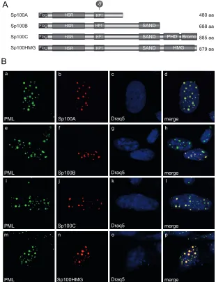
FIG 2Localization of Sp100 isoforms in noninfected cells. (A) Schematic representation of N-terminally Flag-tagged Sp100 isoforms depicting their domain organization. HSR, homogenously stained region; HP1, heterochromatin protein 1 interaction region; SAND, Sp100, AIRE-1, NucP41/45, and DEAF-1 domain; bromo, bromodomain; PHD, plant homeodomain; HMG, high-mobility-group domain; Su, SUMO conjugation motif. Amino acid (aa) lengths are indicated
on the right. (B) HepaRG cells were transfected with 1.5g of pFlag-Sp100A, -B, -C, or -HMG. Cells were fixed at 48 h posttransfection with 4% PFA and double
labeled with MAb Flag-M2 (anti-Flag) and pAb NB 100-59787 (anti-PML). Primary Abs were detected with Cy3 (anti-Flag; red)- and Alexa 488 (anti-PML; green)-conjugated secondary Abs. The DNA-intercalating dye DRAQ5 (Biostatus) was used for nuclear staining. Anti-PML (green; panels a, e, i, and m) and anti-Flag-Sp100 (red; panels b, f, j, and n) staining patterns representative of at least 50 analyzed cells are shown. Overlays of the single images (merge) are shown
in panels d, h, l, and p. Magnification,⫻7,600.
on November 7, 2019 by guest
http://jvi.asm.org/
[image:4.585.135.445.68.473.2]A
merge PML
a b c d
Draq5
e f g h
i j k l
24 h p.i.
Sp100A
merge
PML Sp100B Draq5
merge
PML Sp100C Draq5
m n o p
merge
PML Sp100HMG Draq5
merge
E2A Sp100HMG Draq5
merge E2A
a b c d
Draq5
i j k l
m n o p
48 h p.i.
Sp100A
merge
E2A Sp100B Draq5
merge
E2A Sp100C Draq5
q r s t
B
merge PML
e f g h
Draq5 Sp100A
on November 7, 2019 by guest
http://jvi.asm.org/
Sp100B, -C, and -HMG are relocalized from PML-NBs to
vi-ral replication centers during Ad infection.
Humans are so far
known to express at least four different Sp100 isoforms: Sp100A,
Sp100B, Sp100C, and Sp100HMG. To investigate the intracellular
localization of the different Sp100 isoforms in noninfected
HepaRG cells, we transfected single Flag-tagged Sp100 isoforms
(
Fig. 2A
) prior to immunofluorescence analysis. Costaining of
Flag-tagged Sp100 isoforms with endogenous PML revealed
local-ization of all Sp100 isoforms to PML-NBs (
Fig. 2B
). Although
some PML-NBs seemed to lack Flag-Sp100B, -C, and -HMG
staining in particular, and although higher expression levels
duced the formation of larger nuclear aggregates, with an
in-creased intensity of diffuse nuclear labeling, mild overexpression
resulted in colocalization in at least about 50% of the detected
PML-NBs. These observations are in line with the reports of Seeler
and coworkers (
11
).
In 1996, Doucas and coworkers described the relocalization of
endogenous Sp100 during Ad infection. In the early phase, PML
and Sp100 are reorganized into so-called track-like structures
prior to Sp100 being sequestered to the early viral replication
cen-ters (
74
). Therefore, we evaluated whether Sp100 colocalized to
Ad-induced PML tracks or juxtaposed with established viral
rep-lication centers is affected in an isoform-dependent manner.
HepaRG cells were transfected with the single Sp100 isoforms and
superinfected with Ad wt virus (
Fig. 3
).
Intriguingly, we detected only the Sp100A isoform efficiently
colocalizing with the PML-containing tracks (
Fig. 3A
, panel d). In
contrast, Sp100B, Sp100C, and Sp100HMG formed dot-like
structures not associated with PML (
n
⫽
50) (
Fig. 3A
, panels h, l,
and p). After late replication centers, marked by the Ad DNA
binding protein E2A-DBP (
Fig. 3B
, panels a, i, m, and q), were
established at 48 h p.i., the majority of Sp100A was still retained in
the PML-containing tracks (
Fig. 3B
, panels d and h). Our data
revealed that a substantial number of these Ad-induced tracks
were closely associated with the outer rims of the viral replication
centers (
Fig. 3B
, panels b and d and inset), whereas Sp100B, -C,
and -HMG accumulated entirely in the replication centers (
Fig.
3B
, panels l, p, and t). Based on these observations, we concluded
that in contrast to Sp100A, the B, C, and HMG isoforms are
sep-arated from the PML tracks during Ad infection. This is likely due
to different protein properties conferred by the additional
C-ter-minal domains, e.g., the SAND domain.
Next, we investigated the subcellular location of
de novo
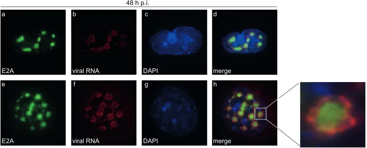
-syn-thesized viral RNA in relation to Ad replication centers. E2A was
visualized by immunofluorescence staining after
in situ
hybridiza-tion with fiber-specific probes (
Fig. 4
). Our data show that viral
RNA aggregated significantly at the periphery of the
E2A-contain-ing complexes (
Fig. 4d
and
h
). Additionally, we identified Sp100A
encircling these E2A-labeled replication centers (
Fig. 3B
, panel d),
colocalizing with arising PML tracks (
Fig. 3B
, panel h). We
dem-onstrated conclusively that
de novo
-synthesized viral RNA was
found mainly at the outer rims of Ad replication centers (
Fig. 4
)
juxtaposed on PML tracks containing Sp100A (
Fig. 3B
). At the
same time, Sp100B, -C, and -HMG were recruited to viral
repli-cation sites (
Fig. 3B
, panels l, p, and t) as seen for cellular
restric-tion factors, such as the recently identified Ad repressor SPOC1
(
75
).
SUMO-2 chains of Sp100 are shortened during Ad infection.
Since Ad apparently manipulates association of Sp100 isoforms
with PML-NBs, we monitored whether SUMO modification of
Sp100A is altered during infection. First, HeLa cells expressing
His-SUMO-1 and -2 were transfected with Sp100A (
Fig. 5A
) and
superinfected with wt Ad at 8 h posttransfection (
Fig. 5A
and
B
).
Immunoblotting of Ni-NTA-purified His-SUMO conjugates
FIG 3Ad-dependent relocalization of Sp100B, -C, and -HMG from PML-NBs to viral replication centers. HepaRG cells were transfected with 1.5g of
pFlag-Sp100A, -B, -C, or -HMG and superinfected with wt virus (H5pg4100) at a multiplicity of infection of 50 FFU/cell at 8 h posttransfection. (A) The cells were
fixed with 4% PFA at 24 h p.i. and double labeled with MAb Flag-M2 (anti-Flag) and pAb NB 100-59787 (anti-PML). (B) The cells were fixed with 4% PFA at 48 h p.i. and double labeled with MAb Flag-M2 (anti-Flag) and pAb NB 100-59787 (anti-PML) or rabbit MAb anti-E2A. Primary Abs were detected with Cy3 (anti-Flag; red)- and Alexa 488 (anti-PML/E2A; green)-conjugated secondary Abs. The DNA-intercalating dye DRAQ5 (Biostatus) was used to stain nuclei. Anti-PML (green; A, panels a, e, i, and m, and B, panel e), anti-E2A (green; B, panels a, i, m, and q), and anti-Flag-Sp100 (red; A, panels b, f, j, and n, and B, panels b, f, j, n, and r) staining patterns representative of at least 50 analyzed cells are shown. Overlays of the single images (merge) are shown in panels d, h, l, p, and t.
Magnification,⫻7,600.
merge E2A
a b c d
DAPI 48 h p.i.
viral RNA
merge E2A
e f g h
DAPI viral RNA
FIG 4Ad mRNA surrounds viral replication centers. HepaRG cells were infected with wt virus (H5pg4100) at a multiplicity of infection of 10 FFU/cell. At 24 h
p.i., cells were fixed and hybridizedin situas described in the text prior to labeling with mouse MAb anti-E2A. The primary Ab was detected with an Alexa 488
(anti-E2A; green)-conjugated secondary Ab. DAPI was used to stain nuclei. Staining patterns representative of at least 30 analyzed cells are shown. Overlays of
the single images (merge) are shown in panels d and h. Magnification,⫻7,600.
on November 7, 2019 by guest
http://jvi.asm.org/
[image:6.585.112.474.69.215.2](
Fig. 5A
, left panel, lane 6) and crude lysates (
Fig. 5A
, right
panel, lane 6) revealed that Sp100A SUMO-2 modification was
reduced during Ad infection. However, SUMO-1 conjugation
on Sp100A was not affected at this time point (
Fig. 5A
, left
panel, lane 4). A similar effect could be observed for
endoge-nous Sp100 (
Fig. 5B
).
To investigate whether the reduction of
high-molecular-weight forms of SUMO-2-modified Sp100A depends on the viral
early proteins E1B-55K, E4orf6, and/or E4orf3, His-SUMO-2
conjugates were purified (Ni-NTA) after Sp100A transfection and
superinfection with wt virus or mutant viruses lacking either
E1B-55K (H5
pm
4149), E4orf6 (H5
pm
4154), or E4orf3 (H5
pm
4150)
(
Fig. 5C
). Significantly lower levels of high-molecular-weight
SUMO-2-modified forms of Sp100 were detected with all viruses,
irrespective of E4orf3, E1B-55K, or E4orf6 expression.
It has previously been shown that the viral early protein E1A
represses SUMO modification of the retinoblastoma protein and
interacts with the SUMO-conjugating enzyme Ubc9 to likely
in-terfere with polySUMOylation (
76
,
77
). Therefore, we tested if
E1A triggers reduced SUMO-2 modification of Sp100 (
Fig. 5D
).
As a control, we included Ubc9 binding mutants of the two major
Ad5 E1A proteins, E1A-12S and E1A-13S (E1A-12S-D121A and
E1A-13S-D121A mutants), with a point mutation in the
con-served Ubc9 interacting motif (EVIDLT). This mutation has
pre-viously been shown to prevent E1A interaction with Ubc9 (
Fig.
5D
) (
76
). Analysis of Ni-NTA-purified SUMO-2 conjugates after
coexpression of Sp100A with 13S, 13S-D121A,
E1A-12S, and E1A-12S-D121A revealed that both wt E1A isoforms
(
Fig. 5D
, left panel, lanes 2 and 4), but not the Ubc9 binding
mutants (E1A-13S-D121A and E1A-12S-D121A), repressed
SUMO-2 modification of Sp100A (
Fig. 5D
, left panel, lanes 3 and
5). We also noticed a slight reduction in steady-state levels of
Sp100A after cotransfection with wt E1A-12S or E1A-13S and
as-sumed that SUMO modification affects the stability of Sp100 (
Fig.
5D
, right panel, lanes 2 and 4). As a control, we included PML but
could detect neither a change in steady-state levels nor a
signifi-cant change in SUMO modification, implying a specific effect on
Sp100. Since transfection of E1A-12S or -13S alone did not
com-pletely abolish Sp100 SUMO modification, it is likely that other
viral regulatory proteins affect SUMO modification of Sp100
dur-ing infection.
HP1
␣
interaction with Sp100A is reduced during Ad
infec-tion.
Sp100 has been shown to interact with HP1 via its PXVXL
A
1 2 3 4 5 6
HeLa par mock HeLa-Su1 moc k
HeLa-Su1 48 hpiHeLa-Su2 48 hpiHeLa-Su2 moc k
HeLa par 48 hpi
+ Sp100A kDa 250 55 Sp100A 95 130 95 72 130 36 28 His (SUMO) 250 11 Ni-NTA pulldown E2A Sp100A β-actin + Sp100A kDa
1 2 3 4 5 6
His (SUMO) 72 72 95 130 250 250 55 95 72 130 36 28 11
HeLa par mock HeLa-Su1 moc k
HeLa-Su1 48 hpiHeLa-Su2 48 hp i
HeLa-Su2 moc k
HeLapar 48 hpi input
C
Ni-NTA pulldown HeLa-Su2 + Sp100AkDa Sp100A 95 130 250 mock
1 2 3 4 5
H5pm 4149 (E1B-55K -) H5pg 4100 (wt) H5pm 4154 (E4or f6 -)
H5pm
4150
(E4orf3 -)
E2A Sp100A
β-actin kDa
1 2 3 4 5
72 72 95 130 55 28 E1B-55K E4orf6
input 24 h p.i.
HeLa-Su2 + Sp100A
E4orf3
mock H5pm
4149 (E1 B-55 K-) H5pg 410 0 (w t)
H5pm
415 4 (E4
orf6 -)
H5pm
4150 (E4o
rf3 -)
11
B
1 2 3 4 5 6
HeLa par mock + HeLa-Su1 mock+ HeLa-Su
1 48 hpi
+ HeLa-Su 2 48 hpi
+ HeLa-Su2 mock HeLa
par 48 hp i Ni-NTA pulldown kDa 250 55 72 95 130 95 72 130 36 28 His 250 11 Sp100 endo. E2A input β-actin kDa
1 2 3 4 5 6
His 95 72 72 95 130 250 55 95 72 130 36 28 11 Sp100 endo.
HeLa par moc k
+ HeLa-Su1 mock+ HeLa-Su1 48 hpi+ HeL a-Su2 48 hpi
+ HeLa-Su2 mock
HeLa par 48 hpi
Sp100A
E1A
β-actin kDa
HeLa-Su2 + Sp100A
empty
vector
E1A
-13S D121A
E1A-13S
95
130
55
1 2 3
input E1A -12S D1 21A E1A -12S 4 5 PML 95 130 72 His (SUMO) 55 72 130 28 11 kDa Ni-NTA pulldown HeLa-Su2 + Sp100A
Sp100A
95
130
250
empty vector
E1A-13S
D121A
E1A-13S
E1A-1
2S D121A
E1A-1 2S PML 130 250 His (SUMO) 55 72 130 28 11
1 2 3 4 5
D
FIG 5SUMO-2 chains of Sp100 are shortened during Ad infection. Parental HeLa cells and HeLa cells stably expressing 6His-SUMO-1 or 6His-SUMO-2
were transfected with 2g of pFlag-Sp100A and then superinfected with wt
virus (H5pg4100) (A) or a mutant virus lacking E4orf3 (H5pm4150), E4orf6
(H5pm4154), or E1B-55K (H5pm4149) (C) at a multiplicity of infection of 20
FFU/cell at 8 h posttransfection, as indicated, or cotransfected with 3g
E1A-13S, E1A-12S, E1A-13S-D121A, or E1A-13S-D121A (D). (B) To monitor en-dogenous Sp100 (Sp100 endo.), the cells were infected with wt virus
(H5pg4100) at a multiplicity of infection of 20 FFU/cell without transfection.
For panels A to C, whole-cell lysates were prepared with guanidinium chloride buffer at 48 h posttransfection, subjected to Ni-NTA purification of 6His-SUMO conjugates, and fractionated in a 4 to 12% gradient gel before immu-noblot analysis. Input levels of whole-cell lysates and Ni-NTA-purified pro-teins were detected using pAb GH3 (anti-Sp100), pAb PML NB100-59787,
MAb M-58 (anti-E1A), MAb B6 (anti-E2A), MAb 6⫻His, MAb Flag-M2
(an-ti-Flag), MAb AC-15 (anti--actin), MAb 2A6 (anti-E1B-55K), MAb RSA3
(anti-E4orf6), and MAb 6A11 (anti-E4orf3). Molecular masses in kDa are indicated on the left, and specific proteins are indicated on the right.
on November 7, 2019 by guest
http://jvi.asm.org/
[image:7.585.43.282.65.721.2]motif (
18
). Overexpressed or IFN-induced Sp100 enhances
re-cruitment of endogenous HP1 protein to PML-NBs, mainly
local-izing to the interior of these subdomains (
18
,
46
). Additionally,
the Sp100 SUMO conjugation site (SCS) is located close to the
HP1 interaction motif, and SUMOylated Sp100 is known to
sta-bilize its binding to HP1
in vitro
(
11
). To test whether
Ad-medi-ated loss of Sp100A-SUMO-2 affects the HP1 interaction
in vivo
,
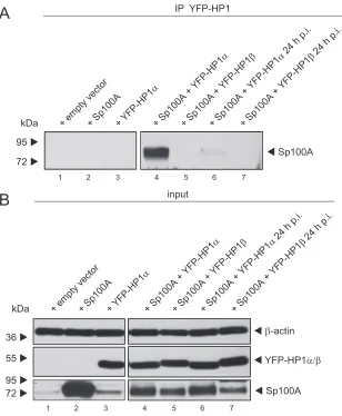
we performed binding assays with infected,
Sp100A-overexpress-ing cells cotransfected with either HP1
␣
or HP1

(
Fig. 6
). We
could not detect an interaction between Sp100A and HP1

.
How-ever, we noticed that HP1
␣
binding to Sp100A was significantly
reduced in infected cells (
Fig. 6A
, lane 6).
Sp100A-specific activation of Ad promoter activity.
Recently,
Newhart et al. reported that Sp100A promotes chromatin
decon-densation at cytomegalovirus (CMV) promoter-regulated
tran-scription sites. In contrast, Sp100B, containing an additional
SAND domain, promotes chromatin condensation, and thus
transcriptional repression (
20
). To test whether Sp100 also affects
transcription from Ad promoters, we investigated Ad E1A,
E2early, pIX, and E3 promoter activity in the presence of either
Sp100A or Sp100B, as stable knockdown of each isoform was not
feasible due to sequence similarity (
Fig. 7
).
Since Sp100A was recently reported not to overcome Daxx/
ATRX-mediated transcriptional repression (
20
), we used U2OS
cells, which have reduced ATRX expression, to minimize
addi-tional effects mediated by these transcription factors.
Further-more, we employed Daxx-depleted U2OS cells (
78
) to
addition-ally diminish the repressive effect of the Daxx-ATRX chromatin
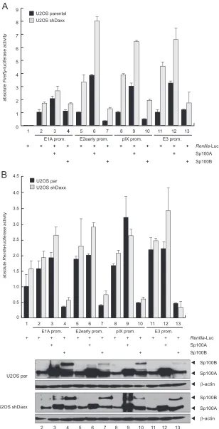
remodeling complex. We observed that Sp100A stimulated
tran-scription from the Ad promoters
⬃
2- to 3-fold in both cell lines.
As anticipated, basal activity of the promoters was higher in
Daxx-depleted cells, implying an additional repression mechanism of
Daxx that is independent of the Daxx-ATRX complex. In contrast
to Sp100A, the B isoform reduced E2early, pIX, and E3 promoter
activity
⬃
0.5- to 3-fold (
Fig. 7A
).
Additionally, we measured
Renilla
luciferase activity under the
control of the HSV-TK promoter, which is normally used as an
internal normalization control. We observed that Sp100 also
β-actin 36
55
Sp100A
kDa
YFP-HP1α/β
72 95
input
1 2 3 4
B
72 95
Sp100A
5 6 7
1 2 3 4 5 6 7
kDa
IP YFP-HP1
A
+ empty vector + Sp100A +
YFP-HP1 β 24 h p.i.
+ Sp100A+ YFP-HP1
α
+ Sp100A + YFP-HP
1α
+ Sp100A + YFP-HP1
β
+ Sp100A + YFP-HP
1α 24 h p.i.
+ empty vector + Sp100A +
YFP-HP1 β 24 h p.i.
+ Sp100A+ YFP-HP1
α
+ Sp100A + YFP-HP1
α
+ Sp100A + YFP-HP
1β
+ Sp100A + YFP-HP
1α 24 h p.i.
FIG 6HP1␣interaction with Sp100A is reduced during Ad infection. H1299 cells were superinfected with wt virus (H5pg4100) at a multiplicity of infection of
20 FFU/cell 8 h after transfection of 2g of pFlag-Sp100A mutant and 3g of pYFP-HP1␣/pYFP-HP1, as indicated. Cells were harvested at 24 h p.i., prior to
preparation of coimmunoprecipitation (IP) samples and whole-cell extracts. Coprecipitated proteins (A) and input levels (B) of whole-cell lysates were detected
using pAb GH3 (anti-Sp100), AC-15 (anti--actin), and YFP antibody (anti-YFP-HP1␣/pYFP-HP1). Molecular masses in kDa are indicated on the left, and
specific proteins are indicated on the right.
on November 7, 2019 by guest
http://jvi.asm.org/
[image:8.585.137.445.65.439.2]Sp100A Sp100B
Renilla-Luc +
+
+ + + +
+
+ +
+
absolute
Firefly
-luciferase activity
+ +
3 2
1 4 5 6 7 8 9 10 11
+ + + 0
1 2 3 4 5 6 7 8
U2OS parental U2OS shDaxx 9
12 13
+ + +
+
+ + E2early prom.
E1A prom. pIX prom. E3 prom.
A
B
0 0.5 1.0 1.5 2.0 2.5 3.0
absolute
Renilla
-luciferase activity
Sp100A Sp100B
Renilla-Luc +
+
+ + +
+ +
+ + 3
2
1 4 5 6 7 8 9 10 11 + + +
12 13 + +
+
+ + E2early prom.
E1A prom. pIX prom. E3 prom. U2OS par
U2OS shDaxx
3.5 4.5
4.0
3
2 4 5 6 7 8 9 10 11 12 13 U2OS par
U2OS shDaxx
β-actin Sp100A Sp100B
β-actin Sp100A Sp100B +
+ + +
FIG 7Sp100 isoform-specific regulation of Ad gene expression. (A) U2OS and U2OS shDaxx cells were transfected with 0.5g pRenilla-Luc, 0.5g
pGL-E1A-promoter, pGL-E2early-pGL-E1A-promoter, pGL-E3-pGL-E1A-promoter, or pGL-pIX-pGL-E1A-promoter, and 0.5g pSp100A or pSp100B in the combinations indicated (⫹). Total cell
extracts were prepared and luciferase activities determined at 24 h posttransfection. Absolute firefly luciferase activities are shown. Means and standard deviations
for two independent experiments are presented. (B) Means and standard deviations of absoluteRenillaluciferase activities are presented for two independent
experiments. Input levels of total cell lysates were detected using pAb GH3 (anti-Sp100) and MAb AC-15 (anti--actin).
on November 7, 2019 by guest
http://jvi.asm.org/
[image:9.585.140.448.66.670.2]fected
Renilla
luciferase expression, either transactivating it via
Sp100A or repressing it via Sp100B (
Fig. 7B
).
Such effector-dependent activation or repression of control
genes included for normalization was previously reported by
other groups and was circumvented by use of a GFP expression
vector for internal transfection control (
79–81
). Hence, we
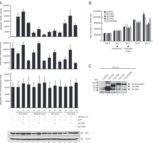
in-cluded a GFP-expressing vector as an additional control to
deter-mine transfection efficiencies (
Fig. 8A
) as previously described
(
82
). In contrast to the
Renilla
luciferase activity, the GFP signal
was not increased by coexpression of Sp100A or decreased by
Sp100B (
Fig. 8A
). This was also validated by immunoblot analysis
of the crude extracts (
Fig. 8A
, bottom panel).
Furthermore, we monitored cell growth of the U2OS cells via
trypan blue exclusion after transfection of Sp100A, -B, -C, or
-HMG or an empty vector to exclude that changes in cell viability
account for the different effects of the Sp100 isoforms on
lucifer-A
B
Sp100A Sp100B
Renilla-Luc +
+ + + +
+ +
+ + +
3 2
1 4 5 6 7 8 9 10 11
+ +
12 13
+ +
+ +
+ + E2early prom.
E1A prom. pIX prom. E3 prom. 0
50000 100000 150000 200000
0 5000 10000 15000 20000
+ +
GFP
+ +
+ + + + +
+ + + + + + +
absolute
Renilla
activity
absolute GFP fluorescence units
25000
pcDNA Sp100A Sp100B
0 300000 600000 900000 1200000 1500000
Sp100C Sp100HMG
total number of viable cells
day 0 day 1 day 2 day 3 day 4
transfection
luciferase assay
72 95 130 kDa
Sp100A
+ pcDNA+ Sp100 A
+ Sp100C+ Sp100HMG
Sp100B
Sp100C/HMG
+ Sp100B
1 2 3 4 5
250
C
24 h p.t.3 2
1 4 5 6 7 8 9 10 11 12 13
absolut
e
Firefl
y
activity
β-actin GFP 0
20000 40000 60000 80000 100000 120000
FIG 8Differences in transfection efficiency do not account for the Sp100 isoform-specific effect on gene expression. (A) U2OS cells were transfected with 0.5g pRenilla-Luc, 0.5g pGFP, 0.5g pGL-E1A-promoter, pGL-E2early-promoter, pGL-E3-promoter, or pGL-pIX-promoter, and 0.5g pSp100A or pSp100B in
the combinations indicated (⫹). GFP fluorescence signals were measured using a Tecan Infinite 200 Pro plate reader before total cell extracts were prepared and
luciferase activities determined at 24 h posttransfection. Absolute firefly luciferase activities andRenillaluciferase activities are shown. Means and standard
deviations for three independent experiments are presented. Input levels of total cell lysates were detected using MAb AC-15 (anti--actin) and a GFP epitope
MAb. (B) U2OS cells were transfected with 3g of pcDNA, pSp100A, or pSp100B, and total cell numbers were determined by trypan blue exclusion at the
indicated time points. Time points of transfection and luciferase assay are indicated at the bottom. (C) U2OS cells were transfected with 3g of pcDNA, pSp100A,
or pSp100B, and total cell extracts were prepared at 24 h posttransfection. Proteins were separated by SDS-PAGE and subjected to immunoblotting using pAb GH3 (anti-Sp100).
on November 7, 2019 by guest
http://jvi.asm.org/
[image:10.585.47.543.65.527.2]ase gene expression (
Fig. 8B
). Cell growth was not altered upon
transfection of the Sp100 isoforms compared to the empty vector
control until 72 h posttransfection. Since the reporter gene assays
were performed at 24 h posttransfection, changes in cell viability
after transfection of the Sp100 isoforms can be excluded for the
time point of luciferase assay measurement (
Fig. 8B
). In addition,
we also compared exogenous Sp100 protein expression at 24 h
posttransfection, a time point equal to that of the reporter assays
presented above (
Fig. 8C
).
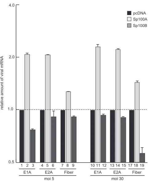
To further confirm our data with the virus background, we
measured Ad early and late mRNA expression in infected cells
expressing either Sp100A or Sp100B (
Fig. 9
). Viral early E1A and
E2A mRNA production was stimulated in infected cells expressing
Sp100A compared to cells treated with the empty vector control
(
Fig. 9
). Additionally, late fiber transcript synthesis was enhanced
in cells cotransfected with Sp100A, suggesting an impact of either
enhanced synthesis of early viral gene products or direct
stimula-tion of the late promoter (
Fig. 9
). Consistent with data obtained in
the reporter assays described above, Sp100B negatively affected
viral early and late mRNA expression, although the effect was not
as strong as that observed for Sp100A-dependent activation.
Sim-ilar results were obtained for early and late mRNA stimulation by
Sp100A in cells infected at a higher multiplicity of infection
(
Fig. 9
).
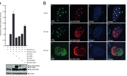
The Sp100A SIM promotes transcriptional activation and
lo-calization to track-like structures.
The transactivating properties
of Sp100 on transcription have previously been mapped to amino
acids 333 to 407, a region immediately downstream of the HP1
interaction region (
9
). The SUMO-interacting motif (SIM) has
been described to be important for Sp100 recruitment to HSV-1
genomes (
83
). Given the fact that the SIM is located within the
transactivating region of Sp100, we investigated the
transcrip-tional properties of an Sp100A nonfunctranscrip-tional SIM derivative
(Sp100A-I323I324K) on Ad promoters (
Fig. 10A
). Reporter gene
assays with the Ad E2early promoter revealed that the
Sp100A-SIM mutant was no longer able to efficiently stimulate
transcrip-tion from the viral promoter (
Fig. 10A
, lane 7), although
Sp100A-wt and the Sp100A-SIM mutant were expressed in similar
amounts (
Fig. 10A
, lower panel).
Next, we tested whether the SIM mutation would alter the
intracellular localization of Sp100A (
Fig. 10B
). In noninfected
cells, the Sp100A-SIM mutant localized to the PML-NBs (
Fig.
10B
). Costaining with Ad E2A-DBP revealed that after PML track
formation, the Sp100A-SIM mutant was diffusely distributed in
the host cell nucleus, localizing neither to the tracks nor to the viral
replication centers in 80% of the cells investigated (
n
⬎
50) (
Fig.
10B
, panels h and m). However, in 20% of the cells, the
Sp100A-SIM mutant localized to PML tracks and/or viral replication
cen-ters, likely due to dimerization with endogenous wt Sp100 via the
HSR domain (
n
⬎
50) (data not shown).
DISCUSSION
Nuclear-replicating DNA viruses antagonize intrinsic antiviral
defense mechanisms to replicate efficiently; however, the
func-tional mechanisms behind this phenomenon are diverse and not
well understood. Here we unravel some more pieces of the puzzle,
using Ad as a model virus, and propose a novel Ad evasion strategy
that involves isoform-specific, active manipulation of the host
re-striction factor Sp100 as a prerequisite for efficient viral
replica-tion.
Initial experiments demonstrated that during the first hours of
Ad infection, E1A orchestrates the temporally regulated
expres-sion of viral proteins. Simultaneously, the early adenoviral
pro-teins E1B-55K and E4orf3 are expressed (
84
). First, E4orf3
in-duces the reorganization of PML nuclear bodies by interacting
with PML-II, which leads to the formation of track-like structures
in the nuclei of cells (
Fig. 11
) (
74
,
85–90
). This reorganization of
PML-NBs is highly conserved among most Ad species (
91
) and
therefore suggests an important function during Ad infection,
presumably in eliminating intracellular viral defense barriers (
35
,
37
,
74
,
92
). In parallel, SUMOylated E1B-55K is localized to the
PML-NBs, presumably by interacting with PML-IV (
93
). Notably,
the presence of E4orf3 and E1B-55K synergistically modulates p53
transcriptional activity (
94
), which might come about by altering
PML-IV function and consequently inducing
E1B-55K-depen-dent SUMOylation of the p53 protein (
95
,
96
). During the onset of
viral infection, E4orf6, which is expressed with a delay of 6 h
com-pared to E1B-55K/E4orf3, facilitates nuclear matrix release of
E1B-55K via deSUMOylation (
93
). Finally, E1B-55K–E4orf6
complex formation induces proteasomal degradation of
repres-sive factors, such as p53, ATRX, and SPOC1 (
Fig. 11
) (
97–101
).
Based on available data obtained with different DNA viruses, it
is reasonable to conclude that diverse molecular mechanisms have
evolved to counteract PML-NB-mediated antiviral activities.
In-pcDNA Sp100A Sp100B
3 2
1 4 5 6 7 8 9 101112 131415171819 E2A
E1A Fiber E1A E2A Fiber moi 5 moi 30
relative amount of viral mRNA
0.5 1.0
2.0 4.0
FIG 9Sp100 isoform-specific regulation of Ad mRNA synthesis.
Subconflu-ent U2OS cells were transfected with 5g pcDNA, pSp100A, or pSp100B and
superinfected with wild-type virus (H5pg4100) at a multiplicity of infection
(MOI) of 5 or 30 FFU/cell at 48 h posttransfection. The cells were harvested at 24 h p.i., and total RNA was extracted, reverse transcribed, and quantified by RT-PCR analysis using primers specific for Ad5 E1A, E2A, and fiber. The data were normalized to 18S rRNA levels. The data are presented on a log axis relative to the amount of RNA detected in control cells expressing empty vector.
on November 7, 2019 by guest
http://jvi.asm.org/
[image:11.585.44.286.63.358.2]activation of PML-NBs during early stages of HSV-1 infection
depends exclusively on the expression of the immediate early
pro-tein ICP0, which rapidly localizes to these subnuclear structures
and disrupts them completely (
102–107
). Interestingly, ICP0
leads to the rapid loss of high-molecular-weight PML in a
protea-some-dependent manner, and subsequently results in
disintegra-tion of PML-NB structures (
108
,
109
). Since ICP0-mediated
pro-teasomal depletion also includes the PML-NB-associated protein
Sp100, it has been suggested that degradation occurs via
interfer-ence with a common SUMOylation pathway rather than by
spe-cific targeting of individual proteins (
110
). Another study
pro-vides evidence that the formation of repressive PML-NB-like
structures in association with incoming viral DNA depends on the
respective SIMs of antiviral proteins, such as PML, Sp100, and
Daxx (
111
). This observation provides a convincing explanation
for how early viral proteins may inhibit the antiviral functions of
these proteins by depleting PML-SUMO conjugates and,
conse-quently, eliminating their SIM-dependent recruitment.
For Ad, the functional consequences of E4orf3-induced PML-NB
reorganization prior to formation of track-like structures in the
nuclei of cells still remain unclear. However, here we provide
ev-idence that Ad selectively counteracts antiviral immune responses
and, at the same time, benefits from other PML-NB-associated
components by actively recruiting them to PML track-like
struc-tures, the sites of viral transcription in the host cell nucleus.
De-pletion of the PML-associated factor Sp100 results in significantly
increased Ad progeny production (
Fig. 1
). According to our data,
Ad counteracts this antiviral measure by specifically targeting the
repressive isoforms Sp100B, Sp100C, and Sp100HMG. However,
the negative impact of the B, C, and HMG isoforms together seems
to be more potent in the repression of Ad replication than the
activating effect of Sp100A, as knockdown of all isoforms causes
an overall negative effect of the PML-NB component Sp100.
The transcription factor Sp100 has been reported to modulate
the replication programs of several DNA viruses. Human CMV
(HCMV) and herpesvirus saimiri (HVS) target Sp100 for
protea-somal degradation; depletion of Sp100 by RNA interference
en-hances HCMV replication and gene expression (
83
,
112
,
113
).
Similar to our findings, Maul and coworkers previously
demon-strated that Sp100 isoforms B, C, and HMG, but not Sp100A,
suppressed HSV-1 immediate early gene expression and repressed
the ICP0 promoter and that this was dependent on the SAND
domain (
114
). In 1996, Doucas et al. had already described
Ad-dependent relocalization of endogenous Sp100 into the
E4orf3-induced track-like PML structures at early times postinfection
(
74
). During late stages, PML and Sp100 segregation was
ob-A
Sp100A Sp100B
Renilla-Luc +
+ + + + +
+ +
+
absolute
Firefl
y
-luciferase activity
+ 3 2
1 4 5 6 7 0
0.5 1.0 1.5 2.0 2.5 3.0 3.5
+ + + + +
+ E2early prom
Sp100C Sp100HMG Sp100A-SIM +
+
B
merge
E2A Sp100A SIM Draq5
i k l m
mock
24 hpi
48 hpi
merge
PML Sp100A SIM Draq5
merge
PML Sp100A SIM Draq5
a b c d
e f g h
β-actin
3 2
1 4 5 6 7
Sp100A/SIM
Sp100B
Sp100C/HMG
FIG 10Sp100A transcriptional activation and localization depend on a functional SIM. (A) U2OS cells were transfected with 0.5g pRenilla-Luc, 0.5g
pGL-E2early-promoter, and 0.2g pE1A-13S, pSp100A, pSp100B, pSp100C, pSp100HMG, or pSp100A-SIM in the combinations indicated (⫹). Absolute firefly
luciferase activities are shown, with means and standard deviations for three independent experiments. Data are presented uncorrected for transfection controls
as explained in the legend toFig. 8. Input levels of total cell lysates were detected using pAb GH3 (anti-Sp100) and MAb AC-15 (anti--actin). (B) HepaRG cells
were transfected with 1.5g of pFlag-Sp100A SIM and superinfected with wild-type virus (H5pg4100) at a multiplicity of infection of 50 FFU/cell at 8 h
posttransfection. The cells were fixed with 4% PFA at 48 h p.i. and double labeled with pAb NB 100-59787 (anti-PML), MAb Flag-M2 (anti-Flag), and rabbit MAb anti-E2A. Primary Abs were detected with Cy3 (anti-Flag; red)- and Alexa 488 (anti-E2A; green)-conjugated secondary Abs. The DNA-intercalating dye DRAQ5 (Biostatus) was used for nuclear staining. Anti-PML (green; panels a and e), anti-E2A (green; panel i), and anti-Flag-Sp100 (red; panels b, f, and k) staining
patterns representative of at least 50 analyzed cells are shown. Overlays of the single images (merge) are shown in panels d, f, and m. Magnification,⫻7,600.
on November 7, 2019 by guest
http://jvi.asm.org/
[image:12.585.50.539.65.359.2]served, with Sp100 relocalizing from these Ad-induced tracks to
the early viral replication centers (
114
). Our novel findings now
illustrate that the activating transcription isoform Sp100A is
found mainly at the Ad-induced PML tracks, while only the
re-pressive Sp100B, -C, and -HMG proteins are displaced from these
nuclear structures to the Ad replication centers (
Fig. 3
and
11
).
Besides these repressive Sp100 variants, other repressive factors or
proteins participating in the DNA damage response (DDR) were
already shown to be recruited to these nuclear loci, suggesting a
common phenomenon during Ad infection (
74
). Based on our
results, we believe that Ad-dependent loss of PML-NB integrity by
redistribution into track-like structures consequently induces the
dispersal of PML-associated repressive transcription factors in the
nucleus. These antiviral proteins, e.g., Daxx, ATRX, and p53, are
immediately targeted by the newly synthesized viral gene products
(
Fig. 11
).
As described for herpesviruses (
112
,
115
,
116
), we observed
modulation of SUMOylated Sp100 forms during Ad infection, a
prerequisite for altered intranuclear localization of the different
Sp100 proteins. Indeed, we observed that Sp100A activates
tran-scription from Ad promoters (
Fig. 7
to
9
), most likely when it is
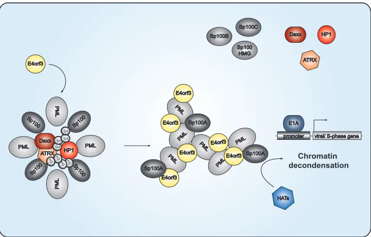
kept at the PML tracks, where it may recruit histone acetylases to
create a favorable environment for Ad gene expression (
Fig. 11
).
It has been proposed that HP1 and Sp100 form a
SUMO-de-pendent complex to regulate chromatin remodeling (
110
),
al-though the exact functionality of the complex is still unknown.
HP1 proteins are phosphorylated in response to DNA damage and
appear to be important for recruiting DDR factors and
dynami-cally reorganizing chromatin (
3
,
117
). For Epstein-Barr virus
(EBV), EBNA-LP, the coactivator of the EBV transactivator
EBNA2, has been shown to interact with Sp100 and to displace the
Sp100-HP1
␣
complex from PML-NBs (
118
). Consistent with
these observations, Ad disrupts the Sp100-HP1
␣
interaction,
likely inducing chromatin decondensation (
Fig. 6
). Ad disrupts
this interaction, suggesting that this cellular chromatin
remodel-ing complex is repressive for virus replication by chromatin
con-densation, providing evidence for the general model that Ad
promotes deSUMOylation of PML-NB-associated proteins to
prevent recruitment of certain repressive factors to the PML
track-like structures in infected cells (
Fig. 11
). This is further supported
by the fact that poly-SUMO-2/3 chains protrude into the interior
of PML-NBs, where they mediate the enriched interactions with
HP1, Daxx, or ATRX.
The predominant opinion in the field is that PML-NBs are part
of an IFN-induced antiviral defense mechanism (
36–38
,
119
).
However, new evidence also points to a proviral function (
35
).
Specifically, DNA viruses require PML-NBs and associated
pro-teins to establish efficient replication and transcription. We now
show that this concept is more intricate than originally believed,
since viruses apparently take advantage of specific
PML-NB-asso-ciated proteins to actively establish a proviral environment in the
host cell, while eliminating and inhibiting repressive factors.
Si-multaneously, antiviral measures are efficiently inhibited to
main-tain the viral infectious program. Obviously, ongoing studies on
virus-host interactions and future insights into novel immune
evasion strategies acquired by pathogenic viruses will contribute
to identifying new therapeutic strategies and targets to limit or
prevent virus-mediated diseases and mortality of patients.
PML PML
PML
PML
Sp100B
PML PM
L
PML
PM L
PML
PM L
E4orf3
E4orf3 E4orf3
E4orf3
E4orf3
E4orf3
Sp100
Sp10 0
Sp100
Sp10 0
HP1 Sp100A Sp100C
Sp100 HMG
E1A
promoter viral/ S-phase gene
Chromatin decondensation
HATs
Daxx HP1
E4orf3
Sp100A
Sp100A
ATRX Daxx
ATRX
Su SuSu
Su
Su Su Su Su
FIG 11PML-NB-mediated transcriptional regulation of Ad gene expression. The schematic representation depicts the proposed model of Ad-mediated relocalization of antiviral factors associated with PML nuclear bodies. In noninfected cells, transcriptional activators and repressors, such as the Sp100 isoforms, HP1, Daxx, and ATRX, are clustered within the PML-NBs. Sp100A promotes chromatin decondensation and histone acetyltransferase (HAT) recruitment, whereas Sp100B, Sp100C, and Sp100HMG prevent these events. HP1 and the Daxx-ATRX complex function in gene silencing by organizing higher-order chromatin structures. Poly-SUMO-2/3 (Su) chains protrude into the interior of the PML-NB to mediate interactions with the HP1, Daxx, or ATRX enriched there. At early time points after infection, E4orf3 targets PML isoform II to reorganize PML-NB into track-like structures. SUMO-2/3 chains are shortened, causing release of the repressive factors HP1, Daxx, and ATRX from the PML-NBs. Under these conditions, Sp100A, associating with the PML tracks, is able to recruit HATs, thereby creating a favorable environment for activation of Ad gene expression.
on November 7, 2019 by guest
http://jvi.asm.org/
[image:13.585.102.484.65.309.2]ACKNOWLEDGMENTS
We thank Roger Everett and Hans Will for kindly providing reagents, and we greatly appreciate the critical comments and scientific discussions of Gregory Fonseca and Nicole Fischer.
The Heinrich Pette Institute, Leibniz Institute for Experimental Virol-ogy, is supported by the Freie und Hansestadt Hamburg and the Bundesministerium für Gesundheit (BMG). S.S. was supported by the Peter und Traudl Engelhorn Stiftung and additional grants from the Erich und Gertrud Roggenbuck Stiftung and the Else Kröner-Fresenius Stif-tung. T.D. was supported by the Deutsche Forschungsgemeinschaft (DFG) and the Wilhelm Sander Stiftung. Part of this work was supported by the B. Braun Stiftung and the Fonds der Chemischen Industrie.
REFERENCES
1.Guldner HH, Szostecki C, Grotzinger T, Will H.1992. IFN enhance expression of Sp100, an autoantigen in primary biliary cirrhosis. J.
Im-munol.149:4067– 4073.
2. Grotzinger T, Sternsdorf T, Jensen K, Will H. 1996. Interferon-modulated expression of genes encoding the nuclear-dot-associated pro-teins Sp100 and promyelocytic leukemia protein (PML). Eur.
J. Biochem. 238:554 –560.http://dx.doi.org/10.1111/j.1432-1033.1996
.0554z.x.
3.Lehming N, Le Saux A, Schuller J, Ptashne M. 1998. Chromatin components as part of a putative transcriptional repressing complex.
Proc. Natl. Acad. Sci. U. S. A.95:7322–7326.http://dx.doi.org/10.1073
/pnas.95.13.7322.
4.Szostecki C, Guldner HH, Netter HJ, Will H. 1990. Isolation and characterization of cDNA encoding a human nuclear antigen predomi-nantly recognized by autoantibodies from patients with primary biliary
cirrhosis. J. Immunol.145:4338 – 4347.
5.Szostecki C, Krippner H, Penner E, Bautz FA.1987. Autoimmune sera recognize a 100 kD nuclear protein antigen (sp-100). Clin. Exp. Immu-nol.68:108 –116.
6.Szostecki C, Will H, Netter HJ, Guldner HH.1992. Autoantibodies to the nuclear Sp100 protein in primary biliary cirrhosis and associated diseases: epitope specificity and immunoglobulin class distribution.
Scand. J. Immunol.36:555–564.http://dx.doi.org/10.1111/j.1365-3083
.1992.tb03224.x.
7.Pandolfi PP, Alcalay M, Fagioli M, Zangrilli D, Mencarelli A, Diverio D, Biondi A, Lo Coco F, Rambaldi A, Grignani F.1992. Genomic variability and alternative splicing generate multiple PML/RAR alpha transcripts that encode aberrant PML proteins and PML/RAR alpha
iso-forms in acute promyelocytic leukaemia. EMBO J.11:1397–1407.
8.Fagioli M, Alcalay M, Pandolfi PP, Venturini L, Mencarelli A, Simeone A, Acampora D, Grignani F, Pelicci PG.1992. Alternative splicing of PML transcripts predicts coexpression of several carboxy-terminally
dif-ferent protein isoforms. Oncogene7:1083–1091.
9.Xie K, Lambie EJ, Snyder M.1993. Nuclear dot antigens may specify
transcriptional domains in the nucleus. Mol. Cell. Biol.13:6170 – 6179.
10. Dent AL, Yewdell J, Puvion-Dutilleul F, Koken MH, de The H, Staudt
LM.1996. LYSP100-associated nuclear domains (LANDs): description
of a new class of subnuclear structures and their relationship to PML
nuclear bodies. Blood88:1423–1426.
11. Seeler JS, Marchio A, Losson R, Desterro JM, Hay RT, Chambon P, Dejean A.2001. Common properties of nuclear body protein SP100 and TIF1alpha chromatin factor: role of SUMO modification. Mol. Cell. Biol. 21:3314 –3324.http://dx.doi.org/10.1128/MCB.21.10.3314-3324.2001. 12. Guldner HH, Szostecki C, Schroder P, Matschl U, Jensen K, Luders C,
Will H, Sternsdorf T.1999. Splice variants of the nuclear dot-associated Sp100 protein contain homologies to HMG-1 and a human nuclear
phosphoprotein-box motif. J. Cell Sci.112:733–747.
13. Sternsdorf T, Jensen K, Reich B, Will H.1999. The nuclear dot protein sp100, characterization of domains necessary for dimerization, subcellular localization,
and modification by small ubiquitin-like modifiers. J. Biol. Chem.274:12555–
12566.http://dx.doi.org/10.1074/jbc.274.18.12555.
14. Bottomley MJ, Collard MW, Huggenvik JI, Liu Z, Gibson TJ, Sattler
M.2001. The SAND domain structure defines a novel DNA-binding fold
in transcriptional regulation. Nat. Struct. Biol.8:626 – 633.http://dx.doi
.org/10.1038/89675.
15. Isaac A, Wilcox KW, Taylor JL. 2006. SP100B, a repressor of gene
expression preferentially binds to DNA with unmethylated CpGs. J. Cell.
Biochem.98:1106 –1122.http://dx.doi.org/10.1002/jcb.20841.
16. Fish PV, Filippakopoulos P, Bish G, Brennan PE, Bunnage ME, Cook AS, Federov O, Gerstenberger BS, Jones H, Knapp S, Marsden B, Nocka K, Owen DR, Philpott M, Picaud S, Primiano MJ, Ralph MJ, Sciammetta N, Trzupek JD.2012. Identification of a chemical probe for bromo and extra C-terminal bromodomain inhibition through
optimi-zation of a fragment-derived hit. J. Med. Chem.55:9831–9837.http://dx
.doi.org/10.1021/jm3010515.
17. Baker LA, Allis CD, Wang GG.2008. PHD fingers in human diseases: disorders arising from misinterpreting epigenetic marks. Mutat. Res. 647:3–12.http://dx.doi.org/10.1016/j.mrfmmm.2008.07.004.
18. Seeler JS, Marchio A, Sitterlin D, Transy C, Dejean A.1998. Interac-tion of SP100 with HP1 proteins: a link between the promyelocytic leu-kemia-associated nuclear bodies and the chromatin compartment. Proc.
Natl. Acad. Sci. U. S. A.95:7316 –7321.http://dx.doi.org/10.1073/pnas
.95.13.7316.
19. Burnett E, Christensen J, Tattersall P.2001. A consensus DNA recog-nition motif for two KDWK transcription factors identifies flexible-length, CpG-methylation sensitive cognate binding sites in the majority
of human promoters. J. Mol. Biol.314:1029 –1039.http://dx.doi.org/10
.1006/jmbi.2000.5198.
20. Newhart A, Negorev DG, Rafalska-Metcalf IU, Yang T, Maul GG, Janicki SM.2013. Sp100A promotes chromatin decondensation at a cytomegalovirus-promoter-regulated transcription site. Mol. Biol. Cell 24:1454 –1468.http://dx.doi.org/10.1091/mbc.E12-09-0669.
21. Sternsdorf T, Jensen K, Will H.1997. Evidence for covalent modifica-tion of the nuclear dot-associated proteins PML and Sp100 by PIC1/
SUMO-1. J. Cell Biol.139:1621–1634.http://dx.doi.org/10.1083/jcb.139
.7.1621.
22. Muller S, Matunis MJ, Dejean A.1998. Conjugation with the ubiquitin-related modifier SUMO-1 regulates the partitioning of PML within the
nucleus. EMBO J.17:61–70.http://dx.doi.org/10.1093/emboj/17.1.61.
23. Ascoli CA, Maul GG.1991. Identification of a novel nuclear domain. J.
Cell Biol.112:785–795.http://dx.doi.org/10.1083/jcb.112.5.785.
24. Chang KS, Stass SA, Chu DT, Deaven LL, Trujillo JM, Freireich EJ. 1992. Characterization of a fusion cDNA (RARA/myl) transcribed from the t(15;17) translocation breakpoint in acute promyelocytic leukemia.
Mol. Cell. Biol.12:800 – 810.
25. Kakizuka A, Miller WH, Jr, Umesono K, Warrell RP, Jr, Frankel SR, Murty VV, Dmitrovsky E, Evans RM.1991. Chromosomal transloca-tion t(15;17) in human acute promyelocytic leukemia fuses RAR alpha
with a novel putative transcription factor, PML. Cell66:663– 674.http:
//dx.doi.org/10.1016/0092-8674(91)90112-C.
26. Dyck JA, Maul GG, Miller WH, Jr, Chen JD, Kakizuka A, Evans RM. 1994. A novel macromolecular structure is a target of the
promyelocyte-retinoic acid receptor oncoprotein. Cell76:333–343.http://dx.doi.org
/10.1016/0092-8674(94)90340-9.
27. de The H, Lavau C, Marchio A, Chomienne C, Degos L, Dejean A. 1991. The PML-RAR alpha fusion mRNA generated by the t(15;17) translocation in acute promyelocytic leukemia encodes a functionally
altered RAR. Cell 66:675– 684. http://dx.doi.org/10.1016/0092-8674
(91)90113-D.
28. Kastner P, Perez A, Lutz Y, Rochette-Egly C, Gaub MP, Durand B, Lanotte M, Berger R, Chambon P.1992. Structure, localization and transcriptional properties of two classes of retinoic acid receptor alpha fusion proteins in acute promyelocytic leukemia (APL): structural
simi-larities with a new family of oncoproteins. EMBO J.11:629 – 642.
29. Goddard AD, Borrow J, Solomon E.1992. A previously uncharacter-ized gene, PML, is fused to the retinoic acid receptor alpha gene in acute
promyelocytic leukaemia. Leukemia6(Suppl 3):117S–119S.
30. Melnick A, Fruchtman S, Zelent A, Liu M, Huang Q, Boczkowska B, Calasanz M, Fernandez A, Licht JD, Najfeld V.1999. Identification of novel chromosomal rearrangements in acute myelogenous leukemia
in-volving loci on chromosome 2p23, 15q22 and 17q21. Leukemia13:
1534 –1538.
31. Melnick A, Licht JD.1999. Deconstructing a disease: RARalpha, its fusion partners, and their roles in the pathogenesis of acute
promyelo-cytic leukemia. Blood93:3167–3215.
32. Koken MH, Puvion-Dutilleul F, Guillemin MC, Viron A, Linares-Cruz G, Stuurman N, de Jong L, Szostecki C, Calvo F, Chomienne C.1994. The t(15;17) translocation alters a nuclear body in a retinoic
acid-reversible fashion. EMBO J.13:107