by
JOSEPH ANTHONY JAKUBOWSKI
A thesis submitted for the degree of Doctor of Philosophy of the Australian National University
STATEMENT
Except where acknowledged, the investigations described in this thesis are my own original work.
J. A. JAKUBOWSKI
ACKNOWLEDGEMENTS
My sincere thanks go to my supervisor, Neville Ardlie, for his endless encouragement, advice and tolerance over the past three years.
I also wish to thank Carmel Boatwright and Veronica Frewin for their excellent technical assistance during the course of this study; Nathalie Quinlivan, Helen Roxburgh and Janine Lewis for their dietary advice and assistance; Margo Bremner and colleagues for their invaluable assistance in the library; the staff of the Department of Medicine, University of Melbourne, for their fruitful co-operation with PF4 and B-TG assays;
Dr P. J. Nestel for lipoprotein characterization; Alan Tayt and staff at Neu-Tech Document Processing for typing this thesis; and Jenni Eason, John Sullivan, Brian Creese, Howard Mitchell and Bryan Crocker for their friendship, constructive discussions and blood donations.
I V
These are palmy days for those of us concerned with platelet research"
PREFACE
There is growing evidence that blood platelets are involved in a number of pathological conditions, the best characterized of these being
atherosclerosis which results in narrowing of the coronary arteries and is the lesion underlying clinically overt coronary heart disease. Coronary heart disease is a multifactorial disease and has at least eighteen reported risk factors associated with its development. Although
controversy exists over some of these factors a number are well documented. If the platelet does play a key role in the initiation and progression of atherosclerosis, the determination of the effects of both positive and negative risk factors on platelet function may provide much needed
information on effective means to control coronary heart disease which has reached epidemic proportions in many developed societies. This thesis contains such investigations and includes the examination of four dietary-related factors on platelet function.
The introductory Chapter includes a precis of the discovery and
elucidation of platelet function and is followed by a detailed discussion of platelet composition, function and factors involved in regulating their activity; knowledge and understanding of such factors is a necessary prerequisite for the investigation of altered platelet function and the interpretation of the findings. The latter part of Chapter 1 describes certain aspects of coronary heart disease and the evidence for platelet involvement. Chapter 2 describes the materials and methods employed in the course of the investigations carried out.
Chapter 3 examines the effects of diets enriched in either saturated or polyunsaturated fats on human platelet function. The findings support the existing evidence for saturated fats being conducive to platelet
vi
phenomenon.
The experiments described in Chapter 4 were conducted in order to examine the possibility that platelets are affected by postprandial
alimentary lipaemia which has been reported to have atherogenic properties; however, no evidence of altered platelet activity could be found during the postprandial state.
Chapter 5 examines the effects of hypercholesterolaemia on platelet composition and their ability to accumulate cyclic AMP in response to three prostaglandins. Although no evidence of altered platelet composition was found, impaired responses to the three prostaglandins was demonstrated.
Chapter 6 examines the effect of eicosapentaenoic acid on platelet function. This polyunsaturated fatty acid, which is structurally similar to arachidonic acid, was found to inhibit aggregation directly and also acted as a substrate for arterial tissue which converted it into a
prostacyclin-like substance which also resulted in inhibition of platelet aggregation and elevation of platelet cyclic AMP.
CONTENTS
page
STATEMENT Ü
ACKNOWLEDGEMENTS ill
QUOTATION iv
PREFACE v
LIST OF FIGURES xi
LIST OF TABLES xii
ABBREVIATIONS xiii
CHAPTER 1 - INTRODUCTION
1.1 THE PLATELET 2
1.1.1 Milestones in platelet studies 2
1.1.2 Ultrastructure and composition 4
1.1.2.1 The peripheral zone 4
1.1.2.2 The sol-gel zone 6
1.1.2.3 The organelle zone 7
1.1.3 Platelet function 10
1.1.3.1 Haemostasis 10
1.1.3.2 Restoration and maintainance
of vascular integrity 12
1.1.4 Platelet dysfunction 14
1.2 MODULATION OF PLATELET ACTIVITY 14
1.2.1 Connective tissue 14
1.2.1.1 Collagen 14
1.2.1.2 Elastin 16
1.2.1.3 Glycoproteins 16
1.2.1.4 Proteoglycans 16
1.2.2 Coagulation factors 16
1.2.3 Anticoagulants 17
1.2.4 Calcium 18
1.2.5 Biogenic amines 19
1.2.5.1 5-HT 19
1.2.5.2 Catecholamines 19
1.2.6 Adenine nucleotides 20
1.2.6.1 ATP 20
1.2.6.2 ADP 21
1.2.7 Prostaglandins and thromboxanes 21
1.2.7.1 Platelet-derived prostaglandins and
thromboxanes • 23
1.2.7.2 Vasculature-derived prostaglandins 27 1.2.7.3 Inhibitors of prostaglandins and
viii
page
1.2.8 Cyclic nucleotides 29
1.2.8.1 Cyclic AMP 29
1.2.8.2 Cyclic GMP 30
1.3 PLATELETS, ATHEROSCLEROSIS, THROMBOSIS AND
CORONARY HEART DISEASE 31
1.3.1 Coronary heart disease 31
1.3.1.1 Epidemiology of CHD 31
1.3.1.2 Nature of the atheromatous lesion 33 1.3.2 Theoretical models of atherosclerosis 34 1.3.2.1 The response to injury hypothesis 34 1.3.2.2 The monoclonal hypothesis 35
1.3.2.3 The thrombogenic theory 36
1.3.2.4 Other theories 36
1.3.3 Platelet involvement in CHD 36
1.3.3.1 Platelets in the pathogenesis of
atherosclerosis 37
1.3.3.2 Platelet function and risk factors 38 1.3.3.3 Antiplatelet agents in the control
of atherosclerosis and thrombosis 39
1.4 SUMMARY 39
1.5 AIMS OF THE PROJECT 40
CHAPTER 2 - MATERIALS AND METHODS
2.1 METHODS 42
2.1.1 Platelet function studies 42
2.1.1.1 Citrated platelet-rich and
platelet-poor plasma 42
2.1.1.2 ETP and CTP platelet-poor plasma 43
2.1.1.3 Platelet washing 43
2.1.1.4 Platelet aggregation and secretion 43 2.1.1.5 Platelet adherence to collagen 44 2.1.1.6 Circulating platelet aggregates 44 2.1.1.7 MDA production by platelets 45
2.1.1.8 Platelet cyclic AMP 45
2.1.1.9 Platelet-specific proteins 46
2.1.1.10 Blood cell counts 46
2.1.2 Clotting assays 46
2.1.2.1 The heparin-thrombin clotting time 46 2.1.2.2 Factor Xa inhibitory activity 47
2.1.3 Lipid studies 47
2.1.3.1 Platelet lipids 48
page
2.2 MATERIALS 49
2.2.1 Buffers 49
2.2.2 Aggregating agents 50
2.2.3 Agents used in clotting assays 51
2.2.4 Miscellaneous 52
2.2.4.1 Aspirin 52
2.2.4.2 Eicosapentaenoic acid 52
2.2.4.3 Prostaglandins 52
CHAPTER 3 - THE EFFECTS OF A DIET ENRICHED IN SATURATED
OR POLYUNSATURATED FAT ON HUMAN PLATELET FUNCTION AND COMPOSITION
3.1 INTRODUCTION 54
3.2 MATERIALS AND METHODS 56
3.2.1 Subjects 56
3.2.2 Diet 56
3.2.3 Analysis of data 57
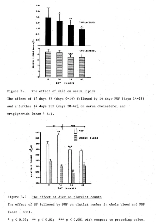
3.3 RESULTS 58
3.3.1 Serum lipids 58
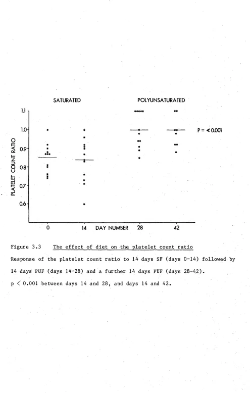
3.3.2 Platelet counts 58
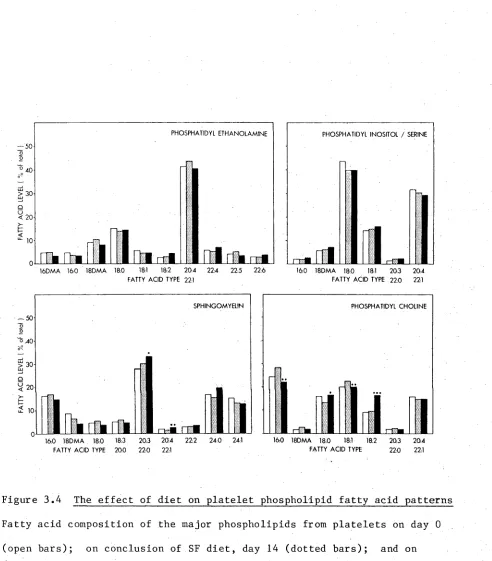
3.3.3 Platelet count ratio 59
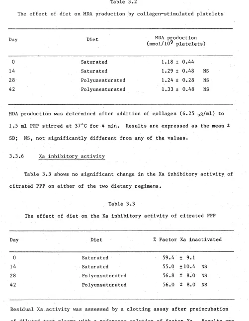
3.3.4 Platelet lipids 59
3.3.5 MDA production 59
3.3.6 Xa inhibitory activity 63
3.3.7 Platelet release products 64
3.4 DISCUSSION 68
3.5 SUMMARY 7 3
CHAPTER 4 - THE EFFECTS OF POSTPRANDIAL ALIMENTARY LIPAEMIA ON HUMAN PLATELET FUNCTION
4.1 Introduction 74
4.2 Materials and Methods 75
4.2.1 Subjects 75
4.2.2 Diet 75
4.2.3 Experimental design 76
4.2.4 Sampling of PPP 76
4.2.5 Analysis of data 77
4.3 RESULTS 77
4.3.1 Serum lipids 78
4.3.2 Platelet counts 78
4.3.3 Platelet count ratio 78
4.3.4 HTCT of citrated and CTP PPP 78
4.3.5 PF^ levels of citrated, CTP and ETP PPP 78 4.3.6 ß-TG levels of citratd, CTP and ETP PPP 82
4.4 DISCUSSION 82
X
page CHAPTER 5 - THE EFFECT OF TYPE H a HYPERLIPIDAEMIA ON THE
COMPOSITION AND CYCLIC AMP STATUS OF HUMAN PLATELETS
5.1 INTRODUCTION 88
5.2 MATERIALS AND METHODS 90
5.2.1 Subjects 90
5.2.2 Platelet handling 91
5.2.3 Platelet incubations 91
5.2.4 Prostaglandins 91
5.2.5 Analysis of data 91
5.3 RESULTS 91
5.3.1 Platelet lipids 91
5.3.2 Basal platelet cyclic AMP levels 92 5.3.3 PGl2-stimulated platelet cyclic AMP levels 93
5.3.4 PGD2-stimulated platelet cyclic AMP levels 93
5.3.5 PGE^-stimulated platelet cyclic AMP levels 93
5.4 DISCUSSION 93
5.5 SUMMARY 99
CHAPTER 6 - EICOSAPENTAENOIC ACID AND HUMAN PLATELET FUNCTION
6.1 INTRODUCTION 101
6.2 MATERIALS AND METHODS 102
6.2.1 Platelet aggregation and secretion 102
6.2.2 Preparation of arterial tissue 103
6.3 RESULTS 104
6.3.1 The effect of EPA on platelet aggregation
and secretion 104
6.3.2 The effect of EPA on aggregation and cyclic
AMP content of washed platelets 106 6.3.3 The metabolism of EPA by arterial tissue 106
6.4 DISCUSSION 109
6.5 SUMMARY 114
CHAPTER 7 - GENERAL DISCUSSION AND CONCLUSIONS
7.1 DISCUSSION 115
7.1.1 Factors affecting human platelet function
and their implications 115
7.1.2 Methodology 122
7.2 CONCLUSIONS 123
REFERENCES 124
LIST OF FIGURES
page
1.1 Sources and metabolism of fatty acids 22
1.2 The metabolism of arachidonic acid by platelets and endothelial
cells 24
3.1 The effect of diet on serum lipids 60
3.2 The effect of diet on platelet counts 60
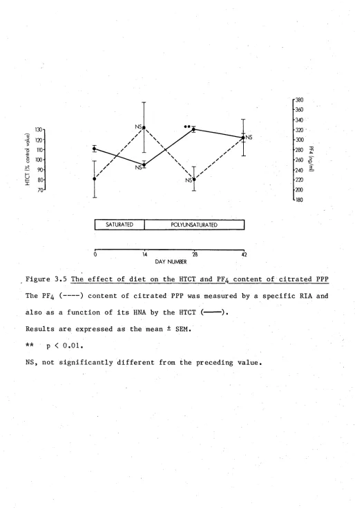
3.3 The effect of diet on the platelet count ratio 61 3.4 The effect of diet on platelet phospholipid fatty acid patterns 62 3.5 The effect of diet on the HTCT and PF^ content of
citrated PPP 65
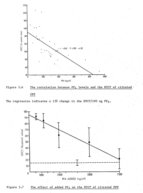
3.6 The correlation between PF^ levels and the HTCT of
citrated PPP 66
3.7 The effect of added PF^ on the HTCT of citrated PPP 66
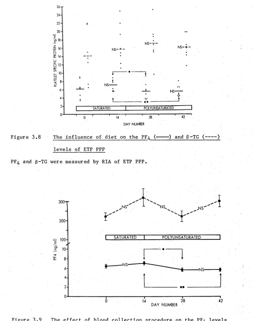
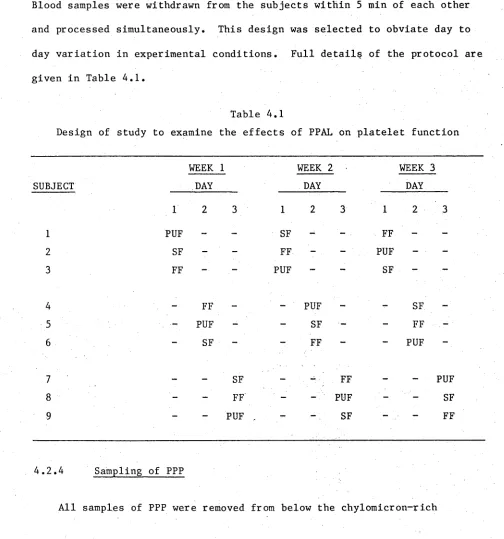
3.8 The influence of diet on the PF^ and ß-TG levels of ETP PPP 67 3.9 The effect of blood collection procedure on the PF^ levels
of PPP 67
4.1 The effect of PPAL on platelet counts in whole blood 79 4.2 The effect of PPAL on the platelet count ratio 79 4.3 The effect of PPAL on the HTCT of citrated PPP 80 4.4 The effect of PPAL on the HTCT of CTP PPP 80 4.5 The effect of PPAL on the PF^ level of citrated PPP 81 4.6 The effect of PPAL on the PF4 level of ETP PPP 81 4.7 The effect of PPAL on the PF^ level of CTP PPP 81 4.8 The effect of PPAL on the ß-TG level of citrated PPP 83 4.9 The effect of PPAL on the ß-TG level of ETP PPP 83 4.10 The effect of PPAL on the ß-TG level of CTP PPP 83 5.1 The response of platelet cyclic AMP levels to PGI2
stimulation 94
5.2 The response of platelet cyclic AMP levels to PGD2
stimulation 95
5.3 The response of platelet cyclic AMP levels to PGE^
stimulation 96
6.1 The effect of EPA on platelet aggregation and secretion 105 6.2 The effect of EPA on the first phase of ADP-induced
aggregation 107
6.3 The effect of EPA on aggregation and secretion induced
by U46619 107
6.4 The effect of EPA on collagen-induced aggregation and
cyclic AMP 108
6.5 The effect of EPA and IB on collagen-induced aggregation
and cyclic AMP levels of washed platelets 110
6.6 The effect of IB containing EPA on collagen-induced
xii
LIST OF TABLES
page 2.1 Composition of modified Tyrode’s solutions 53 3.1 Approximate fatty acid composition of butter and margarine 58 3.2 The effect of diet on MDA production by collagen-stimulated
platelets 63
3.3 The effect of diet on the Xa inhibitory activity of
citrated PPP 63
4.1 Design of study to examine the effect of PPAL on platelet
function 76
4.2 The effect of PPAL on serum lipids 76
5.1 Details of normal and type Ila subjects 90
AA arachidonic acid
ACD acid-citrate-dextrose
ADP adenosine diphosphate
ADR adrenaline
ASA acetylsalicylic acid (aspirin)
AT III antithrombin III
ATP adenosine triphosphate
BHT butylated hydroxytoluene
B-TG B-thromboglobulin
CBH carbohydrate
CHD coronary heart disease
COLL collagen
C/Pl ratio ratio of cholesterol to phospholipid
CTP citrate-theophylline-PGE^
cyclic AMP cyclic adenosine 3',5'-monophosphate cyclic GMP cyclic guanosine 3 ’,5'-monophosphate DEGS-PS diethylene glycol succinate-HßPO^
DHGLA dihomo-y-linolenic acid
EDTA ethylendiamine tetracetate
EPA eicosapentaenoic acid
ETP EDTA-theophylline-PGE
y
FF fat free
5-HT 5-hydroxytryptamine (serotonin)
GLC gas-liquid chromatography
HDL high density lipoprotein
HETE 12-hydroxyeicosatrienoic acid
HHT 12-hydroxyheptadecatrienoic acid
HNA HPETE
HTCT heparin-thrombin clotting time
IB incubation buffer
IBMX 3-isobutyl-l-methylxanthine
i.d. internal diameter
LDL low density lipoprotein
MDA malondialdehyde
PCR platelet count ratio
PDGF platelet-derived growth factor
p f3 platelet factor 3
p f4 platelet factor 4
PG (E^,E2 etc) prostaglandin (E^, E 2 etc)
PPAL postprandial alimentary lipaemia
PPP platelet-poor plasma
PRP platelet-rich plasma
p.s.i.g. pounds per square inch of gas
P/S ratio ratio of polyunsaturated to saturated fat
PUF polyunsaturated fat
PUFA polyunsaturated fatty acid
RD reagent diluent
RIA radioimmunoassay
SF saturated fat
SMC smooth muscle cells
TCA trichloroacetic acid
THETE 8,11,12-trihydroxyeicosatrienoic acid
THR thrombin
TLC thin-layer chromatography
TMB-8 8-(N,N-diethylamino)-octyl-3,4,5-trimethoxybenzoate TX (A2 , B2 etc) thromboxane (A2 , B2 etc)
U46619 (15S)-hydr oxy-11a ,9a-(epoxyme thano)pr os ta-5z ,
13e-VLDL
dienoic acid
2
1.1 THE PLATELET
1.1.1 Milestones in platelet studies
"On March 7, 1842 at a session of the "Academie des Sciences" of Paris during an account of studies on the microscopy of blood Donne stated in clear terms that there existed in the blood red and white globules and little globules ("globulins"). There seems to be no earlier record of the recognition of the platelet as a formed element of the blood" (Tocantins,
1948).
In the same year three other investigators independently reported the presence in blood of what we now know as platelets (Addison, 1842; Gerber, 1842; Simon, 1842). Thus 1842 may be regarded as the inceptive year of platelet studies. There followed forty years of observation and
speculation on the activity and function of platelets, but it was
Bizzozero, who in 1882 put matters into perspective by describing in detail the adhesion of "plättchen" to damaged arterial tissue and the formation of a thrombus, the white portion of which was predominantly platelets. In
1886 Eberth and Schimmelbusch coined the term "viscous metamorphosis" to describe the transformation of platelets from a labile state to an active adhesive state which was a prerequisite for aggregation.
In the following nineteen years the role played by platelets in clot formation was further documented and in 1905 Morawitz delineated
haemostasis with the platelet playing a central role in thrombin formation and hence, blood coagulation. After Morawitz’s treatise there followed a period of almost fifty years during which few publications appeared
The release of 5-hydroxytryptamine (5-HT, serotonin) from platelets during thrombin induced viscous metamorphosis was originally reported by Bigelow (1954) and subsequently demonstrated to be a specific "release reaction" by Grette (1962). The selective release of platelet
constituents, rather than lysis, was confirmed by electron microscopy (Kjaerheim and Hovig, 1962). In the same year Gaarder et al. (1962) demonstrated that a previously described factor released from red blood cells and platelets (Hellem, 1960; 011gard, 1961), which would induce platelet aggregation, was in fact adenosine diphosphate (ADP). Born (1962) and Born et a l . (1963) confirmed these observations and also described the fundamental requirements and kinetics of platelet aggregation. He made this possible by introducing the platelet aggregometer (a slightly modified Unicam spectrophotometer) and in doing so instigated the most useful and widely used instrument in platelet function studies to the present day.
Since the early 1960s a vast amount of literature has been published on the physiology and pathology of the platelet. I have selected two areas for brief discussion which are particularly relevant to this thesis.
(i) In 1974 Hamberg and Samuelsson described the prostaglandin endoperoxides PGG2 and PGH2. The selective metabolism of PGH2 to thromboxane A2 by the platelet and to prostacyclin by arterial tissue have probably been the most significant
discoveries of the decade in terms of the control of platelet function.
4
an absolute requirement for the proliferation of arterial smooth muscle cells (SMC) grown in culture. In vivo arterial SMC
proliferation is an important characteristic of the atherosclerotic plaque. Hence, evidence for platelet involvement in the atherogenic process was greatly reinforced. Detailed discussion of these observations will follow in subsequent chapters.
1.1.2 Ultrastructure and composition
Blood platelets vary from 2-5 pm in diameter and from 2-4 x 10^/pl blood, and they are derived from megakaryocytes of bone marrow (Behnke,
1970).
The structural physiology of the platelet is a field that has contributed greatly to our understanding of platelet function and
dysfunction. Determination of the structural physiology of the platelet has been made possible by use of the highly specialized techniques of electron microscopy, cytochemistry, immunocytochemistry, biochemistry and physiology. For convenience of discussion the platelet may be divided into three zones (White, 1979).
1.1.2.1 The peripheral zone
The peripheral zone may be regarded as having three distinct areas: the outer plasmatic atmosphere, the glycocalyx, and the inner platelet membrane (Jamieson and Smith, 1976).
The outer plasmatic atmosphere is composed of a number of coagulation factors which are thought to be involved in platelet activation. A number are loosely bound and may be removed by washing procedures (Ardlie and Han,
1974).
major glycoproteins have been identified (Nachman and Ferris, 1972; Phillips, 1972; Phillips et al., 1975). It is thought to be the carbohydrate residues of these glycoproteins that are observed as the "fluffy" coat at the platelet periphery (Holmsen, 1972; Jenkins and
Clemetson, 1977). The glycocalyx, in addition to accommodating a thrombin receptor (Tollefsen et al., 1974), is thought to be the portion of the platelet that is transformed from a non-adhesive to a "sticky" surface which is a prerequisite for platelet adhesion and aggregation. Evidence for the role of the glycocalyx in this process is threefold: the outer fluffy coat and ability to aggregate are specific characteristics of the platelet (Holmsen, 1972); sites of platelet-platelet adhesion stain
specifically for carbohydrate (Holmsen, 1972); thirdly, studies of certain haemorrhagic disorders have demonstrated a reduction or absence of specific surface glycoproteins resulting in defective platelet adhesion, with no apparent effect on other platelet functions (Nurden and Caen, 1974; 1975; Phillips et al., 1975). (Section 1.1.4).
The platelet membrane is similar in most respects to the basic plasma membrane of most cells, having a trilaminar appearance and being 7-9 nm in
thickness. The presence of the open, or surface-connected, canalicular system results in the characteristic sponge-like appearance of the platelet surface. The surface-connected canalicular system is a tortuous system of membrane-lined channels that invaginate deep into the platelet and perhaps allows more efficient communication between the platelet and its external environment.
6
the following substances have been reported to be present on the platelet surface: ADP, 5-HT, catecholamines, thrombin, collagen, vasopressin,
acetylcholine, prostaglandins, adenosine and isoprenaline. For a review of platelet receptors see Mills and Macfarlane (1976). More recently,
receptors for the coagulation factor Xa (Miletich et al., 1977) and insulin (Hajek et al., 1979) have been reported.
Numerous enzymes have also been reported to be associated with the platelet membrane system such as phospholipases, cyclo-oxygenase and
adenylate cyclase. In addition to providing a site for enzyme activity the membrane may also provide the substrates for certain enzymes, eg. the DTS membranes contains substantial amounts of arachidonic acid which may be liberated by specific lipases and act as substrate for the enzymes concerned with prostaglandin synthesis.
Further details of membrane-associated enzymes and substrates are discussed in subsequent sections.
1.1.2.2 The Sol-gel zone
The sol-gel zone of the platelet is a viscous matrix containing cytoplasmic enzymes eg. lactate dehydrogenase; metabolic substances eg. ADP, ATP (adenosine triphosphate) and glycogen granules; storage
organelles; a variety of fibre systems and non-specific particles which have been phagocytosed by the platelet (Holmsen, 1972).
1.1.2.3 The organelle zone
The organelle zone is not a discrete area as such, but is distributed throughout the sol-gel zone in the form of storage organelles,
mitochondria, an "occasional" Golgi complex and the DTS. Platelets contain no nuclei, and mitochondria vary in number from 1-6 per platelet, are
small, and contain few cristae relative to other cells (Hovig, 1968).
The DTS is a membranous structure found at the periphery of the sol-gel zone and plays an important role in modulating platelet activity. This is achieved by two closely associated, yet distinguishable mechanisms. One important function of the DTS is thought to be its ability to control the cytoplasmic level of calcium ion (Ca^+) level. Ca^+ is involved in several aspects of platelet aggregation, and the appearance of Ca^+ in the cytoplasm may account for the morphological manifestations of platelet activation (Lüscher and Massini, 1975). The DTS, by its ability to bind and release Ca^+, controls the cytoplasmic concentration of Ca^+ and
thereby aggregation. The presence of Ca^+ storage vesicles may explain the ability of the DTS to control intracellular Ca^+ levels (Statland et^ al. , 1969; Robblee, 1973; White, 1979).
The second important aspect of the DTS is concerned with the
metabolism of arachidonic acid. The platelet membrane systems, including the DTS, are a rich source of arachidonic acid (Marcus et al., 1969) which may be released by Ca^+-dependent phospholipases and metabolized by DTS associated enzymes (Gerrard et al., 1976) to prostaglandins and
thromboxanes which induce further aggregation and release. This action has been hypothesized to be a function of their Ca^+-mobilizing properties
(Gerrard e t a l . , 1977, 1978). The DTS may therefore be regarded as
8
Platelet storage organelles are distinguished from glycogen granules by the presence of a surrounding trilaminar membrane. Release of their constituents is by specific secretion involving membrane fusion and
exocytosis and is a characteristic of the second phase of adrenaline- and ADP- induced aggregation.
Based on electron microscopy, platelet storage organelles were classically divided into two classes, namely dense bodies and a-granules (Holmsen, 1972; Weiss, 1975). However, with the development and
application of more refined techniques it is now possible to distinguish at least four types of storage organelles (Fukami and Salganicoff, 1977). The most dense, both visually (after electron microscopy) and physically (by relative sedimentation rate), are known as dense bodies and contain non-metabolic ADP and ATP, 5-HT, catecholamines, Ca^+ and pyrophosphate
(Fukami and Salganicoff 1977; Da Prada and Picotti, 1979). The functions of these released substances are be discussed in subsequent sections.
Human platelet dense bodies have not been reported to contain any of the platelet-secreted proteins; however, Joist et al. (1976a) have reported antiplasmin activity associated with the release of 5-HT from pig
platelets.
The second class of storage organelles found in platelets are also electron dense, but less so than dense bodies, and are regarded as non-lysosomal a-granules. These organelles contain "platelet-specific proteins" which occur predominantly in the platelet (Niewiarowski, 1977). The development of highly sensitive and specific radioimmunoassays (RIA) has qualified use of the term platelet-specific (Niewiarowski et a l ., 1976; Ludlam, 1979) and has enabled extraplatelet concentrations of the proteins
probably the terms most familiar to workers in the field. However, based on heparin-binding studies Niewiarowski (1977) has proposed the terms high-affinity and low-affinity PF4 for PF4 and 3-TG respectively.
The earliest and perhaps best defined property of PF4 is its ability to neutralize the anticoagulant activity of heparin. Based on observations by Conley et al. (1948) that heparin activity was related to the platelet count, van Creveld and Paulssen (1951) demonstrated that platelets contain heparin-neutralizing activity (HNA). PF4 has also been described as
having paracoagulant properties but this is a point of conjecture (Gjesdal, 1977). More recently, PF4 has been described as having anti-collagenase activity (Hiti-Harper et al., 1978), but as yet, no well defined
physiological role for PF4 has been documented. This is also true for
8“TG, which is stored and released together with PF4 (Dawes et a l ., 1978; Witte et al., 1978; Fukami et a l ., 1979). However, a recent report on the ability of 8~TG to specifically bind to the endothelial cell and inhibit the release of prostacyclin may lead to exciting developments (Hope et al.,
1979).
A third protein, which is thought to be stored and released together with PF4 and 8-TG is the PDGF. Although the evidence for the precise site of storage of the PDGF is not unequivocal, it is highly suggestive (Weiss et al., 1977; Witte et a l ., 1978; Linder et al., 1979). The PDGF is an absolute requirement for the proliferation of cultured arterial SMC (Ross et al., 1974). The relevance of this observation to the role of platelets in haemostasis and atherosclerosis will be discussed in subsequent sections (1.1.3.2; 1.3.2.1; 1.3.3.1)
10
the relevance of fibrinogen secretion by the platelet is not completely understood.
The second class of a-granules found in the platelet are lysosomal in nature and contain numerous hydrolytic enzymes including glycosidases, proteinases, phosphatases and sulphatases. Enzyme distribution data
indicate that platelet lysosomes may be subdivided into two distinct types (Fukami and Salganicoff, 1977). The roles of released platelet lysosomal enzymes have not been fully established, but they may be involved in both physiological and pathological processes. Lysosomal enzymes have been implicated in the phagocytic and inflammatory aspects of platelet function (Gordon, 1975; Ehrlich and Gordon, 1976).
"There may well be other unresolved substances stored in other types of platelet granules, since it is axiomatic that one usually sees only that for which one searches" (Fukami and Salganicoff, 1977).
1.1.3 Platelet function
1.1.3.1 Haemostasis
An important and well documented aspect of platelet function is
haemostasis, i.e. the cessation of blood loss from a damaged blood vessel. Within seconds of disruption of the endothelium the subendothelial tissues become covered with a single layer of platelets. The subendothelial
tissues which induce platelet adhesion include collagen, basement membrane and the microfibrils around elastin (Baumgartner et al., 1976); however, it appears that of these tissues only collagen is capable of initiating subsequent platelet events (Section 1.2.1)
microtubules and microfilaments migrate towards the centre of the platelet encircling the storage organelles (White, 1968; Gerrard and White, 1976). Centralization of the storage organelles is accompanied by secretion of their contents into the surface connecting open canalicular system and consequently into the external environment, i.e. the release reaction. However, the release reaction should not be regarded as an absolute
phenomenon as sequential release of individual storage organelle contents may occur depending on the intensity of stimulus occuring (Holmsen et al.,
1969; Holmsen, 1977a; Kinlough-Rathbone et al., 1977a).
In addition to the release of storage organelle contents the activated platelet also releases metabolites of arachidonic acid in particular
PGG2/H2 (Hamberg et a l ., 1974; Smith et a l . , 1974) and TXA2 (Hamberg et al. , 1975; Smith et al., 1976), (Section 1.2.7.1). These substances, together with a number of dense body constituents, notably ADP, Ca^+ , adrenaline and 5-HT, result in further platelet activation and adhesion to the layer of platelets covering the subendothelium; this is the process of platelet aggregation.
During the morphological changes associated with adhesion and the contractile phase the platelets develop coagulant activity, van Creveld and Paulssen (1951) originally named this, and associated anti-heparin activity, platelet factor 3 (PF3). However, it was subsequently shown that these two properties could be divorced; the coagulant activity retained the term PF3, while anti-heparin activity was renamed PF4
(Deutsch et al., 1955). PF3 has been demonstrated to be a
phospholipoprotein which becomes available on the surface of activated platelets (Hardisty and Hutton, 1966; Joist et al., 1974).
12
may b e i n i t i a t e d by c o n t a c t w i t h t h e e x p o s e d s u b e n d o t h e l i u m and a c t i v a t e d
p l a t e l e t s ( t h e i n t r i n s i c p a t h w a y ) a n d / o r t h e r e l e a s e o f t h r o m b o p l a s t i c
t i s s u e f a c t o r f rom t h e damaged v a s c u l a t u r e ( t h e e x t r i n s i c p a t h w a y ) .
P l a t e l e t c o a g u l a n t a c t i v i t y i s i n v o l v e d i n t h e a c t i v a t i o n o f f a c t o r X t o Xa
and I I t o I l a ( t h r o m b i n ) . T h e r e a r e t h r e e m a j o r c o n s e q u e n c e s o f t h r o m b i n
f o r m a t i o n :
( i ) I n i t i a t i o n o f a d d i t i o n a l p l a t e l e t a g g r e g a t i o n and r e l e a s e ;
( i i ) C a t a l y s i s o f t h e f o r m a t i o n o f f i b r i n monomers f rom f i b r i n o g e n ;
( i i i ) A c t i v a t i o n o f f a c t o r X I I I t o X H I a w h i c h r e s u l t s i n t h e
p o l y m e r i z a t i o n and s t a b i l i z a t i o n o f f i b r i n ( S c h w a r t z e t a l . ,
1 9 7 3 ) .
T h e s e a c t i o n s r e s u l t i n a d d i t i o n a l p l a t e l e t d e p o s i t i o n and t h e
p l a t e l e t p l u g be c omi ng enmes hed w i t h p o l y m e r i z e d f i b r i n . I t h a s b e e n
d e m o n s t r a t e d t h a t p l a t e l e t s a d h e r e t o p o l y m e r i z i n g f i b r i n and t h i s may l e a d
t o f u r t h e r p l a t e l e t d e p o s i t i o n ( N i e w i a r o w s k i e t a l . , 1 9 7 2 ) .
The p l a t e l e t - f i b r i n mass may now be r e g a r d e d a s a c o n s o l i d a t e d
h a e m o s t a t i c p l u g , t h e f u l l y p o l y m e r i z e d f i b r i n p r e s e n t i n g a r e l a t i v e l y
i n e r t s u r f a c e t o c i r c u l a t i n g p l a t e l e t s ( H ov i g e t a l . , 1 9 6 8 ) . V a r i o u s
a s p e c t s of h a e m o s t a s i s h a ve b e e n r e v i e w e d i n d e t a i l by o t h e r s ( W a l s h , 1974;
M u s t a r d an d Packham, 1977; Mason and S a b a , 1 9 7 8 ) .
1 . 1 . 3 . 2 R e s t o r a t i o n and m a i n t e n a n c e o f v a s c u l a r i n t e g r i t y
The h a e m o s t a t i c m e c h a n i s m s j u s t d e s c r i b e d may o n l y be v i e w e d a s a
t e m p o r a r y m e a s u r e t o p r e v e n t b l o o d l o s s . I n o r d e r t o r e t u r n t o a n o r m a l
c o n t r a c t i l e o r g a n t h e b l o o d v e s s e l m us t u n d e r g o r e p a i r p r o c e s s e s wh i c h
r e q u i r e f u r t h e r p l a t e l e t i n v o l v e m e n t .
W e k s l e r and Co up a l ( 1 9 7 3 ) h a ve r e p o r t e d t h a t a p l a t e l e t d e p e n d e n t
tissue damage. Factors such as TXB2 and HETE (Section 1.2.7.1), which are produced during platelet aggregation, have been reported to have chemotactic activity (Turner et a l ., 1975; Kitchen et al., 1978). Other factors which mediate chemotaxis and subsequent inflammation have been reviewed by Ryan and Majno (1977) and platelets may be a source of such factors. Neutrophils and macrophages ultimately remove platelet debris and fibrin (Jorgensen et a l ., 1967).
Subsequent repair of the vascular lesion is poorly understood (Mason and Saba, 1978); however, the release from platelets of factors which stimulate the growth of cultured fibroblasts and SMC (Ross et al., 1974; Rutherford and Ross, 1976; Witte et al., 1978; Pohjanpelto, 1979) may be of importance since these cells are major structural components of the vessel wall which require regeneration in the damaged area. Endothelial cells may also respond in a similar manner (Maca et al., 1977; D ’Amore and
Shepro, 1977). In addition, platelet factors appear to cause migration of endothelial cells, possibly to the de-endothelialized area resulting in restoration of the normal endothelial barrier (Maca et al., 1977; Wall ejt al., 1978).
The proliferative and migratory responses described are also thought to maintain chronic vascular integrity. A number of in vivo and in vitro studies have demonstrated that in the absence of platelets vascular
homeostasis is disrupted in other ways eg. development of endothelial gaps, increased permeability and increased vascular fragility (Gimbrone et a l ., 1969; Gore et al., 1970; Kitchens and Weiss, 1975).
14
considered to be "an integral part of the entire defense system of the body" (Caen et al., 1977).
1.1.4 Platelet dysfunction
On consideration of the major role played by the platelet, namely haemostasis, it is not surprising that the best characterized disorders of platelet function are haemorrhagic or bleeding disorders resulting from hypofunction. Platelet disorders may be qualitative or quantitative in nature (Weiss, 1975; Lüscher and Barnhart, 1977; Mason and Saba, 1978; White and Gerrard, 1978).
In recent years, with a greater understanding of the pathogenesis of obstructive vascular diseases, it has become apparent that the haemostatic process may be disordered and diseases such as thrombosis and
atherosclerosis may be considered to be aberrant forms of haemostasis involving platelet "hyperfunction" and other factors.
1.2 MODULATION OF PLATELET ACTIVITY
A knowledge of factors controlling platelet activity is fundamental to understanding the role played by platelets in both physiological and
pathological processes. The following section summarizes factors known to be involved in the control of platelet activity.
1.2.1 Connective tissue
As described earlier, platelet interaction with subendothelial
connective tissue is the initial event leading to platelet activation and subsequent haemostasis.
1.2.1.1 Collagen
helical in structure, and platelet release and aggregation appears to be related to its quaternary structure (Puett et al., 1973; Brass and Bensusan, 1974; Jaffe and Deykin, 1974).
Four types of collagen have been identified and are characterized by their amino acid sequence and subunit number. Type III appears to be the most potent platelet activator (Balleisen et a l ., 1975) and is the major collagen type found in the internal elastic lamina (Gay et al.,
1975). Adhesion of human platelets to collagen does not have an absolute requirement for plasma cofactors, but is enhanced in the presence of Ca^+ and fibrinogen (Lyman et a l . , 1971; Baumgartner, 1972; 1974; Cazenave e_t a l . , 1973). Adhesion involves glycoproteins of the glycocalyx, and
possibly the von Willebrand factor. The absence of these factors in the Bernard-Soulier syndrome and von Willebrand's disease is associated with bleeding disorders resulting from defective platelet adhesion (Tschopp jrt al. , 1974; Weiss et al., 1974; Baumgartner et al., 1976). More recently, fibronectin, a glycoprotein found in platelets and plasma which binds
collagen, has been implicated in platelet-collagen interaction (Plow e_t a l . , 1979; Ruoslahti and Hayman, 1979), but may only play a minor role (Santoro and Cunningham, 1979). Although Ca^+ is not required for
adhesion or release, subsequent aggregation is dependent upon extracellular Ca^+ and is mediated by the release of arachidonic acid metabolites and dense body constituents (Holmsen, 1977). Aspirin and other non-steroidal anti-inflammatory agents can inhibit collagen-induced aggregation, but this may be overcome by higher concentrations of collagen, suggesting that
collagen may initiate release and aggregation by at least two pathways, one of which is cyclo-oxygenase independent (Zucker and Peterson, 1970; Nyman,
16
1.2.1.2 Elastin
Elastin is a component of both the internal elastic lamina and the intima (Jaffe, 1976) and when exposed by collagenase and trypsin digestion appears to be relatively unreactive to platelets (Baumgartner, 1974a).
1.2.1.3 Glycoproteins
Glycoproteins are found throughout the blood vessel wall and may be organised into non-collagenous microfibrils (Ross and Bornstein, 1969). Evidence suggests that platelets will adhere to such microfibrils but subsequent aggregation does not occur (Baumgartner and Muggli, 1976).
1.2.1.4 Proteoglycans
Proteoglycans are found throughout the vessel wall and appear to be non-reactive to platelets although platelet-proteglycan interaction has not been systematically studied (Jaffe, 1976).
1.2.2 Coagulation factors
Platelet-coagulation factor interaction is central to haemostasis and there is growing evidence that coagulation factors mediate several aspects of platelet function; the von Willebrand factor, and possibly fibrinogen, are necessary for the normal adhesion of platelets to collagen. In
addition fibrinogen is required for subsequent aggregation induced by all aggregating agents (Deykin et a l ., 1965; Inceman et al., 1966).
et al., 1976; Shuman et a l . , 1976). These small amounts of thrombin may be a stimulus for the release of platelet constituents with consequent aggregation. Huzoor-Akbar and Ardlie (1977) have suggested that thrombin formation on the platelet surface mediates platelet aggregation induced by collagen and ADP.
Platelets have been demonstrated to have specific thrombin receptors which are located in the glycocalyx (Tollefsen et al. , 1974). Binding to this receptor appears to be the primary event leading to platelet
activation by thrombin (Shuman and Majerus, 1975); after binding, thrombin may induce aggregation by at least three pathways which are dependent on:
(i) Formation of arachidonic acid metabolites (Malmsten et al., 1975).
(ii) Release of ADP (Haslam, 1974);
(iii) Neither (i) or (ii), and possibly involving direct Ca^+ mobilization (Macfarlane and Mills, 1975; Kinlough-Rathbone et al., 1977b; Packham et al., 1977; Lapetina et al., 1978).
1.2.3 Anticoagulants
As several coagulation factors are involved in the activation of platelets it is not surprising that a number of anticoagulants have
antiplatelet action. The most commonly used anticoagulant is heparin which activates antithrombin III (AT III) a naturally occuring inhibitor of the following coagulation factors: IXa, X a , XIa, Xlla and Ila (thrombin). In the absence of heparin AT III has very low activity, but in the presence of trace amounts of heparin the inhibitory activity is greatly enhanced.
(Damus et al., 1973; Rosenberg and Damus, 1973).
18
prevents or facilitates platelet aggregation". The controversy would appear to be a function of one or more of the following factors: heparin source and concentration, or the presence of other anticoagulants eg. citrate.
A second question concerning heparin is whether or not it is a
naturally circulating anticoagulant. It is associated with various organs of the body, but no reports to date have demonstrated conclusively that free heparin is present in the blood. Related sulphated
mucopolysaccharides do exist, notably on the endothelial cell (Buonassi, 1973; Moritani and Ohta, 1973; Berenson et al., 1974) and have
demonstrable heparin-like activity (Teien et al., 1976; Hatton et al., 1978) and may play physiological roles in fibrin- or platelet-endothelial cell interaction (Teien, 1979).
1.2.4 Calcium
Extracellular Ca^+ is not required for platelet adhesion to
collagen, shape change (Skoza et al., 1967; Born, 1970) or the release reaction induced by collagen or thrombin (Spaet and Zucker, 1964;
Mueller-Eckhardt and Lüscher, 1968; Roblee et al., 1973); however,
subsequent aggregation does have an absolute requirement for Ca^+ (Hovig, 1964). Other agents, such as ADP and adrenaline, do require Ca^+ for the induction of release (Massini, 1977).
In contrast to extracellular Ca^+ , intracellular Ca^+ appears to be involved in each step of platelet activation. This has been demonstrated by use of ionophores, agents which aid the flux of Ca^+ across membranes (Pressman, 1976). Ionophores such as A23187 increase platelet cytoplasmic Ca^+ and the following aspects of platelet
activation may be induced: shape change (Gerrard et a l ., 1974);
(Kinlough-Rathbone et a l ., 1977a); the release reaction (White et al., 1974) and clot retraction (Massini and Löscher, 1974). Use of ionophores has also demonstrated that sufficient intracellular Ca^+ may be derived from sites independent of the dense bodies (namely the DTS) as ionophore- induced aggregation in the absence of extracellular Ca^+ is normal in patients whose platelets are deficient in these Ca^+ storage organelles (White et al. , 1974).
Ca^+ antagonists such as TMB-8 (8-(N,N-diethylamino)-octyl-3,4,5- trimethoxybenzoate) have also been useful in elucidating the role of intracellular Ca^+ in platelet function (Rittenhouse-Simmons and Deykin, 1978).
1.2.5 Biogenic amines
1.2.5.1 5-HT
Platelets are the major source of 5-HT in the blood and it is released from the dense bodies during platelet aggregation. The platelet is
efficient at accumulating 5-HT which is achieved by both active and passive transport mechanisms (Drummond, 1976). The major haemostatic functions of 5-HT are twofold; firstly, it is a potent vasoconstrictor and this action may aid haemostasis; secondly, 5-HT may induce platelet aggregation. In vitro aggregation of human platelets by 5-HT is generally weak and
reversible in nature (Mitchell and Sharp, 1964) and does not appear to induce the release reaction, but will potentiate aggregation induced by ADP or adrenaline (Baumgartner and Born, 1968).
1.2.5.2 Catecholamines
20
Da Prada and Picotti, 1979).
The evidence for platelet shape change induced by catecholamines is controversial and awaits further study (Drummond, 1976); however,
adrenaline and noradrenaline will induce biphasic aggregation (O'Brien, 1963; MacMillan, 1966)
The primary phase of aggregation induced by catecholamines is believed to be mediated by an ot-adrenergic receptor which may be blocked
competitively by a-antagonists such as phentolamine and dihydroergotamine (Mills and Roberts, 1967; Rysanek et al., 1968). Subsequent secondary aggregation is dependent upon released ADP and other substances, and once initiated is independent of adrenaline and the a-receptor (MacMillan, 1966; Mills et al., 1968; Rossi and Levin, 1973; Macfarlane and Mills, 1975). Catecholamines can potentiate platelet aggregation induced by other agents such as ADP (Ardlie et al., 1966; Mills and Roberts, 1967), 5-HT, thrombin and collagen (Baumgartner and Born, 1968; Thomas, 1968).
1.2.6 Adenine nucleotides
The major non-cyclic adenine nucleotides found in, and affecting, platelets are ATP and ADP. Approximately two thirds of the ADP and ATP are metabolically non-active and are stored in the dense bodies; the remainder is found in a metabolically active pool distributed in the cytoplasm, mitochondria and membranes (Holmsen, 1972).
1.2.6.1 ATP
ATP is involved in both resting and-active aspects of platelet function. It is required by the Ca^+ extrusion pump which is thought to maintain the platelet in a non-reactive state. ATP may be converted to
source for the following aspects of platelet function: shape change, aggregation, secretion and clot retraction (Holmsen, 1977b).
1.2.6.2 ADP
In contrast to ATP, the most important pool of ADP appears to be that found in, and released from, dense bodies. The release of ADP is an
important, although non-absolute requirement for platelet aggregation; as thrombin may induce aggregation that is independent of the release of ADP (Section 1.2.2). Under normal physiological conditions the release of ADP induces the activation and deposition of platelets, indeed subjects with storage pool disease, in which ADP is either absent or unreleasable from the dense bodies, demonstrate a mild bleeding disorder (Hardisty, 1977).
1.2.7 Prostaglandins and thromboxanes
Prostaglandins and thromboxanes are oxygenated metabolites of polyunsaturated fatty acids (PUFA) containing twenty carbon atoms (C2o)« The C20 PUFA may be obtained directly from the diet but they are usually synthesized in the body by chain elongation and desaturation of essential dietary precursors (Figure 1.1). Depending upon the number of double bonds associated with the C20 PUFA, which predetermines the resulting number in
the product, three classes of derivatives may be produced (Figure 1.1): (i) The monoenoic series (PG^/TX^);
(ii) The dienoic series (PG2/TX2); (iii) The trienoic series (PG3/TX3).
22
Figure 1.1 Sources and metabolism of fatty acids
All the fatty acids shown may be obtained from the diet. The non-essential fatty acids (centre pathway) may be produced de-novo by
mammals from protein or carbohydrate (CBH) sources. Linoleic and linolenic acids, the parent fatty acids of each series (upper and lower pathways), may only be produced by non-animal species but subsequent chain elongation and desaturation may occur in humans and the majority of other animal species.
dienoic prostaglandins and thromboxanes
monoenoic prostaglandins and thromboxanes
18:2*
DHCLA*** AA**** n-6**
linoleic acid
1 8:2— -2 0:2— *2 0:3
18:0 DIET-- ►PROTEIN
linolenic acid
trienoic prostaglandins and thromboxanes ___
*Each fatty acid is shown as carbon number followed by number of double bonds; thus, linoleic acid is an 18 carbon fatty acid with 2 double bonds **n-3; n-6; n-9 Refer to the position of the first double bond from the methyl terminal
*** DHGLA Dihomo-y-1inolenic acid (8,11,14-eicosatrienoic acid) **** AA Arachidonic acid (5,8,11,14-eicosatetraenoic acid)
[image:36.541.19.530.24.636.2]phospholipids. The ubiquity of arachidonic acid in tissues reflects the involvement of prostaglandins in a vast number of physiological processes (Horrorbin, 1978). Prostaglandins and thromboxanes are also intimately involved in the control of platelet activity, indeed Gerrard and White
(1978) considered them to be "middlemen" modulating platelet function. Arachidonic acid metabolites involved in platelet function are presented in Figure 1.2.
1.2.7.1 Platelet-derived prostaglandins and thromboxanes
Prostaglandin involvement in platelet function was first proposed by Kloeze (1967; 1969) who demonstrated PGE^-induced inhibition and
PGE2-induced enhancement of platelet aggregation. It was subsequently demonstrated that platelets manufactured and released PGE2 and PGF2a during aggregation (Smith and Willis, 1970; Silver et a l . , 1972).
Although these early studies initiated a very important aspect of platelet research it has become apparent that PGE2, PGF2 and PGE^ play only
a minor role in modulating platelet activity.
More important discoveries of prostaglandin involvement in platelet function were initiated by the demonstration that platelets produce and release the prostoglandin cyclic endoperoxides PGG2 and PGH2 (Hamberg and Samuelsson, 1974; Hamberg et al., 1974; Smith et al., 1974); in addition, these were highly potent inducers of platelet aggregation. The enzyme responsible for the production of the cyclic endoperoxides was named cyclo-oxygenase. The same groups subsequently demonstrated the production, release and proaggregatory properties of thromboxane A2 (TXA2) which is a metabolite of PGH2 (Hamberg et al., 1975; Smith et al., 1976a).
TXA2 is much more potent and unstable than its precursors and is hydrolyzed to the non-active, stable metabolite TXB2.
24
PLATELET STIMULATION
l
"ACTIVATION" OF PHOSPHOLIPASE
FREE ARACHIDONIC ACID
ENDOPEROXIDE PGH2
THROMBOXANE B2
ENDOTHELIAL CELLS
PROSTACYCLIN \S Y N T H E T A S E
PROSTACYCLIN PGI2
REDUCTASE ISOMERASE
PGD2
Figure 1.2 The metabolism of arachidonic acid by platelets and endothelial cells
Platelet stimulation results in the liberation of arachidonic acid from platelet phospholipids, and is subsequently metabolized by the
cyclo-oxygenase and lipoxygenase pathways.
Endothelial cells may produce prostacyclin from endogenous or exogenous (platelet-derived) arachidonic acid or cyclic endoperoxides.
HPETE, 12-hydroperoxyeicosatetraenoic acid. HETE, 12-hydroxyeicosatetraenoic acid. MDA, malondialdehyde.
[image:38.541.35.529.24.692.2]the second phase of aggregation and release induced by ADP and adrenaline and the monophasic aggregation pattern exhibited by collagen and thrombin (Smith et al., 1974; Malmsten et al. , 1975). It has also been shown that PGG2/H2 and/or TXA2 initiate contraction and the release reaction
(Gerrard and White, 1975; Gerrard et al., 1977; Gorman, 1979) and
patients with platelet cyclo-oxygenase deficiency have a storage pool type disease as a result (Malmsten et al. , 1975).
Whether PGG2/H2 or their metabolite TXA2 bring about aggregation and release is a moot point resulting from conflicting reports on the necessity for TXA2 production from PGG2/H2 prior to platelet
responses (Gerrard and White, 1978; Horrorbin, 1978). Elucidation of the possible individual roles played by these substances awaits the production and characterization of more specific inhibitors of thromboxane synthetase.
The mechanism of action of PGG2/H2 and TXA2 is unclear. Based on structural considerations and subsequent experimental findings TXA2 , but not PGG2 , has been ascribed with Ca^+ ionophoretic properties and if manifested in vivo may mediate the increase in cytoplasmic Ca^+ that is associated with platelet activation (Gerrard and White, 1978).
In addition to producing proaggregatory metabolites of arachidonic acid during aggregation, platelets are capable of producing inhibitory metabolites. PGD2 is produced during aggregation, possibly in amounts
sufficient to inhibit aggregation (Smith et al., 1976b; Oelz et al., 1977) and may represent a negative feedback control mechanism.
has been discounted from playing a major role in platelet function (Smith and Silver, 1976). A more recent report shows there to be more PGE^ than PGE2 in unstimulated platelets and suggests that it may play a role in the control of basal activity. (Lagarde and Dechavanne, 1979).
It is well established that PGEj and PGD2 inhibit platelet
aggregation by elevation of platelet cyclic AMP; however, one report suggests that PGE^ may also inhibit aggregation independently of platelet cyclic AMP production by interaction with a plasma cofactor. (Sinha and Colman, 1978).
In addition to the cyclo-oxygenase pathway of arachidonic acid
metabolism, platelets possess the lipoxygenase pathway (Figure 1.1). The relevance of this pathway in human platelets is poorly understood, but evidence suggests that lipoxygenase products play important roles in platelet function; thus HPETE has been reported to inhibit thromboxane synthesis (Hammarström and Folardeau, 1977). Consistent with this finding is the observation that patients with decreased platelet lipoxygenase activity have hypersensitive platelets (Okuma and Uchino, 1977); however, Dutilh et a l . (1979) have reported that HETE is essential for rat platelet aggregation; HETE also has chemotactic properties for neutrophilic
leucocytes (Turner et al., 1975). A third lipoxygenase product 8,11,12- trihydroxyeicosatrienoic acid (THETE) has recently been reported to be formed during platelet aggregation (Bryant and Bailey, 1979).
The sources of arachidonic acid are the platelet membrane
implicated in its liberation from phosphatidyl inositol.
1.2.7.2 Vasculature-derived prostaglandins
It has been acknowledged for many years that vascular endothelium presents a non-reactive surface towards platelets. Recently our
understanding of this property has been furthered by the discovery that prostaglandin cyclic endoperoxides may be transformed by human vascular tissue into a substance with potent antiaggregatory activity. The
substance was originally named PGX, but subsequently renamed prostacyclin (PGI2; Johnson et al., 1976; Moncada et a l ., 1976, 1977a). The
discovery that vascular tissue could convert potent proaggregatory substances released from platelets into a potent antiaggregatory agent initiated a very important and fruitful era of reseach on platelet-vessel wall interaction in the physiology and pathology of vascular homeostasis. Clearly, factors which pertubate the balance of metabolites produced from the common precursors may result in pathological events.
The mode of action of PGI2 was clarified by demonstration that it was the most potent stimulator of cyclic AMP accumulation in platelets known at the time (Best et al., 1977; Gorman et a l ., 1977; Tateson et al. , 1977). The vascular endothelial cell appears to be the most active producer of PGI2 (MacIntyre et a l ., 1978), although production by other vascular cell types has been reported (Baenziger et al., 1977; Moncada ejt al. , 1977b; Tansik et a l ., 1978). Endothelial cells have the ability to produce PGI2 both endogenously and from exogenously provided substrates (Weksler et a l ., 1977; MacIntyre et al., 1978), although the ability of endothelial cells to produce PGI2 from platelet-derived PGH2 has
28
The inhibition of platelet function by PGI2 is not an absolute phenomenon and it appears that in the presence of low levels of PGI2 platelets lose their ability to aggregate, but are still able to adhere; only at higher concentrations does PGI2 inhibit adherence (Higgs et al., 1978). These responses may allow the platelet to interact with intact endothelium and maintain vascular integrity without leading to thrombus formation.
PGI2 has been described as a "circulating hormone" produced, but not degraded, by the lung vasculature (Gryglewski et al., 1978; Moncada et al., 1978). However, other groups have failed to demonstrate circulating
PGl2-like activity (Smith et al., 1978, 1979; Haslam and McClenaghan, 1979; Steer et a l . , 1980).
1.2.7.3 Inhibitors of prostaglandins and thromboxanes
Inhibitors of arachidonic acid release, metabolism and action of metabolites are useful tools for studying the roles of such products in regulating platelet function and may also have important clinical
ramifications.
The best documented inhibitors of prostaglandin synthesis are the non-steroidal anti-inflammatory drugs, in particular acetylsalicylic acid (ASA, aspirin). These agents act by inhibiting cyclo-oxygenase, but this is not necessarily achieved by similar mechanisms. Aspirin inactivates cyclo-oxygenase by acetylation of its active site (Roth et al., 1975). As a result of the platelet's inability to resynthesize enzymes the aspirin effect is a function of platelet turnover, and under normal conditions lasts for seven to ten days. Aspirin also inhibits endothelial cell