Int. J. Electrochem. Sci., 10 (2015) 7794 - 7802
International Journal of
ELECTROCHEMICAL
SCIENCE
www.electrochemsci.org
The Study of Urea Nitrogen Biosensor Based on the Disposable
Screen-Printed Carbon Electrode
Jie Chen, Yan-Ping Chen and Ping Chen*
College of Chemistry and Biological Engineering, Changsha University of Science and Technology, Changsha, Hunan 410114, P. R. China.
*
E-mail: [email protected]
Received: 5 June 2015 / Accepted: 12 July 2015 / Published: 28 July 2015
A novel urea nitrogen(UN) biosensor based on disposable screen-printed carbon electrode mixed with potassium ferrocyanide was developed. The biosensor was covered with enzyme ink contained urease, glutamate dehydrogenase and reduced nicotinamide adenine dinucleotide(NADH). Then the electrochemical behavior was studied according to the electrochemical methods and the results revealed that the electrochemical method for detection of UN is feasible. Under optimized conditions, there was a good linear relationship of the UN concentration from 0.05 to 40 mM and the detection limit was 12 μM. The UN biosensor revealed good anti-interference ability and storage stability for more than 6 months when kept at room temperature. Moreover, the UN biosensor was employed to measure the real samples compared with automatic biochemical analyzer, the results suggested the UN biosensor could be applied to UN measurement in renal function.
Keywords: Urea nitrogen; screen printed carbon electrode; reduced nicotinamide adenine dinucleotide
1. INTRODUCTION
Urea is one of the end-product of protein metabolism in human body, and the determination of urea is one of the most frequent analyses in clinical diagnosis. The concentration of urea nitrogen(UN) in normal people blood is 1.8~7.1 mM related to the concentration of urea is 8~20 mg/dl[1]. It is one of diagnostic indicators of uremia when the concentration of UN reached to 21.4 mM (60 mg/dl)[2]. Thus determination of the UN concentration has great significance in nephropathy clinical diagnosis, treatment and prognosis[3,4].
laboratories employ urease to hydrolyze UN and the products, especial of ammonium, is determined at present[7,8].
The ammonium ion can be detected using electrochemical technology of potentiometric[9-12], amperometric urea biosensor [13,14] and conductometric urea biosensor [15,16]. The above method is basically used the traditional electrode, such as PH electrode[17] or the ammonia sensitive electrode[18], which have problems of bad selectivity, slow response and enzyme instability. The development of analytical methods that respond to the growing need to perform rapid in situ analyses shows the disposable screen-printed electrodes (SPEs) as an alternative to the traditional electrodes[19,20]. Compared to the common electrode, SPEs, low cost, easy to prepare and high reproducibility, have attracted extensive interest in various field.
The aim of this work is to introduce a simple, rapid and sensitive UN detection method based on the disposable screen-printed carbon electrode technology. In this method, urease is employed to hydrolyze UN into ammonia and carbon dioxide, then the ammonia and α-ketoglutaric acid react to glutamic acid under the catalytic action of glutamate dehydrogenase, and reduced nicotinamide adenine dinucleotide(NADH). In this process, NADH is oxidized into NAD+, and the rest of the NADH can react with potassium ferrocyanide at the electrode surface. The consumption of NADH is proportional to the UN concentration in sample, thus the UN concentration in sample can be measured according to measure the oxidation current changes of potassium ferrocyanide. In this paper, such a method was applied to UN determination of real samples in whole blood and it can be applied in the portable system to renal function.
2. EXPERIMENTAL
2.1 Materials
Carbon ink and insulation ink were acquired from JUJO (Tokyo, Japan). The hydrophilic film and double sided adhesive tape were from 3M China Co., Ltd(Shenzhen, China). Urease, glutamate dehydrogenase were purchased from TOYOBO(Tokyo, Japan). NaCl, CaCl2, potassium hydrogen
phosphate, potassium dihydrogen phosphate, Potassium perchlorate, polyethylene glycol(PEG-400), Cabosil M5 amorphous untreated fumed silica powder and hydroxyethyl cellulose(HEC), uric acid, ascorbic acid(AA), potassium ferrocyanide, glutathione(GSH), Acetaminophen(ACP), 4-Aminoantipyrine were purchased from Sigma-Aldrich (St. Louis, MO, USA). Aqueous solutions were prepared using Millipore water (Simplicity Model, Billerica, MA, USA).
2.2 Electrode Preparation
The enzyme ink was prepared as followed procedure. 1 wt% CaCl2, 1 wt% PEG-400, 4 wt%
Cabosil M5 amorphous untreated fumed silica powder and 2.5 wt% hydroxyethyl cellulose (HEC) were added into phosphate buffer solution (0.1M, pH=7.4) and stired for 2 h at 2000 r/min, then hydrated for 6 h at room temperature. The prepared enzyme ink in addition of 0.2 g potassium ferrocyanide, 1.5 k units urease, 1 k units glutamate dehydrogenase, 0.2 g NADH and 0.1 g α-ketoglutaric acid to each gram of reagent ink, mixed for 15 minutes. The viscosity of enzyme ink was 5400 mPas by this method.
2.3 Detection of UN at UN biosensor
All electrochemical measurements were carried out with a model CHI832C Electrochemical Workstation(CH Instruments, Austin, TX, USA). The UN biosensor contained two carbon electrode, one printed enzyme ink as the working electrode, and the other as the reference electrode, then we used the electrochemical workstation to measure. The electrochemical behaviors of disposable screen-printed carbon electrode of UN was acquired from cyclic voltammetry(CV) and chronoamperomerty, and the electrolyte was 0.1 M NaCl in PBS solution(pH=8.0). For detection of UN at UN biosensor, the applied potential was set at 0.20 V and the value of the current was obtained at 10 s at I-t curve after waiting 40 s when the sample was added. All experiments were carried out at room temperature unless otherwise stated.
2.4 Real sample preparation and analysis
Blood samples were acquired from venous blood collected in heparin anticoagulant tube. 100 μL serum samples were tested by automatic biochemical analyzer(Beckman Instruments, Inc., California, USA), another 2 μL whole blood samples were measured by UN biosensor without any treatment.
In order to determine the recovery of electrodes, UN was dissolved in PBS solution to prepare 1 mM and 5 mM UN stock solution, and the stock solution were immediately mixed to the same volume of blood samples without any treatment. For the stability of UN biosensor, the same batch of electrode was conserved in a sealed packaging at room temperature, then electrodes were investigated using 5 mM UN every month.
3. RESULTS AND DISCUSSION
3.1 Electrochemical analysis of UN biosensor
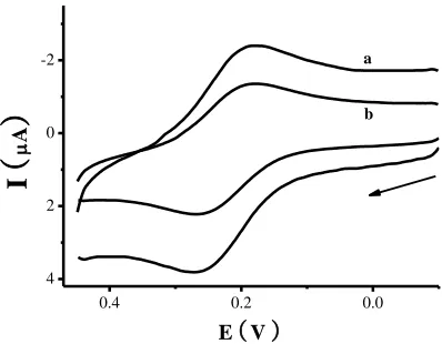
electrode. From curve b in figure 1, the oxidized and reduced current of potassium ferrocyanide obviously reduced when 5 mM UN added to phosphate buffer solution.
0.4 0.2 0.0
4 2 0 -2
I
(
A
)
bE (V )
[image:4.596.200.399.132.287.2]a
Figure 1. Cyclic voltammetry of different UN concentrations at UN biosensor, the arrow indicates the start scanned direction. The scan rate was 100 mV/s.
In order to verify electrochemical properties of potassium ferrocyanide and NADH at the electrode surface, CV behavior of basic electrode mixed with potassium ferrocyanide was researched in different NADH concentrations. When the test solution contained different concentrations of NADH (0, 0.1, 0.2, 0.3 and 0.5 mM), the oxidized current of potassium ferrocyanide increased with the increasing of NADH as shown in figure 2. The results showed that the redox reaction of NADH was effectively catalyzed by the basic electrode doped potassium ferrocyanide, also proved that the electrochemical method to detect UN was feasible.
Figure 2. Cyclic voltammetry of various concentrations of NADH at basic electrode doped potassium ferrocyanide. The scan rate was 100 mV/s.
3.2 Detection of UN at UN biosensor
Chronoamperomerty was employed to measure oxidized current of UN at UN biosensor mixed
0.4 0.2 0.0 4
2 0 -2
I
(
A
)
0.5 mM
[image:4.596.166.409.484.662.2]
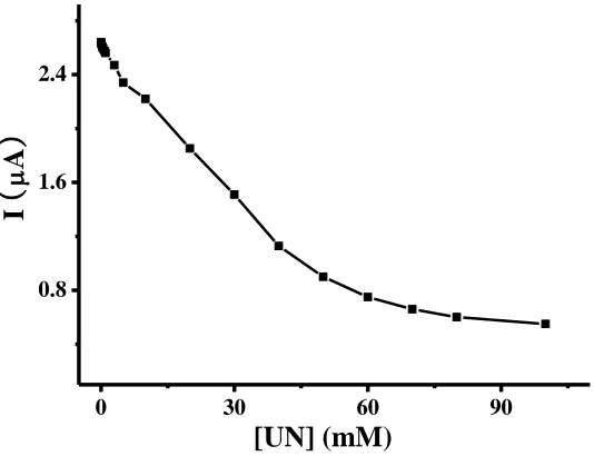
with potassium ferrocyanide. The applied potential catalyzed NADH was set at 0.2 V, the value of the oxidized current was obtained at 10s after adding different concentrations of UN(0-100 mM) and waiting 40 s. The result of oxidized current related with concentration of UN was shown in figure 3.
0 30 60 90
0.8 1.6 2.4
[UN] (mM)
I
(
A
[image:5.596.167.434.154.359.2])
Figure 3. The relationship between different UN concentration and current with the potential of 0.2 V, and the test started after waiting 40 s when sample was added.
0 20 40
1.2 1.6 2.0 2.4 2.8
I
(
A
)
[image:5.596.147.453.420.649.2][UN] (mM)
Figure 4. The calibration curve of UN concentration of 0.05-40 mM with current, the test started after waiting 40 s when sample was added, and the values were acquired at 10 s.
UN concentration and exhibited a good linear relationship with the UN concentration in the range from 50 μM to 40 mM with the linear equation was I(μA)=2.62-0.04[UN](mM) and the limits of detection were calculated on the basis of three times of the background noise and the value was found to be 12 μM.
3.3 Selectivity and specificity of the UN biosensor
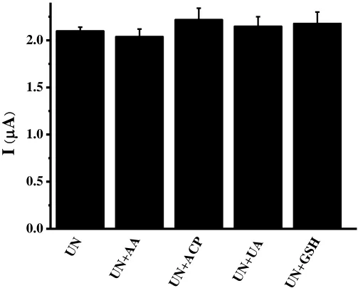
Figure 5. Selectivity of the UN sensor for UN detection in the absence or presence of redox-active species (AA, ACP, UA and GSH).
The electrochemical detection of UN were often affected by some interferences such as glutathione(GSH), glucose, uric acid(UA), acetaminophen(ACP) and ascorbic acid(AA). Therefore, we observed current changes at UN biosensor after adding different redox-active species of UA, AA, ACP and GSH. The results revealed that UN biosensor was basically not affected by the above species and had good selectivity and specificity(figure 5).
3.4 Reproducibility and stability of the UN biosensor
To examine the repeatability of the UN biosensor, 5 mM UN in PBS solution was detected. The results showed that the coefficient of variation of intra-assay and inter-assay were 1.44% and 5.67%, respectively, which suggested that UN biosensor had good reproducibility.
For assessment of the stability of UN biosensor, the electrodes were extracted to measure the current intensity with 5 mM UN in PBS solution every month, the electrodes were acquried from the same batch and stored at room temperature in a closed environment. As shown in figure 6, the catalytic current of electrodes still maintained 84.2% of the initial current after 6 months. The experimental results revealed that structure and activity of enzymes remained stable, which may due to the physical
0.0 0.5 1.0 1.5 2.0
UN +U
A
UN +A
CP
UN +G
SH
I
(
A
)
UN +A
[image:6.596.174.429.217.419.2]
adsorption, and adjunction of hydroxyethyl cellulose(HEC) and activators of enzymes such as CaCl2.
Meibodi et al. developed a Amperometric urea biosensor based on covalently immobilizedurease on an electrochemically polymerized film of polyanilinecontaining MWCNTs. And polyaniline-multiwalled carbon nanotubes (PANI/MWCNTs) composite were fabricated byelectropolymerization method as a matrix for entrapment of enzyme. The optimized urea biosensor shows a good sensitivity from 10−2 M to 10−5 M urea concentration range and a response time of about 50 s. The proposed biosensor retained 50% of its original response after 15 days[21]. Compared these results, our method is more simple and has a better storage stability.
0.0 0.3 0.6 0.9
R
e
la
ti
v
e
c
a
ta
ly
ti
c
a
c
ti
v
it
y
(%
)
180 150
Time (days)
[image:7.596.188.450.237.432.2]30 60 90 120
Figure 6. Stability of the UN biosensor. 3.5 Recovery of UN biosensor
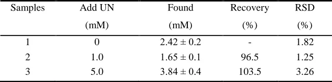
The accuracy of UN biosensor can be reflected by the recovery. UN dissolved in PBS to prepare 1 mM and 5 mM as the stock solution, then added the same volume of whole blood specimens without any treatment, the recovery rate was in the range from 96.5% to 103.5% (cf. Table 1). The data suggested that UN biosensor had excellent accuracy, and the proposed screen-printed method was feasible for the detection of UN without the interference of other redox-active species in blood samples.
Table 1. Recovery of UN in blood sample
Samples Add UN Found Recovery RSD
(mM) (mM) (%) (%)
1 0 2.42 ± 0.2 - 1.82
2 1.0 1.65 ± 0.1 96.5 1.25
[image:7.596.135.461.680.761.2]
3.6 Real sample measurement
Automatic biochemical analyzer was usually employed by hospital to determine UN and the principle of measurement was showed as follows. UN was hydrolyzed by urease into ammonia and carbon dioxide, then the ammonia and α-ketoglutaric acid react to glutamic acid under the catalytic action of glutamate dehydrogenase, and NADH. In this process, NADH is oxidized into NAD+ and has absorption peak in the wave of 340 nm, the decreasing rate of absorbance is proportional to the concentration of UN in the sample.
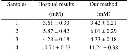
[image:8.596.187.407.415.507.2]When we test the real samples, each sample was respectively measured three times and all the results were acquired by our electrochemical and hospital methods. In table 2, samples 1, 2 and 3 were collected from healthy donors, sample 4 was from patients with disease of renal function whose BUN concentration was higher than normal level (1.78-7.14 mM). The results revealed that the two methods had good consistency and there was no significant difference. Soldatkin et al. developed a enzyme/zeolite sensor for urea analysis in serum. The linear range of urea determination by using the biosensor was 0.003-0.75 mM. The method of standard addition was used for analysis of serum samples with 500-fold dilution. Total time of analysis was 10 min[22]. Compared these methods, our method has shorter response time and doesn't need any treatment, our method is more suitable for testing the real sample.
Table 2. Comparison of UN concentrations determined by hospital and our method
Samples Hospital results Our method
(mM) (mM)
1 3.61 ± 0.30 3.42 ± 0.21
2 5.87 ± 0.42 6.01 ± 0.29
3 4.28 ± 0.18 4.33 ± 0.18
4 10.71 ± 0.23 11.24 ± 0.38
4. CONCLUSIONS
References
1. T. Ahuja, I. A. Mir and D. Kumar, Sens. Actuat. B: Chem., 134 (2008) 140.
2. R. Vanholder, R. D. Smet, G. Glorieux, A. Argilés, U. Baurmeister, P. Brunet, W. Clark, G. Cohen, P. P. D. Deyn and R. Deppisch, Kidney Int., 63 (2003) 1934.
3. P. S. Francis, S. W. Lewis and K. F. Lim, TrAC, Trends Anal. Chem., 21 (2002) 389. 4. G. G. Guilbault, J. G. Montalvo Jr, J. Am. Chem. Soc., 91 (1969) 2164.
5. N. Das, A. M. Kayastha and O. P. Malhotra, Biotechnol. Appl. Biochem., 27 (1998) 25. 6. M. Kitamura, I. Iuchi, Clin. Chim. Acta, 4 (1959) 701.
7. R. Ahmad, N. Tripathy and Y. B. Hahn, Sens. Actuat. B: Chem., 194 (2014) 290. 8. Z. P. Yang, C. J. Zhang, Sens. Actuat. B: Chem., 188 (2013) 313.
9. C. C. Buron, M. Quinart, T. Vrlinic, S. Yunus, K. Glinel, A. M. Jonas and B. Lakard, Electrochim. Acta, 148 (2014) 53.
10.D. Chirizzi, C. Malitesta, Sens. Actuat. B: Chem., 157 (2011) 211.
11.B. Lakard, G. Herlem, S. Lakard, A. Antoniou and B. Fahys, Biosens. Bioelectron., 19 (2004) 1641.
12.T. K. V. Krawczyk, M. Moszczyńska and M. Trojanowicz, Biosens. Bioelectron., 15 (2000) 681. 13.V. Bisht, W. Takashima and K. Kaneto, Biomaterials, 26 (2005) 3683.
14.I. Vostiar, J. Tkac, E. Sturdik and P. Gemeiner, Bioelectrochemistry, 56 (2002) 113.
15.W. Y. Lee, S. R. Kim, T. H. Kim, K. S. Lee, M. C. Shin and J. K. Park, Anal. Chim. Acta, 404 (2000) 195.
16.N. F. Sheppard, D. J. Mears and A. Guiseppi-Elie, Biosens. Bioelectron., 11 (1996) 967. 17.R. Sahney, S. Anand, B. K. Puri and A. K. Srivastava, Anal. Chim. Acta, 578 (2006) 156. 18.G. Palleschi, M. Mascini, E. Martinez-Fabregas and S. Alegret, Anal. Lett., 21 (1988) 1115. 19.O. D. Renedo, M. A. Alonso-Lomillo and M. J. A. Martínez, Talanta, 73 (2007) 202.
20.P. Chen, Y. Peng, M. He, X. C. Yan, Y. Zhang and Y. N. Liu, Int. J. Electrochem. Sci., 8 (2013) 8931.
21.A. S. Emami Meibodi, S. Haghjoo, Synth. Met.,194 (2014) 1.
22.O.O. Soldatkin, I.S. Kucherenko, S.V. Marchenko, B. Ozansoy Kasap, B. Akata, A.P. Soldatkin and S.V. Dzyadevych, Mater. Sci. Eng., C, 42 (2014) 155.