White Rose Research Online URL for this paper: http://eprints.whiterose.ac.uk/132676/
Version: Accepted Version
Article:
Chin, Michael, Evans, Karla orcid.org/0000-0002-8440-1711, Wolfe, Jeremy et al. (1 more author) (2018) Inversion Effects in the Expert Classification of Mammograms and Faces. Cognitive research: principles and implications. ISSN 2365-7464
https://doi.org/10.1186/s41235-018-0123-6
[email protected] https://eprints.whiterose.ac.uk/ Reuse
This article is distributed under the terms of the Creative Commons Attribution (CC BY) licence. This licence allows you to distribute, remix, tweak, and build upon the work, even commercially, as long as you credit the authors for the original work. More information and the full terms of the licence here:
https://creativecommons.org/licenses/
Takedown
If you consider content in White Rose Research Online to be in breach of UK law, please notify us by
Inversion Effects in the Expert Classification of Mammograms and Faces
Michael D. Chin1, Karla K. Evans2, Jeremy M. Wolfe3, Jonathan Bowen1 and James W. Tanaka1
University of Victoria1, University of York2, Harvard Medical School3
Correspondence concerning this article should be addressed to James Tanaka, Department of Psychology, University of Victoria, Victoria, BC, Canada. Email: [email protected]
Authors emails:
Karla K. Evans: [email protected] Jeremy M. Wolfe: [email protected] Jonathan Bowen: [email protected]
Abstract
A hallmark of a perceptual expert is the ability to detect and categorize stimuli in their domain of
expertise after brief exposure. For example, expert radiologists can differentiate between
“abnormal” & “normal” mammograms after a 250 msec exposure. It has been speculated that
rapid detection depends on a global analysis referred to as holistic perception. Holistic
processing in radiology seems similar to holistic perception in which a stimulus like a face is
perceived as an integrated whole, not in terms of its individual features. Holistic processing is
typically subject to inversion effects in which the inverted image is harder to process/recognize.
Is radiological perception similarly subject to inversion effects? Eleven experienced radiologists
(> 5 years of radiological experience) and ten resident radiologists (< 5 years of radiological
experience) judged upright and inverted bilateral mammograms as “normal” or “abnormal”. For
comparison, the same participants judged whether upright and inverted faces were “happy” or
“neutral”. We obtained the expected inversion effect for faces. Expression discrimination was
superior for upright faces. For mammograms, experienced radiologists exhibited a similar
inversion effect, showing higher accuracy for upright than for inverted mammograms. Less
experienced radiology residents performed more poorly than experienced radiologists and
demonstrated no inversion effect with mammograms. These results suggest that the ability to
discriminate normal from abnormal mammograms is a form of learned, holistic processing. (219
words)
Significance Statement
Breast cancer is the most prevalent cancer in women, with approximately 250,000 new cases
reported each year. The ability to accurately diagnose breast cancer in an x-ray image is an
essential medical skill for early detection and treatment. However, identifying breast cancer in an
x-ray is not an easy perceptual task and requires many years of experience and practice. One of
the key differences between an expert and novice radiologist is their ability to detect
abnormalities within a medical image in a blink of an eye. It has been speculated that holistic
processing (widely reported in face-recognition) helps enable this rapid detection. In the present
study, we test this claim by employing an inversion paradigm, which has been demonstrated to
disrupt holistic processing. Resident and experienced radiologists were asked to identify
abnormal and normal mammograms presented in their upright and inverted orientations. We
found that experienced radiologists were more accurate at identifying abnormalities in upright
mammograms than in inverted mammograms. By comparison, the radiology residents performed
more poorly than the experienced radiologists overall and their performance was not affected by
inversion. The expert inversion effect indicates that experienced radiologists employ holistic
processes to assist their rapid detection of breast cancer and these processes are disrupted when
the mammogram is turned upside down. Our results suggest that teaching methods in medical
imaging may benefit by including a holistic approach in which students are trained in the rapid
A hallmark of the human visual system is our ability to make rapid visual categorizations
in fractions of a second, whether we are interpreting the meaning of a picture (Potter, Wyble,
Hagmann, & McCourt, 2014), classifying a scene (Schyns and Oliva, 1994) or recognizing a
familiar face (Grill-Spector & Kanwisher, 2005).
In a medical evaluation, diagnosis of chest radiographs and mammograms requires the
detection and localization of the radiological abnormality (Kundel, Nodine, Conant, &
Weinstein, 2007). After an initial glimpse, expert radiologists report that they have an intuition
that a mammogram is likely to be normal or abnormal before any pathology is localized. In
searching for signs of lung cancer, Kundel and Nodine (1975) found that radiologists could
achieve a d’ of about 1.0 after a just 200 msec glimpse of a chest x-ray. This level of
performance was nowhere near the level of d’ of 2.5 obtained during free-viewing conditions of
these stimuli but, nevertheless, comfortably above chance performance. In mammography, Evans
et al. (2013) found a similar level of performance after a 250 msec exposure to mammograms.
The rapid global analysis of the radiological image has been referred to as holistic processing
and is the precursor to the subsequent stage involved in the localization of abnormalities
(Carrigan et al., 2017).
In holistic processing, recognition relies on the integration of individual stimulus parts
into an emergent whole representation that is qualitatively more than the summed representation
of its individual parts. Face recognition is the prime example of holistic processing where
recognition is based on the synthesis of facial features that yields a unique face that is more than
the summed recognition of each individual facial feature. Three tasks have been applied as the
gold standards for testing holistic face processes: the face composite task, the parts/wholes task
find it difficult to selectively attend to one half of a face (e.g., top half) while ignoring
information from the other half (e.g., bottom half) (Young, Hellawell, & Hay, 1987). In the face
composite task, the whole face representation makes it difficult for participants to selectively
attend to one region of the face, isolated from the whole face. In the “parts/wholes task,”
participants exhibit better recognition when a face part (mouth) is displayed in the whole face
than when displayed in isolation (Tanaka & Farah, 1993; reviewed in Tanaka & Simonyi, 2016).
The parts/wholes task demonstrates that facial features are not represented in memory as
individual parts, but are integrated into a whole face representation.
Perhaps, the most widely used test of holistic face processes is the face inversion task
(Yin, 1969). Although all objects are more difficult to recognize when inverted compared to
upright, inversion disproportionately impairs the recognition of faces relative to other object
classes (McKone & Yovel, 2009; Rossion, 2008; Yin, 1969). Turning a face upside down
disrupts the normal holistic face processing and forces the participant to use a less optimal
strategy based on analysis of specific features (wide-set eyes, square jaw, etc). Inversion has
been shown to abolish the holistic interference observed in both the face composite task (Rossion
& Boremanse, 2008; Young et al., 1987) and the whole face recognition advantage in the
parts/whole task (Tanaka & Farah, 1993; Tanaka & Sengco, 1997).
Real world perceptual experts, such as birdwatchers or dog judges, are similar to face
“experts” in that they recognize objects in their domain of expertise quickly, accurately and at a
specific level of categorization (Tanaka & Taylor, 2001). To facilitate their speeded
precognition, it has been hypothesized that expert recognition demands the same kind of holistic
processing that is employed in face processing. Therefore, it follows that expert object
inversion. In a seminal study, Diamond and Carey (1968) tested this prediction by asking dog
judges and control participants to recognize upright and inverted photographs of dogs. They
found that while the novices exhibited an inversion effect only for faces, dog experts showed a
significant inversion effect for both faces and dogs. In other expert object recognition studies,
inversion impairs the speed and accuracy of expert recognition processes (Ashworth, Vuong,
Rossion, & Tarr, 2008; Campbell & Tanaka, 2018; Rossion & Curran, 2010; Rossion, Gauthier,
Goffaux, Tarr, & Crommelinck, 2002) and limits the visual short-term memory capacity of the
expert (Curby, Glazek, & Gauthier, 2009).
Although it has been speculated that mammogram expertise involves holistic strategies
(Kundel et al., 2007), direct tests of holistic processing strategies in radiology have yet to be
conducted. To investigate a possible link between holistic perception and mammogram expertise,
we tested the effects of inversion on a group of experienced radiologists (>5 years of radiology
experience) and radiology residents (<5 years of radiology experience). On average, an
experienced mammographer evaluates between 1,000-15,000 images per year (Evans et al.,
2013) compared to resident radiologists who sees fewer than 300 cases during the course of their
clinical training. Expert mammographers and residents have likely received similar formal
mammography training, but it is the experts, with their extended experience, who exhibit
evidence of rapid detection (Evans et al., 2013; Kundel and Nodine,1975).
In our study, participants made a “normal/abnormal” decision to briefly presented upright
and inverted mammograms. In order to rule out any age-related inversion effect, participants
were asked to judge the facial expressions (e.g., neutral/happy) of briefly presented upright and
inverted faces. Recognition of facial expressions, like facial identity, recruits holistic perception
that virtually everyone is an expert in holistic expression perception, we expected that both the
experienced and resident radiologists would show an inversion effect in their perception of
expression (i.e., better detection of happy expressions in upright faces than inverted faces).
Second, we hypothesized that the experienced radiologists (< 5 years of radiology practice)
would be more accurate in their discriminations of upright mammograms than novice radiology
residents. Finally, as evidence of their holistic strategies, we predicted that the experienced
radiologists should show a greater inversion effect to mammograms (i.e., difference between
upright and inverted recognition) than the resident radiologists.
Method
Participants
Of 21 study participants, 11 were highly experienced radiologists who performed daily
breast radiology screening and had at least 5 years of experience, (8 female, 3 male; average age,
56 y), average 18 years in practice (range 6 to 37 years) reading, on average, 6045 cases in the
last year (range 1000 to 10000). The other 10 participants were radiology residents who had
fewer than 5 years of experience, (5 female, 5 male; average age, 34 y), average 3 years in
practice (range 2 to 5 years) reading, on average, 297 cases in the last year (range 20 to 500). The
expertise cut-off was based on previous studies (Nodine et al., 1999; Evans, 2013) which
suggested that radiologists with more than 5 years of experience had significantly better
discrimination on rapidly presented mammograms. All study participants were recruited during
the 2016 Radiology Society of North America (RSNA) conference in Chicago, Illinois, United
States. This study was approved by the Human Research Ethics Board at the University of
vision and gave informed consent. The sample size was dictated by the availability of
participants.
Stimuli & Apparatus
Mammograms. Images were JPEG images of 20 bilateral full-field digital
mammograms. Mammograms were presented side by side and were scaled to 800 x 500 pixels.
Images subtended a visual angle of approximately 7.4º vertically and 11.9 º horizontally with
participants sitting 50 cm from the screen. The images showed either mediolateral oblique
(MLO) views or craniocaudal (CC) views of bilateral breasts. Half of the images were normal
and half showed mammograms with cancerous abnormalities. Images of abnormal cases were
either histologically verified or had visible abnormalities, as determined by a study radiologist.
The abnormalities were “subtle” masses and architectural distortions. Calcifications or more
obvious cancers that could easily be identified by novices were not included in this study. The
average size of the lesions in the test set mammograms was 18 millimeters (range 10 - 48 mm).
Mammograms were obtained from anonymized cases from Brigham and Women’s Hospital,
Boston, United States.
Faces. 20 Morphs were developed from the NimStim Emotional Face Stimuli database
(Tottenham et al., 2009), by overlaying a neutral and happy expression of the same individual
and shifting the opacity. Face were piloted to determine the level of difficulty that was sufficient
to demonstrate an inversion effect. Higher percentage morphs (100% happy expression) were
extremely salient. 40% happy, 60% neutral faces were found to be an optimal difficulty for
demonstrating the face inversion effect. Control points were placed on salient features of each
Abrosoft, http://www.fantamorph.com). At least 50 control points were placed on each face, and
control points were added to remove obvious artifacts in the resulting morph. Hair and clothing
information was removed with Adobe PhotoshopTM graphics program (v7.0, Adobe
http://www.adobe.com/photoshop). Faces were scaled to fit within a frame of 250 x 375 pixels
and pasted on a black background. Images subtended a visual angle of approximately 5.6 º
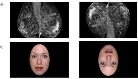
[image:10.612.77.530.239.500.2]vertically and 3.7 º horizontally with participants sitting 50 cm from the screen.
Figure 1. Examples of mammogram and face stimuli in the upright and inverted orientations (a) upright and inverted abnormal mammogram (b) upright and inverted smiling face.
The experiment was conducted on a Macintosh, MacBook Pro using in-house JavaScript
scripts. All participants viewed the experiment on a 13.3 inch, liquid-crystal color screen with a
2560 x 1600 resolution, 227 pixels per inch, and refresh rate of 60Hz
The experiment consisted of one block of 40 face trials (10 neutral, 10 happy: presented
in both upright and inverted conditions) and one block of 40 mammogram trials (10 normal, 10
abnormal: presented in both upright and inverted conditions). Each trial began with a fixation
stimulus, a noise mask (1000 ms), followed by a response screen (Figure 2). In the mammogram
block, the participant’s task was to decide if the viewed mammogram was normal or abnormal
and to indicate their response by pressing the corresponding key. In the face block, the
participant’s task was to decide whether a presented face displayed a happy or neutral expression
and to indicate their response by pressing the corresponding key. Face stimuli were presented for
500 ms. Though previous work shows an ability to distinguish normal from abnormal
mammograms after 250 msec exposure, pilot testing in the present conditions did not produce
reliable performance at that speed. Accordingly, the mammograms were shown for 1000 ms. The
presentation order of the blocks (mammoxgrams or faces) was counterbalanced across
[image:12.612.71.526.349.634.2]participants and participants were given a break halfway through each test
Figure 2. Overview of the alternative forced choice paradigm blocks (a) Mammogram block: determine if the viewed mammogram was normal or abnormal. (b) Face block: decide whether a presented face was happy or neutral.
Results
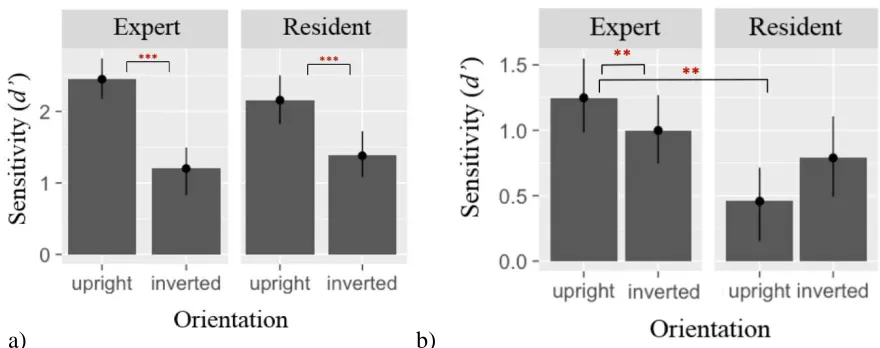
Faces
A 2 x 2 mixed ANOVA was conducted for the face sensitivity (d’), Experience
(experienced radiologists, resident radiologists) was a between group factor and Orientation
(upright, inverted) was a within-group factor. A main effect of Orientation was statistically
significant, F(1, 19) = 36.73, p < .0002, p2 = 0.53, displaying the classical face inversion effect
where expression classification was impaired in the inverted orientation relative to the upright
orientation. With face sensitivity, no Experience effect was found, F(1, 19) = 0.1 , p > 0.76, nor
was there an interaction between Experience and Orientation, (see Figure 3a). Reaction time data
for correct face trials showed a main effect of Orientation, F(1,19) = 8.49, p < .01, p2 = 0.06.
No Experience effect was found, F(1, 19) = 0.86, p > 0.37, p2 = 0.04.
Mammograms
For the ANOVA on the mammogram data, Experience (experienced radiologists, resident
radiologists) was a between group factor and Orientation (upright, inverted) was a within-group
factor. For mammogram recognition sensitivity (d’), there was a main effect of Experience, F(1,
19) = 6.18, p < .02, p2 = 0.22 (experts are better). A direct comparison using paired t-tests
revealed that experts had better performance for upright mammograms, t(10) = 3.6, p = .0019,
Cohen’s d =.45 compared to residents. There was no reliable difference for the inverted
mammograms, p > 0.2. A Group by Orientation interaction was obtained, F(1, 19) = 11.91, p =
.003 , p2 = 0.09, reflecting the presence of an inversion effect for the experts but not residents.
The inversion effect was assessed with paired t-tests. These reveal a significant inversion effect
significant and, in any case, goes in the opposite direction from what would be expected, p >
0.33 (see Figure 3b). Reaction time data for correct mammogram trials showed no Experience,
F(1,19) = 1.03, p > 0.32 , p2 = 0.05 or Orientation effects, F(1,19) = 2.89, p > 0.11 , p2 = 0.04
Faces Mammograms
[image:14.612.86.526.185.359.2]a) b)
Figure 3. Performance d’ scores on forced-choice task for experienced and novice radiology residents for (a) face stimuli and (b) mammogram stimuli. Error bars represent 95% CI for within-subject measures.
Further we explored the differences in mammogram performance, comparing “hit” and
“false alarm” rates. Experienced and resident radiologists did not differ in their ability to classify
an abnormal mammogram as “abnormal” (hit rates) in upright mammograms, t(19) = -0.58, p =
0.56, Cohen’s d = -0.13. However, the experienced radiologists were less likely to misclassify a
normal mammogram as “abnormal” (false alarms), t(19) = -4.74, p < 0.01, Cohen’s d = -1.05.
This means that the criterion of experienced radiologists was more strongly biased toward a
“normal” classification than less experienced radiologists, t(19) = 2.73, p = 0.008, Cohen’s d =
0.6. There were no significant differences in “hit rates”, “false alarms” or biases between experts
and residents for the inverted mammograms, and upright and inverted faces. These results are
Table 1.
Recognition performance of radiology experts and dresidents for upright and inverted
mammogram and face trials in terms of hits (hit), false alarms (fa), sensitivity (d’) and bias (c). ______________________________________________________________________________
Test Group Up (hit)
Up (fa)
Up (d’)
Up (c) Inv (hit) Inv (fa) Inv (d’)
Inv (c)
Up-Inv (d’)
Mam Experts 0.55 0.15 1.25 0.48 0.52 0.18 0.99 0.43 0.26 * Residents 0.52 0.35 0.46 0.18 0.59 0.31 0.79 0.15 -0.33 Faces Experts 0.92 0.17 2.50 -0.18 0.63 0.19 1.36 0.26 1.19 **
Residents 0.94 0.30 2.18 -0.50 0.69 0.24 1.33 0.15 1.16 **
*p< .05. **p < .01. ***p< .001.
Figure 4 plots d’ for upright mammograms as a function of years of experience for all 21
participants. Not unexpectedly, the results showed that mammogram discrimination improved
with years of radiological experience, F(1, 19) = 15.2, p < .001, R2 = .45, 95% CI = 0.018, 06.
However, years of experience was not related to the ability to discriminate inverted
[image:15.612.80.357.472.679.2]mammograms, F(1, 19) = 2.179, p < .16 , R2 = 0.1, 95% CI = -0.006, 0.03 (see Figure 5).
Figure 5. d’ scores of inverted mammogram stimuli across years of radiology experience
There is also a significant correlation of the magnitude of the inversion effect (difference
between upright and inverted mammogram discrimination) with years of experience, F(1, 19) =
8.49, p < .009, R2 = .30, 95% CI =.018, .059 (see Figure 6) suggesting that the use of holistic
Figure 6. An inversion composite score of upright mammogram performance (d’ score) minus the inverted mammogram condition performance across years of radiology experience.
Discussion
In this study, we employed the inversion task to test the holistic hypothesis of
radiological expertise. Experts (>5 years of experience) and residents (< 5 years of experience)
were asked to classify upright and inverted mammograms as either “normal” or “abnormal.” As
comparison stimulus, the same participants judged whether upright and inverted faces displayed
a “happy” or “neutral” expression. For faces, both resident and experienced radiologists
exhibited the classic face inversion effect where face expression discrimination was better in the
upright orientation than the inverted orientation. For mammograms, experienced radiologists
showed superior discrimination relative to resident radiologists for mammograms presented in
their upright orientation. Although the overall performance of resident radiologists was worse
than the experienced radiologists, their detection rates were unaffected by orientation and were
essentially the same for upright and inverted mammograms. When the mammograms were
inverted, their discrimination scores of the experienced radiologists dropped to the same level as
demonstrated by resident radiologists. These results are consistent with previous studies of
perceptual expertise showing that with extensive domain-specific experience, experts access
holistic information in an upright stimulus, but holistic information is impaired when the
stimulus is inverted (Campbell & Tanaka, 2018; Diamond & Carey, 1986; Rossion, Gauthier,
Goffaux, Tarr, & Crommelinck, 2002).
Although inversion impaired mammogram detection for the experienced radiologists, it
did not completely abolish the expertise advantage. The performance of experienced radiologists
studies for upright mammograms (d’=1.14) (Experiment 1, Evans et al., 2016), albeit at a shorter
exposure presentation of 500 msec. The residual expert effect for inverted mammograms suggest
that other types of non-holistic information survived the inversion manipulation. Harley et al,
(2009), for example, presented chest radiographs to experts for 500 ms and compared normal
images to chest radiographs that were scrambled (e.g. segmenting image into 25 squares and
shuffling their positions). Despite disrupting the global structure of the stimulus, the experts’
performance dropped slightly from d’=1.23 for the intact stimulus compared to d’=1.09 for the
scrambled stimulus.
The holistic account of mammogram expertise is consistent with eye-tracking studies
which show that in comparison to non-experts, experts typically perform domain-related tasks
with fewer fixations, longer saccades and less coverage of the image (Manning et al., 2006;
Koncak et al., 2005; Krupinski, 1996). One study directly examined the eye-position of expert
breast radiologists and of novice radiology residents when reading digital mammograms (Kundel
et al., 2007). They found that the median time for the eyes of the experts to reach the location of
a cancerous nodule was 0.96 seconds from image onset whereas for the novices that time was
2.15 seconds. The authors speculated that response time for the experts was too fast to support a
search-to-find strategy implying that their search was being guided by a global representation of
the mammogram. In the current study, the exposure duration was increased to 1000 msec in
order for the experienced and resident participants to perform above chance levels. The extended
exposure duration contrasts to previous “gist” studies where expert radiologists achieved
reasonable detection scores after a much shorter presentation duration (i.e., 250 ms or less)
(Carrigan, Wardle & Rich, 2018; Evans et al., 2016; Kundel & Nodine, 1975). Given that
that additional encoding time was required to first, determine the orientation of the stimulus and
next, to apply the appropriate holistic and non-holistic strategy. In future studies, it would be
useful to block the mammogram stimuli by orientation so participants will have the opportunity
to prepare their detection strategy prior to the onset of the stimulus.
The emergence of a holistic strategy with radiological experience has implications for
training medical students. In our study, the expert radiologists acquired holistic strategies
implicitly over many years of clinical experience, evaluating thousands, even tens of thousands
of mammograms images. It is interesting to speculate whether this perceptual knowledge can be
taught explicitly during medical training by presenting medical students with many images of
abnormal and normal mammograms, asking them to judge the normality of each image and
providing the appropriate feedback. A perceptual expertise protocol might accelerate the learning
process and allow radiological trainees to achieve expert performance more quickly. The
perceptual expertise training approach has been successfully applied in other medical domains
that require visual diagnosis. For example, participants showed reliable gains in their ability to
detect melanoma skin lesions after four, 30 minute sessions of perceptual expertise training (Xu,
O’Rourke, Robinson & Tanaka, 2016).
Conclusion
The goal of this study was to measure the holistic processing of expert mammographers by
employing the inversion test – a standard measure of holistic processing used in the face
recognition research. The main finding was that experienced radiologists exhibited a robust
inversion effect as evidenced by their better discrimination of upright mammograms than
inverted mammograms. In contrast, the less experienced, resident radiologists performed more
inverted mammograms. Critically, detection performance improved and the inversion effect
increased for radiologists who had more experience diagnosing mammogram images suggesting
that holistic abilities developed as a function of perceptual experience. Our results have
implications for training medical students by emphasizing the role of experience and the learning
Declarations
Ethics approval
Signed consent was obtained from human subjects prior to their participation in the study
indicating their knowledge of the goals, publication and possible health risks related to the study. This study was approved by the Human Research Ethics Board at the University of Victoria, Ethics Protocol Number: 16-362.
Consent for publication
Not applicable: Manuscript does not contain individual details, data or photos.
Availability of data and material
The dataset supporting the conclusions of this article is available in the figshare repository, Mammogram and Face Data, https://doi.org/10.6084/m9.figshare.5248513.v1 in CSV format.
Competing interests
The authors declare that they have no competing interests
.
Funding
Source: Natural Sciences and Engineering Research Council of Canada, Recipient: James Tanaka: Travel expenses to the Radiology Conference of North America.
Source: National Eye Institute (US): Award number: EY017001 Recipient: Jeremy W. Wolfe: Supported data collection at the Radiology Conference of North America.
Source: National Cancer Institute: Award number: CA207490 Recipient: Jeremy W. Wolfe: Supported data collection at the Radiology Conference of North America.
Authors’ contributions
Michael Chin: Design and experiment development, collection of data, data analysis, primary author.
Karla Evans: Design, Stimulus development, author. Jeremy W. Wolfe: Design, author.
James Tanaka: Design, author.
Jon Bowen: Experiment development, collection of data, author.
Acknowledgements
References
Ashworth, A. R. S., Vuong, Q. C., Rossion, B., and Tarr, M. J. (2008). Recognizing rotated faces
and greebles: what properties drive the face inversion effect? Vis. Cogn. 16, 754–784.
Calder, A. J., & Jansen, J. (2005). Configural coding of facial expressions: The impact of
inversion and photographic negative. Visual cognition, 12, 495_518.
Campbell, A., Tanaka, J., W., Inversion Impairs Expert Budgerigar Identity Recognition (2018):
A Face-Like Effect for a Nonface Object of Expertise. Perception. 1-13. DOI:
10.1177/0301006618771806
Carrigan, A., J, Wardle, S,. G., Rich, A., N., (2018). Finding cancer in mammograms: if you
know it’s there, do you know where? Cognitive Research: Principles and Implications,
3:10, https://doi.org/10.1186/s41235-018-0096-5
Curby K. M. Glazek K. Gauthier I. (2009). A visual short-term memory advantage for objects of
expertise. Journal of Experimental Psychology: Human Perception and Performance, 35,
94–107.
Diamond, R., & Carey, S. (1986). Why faces are and are not special: an effect of
expertise. Journal of Experimental Psychology: General, 115(2), 107.
Evans, K. K., Georgian-Smith, D., Tambouret, R., Birdwell, R. L., & Wolfe, J. M. (2013). The
gist of the abnormal: above-chance medical decision making in the blink of an eye.
Evans, K. K., Haygood, T. M., Cooper, J., Culpan, A.-M., & Wolfe, J. M. (2016). A half-second
glimpse often lets radiologists identify breast cancer cases even when viewing the
mammogram of the opposite breast. Proceedings of the National Academy of Sciences,
Grill-Spector, K., Kanwisher, N. (2005). Visual recognition: As soon as you know it is there, you
know what it is. Psychological Science, 16, 152–160
Harley, E. M., Pope, W. B., Villablanca, J. P., Mumford, J., Suh, R., Mazziotta, J. C., ... &
Engel, S. A. (2009). Engagement of fusiform cortex and disengagement of lateral occipital
cortex in the acquisition of radiological expertise. Cerebral Cortex, 19(11), 2746-2754.
Krupinski, E. A. (1996). Visual scanning patterns of radiologists searching
mammograms. Academic radiology, 3(2), 137-144.
Kundel, H. L., & Nodine, C. F. (1975). Interpreting Chest Radiographs without Visual Search
1. Radiology, 116(3), 527-532.
Kundel, H. L., Nodine, C. F., Conant, E. F., & Weinstein, S. P. (2007). Holistic component of
image perception in mammogram interpretation: gaze-tracking study. Radiology, 242(2),
396–402. http://doi.org/10.1148/radiol.2422051997
Manning, D., Ethell, S., Donovan, T., & Crawford, T. (2006). How do radiologists do it? The
influence of experience and training on searching for chest nodules. Radiography, 12(2),
134-142.
McKone, E., & Yovel, G. (2009). Why does picture-plane inversion sometimes dissociate
perception of features and spacing in faces, and sometimes not? Toward a new theory of
holistic processing. Psychon Bull Rev, 16(5), 778–797. Retrieved from
http://www.ncbi.nlm.nih.gov/entrez/query.fcgi?cmd=Retrieve&db=PubMed&dopt=Citatio
n&list_uids=19815781
Nodine CF, Kundel HL, Mello-Thoms C, et al. How experience and training influence
Palermo, R., Willis, M. L., Rivolta, D., McKone, E., Wilson, C. E. and Calder, A. 2011.
Impaired holistic coding of facial expression and facial identity in congenital
prosopagnosia. Neuropsychologia, 49: 1226–1235.
doi:10.1016/j.neuropsychologia.2011.02.021
Rossion B. & Curran T. (2010). Visual expertise with pictures of cars correlates with RT
magnitude of the car inversion effect. Perception, 39, 173–183.
Rossion, B. (2008). Picture-plane inversion leads to qualitative changes of face perception. Acta
Psychologica, 128(2), 274–89. http://doi.org/10.1016/j.actpsy.2008.02.003
Rossion, B. (2008). Picture-plane inversion leads to qualitative changes of face perception. Acta
Psychologica, 128(2), 274–89. http://doi.org/10.1016/j.actpsy.2008.02.003
Schyns, P.G. and Oliva, A. 1994. From blobs to boundary edges: evidence for time- and
spatial-scale dependent scene recognition. Psychological Science, 5:195-200
Tanaka, J. W., & Farah, M. J. (1993). Parts and wholes in face recognition. Quarterly Journal of
Experimental Psychology, 46A(2), 225–245.
Tanaka, J. W., & Sengco, J. A. (1997). Features and their configuration in face recognition.
Memory & Cognition, 25, 583-592
Tanaka, J. W., & Simonyi, D. (2016). The “parts and wholes” of face recognition: a review of
the literature. Quarterly Journal of Experimental Psychology (2006), 218(August), 1–37.
http://doi.org/10.1080/17470218.2016.1146780
Tottenham, N., Tanaka, J. W., Leon, A. C., McCarry, T., Nurse, M., Hare, T. A., ... & Nelson, C.
(2009). The NimStim set of facial expressions: judgments from untrained research
participants. Psychiatry research, 168(3), 242-249.
perceptual expertise training approach. Applied Cognitive Psychology, 30, 750-756.
Yin, R. K. (1969). Looking at upside-down faces. Journal of Experimental Psychology, 81(1),
141–145.
Young, A. W., Hellawell, D., & Hay, D. C. (1987). Configural information in face perception.