Structure-function study of the enzymes
of the cyanuric acid catabolic pathways
A Thesis Submitted
for the Degree of Doctor of Philosophy
of
The Australian National University
© Copyright by Lygie Esquirol
All Rights Reserved
i
Declaration
This research constitutes an original work carried out by myself during my PhD
from February 2015 to June 2018, unless otherwise stated. Results and methods
published or obtained by another person have been duly acknowledged.
The work presented herein has not been submitted as part of any other degree.
Lygie Esquirol
ii
Acknowledgements
I would like to thank sincerely Dr Colin Scott, Dr Matt Wilding and Dr Carol Hartley for their support and encouragements during my PhD, for being patient, positive and for providing support and precious advice during my weekly meetings.
I would like to express my appreciation to Pr Chris Easton for co-supervising my work and for being there to discuss my issues each time I needed it. Thanks a lot to Dr Hideki Onagi for helping me find ways to monitor all my many water-sensitive unstable substrates and products.
A big thank you to Nigel French for teaching me how to express and purify proteins. Without his insight, AtzE would probably still be a recalcitrant enzyme impossible to produce heterelogously!
Thanks a lot to Dr Janet Newman and Dr Tom Peat for being amazingly enthusiastic, persistent and so efficient at crystallising and solving the structures of my enzymes and uncovering the identity of unexpected small proteins.
Thanks a lot to Dr Jian-Wei Liu for providing help with proteomics to confirm the identity of my purified enzymes when I needed it and for revealing the true name of my many unknown bacterial strains.
Thanks you to Dr Chris Coppin for sharing his knowledge of molecular cloning and library screening.
Thanks to all my colleagues and students for discussion and support: Dr Karine Caron, Dr Sahil Balotra, Dr Andrew Warden, Dr Madhura Shettigar, Fizza Modh Pushri, James Antoney, Dr Brendon Lee, Matthias Nachtschatt, Ellen Walsh, Blair Ney, Dr Sarah Rottet, Dr Mihir Shah, Dr Phil Robbins.
Finally thanks to the “coffee team” for the much needed daily coffee and puzzle break.
iii
List of publications
Esquirol L*, Peat TS*, Wilding M, Lucent D, French NG, Hartley CJ, Newman J,
Scott C. (2018) Structural and biochemical characterization of the biuret
hydrolase (BiuH) from the cyanuric acid catabolism pathway of Rhizobium
leguminasorum bv. viciae 3841. PLoS One 13(2): e0192736.
https://doi.org/10.1371/journal.pone.0192736.
Lygie Esquirol*, Thomas S. Peat, Matthew Wilding, Jian-Wei Liu, Nigel G. French,
Carol J. Hartley, Hideki Onagi, Thomas Nebl, Christopher J. Easton, Janet
Newman, and Colin Scott (2018). An unexpected vestigial protein complex
reveals the evolutionary origins of an s-triazine catabolic enzyme. J. Biol. Chem.
2018 293: 7880-7891. Published 05/2018. doi:10.1074/jbc.RA118.001996
http://www.jbc.org/content/293/20/7880
Esquirol L*, Peat TS*, Wilding M, Hartley CJ, Newman J, Scott C (2018) A novel
decarboxylating amidohydrolase involved in avoiding metabolic dead ends during
cyanuric acid catabolism in Pseudomonas sp. strain ADP. PLoS ONE 13(11):
e0206949. Published 11/2018. https://doi.org/10.1371/journal.pone.0206949.
iv
Contents
Declaration ... i
Acknowledgements ... ii
List of publications ...iii
Abbreviation ... vi
Abstract ... viii
1 Introduction ... 1
1.1 s-Triazine usage has led to the evolution of new metabolic pathways in microorganisms ... 2
1.2 Discovery of s-triazine-degrading microorganisms and their catabolic pathways ... 5
1.3 s-Triazine catabolic pathway(s) ... 15
1.3.1 The s-triazine catabolic pathway of Pseudomonas sp. strain ADP 15 1.3.2 Variations to the s-triazine catabolic pathway identified in other bacteria ... 19
1.4 Enzymatic basis of the s-triazine catabolism ... 21
1.4.1 Structure-function of the metalloproteins amidohydrolases of the upper pathway ... 21
1.4.2 Structure-function of the amidohydrolases of the lower pathway .. 29
1.5 Goal of the PhD ... 37
2 Structural and biochemical characterization of the biuret hydrolase (BiuH) from the cyanuric acid catabolism pathway of Rhizobium leguminasorum bv. viciae 3841 ... 39
2.1 Overview ... 40
2.2 Statement of contribution ... 40
v 3 An unexpected vestigial protein complex reveals the evolutionary origins of an s-triazine catabolic enzyme ... 83 3.1 Overview ... 84 3.2 Statement of contribution ... 84 3.3 Publication: An unexpected vestigial protein complex reveals the
evolutionary origins of an s-triazine catabolic enzyme ... 87 4 A novel decarboxylating amidohydrolase involved in avoiding metabolic dead ends during cyanuric acid catabolism in Pseudomonas sp. strain ADP . 105
4.1 Overview ... 106 4.2 Contribution ... 106 4.3 Publication: Structural and genomic insights into AtzH, a protein
dynamo for cyanuric acid catabolism in Pseudomonas sp. strain ADP ... 108 5 Conclusion ... 149
5.1 Update of the cyanuric acid degradation pathway of Pseudomonas sp. strain ADP ... 150 5.2 Insights into the evolutionary origin of the cyanuric acid degradation operon ... 155 5.3 An alternative cyanuric acid degradation pathway in Rhizobium
vi
Abbreviation
µM Micromolar
Å Ångström
AH Allophanate amidohydrolase
AtzA Atrazine chlorohydrolase
AtzB Hydroxy-atrazine N-ethylaminohydrolase AtzC N-ethylaminohydrolase
AtzD Cyanuric acid amidohydrolase AtzE 1-Carboxybiuret hydrolase AtzF Allophanate hydrolase
AtzR Cyanuric acid-responsive regulator regulator
BA Barbituric acid
BAH Barbituric acid amidohydrolase BiuH Biuret amidohydrolase
CA Cyanuric acid
ca. Circa
CAH Cyanuric acid amidohydrolase
DNA Deoxyribonucleic acid
DSF Differential scanning fluorometry DTT Dithiothreitol
E. coli Escherichia coli
E.U. European Union
EDTA Ethylenediaminetetraacetic acid EPA Environmental protection agency FAO Food and agriculture organisation GatCAB Glutamyl-tRNA amidotransferase
GDH Glutamate dehydrogenase
GuaD Guanine deaminase
H-bond Hydrogen bond
HEPES 2-[4-(2-hydroxyethyl)piperazin-1-yl]ethanesulfonic acid IPTG Isopropyl β–D-1-thiogalactopyranoside
IS Insertion sequence
kb Kilobase pairs
kcat Catalytic constant
kDa KiloDalton
KM Michaelis constant
LB Luria-Bertani
LC-MS Liquid chromatography mass spectrophotometry
mer Mercury-resistance gene
MES 2-(N-Morpholino)ethanesulfonic acid
NA Not available
NADH Nicotinamide adenine dinucleotide hydrogen
ND Non determined
NDSB Non-detergent sulfobetaine
vii OriV Origin of replication
PDB Protein data bank
PncA Nicotinamidase
RDX Royal demolition explosive RMSD Root-mean-square deviation
RU Repeat unit
RutB Ureidoacrylate peracid amidohydrolase
SDS-PAGE Sodium dodecyl sulphate-polyacrylamide gel electrophoresis SEC Size exclusion chromatography
SeMet seleno-L-methionine spont. Spontaneous
Tm Melting temperature
tnpA Transposase gene
tra Transfer operon
trb Transfer operon
TriA Melamine deaminase
TrzA Melamine deaminase/ atrazine dechlorohydrolase TrzB Hydroxy-atrazine N-ethylaminohydrolase
TrzC N-ethylaminohydrolase
viii
Abstract
Pseudomonas sp. strain ADP1 mineralises the persistent s-triazine herbicide
atrazine. It has been used as a model organism to study evolution of xenobiotic
degradation pathways in bacteria since its discovery in 1995. Prior to this study it
was thought that the degradation of atrazine involved six enzymes: AtzA, AtzB,
AtzC, which transform atrazine into cyanuric acid, and AtzD, AtzE and AtzF,
which mineralise cyanuric acid to ammonia and carbon dioxide. The genes atzD,
atzE and atzF are organised in an operon that is induced by nitrogen starvation
and presence of cyanuric acid. The cyanuric acid catabolism operon of
Pseudomonas sp. strain ADP1 has been intensively studied; however, the study
of the cyanuric acid degradation pathways in other bacteria has revealed an
unexpected diversity in their composition.
The work presented in this thesis has focused on characterising the penultimate
step of the atrazine degradation pathway: biuret hydrolysis. This step is
performed by quite different enzymes in Pseudomonas sp. strain ADP1 and
Rhizobium leguminasorum bv. viciae 3841. The amidase AtzE was thought to be
the biuret hydrolase of Pseudomonas, whereas in Rhizobium an
isochorismatase-like enzyme, BiuH, performs that function. In Chapter 2, we
solve the structure of BiuH and investigate its catalytic mechanism by
site-directed mutagenesis.
Chapter 3 focuses on the study of AtzE, which had not been characterised
previously because it could not be produced in E. coli. The structure of native
AtzE isolated from Pseudomonas sp. strain ADP1 revealed the presence of an
ix subsequently named AtzG. AtzG is encoded by a previously unreported gene
located in the cyanuric acid catabolism operon between atzD and atzE. The
co-expression of AtzE and AtzG in E. coli allowed production of stable and active
AtzEG. Biochemical characterisation revealed AtzEG does not hydrolyse biuret,
as previously thought, but instead catalyses the hydrolysis of the water-sensitive
product of AtzD, 1-carboxybiuret. Interestingly, the AtzEG complex was found to
be structurally similar to the GatCAB complex, a glutamyl-tRNA
amidotransferase, suggesting the AtzEG complex might have evolved from the
GatCA complex.
Examination of the cyanuric acid operon also revealed the presence of another
small open reading frame located between atzE and atzF, renamed atzH.
Proteomics confirmed that AtzH was expressed along with the rest of the cyanuric
acid catabolism operon. A crystal structure of AtzH was obtained and biochemical
characterisation confirmed it played a role in the degradation of cyanuric acid
(Chapter 4). Docking studies and mutagenesis suggested a catalytic role for
AtzH: an allophanate-forming, decarboxylating 1,3-dicarboxyurea
amidohyrdolase.
Two different cyanuric acid degradation strategies have clearly evolved in
Pseudomomas and Rhizobium. The Pseudomomas strategy is to use enzymes
to limit the formation of metabolic ‘dead-ends’ that spontaneously form from
unstable metabolic intermediates. By contrast, the Rhizobium pathway relies on
1
2
1.1
s
-Triazine usage has led to the evolution of new metabolic
pathways in microorganisms
The anthropogenic symmetrical triazines (s-triazines) form a family of polyvalent
compounds that are used around the world. The first s-triazine compound
synthesised was cyanuric acid (1,3,5-triazinane-2,4,6-trione) in the 1800s (1). All
of the s-triazine compounds share a common core structure of a six membered,
aromatic ring containing three nitrogen and three carbon atoms (Fig. 1-1).
Commonly used s-triazine compounds include: broad-spectrum herbicides (such
as atrazine, simazine, propazine, cyanazine, ametryn and terbutylazine),
melamine (used in the production of certain plastics and polymers) (2), the
military explosive RDX (Royal Demolition eXplosive) (3) and the disinfectant and
swimming pool chlorine stabiliser cyanuric acid (4) (Fig. 1-1). Although s-triazines
are predominantly synthetic, cyanuric acid is also produced naturally. It has been
found to be the main component in the mineral Joanneumite, which was
discovered recently in Chile (5), and it is also formed as a product of oxidative
3
Figure 1-1 s-Triazine compounds cited in this work. Structures of: cyanuric acid a disinfectant and swimming pool chlorine stabiliser (4), the herbicides atrazine, simazine, of melamine, a compound used in the production of certain plastics and polymers (2), of the military explosive RDX (3) and of the herbicides propazine, cyanazine, ametryn and terbutylazine.
Among the most abundant group of s-triazines deliberately introduced into the
environment are the herbicides. The s-triazine herbicides are broad-range
pre-and post-emergence herbicides, which disrupt electron transport during
photosynthesis by binding to the plastoquinone-binding protein in photosystem II
and preventing quinone binding (7-9). The s-triazine herbicide atrazine,
synthetised in the 1950s, has been one of the most used herbicides worldwide
(10, 11) behind glyphosate (12). Based on FAO and EPA data, in 2012 around
1.3 billion tonnes of herbicides were used around the world and atrazine
accounted for around 80,000 to 90,000 million tonnes (12, 13). The use of
N N N OH OH HO
Cyanuric acid Atrazine N N N Cl N H N H N N N NH2 NH2 H2N
[image:15.595.163.491.63.341.2]4
atrazine was found to increase the yield efficiency of maize by 6% and across all
type of crops by an average of 3-4% (14).
Despite the undeniable advantages conferred by their use, there are a number of
concerns regarding the continued use of s-triazine herbicides. These herbicides
are quite persistent, with a half-life in the soil ranging from two months to one
year (15). They have also been detected in both surface and ground waters
beyond regulatory limits, leading to potential human exposure (16-22).
Toxicological studies have suggested that the s-triazines are possible
carcinogens, teratogens and an endocrine disrupters (23-31). These concerns
led to the discontinuation of the use of atrazine in the E.U. in 2003 (20), but they
are still used widely elsewhere (11). There are also potential environmental
effects of atrazine use, including off-target herbicidal activity, with some
atrazine-mediated impacts found on lichen, microalgae and benthic diatom communities
(8, 32-34).
In addition to the immediate effects of s-triazine exposure, the introduction of
s-triazines to the environment has resulted in the introduction of new selection
pressures. This has led to the acquisition of new biochemical functions in soil and
aquatic microorganisms, including the evolution of new metabolic pathways and
5
1.2 Discovery
of s
-triazine-degrading microorganisms and
their catabolic pathways
Work towards the discovery of s-triazine degrading strains of soil microorganism
started in the 1960s, and continues today. In 1950, s-triazines degradation
products were shown to be present in soil that had been exposed to s-triazine
herbicides, albeit it was debated at the time whether these products were the
result of a microbial catabolic activity (40) or the result of a slow abiotic
degradation (41, 42). Between 1960 and 1970, several fungal species that could
transform atrazine were identified (Table 1-1). It was later demonstrated that their
degradation abilities were mediated by non-specific enzymes, such as
cytochromes P450 (15, 43). Between the 1960s and the 1980s, a number of
bacterial species were shown to transform or degrade atrazine and other s
-triazines (Table 1-1), albeit they could only catalyse a small number of
transformative steps and these compounds were not completely catabolised by
any single bacterial species. In 1984, the first pure culture that could completely
degrade an s-triazine (melamine), Pseudomonas sp. strain NRRL B-12228
(44-46), was isolated by Cook and co-workers. Although Cook et al. were able to
isolate the enzymes that were responsible for the degradation of melamine
ammeline, ammelide and cyanuric acid in 1981, it took a further ten years before
the genes encoding the s-triazine catabolic enzymes (triA, trzB, trzC and trzD),
6
In 1994, Pseudomonas sp. YAYA6 was isolated by Yanzekontchou and
Gschwind (49); it was the first isolated bacterial strain able to entirely degrade
atrazine to carbon dioxide and ammonia. The following year, Mandelbaum et al.
discovered another Pseudomonas strain able to entirely degrade atrazine:
Pseudomonas sp. strain ADP (50), which later became the model organism used
to study s-triazine degradation. Since then, a large number of s-triazine
[image:18.595.58.474.325.744.2]catabolising bacterial strains have been isolated (Table 1-1).
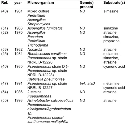
Table 1-1 Isolated s-triazine degrading strains. ND: not determined.
Ref. year Microorganism Gene(s)
present Substrate(s)
(40) 1961 Mixed culture
Penicillium Aspergillus Streptomyces
ND simazine
(51) 1963 Aspergillus fumigatus ND simazine
(52) 1970 Aspergillus
Fusarium Penicillium Trichoderma
ND atrazine,
simazine, propazine
(53) 1982 Nocardia ND atrazine
(45) 1984 Rhodococcus corallinus
Pseudomonas sp. strain
NRRL B-12228
ND melamine,
simazine, atrazine (46) 1985 Pseudomonas strain D (=
Pseudomonas sp. strain
NRRL B-12228)
Klebsiella pneumoniae
ND cyanuric acid
(47) 1991 Pseudomonas sp. strain
NRRL B-12227 triA, atzD melamine, cyanuric acid
(54) 1986 2 strains
Pseudomonas ND atrazine
(55) 1993 Acinetobacter calcoaceticus Pseudomonas
alcaligenes/Agrobacterium sp.
Pseudomonas putida/ xanthomonas maltophilia
7
(56) 1994 Rhodococcus strain B-30 ND atrazine,
propazine, simazine
(49) 1994 Pseudomonas sp. YAYA6 ND atrazine
(50) 1995 Pseudomonas sp. strain ADP ND atrazine
(43) 1995 Rhodococcus NI86/21 P-450 atrazine
(57) 1997 Rhizobium sp. PATR atzA atrazine
(58) 1997 Klebsiella terragena ND melamine
(59) 1999 Pseudomonas sp. strain
NRRLB-12227 trzD cyanuric acid
(60) 2000 14 strains,
Pseudoaminobacter C147 atzA, B, C atrazine
(61) 2000 Nocardia
Nocardioides sp. strain C190 trzN atrazine
(62) 2001 Comamonas acidovorans ND atrazine
(63) 2001 Penicillium steckii DS6F
Moraxella ovis N5C ND simazine
(64) 2001 Chelobacter heintzii
Aminobacter aminovorans Stenotrophomonas
maltophilia
Arthrobacter crystallopoietes
atzA, B, C, trzD
“ “
atzB, C
atrazine
(65) 2002 Arthrobacter aurescens TC1 trzN, atzB,
C atrazine, broad range
(66) 2003 Nocardioides sp. SP12 trzN, atzB,
C atrazine
(67) 2003 Arthrobacter sp. AD1 AtzA atrazine
(68) 2004 Acinetobacter A6 ND atrazine,
broad range (69) 2004 Agrobacterium radiobacter
Bradyrhizobium japonicum ND simazine
(70) 2005 Arthrobacter nicotinovorans
HIM atzA, B, C atrazine, simazine,
propazine, broad range
(71) 2006 Nocardioides trzN atrazine
(72) 2006 Micrococcus sp. strain MF-1 ND melamine
(73) 2007 Arthrobacter sp. strain MCM
B-436 trzN, atzB, C, D atrazine
(74) 2007 6 strains
Nocardioides
Arthrobacter trzN, atzC
atrazine
(75) 2007 Nocardioides sp. NEA-A
Agrobacterium sp. NEA-D
Shinorhizobium sp. NEA-B Polaromonas sp. NEA-C
atzA, B, C, D, E, F atzA, B, C, D, E, F trzN, B, C
8 Nocardioides sp. 1D Arthrobacter sp. 2B Arthrobacter sp. 3A
trzN, B, C trzN trzN trzN (76) 2007 Arthrobacter ATZ1
Arthrobacter ATZ2 Ochrobactrum CA1 Pseudomonas CA2 trzN, atzC trzN, atzB, C trzD trzD atrazine atrazine cyanuric acid cyanuric acid
(77) 2007 Variovorax sp. MD1/MD2 atzA, B atrazine
(78) 2007 Methyloversalis CDB21 atzA, B, C,
D, E, F simazine
(73) 2007 Arthrobacter sp strain MCM
B-436 trzN, atzB, C, D atrazine
(79) 2008 Pseudomonas sp. MHP41 atzA, B, C,
D, E, F simazine
(80) 2008 Stenotrophomonas P51
Stenotrophomonas C53
Arthrobacter P52
atzA, D atzA, D atzD
simazine
(81) 2008 Arthorbacter sp. AD26 trzN, atzB,
C atrazine
(82) 2009 Alcaligenes faecalis Klebsiella ornithinolytica Bacillus megaterium Agrobacterium tumefaciens ND atzA ND atzA atrazine
(83) 2009 Arthrobacter sp. GZK-1 ND atrazine
terbuthylazine (84) 2010 54 strains Arthrobacter
28 strains Nocardioides
Ancylobacter T10AII
trzN trzN
atzA, B, C, D, E, F
NA
(85) 2010 Nocardioides sp. strain DN36 trzN, atzB,
C atrazine simazine propazine (86) 2010 Arthrobacter sp. strain KU001 trzN, atzB,
C (87) 2010 Klebsiella sp. A1
Comamonas sp. A2 ND atzA, B, C,
D, E, F
atrazine atrazine
(88) 2011 Arthorbacter sp. strain
DNS10 trzNC , atzB, atrazine
(89) 2011 Arthrobacter sp. TES6 trzN, atzB,
C atrazine
(90) 2012 Rhodococcus sp. JN201860 trzA melamine
(91) 2012 Nocardioides sp. strain ATD6 triA melamine
(92) 2012 Rhizobium sp. C14
Acinetobacter lwoffii C1 AtzA, B, C atrazine
(93) 2012 Enterobacter cloacae
9
(94) 2012 Arthrobacter sp. strain DAT1 trzN, atzB,
C atrazine
(95) 2013 Arthrobacter trzN, atzB,
C atrazine
(96) 2013 Nocardioides sp. EAA-3
Nocardioides sp. EAA-44 trzN, atzB, C atrazine
(97) 2014 Alcaligenes sp. strain EGD-AK7
Arthrobacter sp. strain AK-YN10
atzA, B, C, D, E, F trzN, atzB, C
(98) 2014 Bacilus subtilis HB-6 trzN,B,C atrazine
(95) 2014 Pseudomonas aeruginosa atzA, B, C,
D, E, F atrazine
(99) 2014 Arthrobacter sp. SD3-25 trzN, atzB,
C simazine
(100) 2015 Sterptomyces sp. Atz2 ND atrazine
(101) 2015 Arthrobacter sp. MCO
Arthrobacter sp. CSP
Microbacterium sp. ZEL
Not found melamine, cyanuric acid
(102) 2016 Variovorax sp. Schlesneria Arthorbacter spp
trzN, atzA,
B, C, trzD atrazine
(103) 2016 Shewanella sp. YJY4 atzA, B,C atrazine
(104) 2017 Ensifer sp. strain CX-T atzA, B, C,
D, E, F atrazine
(105) 2017 Pseudomonas sp. ZXY-1 ND atrazine
(106) 2018 Citricoccus sp. strain TT3 trzN, atzB,
C atrazine
10
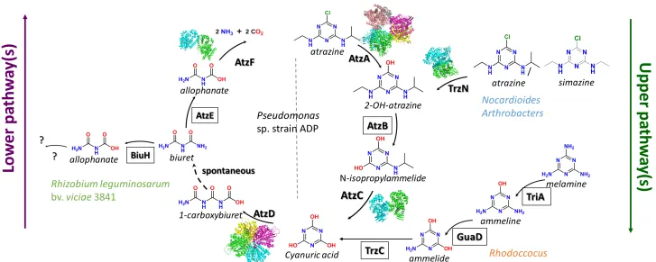
The atrazine degradation pathway is composed of a seven steps process and
can be separated in two parts: the ‘upper degradation pathway’, which transforms
particular s-triazines to cyanuric acid, and the ‘lower degradation pathway’ which
mineralises cyanuric acid to ammonia and carbon dioxide (Fig. 1-2).
In Pseudomonas sp. strain ADP, six enzymes have been identified that are
required for s-triazine catabolism (Section 1.4). Wackett et al. cloned the genes
coding for the enzymes of the first three steps of the degradation pathway,
required to transform atrazine to cyanuric acid: atzA (107), atzB (108) and atzC
(109) (Fig. 1-2, Table 1-2, 3). The atz genes were shown to be localised on an
IncPβ plasmid, pADP1. In 2001, pADP1 was sequenced by Martinez et al. (110).
In addition to atzA, atzB and atzC, Martinez et al. also identified three new genes
atzD, atzE and atzF, which encodes enzymes required to degrade cyanuric acid
to ammonia and carbon dioxide (Fig. 1-2, Table 1-2, 1-3). Interestingly, de Souza
et al. (111) identified atzA, B, C across five different strains of bacteria
(Pseudomonas, Alcaligenes, Clavibacter Ralstonia and Agrobacterium) and
11
11
12
Although the Pseudomonas sp. strain ADP pathway is the best characterised,
several alternative enzymes have been adopted for s-triazine catabolism in other
bacteria. In 2000, Nocardioides sp. strain C190 was demonstrated to perform the
dechlorination of atrazine, the first step in the degradation pathway, without atzA.
Topp et al. cloned, expressed and characterised the enzyme responsible for this
activity: TrzN (61). Subsequently, trzN was found to be present in other Nocardia
(66), in Arthrobacter species(65, 73, 74, 81, 88, 94, 117) and in Bacillus species
(98), revealing a previously unknown diversity in the s-triazine degradation
pathways of bacteria. Cameron et al. identified and characterised a biuH gene
from Rhizobium leguminosarum bv. viciae 3841, encoding for a cysteine
hydrolase enzyme from the isochorismatase family, that was able to degrade
biuret (118), showing that the lower degradation pathway also possesses some
13
Table 1-2 Genes (A) and genomes (B) involved in s-triazine herbicide catabolism.
A.
Ref. year microorganism Genes
(47, 48) 1991 Pseudomonas sp. strain NRRL B-12228/27/99
Klebsiella pneumoniae
triA, trzB, trzC, trzD
(119) 1995 Pseudomonas sp. strain ADP atzA
(120) 1995 Rhodococcus corallinus trzA
(108) 1997 Pseudomonas sp. strain ADP atzB
(109) 1998 Pseudomonas sp. strain ADP atzC
(111,
121) 1998 Pseudomonas Clavibacter michiganensesp. strain CP1 ATZ1 atzAatzC, atzB, (61,
122) 2000/2 Nocardioides sp. strain C190 trzN
(123-128) 2003/5/7/9/10 Pseudomonas sp. strain ADP atzR, atzD, atzE, atzF
(129) 2004 Arthrobacter aurescens TC1 trzN, atzB,
atzC
B.
Ref. year Microorganism
(110) 2001 Pseudomonas sp. strain ADP p-ADP1 plasmid
(130) 2006 Arthrobacter aurescens TC1
(131) 2006 Rhizobium leguminosarum bv. viciae 3841 (132) 2013 Alcaligenes sp. Strain HPC1271
(133) 2013 Arthrobacter sp.FB24
(97) 2014 Alcaligenes sp. strain EGD-AK7
Arthrobacter sp. strain AK-YN10
14
Table 1-3 Biochemical (A) and structural (B) characterisation of the s-triazine enzymes.
A. Biochemical characterisation
Ref Year microorganism Enzyme
(135) 1982 Pseudomonas strain A D F,
2 strains Klebsiella pneumoniae
Not named
(43) 1995 Rhodococcus sp. strain
Ni86/21 Cytochrome P450
(59) 1999 Pseudomonas sp. strain
NRRLB-12227 trzD
(107, 136) 1996/
2002 PseudomonasRhodococcus corallinus sp. strain ADP AtzA TrzA
(137) 2002 Pseudomonas sp. strain ADP AtzC
(115, 116) 2005 Pseudomonas sp. strain ADP AtzF
(138) 2006 Enterobacter cloacae strain 99 TrzF
(139) 2006 Arthrobacter aurescens TC1 TrzN
(112) 2007 Pseudomonas sp. strain ADP AtzB
(118) 2011 Rhizobium leguminosarum bv.
viciae strain 3841 BiuH
B. Structural characterisation
Ref year microorganism Enzyme
(140) 2010 Arthrobacter aurescens TC1 TrzN
(141, 142) 2014/15 Pseudomonas sp. strain ADP AtzF
(143, 144) 2013/14 Azorhizobium caulinodans
ORS571 AtzD
(145) 2013 Pseudomonas sp. strain ADP AtzD
(146) 2015 Pseudomonas sp. strain ADP AtzC
(147) 2015 Pseudomonas sp. strain ADP AtzA
(148) 2017 Acidovorax citrulli TrzD
15
1.3
s
-Triazine catabolic pathway(s)
1.3.1 The s-triazine catabolic pathway of Pseudomonas sp. strain ADP
In Pseudomonas sp. strain ADP the hydrolytic enzymes required for the
transformation of atrazine to cyanuric acid (i.e., the ‘upper pathway’) are: the
atrazine chlorohydrolase AtzA enzyme (E.C. 3.8.1.8) (107, 108), the
hydroxyatrazine amidohydrolase AtzB enzyme (EC 3.5.99.3) (112), and the
isopropylammelide amidohydrolase AtzC enzyme (EC 3.5.99.4)(107-109, 112,
137) (Fig. 1-2, 1-4). The steps of the ‘lower pathway’ are performed by: the
cyanuric acid amidohydrolase AtzD enzyme (E.C. 3.5.2.15) (113, 114), the biuret
amidohydrolase AtzE enzyme (EC 3.5.1.54) and the allophanate amidohydrolase
AtzF enzyme (3.5.1.84) (116). The last three steps are the hydrolysis of the
cyanuric acid ring leading to the production of 1-carboxybiuret, thought to
spontaneously decarboxylate to biuret in the presence of water (113, 114). Biuret
is deaminated to produce allophanate, which is hydrolysed to form ammonia and
carbon dioxide (115, 116). The genes encoding all of the atrazine catabolic
enzymes of Pseudomonas sp. strain ADP are located on the pADP1 catabolic
16
17
The pADP1 sequence revealed the presence of the tra and trb operons, which
encode proteins essential for plasmid conjugation (110). In 1998, pADP has been
shown to be transmissible under laboratory conditions (111), and (as noted
above) the atzA, atzB and atzC genes have been found in a variety of bacteria
and geographic locations (75, 77, 84). The atzA, atzB, and atzC genes were
found to be monocistronic, constitutively expressed, not co-located (Fig. 1-3), and
flanked by transposases and insertion sequences (110). This suggested that
these genes are mobile and explained how they can be found independently from
one another in different bacterial strains (15, 110, 150)(Table 1-1).
The atzD, atzE and atzF genes were shown to be organised as an operon (110).
The regulation of this operon has been studied in detail by Govantes et al.
(123-128, 151). The operon was found to be regulated by a LysR-type transcription
regulator named AtzR (124, 125). The induction of the operon was found to be
upregulated under nitrogen-limiting conditions. Nitrogen starvation in
Pseudomonas sp. strain ADP is sensed via the general nitrogen control circuit
(GlnK-NtrC) and regulates transcription of the cyanuric mineralization operon via
upregulation of atzR expression from a σ54-dependent promoter under nitrogen
limiting conditions (123, 126, 128). AtzR then upregulates the expression of the
the cyanuric acid mineralisation operon, even in the absence of cyanuric acid
(125, 128); however, the presence of cyanuric acid further increases the level of
transcription of the operon by inducing a conformational change in the regulator,
18
[image:30.842.58.785.113.404.2]19
1.3.2 Variations to the s-triazine catabolic pathway identified in other bacteria
Although less extensively studied than the Pseudomonas sp. strain ADP
pathway, several variations on the model s-triazine catabolism pathway have
been described in other bacterial strains (15). As shown in the Table 1-1, s
-triazine degrading bacteria can contain a variety of -triazine catabolism genes:
atzA or trzN only, complete ‘Pseudomonas-type’ pathways (atzA, atzB, atzC,
atzD, atzE, atzF), the ‘Pseudomonas upper degradation-type’ pathway (atzA,
atzB, atzC), or a combination of trzN, atzB and atzC with or without atzD or its
homologue trzD.
The trzN gene encodes a physiological analogue of the enzyme encoded by atzA
and is frequently found in Gram-positive bacteria such as Nocardioides and
Arthrobacter (61, 122). TrzN dehalogenates atrazine simazine, propazine (152),
and some naturally occurring TrzN variants can also hydrolyse less reactive
leaving groups, such as the thiomethyl substituent of ametryn (140, 152). The
structural and mechanistic differences between TrzN and AtzA are explored in
more detail in Section 1.4.
The lower pathway also has some plasticity. TrzD homologues have been found
to encode a cyanuric acid hydrolases. Depending on the trzD homologue,
sequence identity between TrzD and AtzD has been found to vary between
40-60 %, but trzD has never been found to be part of a cyanuric acid degradation
operon (59, 114). This suggests that the polycistronic organisation of cyanuric
acid catabolism genes in Pseudomonas sp. strain ADP is not a ubiquitous
20
been found in association with genes from pathways other than the
Pseudomonas’; for example, in Rhizobium leguminosarum bv. viciae 3841, an
atzD homologue is found immediately upstream of a gene encoding a biuret
hydrolase (biuH). This pathway differs from the Pseudomonas’ because BiuH
belongs to the isochorismatase family, unlike AtzE that belongs to the amidase
family. Additionally, there is no equivalent of atzF found downstream of biuH.
Interestingly, the melamine catabolic pathway parallels the atrazine catabolic
pathway with three hydrolytic steps that sequentially deaminate melamine to
ammeline, ammeline to ammelide and ammelide to cyanuric acid (Fig. 1-4). TriA,
a melamine deaminase, was first identified in 1991 from a Pseudomonas sp.
strain NRRL B-12227 (47). TriA shares 98% sequence identity with AtzA, but was
found to be only a melamine deaminase, unable to degrade atrazine. A second
melamine deaminase TrzA, 50% identical to AtzA, was identified from a
Rhodococcus coralinus NRRL-B-15444R strain in 1994. TrzA can perform both
the dechlorination of atrazine and the deamination of melamine (154).
s-Triazine catabolic genes are usually plasmid-borne (15), but some studies also
report atrazine catabolic genes present in bacterial chromosomes. Cai et al.
found the presence of atzA in the bacterial chromosome of Arthrobacter sp. AD1
strain (67). Devers et al. also observed variation in gene compositions and
locations when they sampled 17 atrazine degrading bacteria from soil (75). The
most frequent pattern of genes they observed were: atzABC-trzD, atzABCDEF
and trzN-atzBC. In some cases genes were found on one plasmid, in others they
were spread across multiple plasmids and they were sometimes found on the
21
copy number also varies. For example, there are multiple copies of trzN in the
Arthrobacter aurescens strain, which contains six identical tandem repeats of
trzN on a 16 kb region of the pTC1 plasmid (130). As yet, the effect of trzN gene
dosage in this strain has not been investigated.
1.4 Enzymatic basis of the
s
-triazine catabolism
1.4.1 Structure-function of the metalloproteins amidohydrolases of the upper pathway
AtzA (E.C. 3.8.1.8), TrzN (E.C. 3.8.1.8), AtzB (EC 3.5.99.3) and AtzC (EC
3.5.99.4) are all metal-dependant amidohydrolase family enzymes that share a
common fold known as the (α/β)8 barrel (140, 146, 147, 155) (Fig. 1-5). The
substrate specificity of these enzymes depends on the steric, conformational and
electronic restrictions imposed by the eight loops that form the active site, located
at the end of the eight β-strands (155). Although they catalyse a broad range of
reactions, the amidohydrolase mechanism usually involves the activation of a
nucleophilic water molecule through the complexation with the metal centre,
followed by nucleophilic attack on the substrate by the activated water. In some
cases, a proton transfer from an active site amino acid to the substrate is also
required to activate the substrate (155). The characteristics of each of these four
22 Table 1-4 Characteristics of the amidohydrolases of the upper pathway.
E.C. No. Enzyme Physiological Function Kinetic Values1 Approx. Size PFam Family Accession2 Mechanism Refs
3.8.1.8 TrzN s-Triazine hydrolase
KM= 20 kcat= 2 kcat/KM= 1 x 10⁵
Dimer (2 x
54,000 Da) Amidohydrolase
UniProt: A1RCJ9, Q8VS01 PDB: 4l9x, 4lh8, 5hmd, 5hme, 5hmf
Zn2+-dependent hydrolase
(122, 139, 140, 152, 156, 157)
3.8.1.8 AtzA s-chlorohydrolaseTriazine
KM = ND kcat = ND KM/kcat = 1.5
x 104
Hexamer (6 x
52,000 Da) Amidohydrolase
UniProt: P72156 PDB: 4v1x, 4v1y
Fe2+-dependent hydrolase
(119, 136, 147, 158, 159)
3.5.99.3 AtzB Hydroxydechloro- atrazine ethylaminohydrolase
KM = 20 kcat= 3 kcat/KM= 1.5 x 10⁵
Dimer (2 x
50,000 Da) Amidohydrolase
UniProt: P95442 PDB: NA
Fe2+/Zn2+ -dependent hydrolase
(108, 112, 160)
3.5.4.42 AtzC N
-Isopropylammelide isopropylamino-hydrolase
KM = 400 kcat = 13 kcat/KM = 3.3 x 10⁴
Tetramer (4 x
45,000 Da) Amidohydrolase
UniProt: O52063 PDB: 2qt3, 4cqb, 4cqc, 4cqd, 5akq
Zn2+-dependent
hydrolase (109, 146)
1Values are given for the physiological substrate only. Units for kinetic values are as follows: KM, µM; kcat, s-1; kcat/KM, s-1.M-1
23
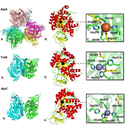
Figure 1-5 Structures of the s-triazine degrading enzymes of the upper pathway. A.
Cartoon representation of the AtzA hexamer (formed by a trimer of dimers (147)); B.
AtzA monomer; C. Zoom onto the AtzA active site with bound Fe2+; D. TrzN dimer; E. TrzN
monomer; F. Zoom on the TrzN active site with bound Zn2+; G. AtzC dimer; H. AtzC
monomer; I. Zoom on the AtzC active site with bound Zn2+. For the quaternary structures,
each monomer is shown in a different colour. For the tertiary structures of the monomers, loops are in green, α-helices are in red and β-strands are in yellow. The black dot indicates the location of the active site, metal cations are represented as spheres (grey for Zn2+and orange for Fe2+). The amino acids involved in metal coordination are
24
AtzA is a hexameric, Fe2+-dependent metalloenzyme (107, 119). The kcat/KM of
this enzyme has been reported at 1.5 x 10⁴ sec-1.M-1 (159). The KM of AtzA
exceeds the water solubility of atrazine (~153 µM) and as a consequence the KM
and kcat of AtzA cannot be directly measured (Table 1-4). The active site of AtzA
is buried at the end of a long hydrophobic channel. A Fe2+, coordinated by four
histidine residues (His66, His68, His243 and His276) and an aspartate (Asp327),
form the metal centre (147). No crystal structure with atrazine in the active site
has been obtained, nor could the substrate be docked into the active site,
suggesting a poor complementarity between the active site and its substrate.
Peat et al. concluded that the high KM of AtzA for atrazine could be explained by
the lack of complementarity. AtzA also has a low affinity for iron, and this was
shown to be due to unusually long bond lengths between the metal and the
histidine ligands. Overall the unusual bonding pocket appears “ill-adapted” for its
function (147, 159). (Fig. 1-5)
TrzN is a dimeric, zinc-dependent amidohydrolase performing the same
physiological function as AtzA. Its metal centre is composed of a Zn2+ coordinated
by four histidines (His63, His65, His238 and His274) and, unusually, a threonine
(Thr325) residue forms a bond to the metal via a bridging water molecule. (140,
152).
Some naturally occurring TrzN variants are strict chlorohydrolases (e.g. the
Nocardioides sp. strain AN3 TrzN) (157, 161, 162), while others (such as the
Nocardioides sp. strain MTD22 TrzN) are capable of hydrolyzing non-halogen
25
ametron, respectively. Interestingly, this biochemical phenotype is controlled by
a small number of amino acid substitutions in the active site: Thr/Pro214,
His/Tyr215 and Gln/Glu241 (157). The strict dehalogenase TrzN contains the
Gln241 mutation, whereas the broader substrate range TrzN enzyme contains a
Glu241. Glu241 residue is thought to protonate the atrazine ring, which then
protonates the poorly reactive leaving groups (e.g. –SCH3). Sugrue et al. showed
that the presence of the two hydrophobic residues Pro214 and Tyr215 contributed
to the creation of a microenvironment, increasing the pKa of the glutamine
side-chain and making Glu241 a more reactive catalytic residue, allowing the efficient
hydrolysis of ametryn (157). Laboratory evolved AtzA variants that can protonate
unreactive leaving groups (such as amines and –SCH3 groups) have been
produced, but such variants have reduced dehalogenase activities (158, 163).
The active site of TrzN is a better fit for its substrate than is the case for AtzA and
atrazine (140, 147), explaining why the KM of TrzN is lower for atrazine(20 μM)
than that of AtzA and the kcat/KM higher (ca. 1 x 10⁵ s-1.M-1)(159). (Fig. 1-5).
The two next enzymes in the pathway are AtzB and AtzC. AtzB, a
metal-dependent amidohydrolase, is a hydroxyatrazine N-ethylaminohydrolase. AtzC,
a zinc-dependent tetramer, catalyzes the hydrolysis of the N-isoproylamine group
from N-isopropylammelide (kcat = 13 sec-1, KM = 400 µM) (137). The Zn2+ metal
centre of AtzC is coordinated by four histidines (His60, His62, His217 and His249)
and an Aspartate, Asp303. In AtzC, while the water molecule is activated by the
metal ion, the substrate is protonated by Gln160, leading to the protonation of the
leaving group (146) (Fig 1-5). AtzC has a relaxed substrate specificity, removing
sidechains from a range of substituted ammelides (137). As yet, there is no X-ray
26
homodimer and has been demonstrated to contain a metal ion, that could be
either Fe2+ and/or Zn2+ (112). Like AtzC, AtzB has been shown to hydrolyse the
side chains from a range of di-N-alkyl-2-hydroxy-s-triazines (112). Interestingly,
AtzB has also been shown to possess chlorohydrolase activity, albeit ten-fold
lower than its physiological deaminase activity with kcat/KM values of 1.5 x 10⁵ and
1.6 x10⁴ M-1.s-1 for deamination and dechlorination, respectively (112). Sequence
alignment of AtzB with the other metallo-enzymes TrzN, AtzA and AtzC shows
conservation of the histidine binding metals and of the asparagine/threonine and
suggests His75, His77, His249, His284 and Asp336 might be the amino acids
28
Figure 1-6 Alignment of AtzA, TrzN, AtzC and AtzB amino acid sequences with T-COFFEE (164-168). The black arrows point towards the four conserved Histidine residues and the aspartate (or threonine in the case of TrzN) involved in the metal binding centre of the enzyme’s active sites. The colour reflects the quality of the alignment and the legend is shown on the top left corner. For reason of readability the C-terminal sequences are truncated at amino acid for 353 AtzA, 351 for TrzN, 330 for AtzC and 359 for AtzB. The Beta The secondary sequences of AtzA, TrzN and AtzC have been added using ESPript (169); The η symbol refers to a 310-helix, α-helices, 310-helices and π-helices are displayed as medium, small and large squiggles, respectively. β-strands are rendered as arrows,
29
1.4.2 Structure-function of the amidohydrolases of the lower pathway
Unlike the enzymes of the upper pathway, the enzymes of the lower pathway do
not belong to the amidohydrolase superfamily, instead they belong to the
amidase and cyclic amide hydrolase families. Their characteristics can be found
in Table 1-5.
AtzD (E.C. 3.5.2.15) produces 1-carboxybiuret by opening the cyanuric acid ring,
its kcat and KM values are 17 sec-1 and 350 µM, respectively (Table 1-5). It forms
a compact tetramer. Each monomer has been found to contain a metal centre
coordinating a Mg2+ metal ion; however, it is involved in structure stabilisation and
is not catalytic (145) (Fig. 1-7). Phylogenetic analysis of the sequences of AtzD
(114) and TrzD (59) with homologous sequences, has revealed that these
enzymes belong to a unique family of enzymes (113, 145, 170), which also
includes barbituric acid hydrolase (BAH), an enzyme involved in the oxidative
pathway of pyrimidines (171). The structural characterisation of AtzD from
Pseudomonas (145) and cyanuric acid hydrolases (CAH) from Frankia sp. strain
EuI1c and Azorhizobium caulinodans ORS 571 (144, 149) confirmed the novelty
of the protein family. AtzD was found to possess a novel fold, named the
“Toblerone fold” in reference to the three-fold pseudo-symmetry of the AtzD
monomer (Fig. 1-7) (145). The catalytic mechanism of AtzD, probed by mutation
30
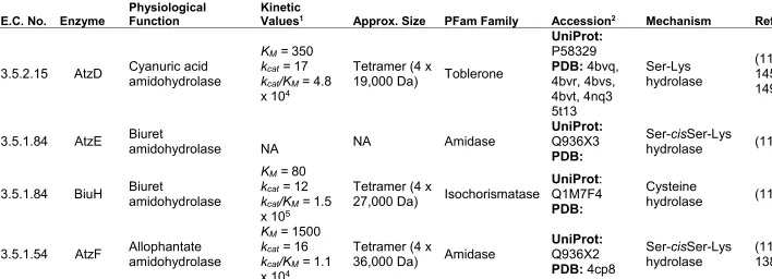
Table 1-5 Characteristics of the amidohydrolases of the lower pathway. NA: not available.
E.C. No. Enzyme Physiological Function Kinetic Values1 Approx. Size PFam Family Accession2 Mechanism Refs
3.5.2.15 AtzD Cyanuric acid amidohydrolase
KM = 350 kcat = 17 kcat/KM= 4.8
x 104
Tetramer (4 x
19,000 Da) Toblerone
UniProt: P58329 PDB: 4bvq, 4bvr, 4bvs, 4bvt, 4nq3 5t13 Ser-Lys hydrolase (113, 144, 145, 148, 149)
3.5.1.84 AtzE Biuret amidohydrolase NA NA Amidase UniProt: Q936X3
PDB:
Ser-cisSer-Lys
hydrolase (115, 118)
3.5.1.84 BiuH Biuret amidohydrolase
KM = 80 kcat = 12 kcat/KM = 1.5
x 105
Tetramer (4 x
27,000 Da) Isochorismatase
UniProt: Q1M7F4 PDB:
Cysteine
hydrolase (118)
3.5.1.54 AtzF Allophantate amidohydrolase
KM = 1500 kcat = 16 kcat/KM = 1.1
x 104
Tetramer (4 x
36,000 Da) Amidase
UniProt: Q936X2 PDB: 4cp8
Ser-cisSer-Lys
hydrolase (115, 116, 138, 142)
1Values are given for the physiological substrate only. Units for kinetic values are as follows: KM, µM; kcat, s-1; kcat/KM, s-1.M-1
31
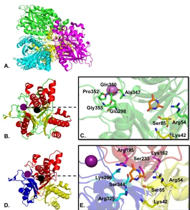
Figure 1-7 Structure of AtzD from Pseudomonas sp. ADP (PDB 5hxu). A. Cartoon representation of the AtzD tetramer; B. Monomer of AtzD showing α-helixes in green,
the loops in red and the β-sheets in yellow; C. Zoom in the active site showing the amino acids coordinating the metal ion in green sticks, the catalytic amino acid of the dyad ser85-Lys42 in yellow stick and the Arg54 in green stick around the inhibitor barbituric acid (145); D. Trimeric monomer of AtzD, each repeat unit (RU) is shown in a different colour; E. Zoom in the active site showing the three sets of potential catalytic amino acids around the barbituric acid inhibitor: in cyan sticks for the blue RU, in pink stick for the red RU, in yellow stick for the yellow RU. The black dot represents the location of the active site, the purple dot the location of the metal ion, the Mg2+ ion is shown as a purple
[image:43.595.129.520.83.516.2]32
Due to the three-fold symmetry of the active site, the AtzD monomer contains
three potential Ser-Lys dyads: Lys42 and Ser85, Lys162 and Ser233, or Lys296
and Ser344 (Fig. 1-7B,C). It is uncertain which of the Ser-Lys dyads is catalytic;
but X-ray data from the Pseudomonas AtzD structure suggests that Ser85-Lys42
may form the catalytic dyad (145), while phylogenetic studies suggest that
Ser226-Lys156 may fulfil this function (144). The active site of AtzD is largely
composed of polar amino acids that stabilise cyanuric acid substrate through an
extensive H-bonding network. The reaction mechanism resembles that of other
serine hydrolases (Fig. 1-8); the catalytic lysine acts as a general base and
deprotonates the serine that in turn attacks cyanuric acid, leading to the formation
of a covalent acyl intermediate. Finally, a water molecule attacks the acyl:enzyme
complex regenerating the active site serine and liberating 1-carboxybiuret (145).
HN N H NH O O O Ser85 O H
H2N
Lys42 HN N H NH O O O Ser85 O N H2 Lys
42 H HN N H NH2 O O Ser85 O
H2N
Lys42 O H O H HN N H NH2 O O Ser85 N H2 Lys
42 O
OH H O
H2O
NH N H NH2 O O O HO HN N H NH2 O O O Ser85 O
H2N
Lys42
33
AtzE and AtzF both belong to the amidase enzyme signature family,
characterised by a glycine serine rich sequence (Fig. 1-9) (115, 172). Up until
now all attempts to produce AtzE in E. coli had failed, and consequently AtzE had
not been characterised prior to the beginning of the investigation reported in
Chapter 3. However, AtzF has already been biochemically characterised. It is an
allophanate amidohydrolase (AH) from the amidase family and possesses the
signature amidase catalytic triad (Lys65, cisSer165 and Ser189), which was
confirmed via mutagenesis and through inhibition studies (115). AtzF is a
homotetramer and its kcat and KM values with allophanate are 16 sec-1 and 1500
µM, respectively (Table 1-5) (115).
34
AtzF is larger than most amidase family enzymes, as it possesses a ca. 15 kDa
C-terminal extension (173). The X-ray structure of N-terminal amidase domain of
AtzF was obtained. This fragment of AtzF was catalytically competent, with
identical KM and kcat values to the full length protein. Indeed, the only function so
far ascribed to the C-terminal extension is to co-ordinate the quaternary structure
of the protein, as the truncated amidase domain is dimeric, not tetrameric (173).
The structure of the AtzF amidase domain, truncated from its C-terminal domain,
confirmed that the enzyme adopts the α-β-α fold common to this enzyme family
(141, 142) (Fig. 1-9). Amidase signature enzymes are found across domains of
life and they are involved in a broad range of reactions (172). The alignment of
five amidases, sharing the same catalytic mechanism as AtzF, are shown in Fig.
1-10: the fatty acid amide hydrolase from the rat (FAAH) (174), the malonamidase
E2 from Bradyrhizobium japonicum (175), the peptide amidase from
Stenotrophomonas maltophilia (176), the AtzF and AtzE from Pseudomonas sp.
strain ADP (110). This illustrates the conservation of the AS signature and of the
35
Figure 1-10 Alignment of five AS signature amidase enzymes with T-COFFEE (162-166).
36
The AtzF mechanism is essentially identical to the mechanisms found in other
amidases such as the FAAH amidase (176). Ser189 is activated by the
Lys-cisSer proton shuttle, and then forms the acyl:enzyme intermediate via the
nucleophilic attack on the substrate (liberating ammonia). Finally, a water
molecule hydrolyses the acyl-enzyme intermediate releasing dicarboxyamonia
and regenerating Ser189 (176, 177).
In addition to its role in cyanuric acid catabolism, allophanate amidohydrolase is
also found as part of a multiprotein complex (urea carboxylase) involved in urea
degradation in some bacteria, algae, fungi and yeast (178). Urea carboxylase
contains two enzymes, urea carboxylase and allophanate amidohydrolase. Urea
carboxylase performs an ATP-dependent carboxylation of urea to form
allophanate and allophanate amidohydrolase deaminates allophanate, forming
ammonia and carbon dioxide (141, 179). Interestingly, Balotra et al. suggest that
like urea carboxylase, AtzD, AtzE and AtzF may also form a multiprotein complex
(141).
As the Pseudomonas sp. strain ADP biuret hydrolase (AtzE) was difficult to
produce in E. coli (110, 116, 118), Cameron et al. identified and characterised
the biuret hydrolase (BiuH) from Rhizobium leguminosarum bv. viciae 3841 BiuH.
Unrelated to the AtzE amidase, BiuH is a member of the isochorismatase family
with homology to cysteine hydrolases. BiuH hydrolyzes biuret to form allophanate
(kcat/KM = 1.5 x 10⁵), but does not accept 1-carboxybiurte as a substrate (118).
Homologs of BiuH have been described in other bacteria, but are infrequently
associated with an AtzD homologue, which may suggest a broader role for biuret
37
1.5 Overview of the PhD
The work reported in this thesis focuses on the variation in the composition of the
cyanuric acid degradation pathways from Rhizobium leguminasorum bv. viciae
3841 and of Pseudomonas sp. strain ADP. AtzE was identified more than twenty
years ago (110), and has been identified in the genomes of many s-triazine
degrading strains (Table 1-1). However, AtzE has remained uncharacterised
biochemically or structurally due to its recalcitrance to heterologous expression.
Its physiological counterpart in Rhizobium leguminosarum bv. viciae 3841, BiuH,
had been partially biochemically characterised (118), but lacked characterisation
at an atomic level.
The goal of my PhD was to obtain and study the X-ray structures of both AtzE
and BiuH, to elucidate the structure-function relationships for these two enzymes.
Previous studies of this type with xenobiotic-degrading enzymes have been
informative about their recent evolution, and it was hoped that this study would
provide insights into the convergent evolution of cyanuric acid degradation
pathways in these two bacteria.
In Chapter two, BiuH is characterised Rhizobium leguminasorum bv. viciae 3841,
with a particular focus on developing a deeper understanding of the
structure-function relationship of this previously under-characterised amidohydrolase. BiuH
was purified to apparent homogeneity, its structure obtained and structure
function analysis performed by mutagenesis and molecular dynamics modelling.
The third chapter of this PhD focuses on gaining a detailed understanding of the
38
previously been identified as a functional analogue of BiuH. Careful and detailed
charactisation of AtzE purified from Pseudomonas led to the revision of the
cyanuric acid catabolism pathway and to the identification of two small and
previously overlooked proteins, the function of one of these new proteins (AtzG)
is also explored in this chapter.
In Chapter four, the function and structure of the second newly identified small
protein in the cyanuric acid catabolism (AtzH) is explored. AtzH is purified to
apparent homogeneity from E. coli, its structure obtained and its potential function
analysed biochemically, through substrate docking and by mutagenesis. From
these data it is inferred that AtzH is like an enzyme acting on a metabolite of the
39
39
2 Structural and biochemical
characterization of the biuret
hydrolase (BiuH) from the cyanuric
acid catabolism pathway of
Rhizobium
40
2.1 Overview
The exploration of cyanuric acid degradation pathways in different bacteria
showed that substantial differences exist in the CA degradation pathways
between microorganisms (153). In Rhizobium leguminasorum bv. viciae 3841, for
example, a biuret hydrolase (BiuH) belonging to the isochorismatase family
performs the deamination of biuret to produce allophanate (118); whereas, in the
model bacterium Pseudomonas sp. strain ADP, it is an amidase that is thought
to perform that step. In this chapter, the structure-function study of the biuret
hydrolase BiuH is presented. The atomic structure of BiuH was solved and
site-directed mutagenesis was used to gain a better understanding of the BiuH
catalytic mechanism. Additionally, molecular dynamics simulations highlighted
the presence of three channels from the active site to the enzyme surface forming
potential substrate channel and a product (ammonia) channel and a
co-substrate (water) channel.
2.2 Statement of contribution
I performed the cloning, mutagenesis, protein expression and enzyme kinetics. I
also analysed and interpreted the data. The crystallography and structural
determination were performed by Dr Tom Peat and Dr Janet Newman. All the
molecular dynamics work was performed by Dr Del Lucent.
Publication status: Published 01/2018.
Esquirol L, Peat TS, Wilding M, Lucent D, French NG, Hartley CJ, Newman J, Scott C. 2018. Structural and biochemical characterization of the biuret hydrolase (BiuH) from the cyanuric acid catabolism pathway of Rhizobium
42
2.3 Publication: Structural and biochemical characterization
of the biuret hydrolase (BiuH) from the cyanuric acid
catabolism pathway of
Rhizobium leguminasorum
bv.
Structural and biochemical characterization of
the biuret hydrolase (BiuH) from the cyanuric
acid catabolism pathway of Rhizobium
leguminasorum bv. viciae 3841
Lygie Esquirol1,2☯, Thomas S. Peat3☯, Matthew Wilding2,3, Del Lucent4, Nigel G. French1, Carol J. Hartley1, Janet Newman3, Colin Scott1
*
1 CSIRO Biocatalysis and Synthetic Biology, Canberra, Australian Capital Territory, Australia, 2 Research School of Chemistry, Australian National University, Canberra, Australian Capital Territory, Australia, 3 CSIRO Biomedical Manufacturing, Parkville, Melbourne, Victoria, Australia, 4 Department of Electrical Engineering and Physics, Wilkes University, Wilkes-Barre, Pennsylvania, United States of America
☯These authors contributed equally to this work.
Abstract
Biuret deamination is an essential step in cyanuric acid mineralization. In the well-studied atrazine degrading bacterium Pseudomonas sp. strain ADP, the amidase AtzE catalyzes this step. However, Rhizobium leguminosarum bv. viciae 3841 uses an unrelated cysteine hydrolase, BiuH, instead. Herein, structures of BiuH, BiuH with bound inhibitor and variants of BiuH are reported. The substrate is bound in the active site by a hydrogen bonding net-work that imparts high substrate specificity. The structure of the inactive Cys175Ser BiuH variant with substrate bound in the active site revealed that an active site cysteine (Cys175), aspartic acid (Asp36) and lysine (Lys142) form a catalytic triad, which is consistent with bio-chemical studies of BiuH variants. Finally, molecular dynamics simulations highlighted the presence of three channels from the active site to the enzyme surface: a persistent tunnel gated by residues Val218 and Gln215 forming a potential substrate channel and two smaller channels formed by Val28 and a mobile loop (including residues Phe41, Tyr47 and Met51) that may serve as channels for co-product (ammonia) or co-substrate (water).
Introduction
The mineralization of cyanuric acid by bacteria is thought to be an ancient metabolic pathway [1]. It is thought that this pathway has been recently ‘co-opted’ into pathways for the degrada-tion of highly funcdegrada-tionalizeds-triazines as they have become environmentally abundant through human activities since the mid-twentieth century [1–3]. The s-triazinemineralization pathways, including the cyanuric acid catabolism pathway, are thought to have evolved in response to an increase in the abundance ofs-triazines in the environment as a result of human activities [2,4,5]. Although most incidentally exposed bacteria are not sensitive to the
s-triazines, these anthropogenic compounds are an excellent nitrogen source and bacteria that
PLOS ONE |https://doi.org/10.1371/journal.pone.0192736 February 9, 2018 1 / 20
a1111111111 a1111111111 a1111111111 a1111111111 a1111111111 OPEN ACCESS
Citation: Esquirol L, Peat TS, Wilding M, Lucent D, French NG, Hartley CJ, et al. (2018) Structural and biochemical characterization of the biuret hydrolase (BiuH) from the cyanuric acid catabolism pathway of Rhizobium leguminasorum bv. viciae 3841. PLoS ONE 13(2): e0192736.https://doi.org/ 10.1371/journal.pone.0192736
Editor: Renwick Dobson, University of Canterbury, NEW ZEALAND
Received: November 20, 2017
Accepted: January 29, 2018
Published: February 9, 2018
Copyright:©2018 Esquirol et al. This is an open access article distributed under the terms of the
Creative Commons Attribution License, which permits unrestricted use, distribution, and reproduction in any medium, provided the original author and source are credited.
Data Availability Statement: Data are available from the Protein Data Bank (accession numbers: 6AZO, 6AZN, 6AZQ, 6AZS, 5BK6).
Funding: The author(s) received no specific funding for this work.