Copyright i 1975 AmericanSociety forMicrobiology Printed in U.S.A.
Simian Virus 40 Gene A Function and Maintenance of
Transformation
MARY OSBORN*lAND KLAUS WEBER'
Cold SpringHarborLaboratory, ColdSpring Harbor, New York 11724
Received forpublication 30 September 1974
Transformants have been isolated after infection of ratembryo cells at 33C
with either wild-type simian virus40 or with the temperature-sensitive geneA
mutants, tsA7 and tsA28. Examination of properties usually associated with
transformationsuch asgrowthin 1%serum, growth rate,saturationdensity,and
morphology show that these properties are temperature dependent in the tsA
transformantscharacterized,butarenottemperaturedependentin thewild-type
transformants that have been examined. In the most thoroughly characterized
tsA transformants the expression of T antigen also appears to be temperature
dependent. These data suggest that an active A function is required for the
maintenance of transformation inthese cells. In thelytic cycle,the A functionis
involved in the initiation of DNA synthesis. Thus transformation by simian virus
40 may be the direct consequence of the introduction of the simian virus 40
replicon and thepresenceof its DNA initiatorfunction, which causesthe cell to
express atransformed phenotype.
Certain viruses have the property that,
al-though they containonly averysmall amount
ofDNA, theyare capable of
transforming
vari-ouis types of cells in culture, thus causing
profound and lasting changes in the growth
properties ofsuch cells.Intryingtounderstand
the virus host relationships that may lead to
such changes, one approach is to attempt to
characterize the viral contributiontothe
trans-formingprocess withtemperature-sensitive
mu-tants of the virus. If transformants can be
isolated that are temperature sensitive for cer-tainpropertiesusually associated with
transfor-mation, it is probable that the product of the temperature-sensitive viral gene is actively
re-quired for the maintenance of transformation.
Such anapproach hasbeen useful inidentifying thosegenes ofthe
oncogenic
RNA viruses whichare associatedwith transformation (14, 24).
That the small DNAviruses, such as simian
virus 40 (SV40), might control transformation
through the continuous production of a gene
product is suggestedbytheobservationthat all
cells transformed by SV40 thus far examined
express early SV40 RNA (9, 19). It seemed
promising, therefore, to construct
transform-ants with temperature-sensitive mutantsofthe
only early complementation group thus far
identified in thelytic cycleofSV40, the A gene
'Present address: Max-Planck Institut fur
Bio-physikalische Chemie,G6ttingen, West Germany.
(3, 23). Since the A function is involved in the
initiation of DNA synthesis, A mutantsdonot
replicateviral DNA or make viralcapsidsat the
nonpermissive temperature (2, 21).
We have used
wild-type
SV40, and two tsAmutants, toisolatetransformants ofratembryo
cells. The transformants have been
character-ized at both permissive and nonpermissive
temperatureswith respect togrowthrate,
satu-rationdensity,morphology, and T antigen. Our
results suggest that these parameters are
tem-perature sensitive inthetsAtransformants but
not in the wild-type transformants. Thus it
seemsthatatleastinthese cells theA function
maybe involved, either directlyorindirectly, in
the maintenance ofthe transformed state.
An-otheraccount ofthe work willappear elsewhere
(M.
Osborn
andK.Weber, ColdSpring
HarborSymp. Quant. Biol., in press).
MATERIALS AND METHODS
Virus.Stocksof theSV40A genemutants tsA7 and tsA28(23)and of theSV40wild-typestrain were very
kindlyprovided by Peter Tegtmeyer (Case Western
Reserve University). Virus stocks were prepared at 33C by infecting confluent monolayers of BSC-1 or
CV-1 cells (at a multiplicity of infection of -1 PFU per cell). When cells showed cytopathic effect they were scrapedoffthe plates, disrupted by two
freeze-thawcycles, and stored at -80 C.
Isolation of transformants. The procedures for
obtaining cells from embryos of CDF albino inbred
6:36
on November 10, 2019 by guest
http://jvi.asm.org/
SIMIAN VIRUS GENE AFUNCTION. III.
rats(Charles River), as well as for the handling of the cells priortoinfectionwith virus, and theprocedures used for transformation arethosedescribedindetail byR. Risser, D.Rifkin, and R. Pollack (18a). Usually cells from15- or16-day-oldratembryoswereused.
Af-terseveral in vitro passages, almost confluent
mono-layers ofratembryo cellswereinfected atlow multi-plicity (- 1 or less) inparallel with either wild-type SV40orwith tsA7ortsA28.Infectionproceededfor3
h at33C withshaking. Afterovernight incubationat 33C the cells were trypsinized and diluted to ap-proximately 4,000 cells per 6-cm plate. After 2 to 3 weeks at 33C therewas a 4- to 10-fold difference in the number of colonies on the virus-infected plate when compared to the mock-infected control plate. Colonies of two types were detected on both wild-type and tsA-infected plates. The first type stained
denselywith Harrishematoxylin,andonexamination
under the microscope cells in these colonies had a
typically transformed morphology, i.e., they were
piled up and disoriented. On subculture, cells from such colonies grew well andwereall T antigen posi-tive at 33C. The second type of colony stained less
well, and the cells had a flat morphology. On
sub-culture, cells from the second type frequently failed
to grow, and furthermore, as shown by Risser et al.
(Cold Spring Harbor Symp. Quant. Biol., in press),
such cells lack T antigen. Mock-infected cells
ap-peared to have only the second type of colony.
Several clones of the dense type were picked from wild-type,infected, and from tsA7- and tsA28-infected cells with steelcloning cylindersandweresubcultured. Growth mediumwasDulbeccomodifiedEaglemedium
(Gibco H-21) containing 10% fetal calf serum
(Re-hatuin) and 150 U ofpenicillin and 150 ggof
strep-tomycin per ml. Thegrowthcurvesand size distribu-tions were measured on a Coulter counter (model ZB). Mediumwaschanged every4days.
Staining of cells. Cells were fixed in
phosphate-buffered saline (PBS) containing 3.5%formaldehyde for 30min,washed with water, and then stained with Harrishemotoxylin (30 mintoovernight). Theywere
thenwashed with water followed by 1%ammonium hydroxide andafinal waterwash.
Morphology. Cells were fixed in PBS containing
3.5% formaldehyde for 30 min, washed with PBS,
mounted inElvanol, and examinedby phasecontrast
withamicroscope(ZeissPMII).
Indirect immunofluoresence. Cells on 12-mm
glass coverslipswere fixed and stainedas indicated below. Incubation with all antibodies was for 1 h at 37C, andthe coverslipswerewashed well with PBS aftereachantibody.Afterafinalrinse,thecoverslips
were mounted in Elvanol and examined under dark
field UV illumination with a microscope (Zeiss PM
II).PhotographsweretakenonPlus-X film.
T antigen. T antigen was assayed by indirect immunofluorescence essentiallyasdescribedbyRobb (17). Hamster anti-T antibody (Flow Laboratories)
was diluted 1:5inPBS. Fluorescein conjugated goat anti-hamsterglobulins (Antibodies Inc.)wereusedat a1:10dilutioninPBS. Thecoverslipswere counter-stained with Evans Blue (1:100 in PBS) for 30 s.
Actin. The expression of actin-containing fibers in cells was assayed by immunofluoresence essentially as described by Lazarides and Weber (12). Preparation oftherabbit anti-actin antibody has been described (12). This antibody was used at a 1:20 dilution in PBS. Fluorescein conjugated goat anti-rabbit globu-lins (Miles Laboratories) wereused at a 1:10 dilution inPBS.
RESULTS
Elegant studies (R. Pollack, R. Risser, S.
Conlon, and D. Rifkin, Proc. Nat. Acad. Sci.
U.S.A., in press; 18a) have established the
conditions necessary for transformation of rat
embryo cells by wild-typeSV40, and have shown
that the transformants obtained can be
characterized in a manner similar to those
ob-tained from established cell lines (18). In
addi-tion, a test of wild-type SV40-transformed rat
embryo cells showed that they would grow at
41C, and we thought this property might be
important since the tsA mutants were
origi-nally isolated at a nonpermissive temperature
of 41C and at least some of the mutants are
known to be leaky belowthis temperature (23).
Transformants were isolated after parallel
infections of ratembryo cells with either
wild-typeSV40,orwithtsA28 or tsA7. Several clones
of the densetype (see above) were picked from
eachplate,andstocksofthetransformantswere
maintained by serial passage at 33C. The
transformants were then examined for
differ-ences in their growth properties, morphology,
and expression of T antigen at 33 and41 C as
described below. Though the results are
de-scribed in terms of asingletsA28transformant,
temperature-dependent effects have been seen
with other tsA28 transformants and with some
tsA7transformants, whereas they havenotbeen
observed withwild-type transformants. At least
some ofthe transformants have a chromosome number close to that ofthe normal rat diploid complement (Pollack et al., Proc. Nat. Acad.
Sci. U.S.A., in press).
Growth rate and saturation density. Cells
wereseeded in 10%fetal calfserum at
approxi-mately 105 cells per 6-cm dish and allowed to
attach overnightat33 C. Half of the plateswere
shifted to 41 C the next morning, and the
remainder were held at 33C. Growthcurves for
a wild-type and for a tsA28 transformant are
shown in Fig. 1. The growth rates of the
wild-type and the tsA transformant wereequal
at 33C. The wild-type transformant grew
slightly faster at 41 than at 33 C. In contrast,
the tsA28 transformant appears to grow slower
637
VOL.15,1975
on November 10, 2019 by guest
http://jvi.asm.org/
100
c 4 la
=
11
E
0
50
Daysafter shift to 41
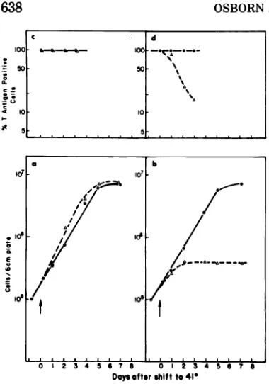
FIG. 1. (a and b) Growth curves of (a) wild-type
transformant WT4 and(b)tsA transformanttsA28.3
weremeasuredat33C(0)orat41 C(A). Cellswere
seeded at10O per6-cmdish and allowedtoattachat 33C.Half the platesweremovedto41 Catzerotime.
Cell number is expressed as the total numberper6
cm-dish andwasmeasuredonthe Coultercounter.(c andd) Thepercentages ofcells thatwere Tantigen
positive in (c) W74 and (d) tsA28.3 cultures were
measured at 33C (0) or at 41 C (A). Cells were
seededonglasscoverslips asdescribed under(aand
b). T antigenwasassayed byindirect immunofluores-cence asdescribedin thetext(seealsoFig. 5).
at41 thanat33C andappearstolimitata
10-to12-fold lowerdensity at 41 than at 33 C (see
Fig. 1). Raising theserumconcentrationto30%
didnotsignificantly changethegrowth
proper-ties of the tsA28 transformant at41 C.
The same experiment was repeated with 1%
fetal calf serum rather than 10% (data not
shown). The wild-type transformant grew well
at both temperatures, as did the tsA28
trans-formant at33C. Again, however, growthof the
tsA28 transformant wasvery much reduced at
41 C.
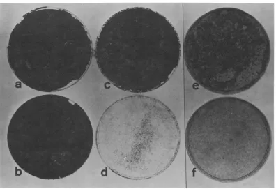
The lower saturationdensityapparentfor the
tsA transformant at 41 C might be caused
either by changed growth properties due to a
shifttothenonpermissivetemperatureortothe
cellsonly completing a singleround of division
after shift(Fig. 1). To distinguish between these
possibilities, the saturation densities of the
wild-type and of the tsA transformant were
examined as afunction of the initial cell
inocu-lum. The characteristic saturation densities of
the wild-type and of the tsA transformant at
41 C were independent of the initial plating
density in therange5 x 105to 3 x 10O cellsper
6-cm plate (Fig. 2). Below a starting inoculum
of 3 x 10⁴cellsperplate, neither the wildtype
northemutantgrowtocoverthe dish. However,
the individual colonies of wild-type cells stain
muchmoreheavily than the individual colonies
of tsA28transformant cells after equal timesat
41 C. The existence of individual colonies for
the tsA transformant at 41 C argues that the
cells, when shifted to 41 C, go through several
cell divisions.
Morphology by phase microscopy.
Wild-typetransformants appeared similaratthetwo
temperaturesand similartothe tsA
transform-ant at 33C. At 41 C the morphology of cells of
the tsA transformant changed, andmostofthe
cells became very large and flat. The pictures
shown inFig. 3weretaken 6 days after the shift
to41 C.
Size. The increase insize of these cells could
also be assayed with the Coulter counter. The
size distributions of the wild-type transform-ants at33and41 C and of the tsA transformant
were very similar. The size distributions were
normaland peakedatanarbitrary setting of 30,
with only 2% of the cells having a size greater
than85. Incontrast,the size distribution of the
tsA transformant 4 days after the shiftto 41 C
wasabnormal; no real peak wasseen, and 30%
ofthe population hada sizegreaterthan 85.
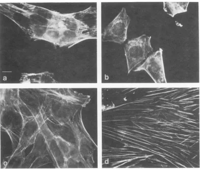
Staining with antibody to actin. A
differ-ence in morphology can also be demonstrated
by indirect immunofluorescence with actin
antibody (12). The wild-type transformant at
both temperaturesand the tsA transformantat
33 C show few thick actin-containing fibers
(Fig. 4). In contrast, the tsA transformant at
41 C showsvery long andverythick fibers (>1
,um in diameter). Similar cables, so-called
"stress fibers," can be seen also in the phase
microscopy picturesofthetsA transformant at
41 C (Fig. 3b).
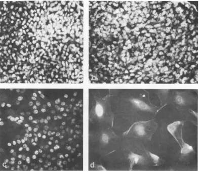
T antigen. T antigen was examined by
in-direct immunofluorescence microscopy in both
thewild-type and the tsA28 transformant as a
function of time afterthe shift to 41 C (Fig. 1,
topframe; Fig. 5). For the wild-type
transform-ant, greater than 98% of the cells of a given
clone hadTantigenat bothtemperaturesatall
times tested (Fig. lc and 5b). For the tsA
transformant shown in Fig. 1, expression of T
antigen appeared to be temperature sensitive.
Thus, at33Cgreaterthan 98% of the cellswere
T antigen positive. However, at 41 C the
per-C d
5
,,,,-,,,, .
1-~~~~~~~~~~~1
on November 10, 2019 by guest
http://jvi.asm.org/
[image:3.504.63.253.57.328.2]SIMIANVIRUS GENE A FUNCTION. III.
FIG. 2. Saturation densitiesofWT4and tsA28.3 cellsas afunction of starting inoculumand temperature.
(a) WT4, 33 C, 105; (b) WT4, 41 C, 10w; (c)tsA28.3, 33C, 101; (d) tsA28.3, 41 C, 105; (e)tsA28.3,33C, 3 x
104; () tsA28.3,41 C, 3 x 10'. Cellswereseededatthe indicated densitiesasdescribed in the legendtoFig.
1and stained with Harrishematoxylin after10days.
FIG. 3. Morphology of (a) WT4at 41 C and(b)tsA28.3 at41 C. CellswereseededasinthelegendtoFig. 1, and themorphologywasexamined6days after theshiftto41C. For(a) thecells werediluted4days after the shiftsocolony morphology could be clearlyseen.Morphologyof WT4 and tsA28.3at 33C is similarto that shownforWT4 at 41 C in(a).
639
VOL.15,1975
on November 10, 2019 by guest
http://jvi.asm.org/
[image:4.504.55.451.78.349.2] [image:4.504.56.449.401.600.2]FIG. 4. Indirectimmunofluorescenceusingactinantibody (12) of (a) WT4,33C; (b) WT4,41 C; (c)tsA28.3,
33C; (d) tsA28.3,41 C. Cellswereseededasin thelegendtoFig. 1 andprocessed for immunofluorescence6
days after theshift to 41 C. For a, b and c, cellsweredilutedafter4dayssostainingpattern could beclearly
seen.Allfourplates were taken at the same magnification, and the barrepresents 10Am. Cellsofthe tsA transformant at41C have verythick cables(d).
centage ofcells having T antigen appeared to
decrease with the lengthof time the cells were
kept at the elevated temperature. Complement
fixation assays of T antigen at 41 and 33 C 4
days aftertheshiftto 41 Calsoshowedthat the
amount of T antigen was
considerably
reducedinthetsA transformant at 41 C.
Both theextentofthemorphological changes
and the fraction of cellsshowingloss of T
anti-genexpressiondependonthe lengthoftimethe
cellsare held at41 C. Severaldaysat 41 Cwere
required to demonstrate the changes shown in
Fig. 4 and 5 for the transformant tsA28.3. In addition, 15% of the cells of the tsA
transform-antstill have T antigen 3daysafterthe shift to
41 C (Fig. 1). Therefore caution is necessary in
interpreting the data and particularly in
decid-ing whether the changes described are fully
reversible.
The following preliminary experiments sug-gest that cells ofthe tsA transformant remain
viable after several days at 41 C. Cells were
plated at approximately 105 cells per plate and
shifted to 33 C after 4 days at 41 C. After
further incubation at 33 C, dense areas of growth were seen. The same experiment was
repeated, but the cells were trypsinized and
replated after the 41 C incubation at dilutions
of
10-1,
10-2, and 10-3 at 33 C. After 10 days,cellsreplated at 33 C formed visiblecolonies.
DISCUSSION
Our results suggest thatwild-type
transform-ants and tsA transformants of ratembryo cells
behave very similarly, if not identically, at
33C, a temperature which is permissive for
bothwild-type SV40 and for SV40 tsA mutants
a
I
on November 10, 2019 by guest
http://jvi.asm.org/
[image:5.504.66.459.73.406.2]SIMIAN VIRUS GENE A FUNCTION. III.
FIG. 5. Tantigen staining of (a) WT4 at 33 C, (b) tsA28.3 at 33 C, (c) WT4at 41 C, and (d)tsA28.3at 41 C. The cells were seeded as in the legend to Fig. 1 and processed forimmunofluorescence 3 days (d) or 5 days(a-c)
aftertheshift to41C. All four plates were taken at the same magnification. Almost all cells of the tsA
trans-formant at41C arenegative for Tantigen (d; see also Fig. lc and ld).
in the lytic cycle. Their properties at 33C are
very similar tothose reported previously for a
series of transformed rat clones isolated after
infection of rat embryo cells with wild-type
SV40 at 37 C (Pollack etal., Proc. Nat. Acad.
Sci., U.S.A.,
in press; 18a).By
criteria showntobe valid for therat
embryo
system(Pollack
et al., Proc. Nat. Acad. Sci.U.S.A.,
in press;18a) and which are
commonly
used tojudge
transformation in vitro
(i.e.,
lowserumrequire-ment, faster growth rate, higher saturation
density,
changed
morphology,
andexpression
ofSV40 T antigen) both WT4 and tsA28.3 cells
are transformed at 33C. However, the
wild-type transformant and the tsA transformants
behave very differently at 41
C,
a temperaturewhich is nonpermissive forthe tsA mutants in the lytic cycle (23). Thus the
wild-type
trans-formants remain transformed at 41C, whereas
the tsA transformants show reduced saturation density, changed morphology, and loss of
ex-pression of Tantigen.
It will be interesting in the future to
deter-mine whether the tsA transformants at 41 C
resemble rat embryo cells prior to transforma-tion. Such a characterization was not done in
the present study because of the difficulty of
defining the properties of a population of
em-bryonic cells. Also, since the transformation of wild-type SV40 and tsA mutants was done in
parallel on the same cells, it seemed sufficient
to use as a criteria for transformation the
behavior of the wild-type cells, particularly
since the behavior ofthese cells has been well
documented by others (Pollack et al., Proc.
Nat. Acad. Sci. U.S.A., in press; 18a).
How-6.41
VOL.15, 1975
on November 10, 2019 by guest
http://jvi.asm.org/
[image:6.504.56.449.78.418.2]ever, the apparent temperature sensitivity of
properties usually associated with
transforma-tion is found notonly inthe tsA transformants
of rat
embryo
cells, but also as shown in theaccompanying manuscripts (1, 15, 22) for tsA
transformants of several established cell lines.
In these cases, the properties ofthetsA
trans-formants at the nonpermissive temperature
appear toresemble those of the cell lines prior
totransformation.
Itis important thatinallcases (1, 15, 22;this
study)
theoriginal
isolation of transformantswas at the permissive temperature and the
differences in properties ofwild-type and tsA
transformants only became apparent after a
shift to the nonpermissive temperature. These combined studies argue that thechange being
observed
isduetoaviralgeneandnot acellularone.TheysuggestthatamutationintheA gene
interferes indirectly with the maintenance of
transformation, or alternatively that a func-tionalgeneA product may
be
directly required forthe maintenanceofthe transformed state.Such
aconclusionwasunexpected inview of the resultsof Kimura and Dulbecco (10) withSV40
and of other earlier results (5-7) with polyoma, where transformants isolated at the permissive temperature after infection of cer-tain cell lines with mutants of the tsA typeappeared not to be temperature dependent for themaintenance of transformation. Additional work is needed to clarify whether the
discrep-ancy
between
the earlier results and theresultsreported here and in the accompanying papers
is duetothe different viralmutantsused,tothe
different cell lines used, orto the tests usedto
assay transformation. For example, it may be
that,
as previously shown with atemperature-sensitivemutantof aviansarcomavirus(8), the host cellcanstrongly influence thetemperature at which temperature-dependent behavior of
transformation isobserved.
The data suggest that transformation
re-quires morethan the presence of an integrated
site of viralreplication in the transformed cell.
The origin of replication ofSV40 is 0.67 map
units from the Eco RI site, whereas the A
mutants mapped to date lie 0.32 to 0.42 map
units from thesame site (11), so the site of the
origin of viral replication is clearly separate
from the site at which the A mutants map.
Therefore, it seems very likely that it is the
product oftheA genethat isactively required for the maintenance oftransformation. In this
context it should be recalled that all SV40
transformants characterized so far by RNA
hybridization
have been found to express theearly SV40 RNA (9, 19) and that recent
map-ping of tsA mutants has shown that these
mutations occur exclusively within the early SV40 RNA (11).
What is the function oftheSV40A
gene?
In thelyticcycle, theAfunction isrequiredforthe initiation ofviral DNA synthesis (2, 21). Ifoneassumes that in transformed cells the A
func-tion is also involved in some way in DNA replication, thenit may be that theAfunction
perturbs
or alters the normal DNA replicationprocess of the host cell. Taking into account
that in transformed cells the SV40 genome is integratedintohost cell DNA (20), thereare two
obvious
waysinwhichavirus-specific influenceonhost DNAsynthesismay occur.
(i)
Thegene A product might act on theSV40
DNA initia-tion site and DNAsynthesis might
bepropa-gated beyond the virus genome into host cell DNA. This interpretation would be analogous
to that given
by
Nishimura et al.(16)
for astrain ofE. coli carrying a
temperature-sensi-tive mutation in DNA initiation. DNA
synthe-sis couldbe renderedtemperatureinsensitive in
this mutant
by
anautonomously
replicating
episomeonly if the episomewas
integrated (i.e.,
onlywhen the bacterial chromosome had come
undercontrol of the
episomal replicon). (ii)
TheA function
might
acton oneor morehost DNA loci of similar sequence to the viral initiation site, thusproducing
new initiator sitesforDNA replicationinthe hostgenome.The latter model would be supportedby
the report that the induction of host DNAsynthesis
in permissivecells seems to
depend
onexpression
of the Afunction (4).
TheA geneproduct might alsoact to
perturb
the normal cellcycle
by
eitheractingdirectly
onhost cell factors normally associated with cell
division,
orindirectly
as a consequence of theintroduction of new sites of DNA replication intothe host cell genome.
Thus,
atransformedcell
might
require
a functional gene Aproduct
to pass
through
aparticular point
on the cellcycle, e.g., from
G,
(or Go) into S, orthroughmitosis. Inthisconnection,itwillbeinteresting
to see ifthe cellsofthetsA transformants that
become very large after the shift to 41 C are
blocked at a particular pointin thecellcycle.
Are the A gene product and T antigen
re-lated?The ubiquitouspresence of T antigen in
the nuclei of cells transformed
by
SV40indi-cates that Tantigen is an expression of
trans-formation ofSV40. It is interesting, therefore,
thatat least some of our rat embryotsA
trans-formants lose T antigen as a function of time
held at 41 C. Parallel to this loss of T antigen
on November 10, 2019 by guest
http://jvi.asm.org/
SIMIAN VIRUS GENEA FUNCTION.III.
the cells
show strongly
reducedgrowth
prop-erties, a
decreased
saturationdensity,
a dras-tically flattened outmorphology,
and an in-creased expression of actin-containing fibers. Thus, in the rat embryo tsA transformantswe describe, expression of T antigen appears to be correlated with the expression of the transformed state. A similar correlation of
ex-pression of T antigen with growth properties associated with transformation has
been
re-ported in other systems (17, 18). In
addition,
lossofTantigen inthe tsA transformantsat 41
C seems to be correlated with a gain in actin
fibers, suggesting that in these cells these properties may be related. K.
Weber,
E. Laza-rides, R. E. Goldman, A. Vogel, and R. Pollack (ColdSpring
Harbor Symp.Quant.
Biol., inpress) have suggested that the decrease of actin-containing fibers may be correlated with transformation,again suggesting
by
this criteria for transformation that the tsA transformantsare temperature-sensitive for the maintenance of transformation. Other evidence suggesting
a possible relationship between A function and T antigen is our report that T
antigen
from cells infected at41 C with certain tsA mutantshas an aberrant sedimentation value
(Osborn
and
Weber,
ColdSpring
HarborSymp. Quant.
Biol.,inpress).
Although the data are consistent with the interpretation that T antigen is the direct product of the A gene ofSV40, several points should be discussed. (i) tsA transformants of
other celltypesdescribed intheaccompanying manuscripts do not appear to lose T antigen
upon
shift-up
experiments (1, 15, 22). Itmaybethat the loss of immunological properties
re-quires a highertemperature than the lossof T
function or loss of A
function,
or that thetemperature
required
for inactivationdepends
onthe host cell.Altematively, the sampleof tsA
transformants wehave characterizedsofarmay
be too small to detect clones which stay T
positiveat 41C. (ii) Robb has reported (17) that
certain
SV40
Dmutantsshowasimilar lossofTantigen when transformantsareshiftedto 41 C, and since thesemutants are nowknownto map
in the late region (11) it has to be explained how such mutants can
regulate
the expressionof an early event. (iii) All the A mutants
mapped thus far lie in the Hin H and Hin I
fragments (11), and this part of theSV40DNA is generally assumed to induce TSTA antigen
rather than T antigen (11, 13). This might be
explained ifthe wholeearlyregion is translated
into a precursor polypeptide chain which is
secondarilycleaved togiverise toT,TSTA,and fl
Uantigens (P. Tegtmeyer, Cold SpringHarbor
Symp. Quant. Biol., in press; Osborn and Weber, Cold Spring Harbor Symp. Quant. Biol., in press). (iv) Analternative explanation
ofthe above data, which is alsoconsistent with
the currently knownproperties ofTantigen, is
that T antigen is not virus coded but is a
conservative host protein modified or induced by early events after SV40 infection. In this case, wewouldexpectthegene A producttobe involved in some way in the modification or induction of T antigen. It is important,
there-fore, to isolate bothT antigen and the gene A
product and see if they are related
biochemi-cally.
Although the datasuggestthat theAfunction is required for the maintenance ofSV40
trans-formation, they do not show that the gene A
product is sufficient for maintenance of
trans-formation or thatthe expression ofthegeneA
product is not regulated by another viral gene
function. Inaddition, by analogy with theRNA
viruses, itmay be that SV40mutants affecting transformation, but not thelytic cycle, remain
to be isolated. However, the finding that theA
function mostprobably affects maintenance of
transformation should serve to focusattention
on the proteins coded by the earlypart of the
SV40 genome. Hopefully, the understandingof
the function of such proteins, and of their interactions with cellular components, may
allow an explanation of transformation by SV40.
ACKNOWLEDGMENTS
This workwassupportedby PublicHealth Service grant CA 13106 from theNational Cancer Institute.
We thank PeterTegtmeyer for giving us the tsA mutants and Rex Risser andBob Pollack for sharing their expertise on the rat embryo system with us. Enjoyable discussions with Peter Tegtmeyer, Janet Butel, and Bob Martin at the Tumour VirusSymposium atCold Spring Harbor in June 1974,where these results and the results in the accompanying paperswerefirst presented,arealso acknowledged.
LITERATURE CITED
1. Brugge, J.S., and J. S. Butel. 1975. Role of simian virus 40gene Afunction in the maintenance of transforma-tion. J. Virol. 15:619-635.
2. Chou,J.Y., J. Avila, and R. G. Martin. 1974. Viral DNA synthesis in temperature-sensitive mutants of simian virus40.J. Virol. 14:116-124.
3. Chou,J.Y., and R.G. Martin. 1974.Complementation analysis of simian virus 40 mutants. J. Virol. 13:1101-1109.
4. Chou,J.Y.,and R.G. Martin. 1975. DNA infectivity and the inductionofhost DNAsynthesiswith temperature-sensitive mutants of simian virus 40. J. Virol. 15:145-151.
5. DiMayorca, G.,J.Callender, G. Marin, and R. Giordano. 1969.Temperature sensitivemutantsofpolyomavirus. Virology 38:126-133.
6. Eckhart,W. 1969.Complementationand transformation
643
VOL. 15,1975
on November 10, 2019 by guest
http://jvi.asm.org/
OSBORN AND WEBER by temperature sensitive mutants ofpolyoma virus.
Virology38:120-125.
7. Fried, M.1965.Cell transforming ability ofatemperature
sensitive mutant ofpolyomavirus. Proc. Nat. Acad. Sci.U.S.A. 53:486-491.
8. Graf, T., and R. R.Friis.1973. Differential expression of transformationin ratandchicken cells infected withan
aviansarcomavirustsmutant.Virology56:369-374. 9. Khoury, G., J. C. Byrne, K. K. Takemoto, and M. A.
Martin. 1973.Patternsofsimian virus40 deoxyribonu-cleic acid transcription. II. In transformed cells. J. Virol. 11:54-60.
10. Kimura, G., and R. Dulbecco. 1973. A temperature
sensitivemutantofsimian virus40affecting transform-ingability. Virology52:529-534.
11. Lai, C-J., and D. Nathans. 1974. Mappingtemperature
sensitivemutantsofsimian virus40:rescueofmutants
by fragments of viral DNA. Virology60:466-475. 12. Lazarides,E., and K. Weber. 1974.Actin antibody: the
specific visualization of actin filaments in non-muscle cells.Proc.Nat. Acad.Sci. U.S.A.71:2268-2272. 13. Lebowitz, P., T. J. Kelly, Jr., D. Nathans, T. N.H.Lee,
and A. Lewis. 1974. Acolinearmaprelating theSV40
DNAsegmentsof six adenovirusSV40 hybridstothe DNA fragments produced by restriction endonuclease cleavage ofSV40DNA. Proc. Nat. Acad. Sci.U.S.A. 71:441-448.
14. Martin, G. S. 1970. Rous sarcoma virus: a function
requiredforthemaintenance ofthe transformedstate.
Nature(London)227:1021-1023.
15. Martin, R. G., and J. Y. Chou. 1975. Simian virus 40
functions requiredforthe establishment and
mainte-nance of malignant transformation. J. Virol. 15:599-612.
16. Nishimura, Y., L. Caro, C. M. Berg, and Y.Hirota. 1971.
Chromosome replication in E. coli. IV. Control of chromosome replication and cell division by an
inte-grated episome. J. Mol. Biol. 55:441-456.
17. Robb, J. A.1973.Simianvirus 40host-cell interactions. I. Temperature-sensitive regulationofSV40 T antigenin 3T3mouse cells transformed by the ts*101
tempera-ture-sensitive early mutant of SV40. J. Virol. 12:1187-1190.
18. Risser, R., and R. Pollack.1974.A nonselectiveanalysis ofSV40 transformation ofmouse 3T3 cells. Virology
59:477-489.
18a. Risser, R., D. Rifkin, and R. Pollack. 1974.The stable classesoftransformed cells induced bySV40 infection of established 3T3 cells and primary rat embryonic cells. ColdSpring Harbor Symp. Quant. Biol. 39:317-324.
19. Sambrook, J., P. A. Sharp, and W. Keller. 1972. Tran-scriptionof simian virus 40.I.Separationof the strands
of SV40 DNA and hybridization of the separated strandstoRNA extracted fromlyticallyinfected and
transformedcells. J. Mol. Biol. 70:57-71.
20. Sambrook, J., H. Westphal, P. R. Srinivasan, and R. Dulbecco. 1968.The integratedstate ofviral DNA in SV40 transformed cells.Proc. Nat.Acad. Sci. U.S.A. 60:1288-1295.
21. Tegtmeyer, P. 1972. Simian virus 40 deoxyribonucleic acidsynthesis: the viral replicon. J. Virol. 10:591-598. 22. Tegtmeyer,P.1975.Function of simian virus 40geneA in
transforminginfection. J. Virol.15:613-618.
23. Tegtmeyer,P.,and H. L.Ozer.1971. Temperature-sensi-tivemutantsof simianvirus 40: infection ofpermissive cells. J. Virol. 8:516-524.
24. Wyke,J.A.,and M. Linial. 1973.Temperaturesensitive avian sarcoma viruses: aphysiological comparison of
twenty mutants.Virology 53:152-161. 644