Short Review: Special Edition
Apicomplexan autophagy and modulation of
autophagy in parasite-infected host cells
Perle Lat
e de Lat
e
b,c, Miguel Pineda
a, Margaret Harnett
a,*,
William Harnett
e, S
ebastien Besteiro
d, Gordon Langsley
b,c,**aInstitute of Infection, Immunity and Inflammation, College of Medical, Veterinary and Life Sciences, Glasgow
Biomedical Research Centre, University of Glasgow, Glasgow, UK
bInserm U1016, Cnrs UMR8104, Cochin Institute, Paris, France c
Comparative Cellbiology of Apicomplexan Parasites, Faculty of Medicine, Paris-Descartes University, Paris, France
d
DIMNP, UMR CNRS 5235, University of Montpellier, Montpellier, France
eStrathclyde Institute of Pharmacy and Biomedical Sciences, University of Strathclyde, Glasgow, UK
a r t i c l e i n f o
Article history:
Received 7 December 2016 Accepted 11 January 2017 Available online 23 March 2017
Keywords:
Autophagy
Plasmodium Toxoplasma Theileria
Cell signalling Host cell
a b s t r a c t
Apicomplexan parasites are responsible for a number of important human pathologies. Obviously, as Eukaryotes they share a number of cellular features and pathways with their respective host cells. One of them is autophagy, a process involved in the degradation of the cell's own components. These intracellular parasites nonetheless seem to present a number of original features compared to their very evolutionarily distant host cells. In mammals and other metazoans, autophagy has been identified as an important contrib-utor to the defence against microbial pathogens. Thus, host autophagy also likely plays a key role in the control of apicomplexan parasites, although its potential manipulation and subversion by intracellular parasites creates a complex interplay in the regulation of host and parasite autophagy. In this mini-review, we summarise current knowledge on auto-phagy in both parasites and their host cells, in the context of infection by three Apicom-plexa:Plasmodium,Toxoplasma, andTheileria.
*Corresponding author. Institute of Infection, Immunity and Inflammation, College of Medical, Veterinary and Life Sciences, Glasgow Biomedical Research Centre, University of Glasgow, Glasgow, G128TA, UK.
**Corresponding author. INSERM U1016, CNRS UMR 8104, Paris-Descartes University, 27, rue du Faubourg-Saint-Jacques, 75014 Paris, France.
E-mail addresses:Margaret.Harnett@glasgow.ac.uk(M. Harnett),gordon.langsley@inserm.fr(G. Langsley). Peer review under responsibility of Chang Gung University.
Available online at
www.sciencedirect.com
ScienceDirect
Biomedical Journal
journal homepage: www .e lsev ie r.com/locate/bjhttp://dx.doi.org/10.1016/j.bj.2017.01.001
Autophagy in
Apicomplexa
The core machinery for autophagy is evolutionarily conserved in most of the eukaryotic phyla, however Plas-modium, Toxoplasma and Theileriapossess a reduced reper-toire of recognizable autophagy-related proteins. Except in
Toxoplasma, they noticeably lack clear orthologues of the initiating kinase ATG1/ULK1/2, and all lack proteins involved the nucleation of autophagosomes [Table 1]. Apicomplexan parasites also lack the equivalent of mammalian lysosomes, so they rather resemble fungi and plants by degrading autophagosome cargo in vacuoles with a proteolytic func-tion. For example, in Plasmodium-infected red blood cells, autophagosomes fuse with the digestive food vacuole that is better known for degrading haemoglobin that the parasite imports from the erythrocyte cytosol[1].Plasmodium sporo-zoites and merosporo-zoites are the developmental stages invasive for hepatocytes and erythrocytes, respectively, but they do not possess a food vacuole. However, it has been proposed that post-invasion of hepatocytes,Plasmodium berghei ATG8-decorated micronemes (an invasion-related organelle) are expelled from the parasite and degraded by enzymes pre-sent in the parasitophorous vacuole (PV) lumen[2]. Propos-ing the PV as a degradative compartment is an interestPropos-ing concept, as invasiveToxoplasmatachyzoites leave behind a residual body of unused material after their division by endodyogeny, which vanishes quite rapidly as parasites develop in the vacuole. Therefore, the PV might be an important interface between the parasite and its host cell for nutrient acquisition, where import of autophagosome-recycled parasite material from the lumen back into the parasite might be facilitated. One should point out that post-invasion of leukocytes or erythrocytes, Theileria parasites reside only very transiently within a PV that is rapidly degraded, leaving the parasites exposed to the host cell cytosol[3]. If secretory autophagy occurs, then lysosomes in the host cell cytosol could be the digestive compartment for
Theileria-derived autophagosome cargo.
Autophagy in
Plasmodium
parasites
A better understanding of autophagy regulation in malaria-causingPlasmodium species has taken on renewed urgency due to the recent description of artemisinin-resistance mu-tations occurring inPlasmodium falciparum Atg18 (PfAtg18)[4]. In addition, previously, resistance to another anti-malaria drug (chloroquine) was associated with alterations in PfATG8 distribution[5]. Although PfATG18 has not yet been characterised, studies on PfATG8 are well documented (14 papers in PubMed). Particularly, a surprisingly common observation was the localisation of PfATG8 on a non-photosynthetic plastid, present in most apicomplexan para-sites, called the apicoplast[6,7]. This led to the proposition that PfATG8 has a non-canonical function in apicoplast biogenesis[1] or, since apicoplasts also bind Phosphatidyli-nositol 3-phosphate (PI3P) produced by Vps34, its membrane might be the site of phagophore formation[2]. Once formed, the maturation of autophagosomes is associated with them
becoming decorated with PfRab7, and then fusing with the food vacuole for degradation of their cargo[1]. PI3P binds to FYVE-domains [8] and the single parasite FYVE domain-containing protein also locates to the food vacuole[9], where it might participate in fusion of the autophagosome with the food vacuole membrane. The function of autophagy in Plas-modiumblood stages is largely unexplored, but one proteomic study suggested that PfATG8 could be involved in parasite ribophagy and piecemeal microautophagy of the nucleus[10]. In the absence of a recognizable ATG1 orthologue (see
Table 1, [11]) it's intriguing as to how malaria parasites regulate the initiation of autophagy and one can only hy-pothesize that another unidentified parasite kinase activity might play an ATG1-like role. Clearly, little is known and one possibility is that post-translational modifications of ATG proteins play a dominant role in regulating autophagy. In
Table 1 we have indicated the phosphorylation status collated from PlasmoDB (http://plasmodb.org) of the different PfATG proteins and most are phosphorylated at more than one site. One can see that only PfATG5, PfATG7 and PfATG12 are not phosphorylated in infected red blood cells. cAMP-dependent protein kinase A (PKA) likely plays an important regulatory role, as PfATG4 (T625), PfATG8 (T83), PfATG11 (S243, S465), PfVps34 (T47, S90, S1036, S1362), PfVps15 (S250) and PfRab7 (S72) are all phosphorylated
in vivoat typical PKA sites. Other phospho-sites and the two in PfATG18 (S42, S375) are not typical of PKA suggesting that additional parasite kinases must be responsible. Clearly then, kinases and phosphatases are likely key players in the regulation of parasite autophagy.
Autophagy in
Plasmodium-
infected host cells
Although autophagy is well studied as reticulocytes develop into normocytes, a process during which organelles including the nucleus are eliminated during erythropoiesis
[12], little is known about host cell autophagy inPlasmodium -infected mature erythrocytes. However, inP. berghei-infected hepatocytes the PV membrane (PVM) is decorated with LC3 (“microtubule-associated protein 1A/1B-light chain 3”, the mammalian orthologue of ATG8), ubiquitin, SQSTM1/p62 and lysosomes in a process resembling selective autophagy
[13]. AsP. berghei development is dampened in host hepa-tocytes deficient in autophagy, it gave rise to the proposition that host cell autophagy was occurring at the PVM to supply the parasite with nutrients necessary for optimal growth
[13,14]. Moreover, in human hepatocytes infected with Plas-modium vivax, interferon-gamma (IFN-g) stimulation also enhances LC3 and lysosome recruitment to the PVM [15]. However, this IFN-g mediated induction of autophagy seemed detrimental to liver-stage P. vivax infection, in contrast to the role described promotingP. berghei develop-ment [13,14]. Moreover, IFN-g mediated induction of auto-phagy appeared non-canonical, as it did not involve activation of the mammalian ATG1 orthologue ULK1. Thus, during liver stage infection the parasite provokes hepatocyte autophagy to help it grow, while the host appears to respond to infection by IFN-gstimulated autophagy to eliminate the parasite.
Functional group Yeast protein T. annulata orthologue
P. falciparum orthologue
Phosphorylation T. gondii
orthologue
Phosphorylation Features and possible function
Atg1 complex Atg1 multiple
possible hits
multiple possible hits
TGME49_316150* No Ser/Thr protein kinase; regulated by the TOR complex; recruitment of Atg proteins to the phagophore assembly site
Atg13 nf nf nf Regulatory subunit through phosphorylation by
TORC1 or PKA
Atg17 nf nf nf Scaffold protein
Atg29 nf nf nf Ternary complex with Atg17 and Atg31; not
found in mammals
Atg31 nf nf nf Ternary complex with Atg17 and Atg29; not
found in mammals
Atg11 nf PF3D7_0216700* Yes nf Scaffold protein for phagophore assembly in
selective autophagy; not found in mammals Atg9 and its
cycling system
Atg2 nf nf TGME49_304630 Yes Interacts with Atg18
Atg9 nf nf TGME49_260640 Yes Transmembrane protein, possible membrane
carrier for phagophore formation Atg18/A tg21 TA03100 PF3D7_1012900 Yes TGME49_288600
TGME49_220160
Yes PtdIns3P-binding protein; potentially involved in driving membrane elongation
PtdIns3K complex Vps34 TA20360 PF3D7_0515300 Yes TGME49_215700 Yes PtdIns 3-kinase Vps15 TA04815 PF3D7_0823000 Yes TGME49_310190 Yes Ser/Thr protein kinase
Vps30/Atg6 nf nf TGME49_221360 Yes Component of the PtdIns3K complex
Atg14 nf nf nf Component of the PtdIns3K complex
Atg8 and Atg12 Ubiquitin-like conjugation systems
Atg8 TA03605 PF3D7_1019900 Yes TGME49_254120 No Ubiquitin-like; conjugated to PE at the autophagosome membrane; involved in autophagosome cargo recognition and possibly in membrane elongation
Atg7 TA06610 PF3D7_1126100 No TGME49_229690 Yes E1-like enzyme
Atg3 TA03605 PF3D7_0905700 Yes TGME49_236110 Yes E2-like enzyme
Atg4 TA13550 PF3D7_1417300 Yes TGME49_206450* Yes Cysteine protease; deconjugating enzyme for Atg8
Atg12 TA11895* PF3D7_1470000 No TGME49_321300 Yes Ubiquitin-like
Atg10 nf nf nf E2-like enzyme
Atg16 nf nf TGME49_200280* Yes Interacts with Atg5 and Atg12
Atg5 TA04165 PF3D7_1430400* No TGME49_230860* Yes Conjugated by Atg12
Other Rab7 TA17640 PF3D7_0903200 Yes TGME49_248880 Yes Late endodomes/lysosomes marker; involved in the final maturation of late autophagic vacuoles in mammals
biomedical
journal
4
0
(
2017)
23
e
30
[image:3.595.51.734.80.477.2]Autophagy in
Toxoplasma
parasites
Toxoplasmatachyzoites (rapidly dividing forms of the parasite) can generate autophagosome-like structures upon experi-encing stress, for instance in the case of nutrient deprivation, both for extracellular [16] and intracellular [17] parasites. Electron microscopy imaging revealed the presence of cytoplasm-containing double-membrane autophagosomes and potential autophagolysosomes in starved parasites
[16,17]. InToxoplasma, GFP-fused TgATG8 was used to detect and quantify autophagic vesicles[16]: upon starvation, the protein re-localizes from the cytosol to punctate structures that by immuno-electron microscopy resemble autophago-somes. Noticeably, prolonged starvation triggers significant parasite mortality and leads to the disruption of the mito-chondrial network inToxoplasmatachyzoites. The fact that this can be prevented by the use of a chemical inhibitor of autophagy suggests that autophagic cell death could be involved[17]. Functional investigation of aToxoplasmaATG9 homologue (a protein potentially important for the early steps of autophagosome formation), revealed a possible role for canonical autophagy in the parasites for surviving stress conditions, either as extracellular parasites or within host immune cells [18]. Altogether, these data suggest that canonical autophagy could be part of an integrated stress response pathway inToxoplasma, although there is no clear demonstration of a fully functional parasite catabolic autophagy.
Surprisingly, under normal intracellular growth conditions, TgATG8 localizes to the membrane of the apicoplast[19,20], as described above forPlasmodium. This peculiar organelle har-bours essential metabolic pathways, and cell lines deficient for TgATG8[21]and related proteins TgATG3[16]and TgATG4[19]
(that regulates TgATG8 membrane association), have converging phenotypes showing loss of both the apicoplast and parasite viability. This illustrates that part of the autophagy machinery is used for associating TgATG8 to the apicoplast, where it plays a vital role in organelle inheritance during cell division[21]. This important function appears clearly distinct from canonical autophagy and highlights that apicomplexan parasites may have subverted at least part of the machinery for performing a specialized non-canonical function[22].
Host cell autophagy for the control of
Toxoplasma
Toxoplasma gondiiis an obligate intracellular parasite that in-vades a wide range of vertebrate host cells. In these, auto-phagy has been identified as an important contributor to the defense against microbial pathogens (including viruses, bac-teria and parasitic protists) [23]. Not only does autophagy allow the selective delivery of intracellular pathogens to the lysosomes for their degradation (a process called xenophagy), but microbial antigens generated through this process can also be used for the activation of innate and adaptive immu-nity. The recruitment of LC3 to single-membrane phagosomes surrounding intracellular bacteria has also been described recently and termed LC3-associated phagocytosis (LAP)
[15,24]. This suggests that observing LC3 around pathogens
can no longer by itself be taken as an evidence for the pres-ence of autophagosomes, and that some other unconven-tional compartments involving autophagic markers might be involved in their elimination.
In mammalian cells, efficient control ofT. gondiiinfections is achieved by IFN-g, a cytokine that triggers the activation of a diverse array of effector pathways, including NO production, nutrient starvation, and the induction of immunity-related GTPase (IRG proteins - rodent-specific GTPases), or GBP (gua-nylate-binding proteins) proteins that damage the PV mem-brane[25]. In recent years, a number of reports have suggested a role for the host cell autophagy machinery in the control of
Toxoplasmatachyzoites. In the mouse model, IRGs promote the elimination ofToxoplasmaby associating with the PV in an IFN-g-dependent way, leading to the disruption of its membrane, and exposure of the parasite to the host cytoplasm and its eventual elimination [26]. Early reports were already describing the recruitment of both IRGs and autophagy pro-tein LC3 to the vicinity of the PV[27] [26]. Since then, numerous studies have convincingly shown that several members of the host autophagy machinery are important for IRG recruitment at the PV membrane and subsequent parasite clearance. Noticeably, ATG3, ATG5, ATG7, ATG12 and ATG16L1, which are all involved in the mechanism of LC3 conjugation to membranes, are also important for IFN-g-inducible IRG localization to the PV membrane[28]. LC3 is thus likely the key player in this, and it was indeed confirmed that this protein, and also its homologues GABARAPL1 and GABARAPL2, are needed for targeting of the IRGs to the PV[29]. Once associated with the PV membrane, LC3 could ‘tag’it for targeting by IRGs, and maybe even act as a scaffold to recruit the GTPases for subsequent parasite elimination[30]. However, to date there is no proof of a direct interaction between LC3 and IRGs. Nevertheless, it seems clear that this mechanism is indepen-dent of canonical autophagy, as interfering with the function of ULK1, ULK2, ATG9 and ATG14 did not impact on LC3 and IRG recruitment to the PVM, or subsequent elimination of para-sites[30,31].
Noticeably, human cells do not express IRGs, but GBPs, which can also be recruited to the PVM through the LC3 conjugation system[31]. However, GBPs are not essential for IFN-g-dependent parasite elimination and a recent study in human cells has shown core autophagy proteins involved in LC3 conjugation are important in the control of parasite growth in a different way: in this case, they seem to be required for a process that results in wrapping the PV in multiple host membranes to limit parasite growth[32].
CD40 [a member of the tumor necrosis factor receptor su-perfamily] signaling has also been shown to trigger autopha-gic elimination of T. gondiiindependently of IFN-gand IRGs
[33e36]. This anti-parasitic activity also depends on proteins of the autophagy machinery, and the sequestration and degradation of intracellular tachyzoites occurs possibly through classical autophagy, although this remains to be more firmly established.
Finally, in parallel with the implication of host autophagy in the control of intracellular microbes, there is also a growing list of pathogens that seem to be able to antagonize the host auto-phagy machinery, or even exploit it to enhance their replication. A couple of reports suggestToxoplasmamight be able to do so in
the case of permissive and non-immune host cells. For example,T. gondiican activate the epidermal growth factor re-ceptor (EGFR)/Akt pathway to prevent the host autophagy ma-chinery from targeting the parasite for lysosomal degradation
[37]. Moreover,T. gondiiis able to induce a significant recruit-ment of the autophagy machinery in HeLa cells or primary fi-broblasts, where genetic inactivation of the autophagic function limits parasite growth, suggesting a beneficial role for host cell autophagy in the development of the parasites[38].
In conclusion, a clearance function of the autophagy ma-chinery enhances pathogen killing in host cells that have been activated for anti-parasitic function, while in permissive host cells tachyzoites may co-opt the autophagy machinery for their own benefit.
Modulation of autophagy by
Theileria:
at the
crossroads of infection, immunity and
tumorigenesis
Theileriabelongs to the sameApicomplexaphylum as Plasmo-diumandToxoplasma, but it has developed distinct mecha-nisms in its parasitic life cycle that make these parasites
unique among the knownApicomplexa[39]. UnlikeToxoplasma
and Plasmodium, Theileria parasites do not reside within a PV within host leukocytes, so are exposed to the host cell's autophagy machinery [3,40]. Nevertheless, the host autophagy machinery does not appear to react to the pres-ence of the parasite that surprisingly persists in the cell in an almost 1:1 leukocyte:parasite ratio. Thus, even when
Theileriais somewhat exposed due to the absence of the PV, infected host cell autophagy is not induced[41]. How these parasites are able to avoid cell autophagy to survive in infected leukocytes is still unclear. Recent work suggests that
Theileriamight block some of the autophagy pathways in the infected host cell, because when Theileriais killed with an anti-parasite drug, dead parasites are immediately engulfed by LC3-positive structures (Latre de Late and Pineda, unpublished).
Another extraordinary difference to Plasmodium and
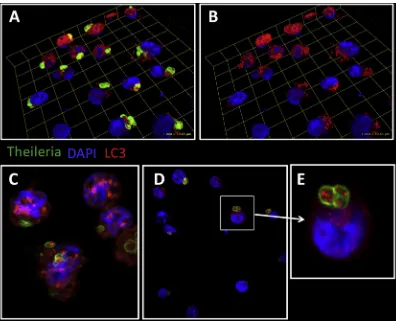
[image:5.595.102.498.64.387.2]macrophages,Theileria annulatamaintains a Hypoxia Inducible Factor (HIF)-1a-driven transcriptional programme typical of Warburg glycolysis[42], a hallmark of cancer cells. Thus, Thei-leriainfection represents a valuable model for studying cancer and lymphoproliferation. In this regard, defects in autophagy have been associated with increased tumorigenesis in some cancers[43], so perhapsTheileria'sregulation of host autophagy is the mechanism underlying parasite survival and leukocyte transformation, events that would not be independent, but closely intertwined, although the exact mechanisms are still unknown.Theileriainduces the oncomiR miR-155, via the c-Jun transcription factor and AP-1 activity [44]. In turn, miR-155 modulates cell autophagy, promoting it to clear intracellular mycobacteria[45], or suppressing it during osteoarthritis and cancer [46,47]. Furthermore, cAMP-PKA signalling is upregu-lated by Theileriainfection [48] and it is well established in different cell types that cAMP-PKA signalling negatively regu-lates autophagy (see accompanying review and[49e52]). cAMP-PKA-mediated phosphorylation of LC3 blocks autophagy. This is mimicked when cAMP production is stimulated by forskolin (an adenylate cyclase activator) and prevented by PKA inhibitor H89. Under conditions of costimulation, LC3 phosphorylation is diminished, leading to autophagy[49]. As augmented cAMP-PKA signaling is characteristic ofTheileria-infected leukocytes, increased LC3 phosphorylation can be observed and this likely contributes to infection, perhaps inducing blockade in the host's autophagy response. It is also well known thatTheileria infec-tion induces increased JNK kinase activities[53,54]. Recently it has been shown that loss of JNK2 leads to accumulation of smARF and lysosomal degradation of the adaptor p62 (seques-tosome-1, SQSTM1)[55]implying that constitutive induction of JNK2 followingTheileria-infection could also contribute to the observed stable levels of p62 that reflect a blockade of the host leukocyte's autophagic response. Consistent with this notion, one can observe a decrease in p62 expression only when infected leukocytes are treated with a pro-oxidant anti-parasite drug (Latre de Late and Pineda, unpublished). Interestingly, when expression of LC3 and phosphorylated LC3 (p-LC3) is evaluated in infected macrophages, a positive staining is also observed within the parasites [Fig. 1]. Whether this is a result of parasite uptake of host LC3 is still unclear and requires further investigation, but it strongly suggests that host and/or parasite autophagy play a relevant role in parasite survival within the infected cell, and perhaps by sequestering host LC3 within itself,
Theileriainhibits autophagy.
In conclusion, to survive, it appears that while exposed within the cytoplasm of host leukcoytes Theileria parasites might modulate the induction of autophagy by targeting at least three different steps in the autophagy induction process: cAMP-PKA-mediated phosphorylation of LC3, JNK2-mediated blockade in p62 degradation, and LC3 sequestration. Under-standing howTheileriaregulates host autophagy will provide novel pathways to improve our current knowledge of auto-phagy both in infection and in cancer.
Conflicts of interest
The authors have no conflict of interest to declare regarding this manuscript.
Acknowledgments
PLdL was supported by a ParaFrap post-doctoral fellowship. MAP is an Arthritis Research UK Career Development Fellow. SB and GL acknowledge support from ANR-11-LABX-0024 and the CNRS; SB acknowledges grant ANR-13-JSV3-0003 and GL acknowledges INSERM support.
r e f e r e n c e s
[1] Tomlins AM, Ben-Rached F, Williams RA, Proto WR, Coppens I, Ruch U, et al.Plasmodium falciparumATG8 implicated in both autophagy and apicoplast formation. Autophagy 2013;9:1540e52.
[2] Voss C, Ehrenman K, Mlambo G, Mishra S, Kumar KA, Sacci Jr JB, et al. Overexpression ofPlasmodium bergheiATG8 by liver forms leads to cumulative defects in organelle dynamics and to generation of noninfectious merozoites. MBio 2016;7:e00682e16.
[3] Shaw MK, Tilney LG. The entry ofTheileria parvamerozoites into bovine erythrocytes occurs by a process similar to sporozoite invasion of lymphocytes. Parasitology 1995;111(Pt 4):455e61. [4] Wang Z, Cabrera M, Yang J, Yuan L, Gupta B, Liang X, et al.
Genome-wide association analysis identifies genetic loci associated with resistance to multiple antimalarials in
Plasmodium falciparumfrom China-Myanmar border. Sci Rep
2016;6:33891.
[5] Gaviria D, Paguio MF, Turnbull LB, Tan A, Siriwardana A, Ghosh D, et al. A process similar to autophagy is associated with cytocidal chloroquine resistance inPlasmodium
falciparum. PLoS One 2013;8:e79059.
[6] Kitamura K, Kishi-Itakura C, Tsuboi T, Sato S, Kita K, Ohta N, et al. Autophagy-related Atg8 localizes to the apicoplast of the human malaria parasitePlasmodium falciparum. PLoS One 2012;7:e42977.
[7] Eickel N, Kaiser G, Prado M, Burda PC, Roelli M, Stanway RR, et al. Features of autophagic cell death inPlasmodium liver-stage parasites. Autophagy 2013;9:568e80.
[8] Raiborg C, Schink KO, Stenmark H. Class III
phosphatidylinositol 3-kinase and its catalytic product PtdIns3P in regulation of endocytic membrane traffic. FEBS J 2013;280:2730e42.
[9] McIntosh MT, Vaid A, Hosgood HD, Vijay J, Bhattacharya A, Sahani MH, et al. Traffic to the malaria parasite food vacuole: a novel pathway involving a phosphatidylinositol 3-phosphate-binding protein. J Biol Chem 2007;282:11499e508.
[10] Cervantes S, Bunnik EM, Saraf A, Conner CM, Escalante A, Sardiu ME, et al. The multifunctional autophagy pathway in the human malaria parasite,Plasmodium falciparum. Autophagy 2014;10:80e92.
[11] Foldvari-Nagy L, Ari E, Csermely P, Korcsmaros T, Vellai T. Starvation-response may not involve Atg1-dependent autophagy induction in non-unikont parasites. Sci Rep 2014;4:5829.
[12] Fader CM, Colombo MI. Multivesicular bodies and autophagy in erythrocyte maturation. Autophagy 2006;2:122e5. [13] Prado M, Eickel N, De Niz M, Heitmann A, Agop-Nersesian C,
Wacker R, et al. Long-term live imaging reveals cytosolic immune responses of host hepatocytes againstPlasmodium infection and parasite escape mechanisms. Autophagy 2015;11:1561e79.
[14] Thieleke-Matos C, Lopes da Silva M, Cabrita-Santos L, Portal MD, Rodrigues IP, Zuzarte-Luis V, et al. Host cell autophagy contributes toPlasmodiumliver development. Cell Microbiol 2016;18:437e50.
[15] Boonhok R, Rachaphaew N, Duangmanee A, Chobson P, Pattaradilokrat S, Utaisincharoen P, et al. LAP-like process as an immune mechanism downstream of IFN-gamma in control of the human malaria Plasmodium vivax liver stage. Proc Natl Acad Sci U S A 2016;113:E3519e28.
[16] Besteiro S, Brooks CF, Striepen B, Dubremetz J-F. Autophagy protein Atg3 is essential for maintaining mitochondrial integrity and for normal intracellular development of
Toxoplasma gondiitachyzoites. PLoS Pathog 2011;7:e1002416.
[17] Ghosh D, Walton JL, Roepe PD, Sinai AP. Autophagy is a cell death mechanism inToxoplasma gondii. Cell Microbiol 2012;14:589e607.
[18] Nguyen HM, El Hajj H, El Hajj R, Tawil N, Berry L, Lebrun M, et al. Toxoplasma gondii autophagy-related protein ATG9 is crucial for the survival of parasites in their host. Cell Microbiol 2017. in press.
[19] Kong-Hap MA, Mouammine A, Daher W, Berry L, Lebrun M, Dubremetz J-F, et al. Regulation of ATG8 membrane association by ATG4 in the parasitic protistToxoplasma
gondii. Autophagy 2013;9:1334e48.
[20] Lavine MD, Arrizabalaga G. Analysis of monensin sensitivity
inToxoplasma gondiireveals autophagy as a mechanism for
drug induced death. PLoS ONE 2012;7:e42107.
[21] Lev^eque MF, Berry L, Cipriano MJ, Nguyen H-M, Striepen B, Besteiro S. Autophagy-related protein ATG8 has a
noncanonical function for apicoplast inheritance in
Toxoplasma gondii. mBio 2015;6:e01446e15.
[22] Lev^eque MF, Nguyen HM, Besteiro S. Repurposing of conserved autophagy-related protein ATG8 in a divergent eukaryote. Commun Integr Biol 2016;9:e1197447.
[23] Gomes Ligia C, Dikic I. Autophagy in antimicrobial immunity. Mol Cell 2014;54:224e33.
[24] Lai S-C, Devenish RJ. LC3-Associated phagocytosis (LAP): connections with host autophagy. Cells 2012;1:396e408. [25] Yarovinsky F. Innate immunity toToxoplasma gondii
infection. Nat Rev Immunol 2014;14:109e21.
[26] Martens S, Parvanova I, Zerrahn J, Griffiths G, Schell G, Reichmann G, et al. Disruption ofToxoplasma gondii parasitophorous vacuoles by the mouse p47-resistance GTPases. PLoS Pathog 2005;1:e24.
[27] Ling YM, Shaw MH, Ayala C, Coppens I, Taylor GA,
Ferguson DJP, et al. Vacuolar and plasma membrane stripping and autophagic elimination ofToxoplasma gondiiin primed effector macrophages. J Exp Med 2006;203:2063e71. [28] Zhao Z, Fux B, Goodwin M, Dunay IR, Strong D, Miller BC,
et al. Autophagosome-independent essential function for the autophagy protein Atg5 in cellular immunity to intracellular pathogens. Cell Host Microbe 2008;4:458e69.
[29] Park S, Choi J, Biering SB, Dominici E, Williams LE, Hwang S. Targeting by AutophaGy proteins (TAG): targeting of IFNG-inducible GTPases to membranes by the LC3 conjugation system of autophagy. Autophagy 2016;12:1153e67.
[30] Choi J, Biering SB, Hwang S. Quo vadis? Interferon-inducible GTPases go to their target membranes via the LC3-conjugation system of autophagy. Small GTPases 2016:1e9. [31] Ohshima J, Lee Y, Sasai M, Saitoh T, Su Ma J, Kamiyama N, et al. Role of mouse and human autophagy proteins in IFN-gamma-induced cell-autonomous responses against
Toxoplasma gondii. J Immunol 2014;192:3328e35.
[32] Selleck EM, Orchard RC, Lassen KG, Beatty WL, Xavier RJ, Levine B, et al. A noncanonical autophagy Pathway restricts
Toxoplasma gondiigrowth in a strain-specific manner in
IFN-g-activated human cells. mBio 2015;6:e01157e15.
[33] Andrade RM, Wessendarp M, Gubbels M-J, Striepen B, Subauste CS. CD40 induces macrophage anti-Toxoplasma
gondiiactivity by triggering autophagy-dependent fusion of
pathogen-containing vacuoles and lysosomes. J Clin Invest 2006;116:2366e77.
[34] Liu E, Van Grol J, Subauste CS. Atg5 but not Atg7 in dendritic cells enhances IL-2 and IFN-gproduction byToxoplasma gondii-reactive CD4þT cells. Microbes Infect 2015;17:275e84. [35] Liu E, Lopez Corcino Y, Portillo J-AC, Miao Y, Subauste CS.
Identification of signaling pathways by which CD40 stimulates autophagy and antimicrobial activity against
Toxoplasma gondiiin macrophages. Infect Immun
2016;84:2616e26.
[36] Van Grol J, Muniz-Feliciano L, Portillo JAC, Bonilha VL, Subauste CS. CD40 induces anti-Toxoplasma gondiiactivity in nonhematopoietic cells dependent on autophagy proteins. Infect Immun 2013;81:2002e11.
[37] Muniz-Feliciano L, Van Grol J, Portillo J-AC, Liew L, Liu B, Carlin CR, et al.Toxoplasma gondii-induced activation of EGFR prevents autophagy protein-mediated killing of the parasite. PLoS Pathog 2013;9:e1003809.
[38] Wang Y, Weiss LM, Orlofsky A. Host cell autophagy is induced byToxoplasma gondiiand contributes to parasite growth. J Biol Chem 2008;284:1694e701.
[39] Dobbelaere D, Heussler V. Transformation of leukocytes by
Theileria parvaandT. annulata. Annu Rev Microbiol
1999;53:1e42.
[40] Shaw MK. Cell invasion byTheileriasporozoites. Trends Parasitol 2003;19:2e6.
[41] Duszenko M, Ginger ML, Brennand A, Gualdron-Lopez M, Colombo MI, Coombs GH, et al. Autophagy in protists. Autophagy 2011;7:127e58.
[42] Metheni M, Echebli N, Chaussepied M, Ransy C, Chereau C, Jensen K, et al. The level of H(2)O(2) type oxidative stress regulates virulence ofTheileria-transformed leukocytes. Cell Microbiol 2014;16:269e79.
[43] Maes H, Rubio N, Garg AD, Agostinis P. Autophagy: shaping the tumor microenvironment and therapeutic response. Trends Mol Med 2013;19:428e46.
[44] Marsolier J, Pineau S, Medjkane S, Perichon M, Yin Q, Flemington E, et al. OncomiR addiction is generated by a miR-155 feedback loop inTheileria-transformed leukocytes. PLoS Pathog 2013;9:e1003222.
[45] Wang J, Yang K, Zhou L, Wu M, Wu Y, Zhu M, et al. MicroRNA-155 promotes autophagy to eliminate
intracellular mycobacteria by targeting Rheb. PLoS Pathog 2013;9:e1003697.
[46] D'Adamo S, Alvarez-Garcia O, Muramatsu Y, Flamigni F, Lotz MK. MicroRNA-155 suppresses autophagy in chondrocytes by modulating expression of autophagy proteins. Osteoarthr Cartil 2016;24:1082e91.
[47] Chen L, Jiang K, Jiang H, Wei P. miR-155 mediates drug resistance in osteosarcoma cells via inducing autophagy. Exp Ther Med 2014;8:527e32.
[48] Guergnon J, Dessauge F, Traincard F, Cayla X, Rebollo A, Bost PE, et al. A PKA survival pathway inhibited by DPT-PKI, a new specific cell permeable PKA inhibitor, is induced byT.
annulatain parasitized B-lymphocytes. Apoptosis
2006;11:1263e73.
[49] Cherra 3rd SJ, Kulich SM, Uechi G, Balasubramani M, Mountzouris J, Day BW, et al. Regulation of the autophagy protein LC3 by phosphorylation. J Cell Biol 2010;190:533e9.
[50] Chang YW, Howard SC, Herman PK. The Ras/PKA signaling pathway directly targets the Srb9 protein, a component of the general RNA polymerase II transcription apparatus. Mol cell 2004;15:107e16.
[51] Budovskaya YV, Stephan JS, Deminoff SJ, Herman PK. An evolutionary proteomics approach identifies substrates of the cAMP-dependent protein kinase. Proc Natl Acad Sci U. S. A 2005;102:13933e8.
[53] Galley Y, Hagens G, Glaser I, Davis W, Eichhorn M, Dobbelaere D. Jun NH2-terminal kinase is constitutively activated in T cells transformed by the intracellular parasite
Theileria parva. Proc Natl Acad Sci USA 1997;94:5119e24.
[54] Chaussepied M, Lallemand D, Moreau MF, Adamson R, Hall R, Langsley G. Upregulation of Jun and Fos family members and permanent JNK activity lead to constitutive AP-1 activation inTheileria-transformed leukocytes. Mol Biochem Parasitol 1998;94:215e26.
[55] Zhang Q, Kuang H, Chen C, Yan J, Do-Umehara HC, Liu XY, et al. The kinase Jnk2 promotes stress-induced mitophagy by targeting the small mitochondrial form of the tumor suppressor ARF for degradation. Nat Immunol 2015;16:458e66.
[56] Shiels BR, McDougall C, Tait A, Brown CG. Identification of infection-associated antigens inTheileria annulata
transformed cells. Parasite Immunol 1986;8:69e77.