Massey University Library
L
New Zealand&
Pacific CollectionGENETIC DIVERSITY OF
DOTHISTROMA PINI
IN NEW ZEALAND
This thesis is presented in fulfilment of the requirements for the degree of Master of Science in Molecular Genetics
at Massey University, Palmerston North New Zealand.
Paul Hirst
ABSTRACT
Dothistroma pini is an important pathogen of Pinus radiata, New Zealand's major exotic forest species. This study was undertaken to elucidate the genetic background of New Zealand's D. pini population as part of a research program which aims to reduce its overall effect.
Two maJor sampling strategies were devised and undertaken. The first involved collection from within an NZFRI field trial in which five year old host clones were available that had been scored for resistance to D. pini over a period of three years. This collection was designed to test the hypothesis that genetic differences would be seen between "resistant" and "susceptible" hosts.
The second collection tested the hypothesis that polymorphisms would be observed between samples from geographically isolated regions, and that more variability would be seen between these regions than within any of them. For this study, samples were collected in a "hierarchy of populations" approach from three New Zealand forests: Kinleith, Kaiangaroa and Golden Downs. Additional samples for analysis included four D. pini samples which were isolated during the l 960's, and DNA obtained from a Guatemalan isolate of the teleomorphic form, Mycosphaerella pini.
PCR amplification using 32 RAPD and 5 RAMS primers revealed no polymorphisms within two sets of five D. pini samples designed to be representative of the New Zealand population. Amplification was repeated with a larger number of D. pini samples using five RAPD and two RAMS primers, again showing no differences between any of the isolates and proving that the two sets of five samples were indeed representative of the population. However, differences were seen between D. pini and the isolate of M pini with all primers used.
lll Growth studies supported the molecular data, showing no differences between the isolates of D. pini. Morphological differences between D. pini laboratory cultures were observed, but these do not appear to correlate with any permanent genetic rearrangement.
ACKNO\VLEDGEMENTS
I would like to express deep gratitude to my supervisor, Rosie Bradshaw, for her never ending support, supervision and advice, and for being a cheery and lovely person.
I am indeed grateful to the Department of Microbiology and Genetics for three years employment as a Graduate Assistant and use of resources.
Thanks to everyone in The Department for help, advice, distractions and friendship given over the years. Anita, Austen, Brendon, Carmel, Carolyn, Chris, Dianne, Karen, Kirsty, Laura, Linda, Mike, Neville, Pete, Peng, Rich, Shalome, Sheralee, Tania, Trish ... too many, I'm bound to miss someone out -you should know who you are.
Scientifically speaking, thanks to: Barry Scott for primers, plasmids and freezers; Tash and Seth for their help with single sporing and growth rate experiments; Shalome for CsCl gradient mtDNA isolation and theological discussions.
Thanks to the NZFRI in Rotorua, without whose facilities it would've taken so much longer. Non-Massey scientific folks whose assistance has been invaluable, and is much appreciated, include (but are not limited to) Sue Carsons, Tom Richardson, John Lee, Luis Gea, Margaret Dick, Alan Goldstein, Raoul Thomas, Paul Bradbury.
Cheers to all my friends and flatmates for providing much needed release of tension in various ways throughout the mission. Promise I'll put less on my plate from now on. Special thanks to Carolyn, Elise, Amanda, Tania and Bannoy.
v
TABLE OF CONTENTS
ABSTRACT 11
ACKNOWLEDGEMENTS IV
CONTENTS v
LIST OF FIGURES
x
LIST OF TABLES XI
CHAPTER 1 INTRODUCTION.
1.1 OVERVIEW
1.2 BIOLOGY AND INFECTION OF DOTHJSTROMA PINI. 2
1.2. l Dothistromin. 3
1.2.2 Host-pathogen Interactions. 4
1.2.3 Occurrence of Dothistroma pini. 10
1.2.4 Overcoming Dothistroma Blight. 11
1.3 DETECTION OF FUNGAL INTRASPECIFIC POLYMORPHISMS 13 1.3.l Sequencing oflntergenic Transcribed Spacer Regions in the
Ribosomal RNA Cluster 14
1.3.2 RFLP Analysis of the Ribosomal RNA Cluster 18
1.3.3 RFLP Analysis of Mitochondrial DNA 19
1.3.4 RAPD Analysis 20
1.3.5 Random Amplification of Microsatellites 21
CHAPTER 2 MATERIALS AND METHODS
2.1
2.2
2.3
2.4
FUNGAL AND BACTERIAL STRAINS, AND PLASMIDS
GROWTH MEDIA
2.2.1 Dothistroma Media
2.2.2 Dothistroma Sporulation Media 2.2.3 LB Media
BUFFERS AND SOLUTIONS 2.3.1 1 x T AE Buffer
2.3.2 1 x TBE Buffer 2.3.3 TE Buffer 2.3.4 Gel Loading Dye 2.3.5 DNA Extraction buffer 2.3.6 Tris-Equilibrated Phenol 2.3.7 20 x SSC
2.3.8 TES
2.3.9 Hybridisation Buffer 2.3.10 10 x Taq PCR Buffer
2.3.11 2 x Binding and Washing Buffer 2.3 .12 Acrylamide Mix
2.3.13 10 x Sequencing TBE Buffer
CUL TURING TECHNIQUES
2.4.1 Isolation of Dothistroma pini from Pinus radiata needles 2.4.1.1 Surface sterilisation
2.4.1.2 Humidity Chamber 2.4.1.3 Antibiotic Selection 2.4.2 Growth of cultures
2.4.3 Storage
2.4.4 Pure Culture Isolation
2.5 2.6 2.7 2.8 2.9 DNA ISOLATION
2.5.1 Plasmid Preparation
2.5.2 Genomic DNA Preparation
DNA QUANTIFICATION
2.6.1 Spectrophotometric Determination 2.6.2 Gel Electrophoresis Determination
AGAROSE GEL ELECTROPHORESIS
RESTRICTION ENDONUCLEASE DIGESTION OF DNA
SOUTHERN BLOTTING 35 35 35 37 37 37 37 38 38
2.9.1 Capillary Blotting 38
2.9.2 Preparation of Radioactive Probe 39
2.9.3 Hybridisation and Detection of Probe DNA on Southern Blots 40
2.10 POLYMERASE CHAIN REACTION 2.10.1 rDNA Primers
40 40 43 44 2.10.2 RAPD Primers
2.10.3 Microsatellite Primers
2.11 DNA SEQUENCING 47
2.11.1 fmol DNA Cycle Sequencing Protocol 47
2.11.2 Sequenase Protocol 47
2.11.2.1 Biotinylated PCR Product Strand Separation 48 2.11.2.2 Sequenase Version 2.0 Sequencing Protocol 48 2.11.3 Polyacrylamide Gel Electrophoresis of Sequencing Products 49
2.12 SAMPLING 2.12.1 Field Trial 2.12.2 National Survey 2.12.3 Others
2.13 GROWTH RA TE ANALYSIS
CHAPTER 3 RESULTS
3.1
3.2
3.3
3.4
ISOLATION OF D. PINI FROM P. RADIATA NEEDLES 3.1.1 D. pini Isolation Techniques
3 .1.1.1 Surface Sterilisation 3.1.1.2 Humidity Chamber 3 .1.1.3 Antibiotic Selection 3.1.2 Sampling
3.1.2.1 Field Trial 3.1.2.2 National Survey
MYCOLOGICAL EXAMINATION
RFLP ANALYSIS 3.3.1 Ribosomal DNA 3.3.2 Mitochondrial DNA
SEQUENCE ANALYSIS
3.4. l l 8S rRNA Fragment Sequencing 3.4.2 ITS 1 Sequencing
3.5 PCR ANALYSIS
3.5.1 RAPD Profiles
3.5.1.1 RAPD Optimisation
3.5.1.2 Selection of Primers
3.5.1.3 RAPD Profiles of Five Representative
New Zealand Samples
3.5.1.4 RAPD Profiles of More D. pini Samples and an
M pini Isolate 3.5.2 RAMS Profiles
3.6 GROWTH STUDY
CHAPTER 4 DISCUSSION
4.1
4.2
VARIATION IN NEW ZEALAND'S D. PINI POPULATION 4.1.1 RAPD's and RAMS
4.1.2 Southern Blotting
4.1.3 Growth Comparisons
IMPLICATIONS OF A SINGLE STRAIN IN NEW ZEALAND
4.3 IMPLICATIONS ON THE BREEDING PROGRAM FOR
DOTHISTROMA RESISTANT P. RADIATA
4.4 FUTURE WORK
CHAPTER 5 SUMMARY
APPENDICES
Appendix 1 Partial Sequence of D. pini ITS 1 Region
Appendix 2 Growth Study Statistical Analysis
x
LIST OF FIGURES
Figure 1.1 Ribosomal DNA Repeat Unit 15
Figure 2.1 Universal Ribosomal RNA PCR Primers for Fungi 41
Figure 3.1 Southern Blot Probed with Heterologous Ribosomal DNA 62
Figure 3.2 Southern Blot Probed with D. pini Mitochondrial DNA 65
Figure 3.3 PCR Amplification Products from the Ribosomal RNA Cluster 68
Figure 3.4 Partial Sequence of D. pini 18s rRNA Subunit 70 Figure 3.5 RAPD Amplification of Set B Samples Using Primer OPA-8 77
Figure 3.6 RAPD Amplification of Set B Samples Using Primer OPB-4 80
Figure 3.7 RAPD Amplification of Set B Samples Using Primer OPC-5 82
Figure 3.8 RAPD Amplification of Set B Samples Using Primer OPC-15 84
Figure 3.9 RAPD Amplification of Set B Samples Using Primer OPF-6 86
Figure 3.10 RAPD Amplification of 30 Samples Using Primer OPD-3 90
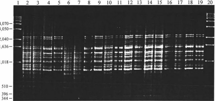
Figure 3.11 RAPD Amplification of 71 Samples Using Primer OPC-5 96
Figure3.12 Amplification of 30 samples using the M 13core Primer 106
XI
LIST OFT ABLES
Table 2.1 Strains and plasmids
26
Table 2.2 Primers Used For RAPD Analysis
45
Table 2.3 Primers Used for Microsatellite Analysis
46
Table 2.4 Field Trial P. radiata Progeny from which D. pini Samples
were Collected
51
Table 2.5 Sites from which D. pini Samples were Collected
54
Table 3.1 RAPD Primers Screened Using Set A and Set B D. pini Samples
75
Table 3.2 Microsatellite Primers Screened Using Set B D. pini Samples
105
CHAPTER 1 INTRODUCTION
1.1 OVERVIEW
The filamentous fungus Dothistroma pini Hulbary is a foliar pathogen of Pinus radiata
and other pine species. Infection leads to premature needle cast, thereby reducing
photosynthesis and wood production. As P. radiata is the predominant forest species in
this country, the control of D. pini infection is of great economic importance to New Zealand.
Current work in breeding Dothistroma resistant (DR) trees has the potential to result in
the selection of more virulent isolates within the pathogen population. Because it is
economically favourable to plant a limited number of pine types in a plantation,
resistance in P. radiata could be overcome if more virulent strains of D. pini evolve; a
distinct possibility considering the fungal life-cycle is a lot shorter than that of its host.
For these reasons, it is important to have an idea of the level of genetic diversity of the
D. pini population within New Zealand.
Current molecular techniques enable the genetic variability of New Zealand's D. pini
populaton to be ascertained. Using this insight from an evolutionary standpoint, a
correlation will be able to be made with the likelihood of a highly pathogenic strain
evolving (Adachi et al. 1993). Knowledge of the genetic background of D. pini in
relation to its geographical distribution will also assist in devising strategies for
deploying genetically improved P. radiata.
It is unclear at this stage whether any sexual isolates are present in New Zealand.
Although no sexual spores have been observed, more studies are required to elucidate
whether there are only asexual forms, only one mating type is here, or whether sexual
fom1s are here but have not been observed. If it is not yet present in New Zealand, the
introduction of the sexual form (Mycosphaere/la pini), or the required mating type for
2
profile of asexual and sexual forms, a successful strategy for monitoring D. pini infection sites could be devised to ensure the exclusion of teleomorphs of D. pini in New Zealand.
1.2 BIOLOGY AND INFECTION OF DOTHISTROMA PINI.
Dothistroma pini (a.k.a. Dothistroma septospora) is of the order Dothideales in the Ascomycotina class. It is the anamorphic (asexual) form of Mycosphaerella pini (a.k.a. Scirrhia pini). Differences in pathogenicity between the two forms have not been reported in any of the wide number of pine species that they infect (Evans 1984).
D. pini is a necrotrophic pathogen which is believed to kill plant tissue and then live saprophytically. Tree infection occurs when D. pini conidia land on the needle surface, and germ tubes grow towards stomata (Peterson and Walla 1978). This growth response in D. pini may be similar to that of Uromyces viciae-fabae which is dependent on pH gradients (Edwards and Bowling 1986). The conidial germ tubes take up to three days to penetrate the stoma, followed by both inter- and intracellular hyphal growth (Gadgil 1967). Within the needle, disruption of the mesophyll tissue occurs well in advance of the developing hyphae as a result of the pathogen toxin, dothistromin. Needle infections are initially seen as yellow areas, developing into characteristic red banding and the formation of a necrotic lesion. Necrosis can continue throughout the needle as the hyphae extend, followed by needle cast.
Infection initially appears as necrosis of the needles on the main stem, and at the base of lower branches, but more severe defoliation occurs under favourable conditions, resulting in tree death in the most extreme cases (Gallagher 1971; Franich et al. 1982; Philips and Burdekin 1983; Gadgil 1984).
3
Asexual conidiospores are multicellular, and it is unknown whether each cell is derived from the same mother cell. Multiple germ tubes can be produced from these spores, up to one per compartment. Sexual wind dispersed spores (in M. pini) are also formed in stromata, but these have never been observed in New Zealand (P. Gadgil. Pers. Comm.) and are never seen in culture (D. Morrison. Pers. Comm).
D. pini is sensitive to environmental conditions, with moisture required for rain splash dispersal of asexual spores (usually within a single tree, although wider distribution occurs when mist clouds are present). Studies to ascertain environmental effect on severity of dothistroma infection show duration of leaf wetness (> 10 hours) to be a crucial factor, athough germination may still occur without water in humidity over 96% (Gadgil 1977). Light is necessary for infection, while temperature is not crucial although stromata appear sooner as optimal conditions ( l 6- l 8°C) are reached (Gadgil 1974). Infection in P. radiata requires 100 conidia/mm2, with mature trees susceptible to infection if the inoculum is high enough. Soil deficiencies in boron and sulphur have also been implicated in disease susceptibility (Ades and Simpson 1991).
1.2.1 Dothistromin
Dothistromin is a red pigmented toxin produced by D. pini, found at high levels in infected needles. It is a difuroanthraquinone which bears structural similarity to the carcinogenic aflatoxins produced by Aspergillus species, sharing many of the same biochemical precursors (Gallagher 1971 ).
4
sufficient to elicit an important plant defence response, <Uld that it plays an integral role in pathogenesis.
Hyphae are not observed in the dark green region (0.1 - 0.5 mm) adjacent to the necrotic region. of infected needles (Gadgil 1967). Benzoic acid bound to lignin polymers is found in this region in disproportionately high amounts, suggesting that the benzoic acid response prevents hyphal extension within the needle. This leads to the proposal that benzoic acid is a phytoalexin which is induced by D. pini invasion and is associated with lesion formation and consequent blight symptoms (Franich et al. 1986).
Phytoalexins are antimicrobial compounds produced by the plant in response to infection (reviewed by Kuc et al. 1976; Cruikshank 1977; Stoessl 1983) which act by inhibiting spore formation, germ tube elongation and mycelial growth of fungal pathogens. However, phytoalexins can also be toxic to plant cells (reviewed by Mansfield 1982; Smith 1982) as seen in P. radiata needles with long lesions which, while restricting infection by creating a highly fungistatic environment, can die if benzoic acid levels are too high and the lesion is not localised.
1.2.2 Host-pathogen Interactions
Resistance of plants to pathogen infection relies on many different mechanisms, the nature of the plant-fungus interaction determining which of these are employed. The host-pathogen interaction can be either compatible (the host is susceptible: the fungus is a virulent pathogen) or incompatible (the host is resistant and/or the fungus is a non-pathogen or an avirulent non-pathogen). A non-non-pathogen is a species incapable of infecting the host. An avirulent pathogen is a member of a species which is unable to infect a host that other members of its species can. A virulent pathogen induces a slow defence response, whereas an avirulent pathogen will induce a rapid one.
5
pathogen, or postinfectional mechanisms that occur near pathogen infection sites. These
mechanisms include structural responses that modify the host cell wall and the synthesis
of defense compounds which act directly against the pathogen or catalyse the synthesis
of antimicrobial compounds. These compounds may elicit hypersensitive responses,
phytoalexin production and the disruption of biochemical pathways. Enhanced protein
(Bowles 1990) and phenolic production via the shikimic acid pathway (Adaskaveg
1992) are responses to infection common to most plants. The precise nature of many of
these proposed mechanisms of resistance is unknown as most studies have been
correlative, so more work is required to elucidate the biochemical nature of many
host-pathogen interactions (Adaskaveg 1992).
The hypersensitive response (HR) is one result of incompatible interactions (Tomiyama
et al. 1979; Bowles 1990). It is a widespread mechanism among plants, involving a
rapid but limited necrosis of a limited number of plant cells at the site of interaction
(Adaskaveg 1992). HR in plants is thought to be orchestrated by an oxidative burst in
response to molecular recognition, specified by avirulence gene interactions (Lamb et al.
1994) as described by Flor's gene-for-gene hypothesis ( 1946). This mechanism involves
rapid H202 accumulation, resulting in the creation of antimicrobial conditions, both
directly and indirectly. Incompatible pathogens and fungal elicitors have been shown to
specifically increase H202 concentration, a large burst occuring 2-5 hours after
infection. The effects of this burst include the crosslinking of cell wall proteins,
apoptosis, the activation of HR genes, and the creation of a generally antimicrobial
environment (Lamb et al. 1994 ). H202 has been shown to have different effects at
various concentrations, causing apoptosis at high levels; and protecting plant cells at
low levels by inducing glutathione-S-transferase (GST) production, thereby detoxifying
lipid peroxides.
Biotrophic pathogens make up the majority of those involved in incompatible reactions.
The gene-for-gene hypothesis refers to these interactions, and states that for each gene
conferring race specific resistance in the host plant, there is a specific gene which
confers avirulence in the pathogen (Flor 1946). The latter genes encode incompatibility
6
example, disruption of the race-specific incompatibility gene avr9 in Cladosporium
fulvum results in loss of the HR in the host tomato plant, leading to virulence of the
fungus. A C. fulvum avirulence gene cloned into a virulent race resulted in avirulence,
leading to the proposal that avirulence gene products are in some way involved in the
initial stages of signal transduction, perhaps by binding to a host race-specific receptor
(De Wit et al. 1992).
Compatible interactions between host and pathogen can evoke responses similar to
hypersensitive responses, induced by cell death rather than elicitors. Following
pathogen invasion, apoptosis in the host results in oxidative burst and the release of cell
components. These effects have been related to the synthesis of phytoalexins,
phenolics, lignin and callose - factors which inhibit microbial growth and result in
limited plant resistance (Heath 1976; Ingham 1978; Bowles 1990). Effects such as rapid
oxidative cross-linking have been implicated in plant interactions with non-pathogens (Lamb et al. 1994 ), but although hypersensitive-like reactions can result from general infection as well as specific disease resistance, it is a crucial response in limiting the spread of disease.
Systemic acquired resistance (SAR) is a broad spectrum resistance triggered throughout
plants by salicylic acid production following HR. SAR results in increased resistance to further infection by pathogens. Both HR and SAR are associated with the synthesis of host encoded pathogenesis related products - a general defense against pathogens. Disease resistance is due at least in part to the accumulation of pathogenesis related
(PR) proteins. Salicylic acid is a signal produced by infected regions of the plant in
response to pathogen, which indicate to uninfected regions that induction of the SAR
response is required. Salicylic acid has been observed to induce the same PR proteins as
are observed after pathogenesis (Ward et al. 1991), and to induce the same broad
spectrum resistance as SAR (Kessmann et al. 1994). Although salicylic acid plays a
necessary role
in
signal transduction leading to SAR, it is not thought to be the long7
In compatible interactions, salicylic acid is also seen to play an important role in disease restriction, as plants with genes producing a salicylic acid degrading enzyme showed
considerably worse symptoms than wild type (Delaney et al. 1994).
Although the defence responses of gymnosperms are not as well characterised as those
in angiosperms, some studies have been done. For example, the inaccessible location of
stomata in resistant pine needles has been proposed as a morphological barrier, as the
position directly affects the success of Cronartium ribicola infection (Spaulding 1925).
Although no such correlation of P. radiata resistance to D. pini infection has been noted
(Peterson and Walla 1978), increased resistance has been observed in older P. radiata trees. Several factors have been proposed to be involved in this apparent resistance.
The presence of wax occlusions in the stomata! pores of older trees has been proposed
to be partly responsible for this effect (Franich et al. 1977). Changes in the composition
of resin acids present in these occlusions have also been implicated, indicating that D.
pini is incapable of metabolising the acids produced by older trees (Franich et al. 1978).
The production of volatile, monoterpene hydrocarbons also appears to have some effect
on tree resistance to D. pini. Twice the amount of these compounds are exuded through
the needle cuticle in young trees, compared to older ones (Franich et al. 1982). Thirteen
different hydrocarbons were observ~d, mixtures of which stimulated pathogen growth,
inhibiting only at concentrations of 1000 ppm or greater (Franich et al. 1983 ). The
"resistance" of mature trees to D. pini has also been suggested to simply be an effect of
the altered microclimate for young needles as trees are pruned at 8-9 years, affecting
conditions so that they are no longer warm, damp and wind-free (P. Debnam. Pers.
Comm.).
Constitutive biochemical compounds found within host tissue have also been implicated
as barriers to microbial infection. For example, the wax fraction of the cuticle in P.
radiata contains fatty and resin acids which are highly fungistatic to D. pini (Franich et
al. 1983). These acids inhibit spore germination and mycelial growth in vitro, and when
8
double that of untreated plants. These compounds have thus been proposed as
preinfectional barriers which contribute to mature tree resistance.
Cronartium ribicola (white pme blister rust) infection is limited by postinfectional
mechanisms in various pine hosts. Foliar lesions have been observed in P. ar111a11dii,
and P. 111011ticola (Hoff and McDonald 1975), while pathogen entry into P. 111011ticola
stem tissue is limited by HR-mediated premature needle shed and a necrotic reaction at
the needle fascicle (Hoff and McDonald 1971). A classical gene-for-gene type HR
occurs in P. lambertiana needle tissue infected with C. ribicola (Kinloch and Littlefield
1977), resistant hosts showing a classical hypersensitive response (small, barely visible
lesions) controlled by a single dominant gene which results in only a small area of tissue
death.
The infection of P. radiata by D. pini appears to be a compatible reaction. Although the
pathogen does not spread far due to the hypersensitive-like response of benzoic acid
production, mycelial penetration is greater than expected in an incompatibility system.
In fact, the pathogen can survive and grow for long enough to produce stroma and
conidiospores. Benzoic acid does not restrict growth to as small an area as seen in a classical HR situation, and the reaction is also a slow response, typical of a compatible
reaction. Benzoic acid is considered an inducible mechanism of resistance involved in
the direct inhibition of the pathogen, as well as being directly involved in lignin
accumulation in tissue adjacent to the infected areas (Franich et al. 1986). Lignification
is a common defence mechanism in dicotyledonous plants, however, in gymnosperm
foliage it results in the cessation of metabolic activity (Adaskaveg 1992), and no work
has been reported as to its role as a preinfectional barrier to pathogen infection. In
addition, since benzoic acid is a precursor of salicylic acid, it may be involved with the
•
induction of SAR, as well as being a late HR-like response. The induced production of benzoic acid has also been implicated as a factor that may involve resistance in other
plants (Adaskaveg 1992). For example, in studies of resistance of apple fruit to
infection by Nectria galligena, Brown and Swinburne (1973) found that benzoic acid
9
produce 2-3 times more of the compound than susceptible cultivars (Noble and Drysdale 1983).
Dothistromin does, however, have some features of an incompatibility factor capable of eliciting a hypersensitive response from the host. It is possible that dothistromin acts as an incompatibility factor (eg. eliciting benzoic acid production), in which case D. pini may become more virulent without it. Alternatively, dothistromin may be necessary for infection, as in the case of Cochliobolus heterostrophus, which can only infect oats if capable of victorin production (S. Briggs. Pers. Comm.). A toxin deficient mutant is currently being developed which will be invaluable in addressing this question.
Likewise in Pseudomonas syringae, which causes halo blight in the common bean, phaseolotoxin deficient mutants have been constructed which multiply at a normal rate in leaves, producing typical lesions. However, these mutants do not cause chlorotic halos or systemic chlorosis, and are unable to produce systemic infections (De La Fuente et al. 1992).
10
1.2.3 Occurrence of Dothistroma pini
D. pini infects a wide range of pine species, with most hard pines being particularly susceptible. For example, P. ponderosa, P. nigra and P. attenuata (nobcone) are extremely susceptible for their entire life, while P. radiata appears to exhibit increased resistance with age (Gadgil 1984).
P. radiata is the most economically important pine species susceptible to D. pini, its natural habitat being North and Central America where it is found in five small separate areas along the western coast: Ano Neuvo Point, Monterey, and Cambia on the mainland, and Guadalupe and Cedros Islands (7284 ha. total). One of New Zealand's
first P. radiata plantations was at Mount Peel Station in South Canterbury in 1859, and since then it has been planted in many locations in New Zealand, growing at a rapid rate (Boyd 1992).
D. pini and M. pini are widely distributed in South America, North America, Europe,
Africa and Australasia (Evans 1984 ). The species are thought to be indigenous to Central America and western North America (Evans 1984), but have never been observed in natural Californian stands. D. pini was first identified in New Zealand in 1964 (Gadgil 1967), and is thought to have been introduced to central North Island forests by forestry officials who had visited East Africa to observe the fungus in 1957.
D. pini is a significant pathogen of young radiata pine (2-15 years), which comprise
I I
Although sexual forms have been reported in other countries, no mating has been
observed in New Zealand isolates of D. pini, suggesting that compatible mating forms
are absent (anamorphic), or that only one mating type is present and the fungus is
obligately heterothallic. However, mating tests carried out at the FRI (P. Gadgil. Pers.
Comm.) have only been performed in culture and it has since been reported that mating
and sexual sporulation are only been observed in planta (D. Morrison, Pers. Comm.). It
is therefore possible that teleomorphic forms are present in New Zealand and that
further work is required to identify them.
1.2.4 Overcoming Blight
Forestry has been predicted to provide 25% of New Zealand's exports by the year 2000,
and over 90% of our 1,330,000 ha. forests are P. radiata (L. Bulman. Pers. Comm.).
Although P. radiata in New Zealand has few diseases, D. pini blight (the most
significant disease) has been estimated to cost over seven million dollars per year (L.
Bulman. Pers. Comm.). If unchecked, it causes wood volume loss directly proportional
to the severity of infection (Carson, S.D. et al. 1991 ), so control of the disease is
obviously important.
Dothistroma blight is currently kept under control by the aerial application of copper
fungicide, a program mounted by Kaiangaroa Forestry Management in the late 1960's
after D. pini forced the clear felling of severely infected Corsican and ponderosa pines
(Boyd 1992). The copper oxide fungicide reacts with aqueous exudates on P. radiata needles to form free or complexed Cu2+ in solution, at concentrations sufficient to
inhibit D. pini germination (Franich 1988). Plantations of under 15 years are aerially
assessed for foliage damage every two to three years and treated when necessary (Dick
1989).
Although copper-based fungicide treatment is effective and the development of more
efficient application methods have led to a reduction in treatment costs (from over
$60/ha to under $15/ha in the twenty years to 1988 (New 1989)), it is still expensive and
12
Trees with natural resistance to dothistroma infection have been observed which
correlate with the formation of longer lesions upon infection (Gallagher 1971 ). Plant
breeding programs have been operating at the FRI in Rotorua for 15 years to develop
DR (Dothistroma Resistant) P. radiata for growth in high risk sites. There has been
considerable success in developing trees with increased resistance to D. pini, with the
most resistant seedlots expected to have a reduced mean stand infection of 15% (Carson,
S.D. et al. 1991 ). This resistance, however, will only be effective for as long as D. pini
maintains its current levels of virulence. Given that the pathogen has a far shorter life cycle than its host, it is possible that strains could evolve which are capable of
overcoming current plant resistance mechanisms. There will be no opportunity for
naturally occurring resistance mechanisms to evolve in the P. radiata population, as the
breeding and planting strategies used are artificial, not relying on evolution for genetic
improvement. However, progeny testing and appropriate deployment strategies could
be used to manage the pathogen population so that current levels of pathogenicity are
maintained. Similar tree breeding programs have been undertaken in pine species for
resistance to Cronartium quercuum f.sp. fusiforme, taking into account the potential for
pathogen adaptation to resistant phenotypes, by analy~ing genetic variability in the
pathogen population (Snow et al. 1976).
A further cause for concern is the current lack of knowledge about the mechanisms by
which DR P. radiata achieve increased resistance. Recent studies of susceptible and
resistant populations showed no correlation between susceptibility and the amount of
dothistromin found in needles (S. Carson. Pers. Comm.), hence the primary effect of
resistance may not be the restriction of toxin from the needle.
The New Zealand P. radiata population is derived from three separate Californian
populations and genetic variability is currently high. However, it is economically
favourable to have lower genetic variation (monoclonal plantations). A reduction in
genetic variation would mean that if a more virulent D. pini strain evolved, or was
introduced, the entire host population may well be destroyed. A compromise of profit
and safety from pathogen attack is being suggested with a strategy involving planting
New Zealand forests with at least fifteen different P. radiata clones in order to retain
13
1.3 DETECTION OF FUNGAL INTRASPECIFIC POLYMORPHISMS
When examining phylogenetic relationships, it is important to select the appropriate
techniques for studying the level of divergence of interest, and also to consider what
techniques are available to analyse the various types of molecular data generated. All
known D. pini isolates within New Zealand are thought to be asexual, so there is no
mechanism for sexual recombination to shuffle marker genes (although parasexual
recombination is a possibility). Divergence is also expected to be relatively low because
of D. pini's recent introduction to New Zealand, with a probable narrow genetic base
due to the small number of introduction sites.
However, several factors which increase genetic variation should be considered, as they
may help create a more genetically diverse D. pini population than initially expected.
Fungal populations have been observed which contain a large amount of chromosomal
polymorphism, inversely correlated with meiosis, meaning that such polymorphism is
expected to occur more frequently in imperfect fungi such as D. pini (Kistler and Miao
1992). Although it has been noted that many fungal phytopathogens are anamorphic in
nature, rapid evolution is still observed in some fungi, as seen by changes in virulence
(Michelmore and Hulbert 1987). Evidence of morphology changes reminiscent of
phenotypic switching has been observed in D. pini in culture, which could be explained
by changes such as loss of nuclei in heterokaryons, extrachromosomal elements or
chromosomal rearrangements. This type of switching has also been observed in other
fungi, associated with chromosome variation (Rustchenko-Bulgac et al. 1990).
Many molecular tools are available to analyse the New Zealand D. pini population.
Techniques routinely used to detect intraspecific variation are sequence analysis,
analysis of restriction fragment length polymorphisms (RFLPs), randomly amplified
polymorphic DNA (RAPD) and randomly amplified microsatellites (RAMS) . Each of
these techniques has their own advantages and drawbacks, and all have been used
extensively in intraspecific studies. A comparison of the use of these techniques in
other fungi is of great interest, in order to see which are most successful at detecting
14
1.3.1 Sequence Variation in the ITS Region of the Ribosomal RNA Gene Cluster.
Most eukaryotes have four ribosomal RNA (rRNA) genes; Ss, 5.8s, 17-18s and 25-28s.
In all fungi, the genes for the three largest rRNA molecules are clustered and repeated in
tandem arrays (Figure 1.1 ), with copy numbers estimated from 100 to 185 in fungi
examined (Garber et al. 1988).
Within the rDNA cluster, the genes are separated by spacer regions. Two of these are
transcribed in the ribosomal primary transcript. These intergenic transcribed spacers
(ITS) flank the 5.8S rRNA gene, separating it from the 18S and 25S rRNA genes. The
other spacer is a non-transcribed (NTS) or intergenic spacer (IGS).
Gene order is universally conserved in filamentous fungi, apart from the 5S rRNA
subunit, which may or may not be found within the repeat (Lockington et al. 1982;
Garber et al. 1988). The rRNA genes are transcribed together, with the ITS regions,
before being processed to give mature rRNA molecules (Perry 1976).
The gene-spacer-gene array provides a wide range of well characterised elements with a
variety of evolutionary rates, within a small region of DNA. Extensive use of the
various components of the ribosomal DNA repeat has been made to determine
relationships between organisms at many phylogenetic levels, including populations of
unknown diversity.
Nuclear ribosomal RNA genes evolve slowly, with some genie regions being highly
conserved and others slightly variable. These genes are therefore useful for analysis of
distantly related organisms. The spacer regions, having fewer functional restraints, are
more variable, often showing significant differences at the intra.specific or population
15
17
In using sequence analysis for phylogenetics, the region of interest must be evolving at a
rate appropriate to the relationship being examined. That is, it must have enough
differences to separate strains into statistically supported groups, but not so many
differences that multiple substitution sites are analysed, or that alignment difficulties
occur. The region should also be known to evolve as a single copy, to avoid paralogous
comparisons.
The ITS regions satisfy these criteria and sequence analysis of these spacers is therefore
often used to investigate intraspecific heterogeneity. The highly conserved nature of the
genie rDNA can be utilised with this technique, as fungal consensus sequences at the
end of the genes can be used as priming targets for amplification of the ITS regions.
Following PCR amplification (Mullis and Faloona 1987; Saiki et al. 1988) of these
spacers using these "universal" primers, direct sequencing can provide data with which
intraspecific variation can be examined (White et al. 1990).
PCR amplification of specific DNA fragments allows direct sequencing m both
directions, reducing error rates from rDNA sequencing. Compared to clone sequencing,
fidelity is of little concern as the error rate for thermostable polymerases is less than
1: 15,000 under optimal conditions, and even with an error in the first round of DNA
replication only 25% of the band density will be erroneous, as the sequence obtained is a
consensus of all molecules present in the reaction (Bruns et al. 1991 ).
Variability has been observed in the ITS 1 region within many phytopathogenic species
(Schardt et al. 1991; Carbone and Kohn 1993; Kasuga et al. 1993). With few
exceptions (O'Donnell 1992; Xue et al. 1992) the amount of variation detected is low.
Sequence variation has generally been found only in discrete regions of the ITS
elements, with possible reasons for this conservation being requirements for specific
rRNA folding for the processing of primary ribosomal transcripts (Garber et al. 1988).
Although it has been used with success in the past, sequence analysis is not generally
used for studying intraspecific variation in species of unknown diversity. This analysis
can be used in future studies to compare New Zealand's D. pini population with isolates
18
t.3.2 RFLP Analysis of the Ribosomal RNA Gene Cluster.
Restriction fragment length polymorphism (RFLP) analysis involves a comparison of DNA fragment sizes produced by cleaving genomic DNA with restriction endonucleases. The DNA is probed for a specific region of interest, detecting different fragment sizes where restriction sites have been altered, or where deletion or insertion events have occurred.
A drawback with RFLP analysis is that the degree of nucleotide divergence cannot be estimated from the number of fragment differences. Bacterial studies using RFLP analysis suggest that the majority of polymorphisms detected in RFLP's are due to large DNA rearrangements (possibly involving mobile elements) rather than point mutations (Hall 1994). Likewise, many mutations in fungal mitochondrial DNA have been shown to be length mutations (Taylor 1986), suggesting that this may also hold true in general for fungi. The predominance of deletion/insertion events poses analysis difficulties, as all restriction enzyme patterns from a particular locus will be altered by one mutation, and each type of mutation should be compared independently. However, by analysing restriction fragment patterns rather than individual fragments, a catalogue of allelic forms of a locus can be built up, which is ideal for population studies (McDonald and Martinez 1990; Michelmore and Hulbert 1987). Apart from the drawbacks outlined, RFLP analysis is fast with simultaneous analysis of multiple isolates possible. Differences between strains and species have been correlated with RFLPs, and unique RFLP patterns can be used for strain identification (fingerprinting). Another advantage of RFLP analysis is that markers are codominant ie. heterozygotes can be distinguished from either homozygote.
19
length between organisms are widespread, no variation is generally observed between
the different repeats within an organism. Unequal crossing over is proposed as the
mechanism by which the multiple NTS copies are eliminated or fixed until all are
identical.
Many rDNA RFLP studies in fungal phytopathogens have revealed polymorphisms
within and between species (Kohn et al. 1988; O'Donnell 1992; Vilgalys and Gonzales
1994). The number of classes produced using RFLP analysis is generally fewer than in
PCR-based techniques (Sections 1.3.4 and 1.3.5) and it is therefore not ideal for the
initial purpose of finding intraspecific polymorphisms. It is a potentially useful
technique for later studies and especially in looking for differences between samples
which are more likely to be polymorphic than New Zealand's D. pini population.
1.3.3 RFLP Analysis of Mitochondrial DNA.
Mitochondrial DNA (mtDNA) is ideal for evolutionary studies because of its size. It is
easy to manipulate and the entire molecule can be studied. Mitochondrial DNA is
circular, varying in size from 18.9 kb to 176 kb in fungi (Taylor 1986), and carrying
almost exactly the same genes as in animals. The larger amount of mitochondrial DNA
in fungi is due mainly to length mutations and the increased occurrence and length of
introns. The rate of mutation in mtDNA in animals is estimated at around ten times
faster than that of nuclear DNA, obviously creating more variability.
Most mtDNA studies in fungi involve RFLP analysis, although sequence analysis of
mitochondrially encoded genes is commonly used in animals (Simon et al. 1994).
RFLPs in mtDNA have a strong correlation with other taxonomic features such as toxin
production, mating type, and host range (Taylor 1986; Jacobson and Gordon 1990; Kim
et al. 1993), making them useful for analysing the possibility of a more virulent fungal
20
RFLP analysis in fungal mtDNA shows a large variation in the level of intraspecific
vm-iation detected, depending on the genus and species in consideration. For example,
little variation has been observed in Aspergillus sp. except for A. niger (Varga et al.
1993), and extensive variation has been found in Agaricus bitorquis, while none was
seen within a population of the closely related A. brunnescens (Hintz et al. 1985).
Despite the advantages of working with mitochondrial DNA, RFLP analysis is not the
most reliable method of detecting intraspecific polymorphisms, but similar to rDNA
studies, could be an informative technique in later studies.
1.3.4 RAPD Analysis
Random amplified polymorphic DNA (RAPD) analysis involves the use of short
primers of random sequence which are annealed to complementary regions of the
genome. Polymerase chain reaction (PCR) amplification produces a specific pattern of
products, usually yielding several fragments of varying intensity. Polymorphisms are
created by deletions or additions in the region between the priming sites, or through
mismatched binding of the primers to template DNA. RAPD analysis produces genetic
markers that are comparable to those found using RFLP analysis, but they are not
restricted to a particular region of the genome. No prior sequence information is
required for RAPDs, so there is therefore no need to find probes and restriction enzymes
that will be informative at a particular locus (Williams et al. 1990; Welsh and
McClelland 1990). Large numbers of markers can be screened rapidly, and only
nanogram amounts of template DNA are required, about 1000-fold less than that
required for RFLP analysis using traditional Southern blotting analysis.
RAPD markers have proven to be valuable in allowing sensitive, repeatable detection of
genetic differences between organisms, being particularly useful in studying organisms
whose genomes are not well characterised. Polymorphisms have been observed in the
vast majority of intraspecific fungal studies using arbitrary primers, showing a greater
21
One of the difficulties associated with RAPD analysis is that the markers are dominant ie. heterozygous and homozygous bands cannot be differentiated as allele differences are seen as a lack of a particular fragment. Also, multiple fragments of a single RAPD can map to the same locus or one nearby, and so loci analysed may not be independent. A lack of bands may be simply be poor PCR amplification, not the true banding pattern. The standardisation of amplification conditions is crucial to the success of this technique, as fragment patterns are very sensitive to conditions such as cycling temperatures, and concentrations of Mg2+, primers and template DNA. Finally, the occurrence of different band intensities can make scoring patterns ambiguous, but these problems can be overcome by using the appropriate controls.
RAPD analysis has been used extensively to analyse intraspecific population structures in fungi, with extensive heterogeneity observed in almost every study done (Kersulyte et al. 1992; Hamelin et al. 1994, 1995; Tham et al. 1994; Sorrell et al. 1996). It has also been developed as an effective fingerprinting technique, and pathogenicity assays in several studies have suggested a correlation between RAPD profiles and pathogenicity (Goodwin and Annis 1991; Nicholson and Rezanoor 1994; Fegan et al. 1993) making this sensitive and rapid technique ideal for assessing the variability of D. pini isolates.
1.3.5 Random Amplification of Microsatellites
Microsatellites are DNA sequences which contain short tandem repeat units (under 10 base pairs), thought to be created by slipped-strand mispairing . during replication (Levinson and Gutman 1987).
22
RFLP studies using microsatellitc probes have shown that these elements are highly
vm·iable, with RFLP phenotypes having been correlated with fungal host range (Kistler
et al. 1991 ). The use of hypervariable repetitive DNA sequences is a very sensitive
technique for detecting intraspecific polymorphisms, however, RFLP analysis requires
large amounts of template DNA and the time-consuming activity of Southern blotting.
Microsatellite analysis can be used to screen specific loci for polymorphisms, in particular to determine the number of repeats of a simple sequence. This can be
performed using PCR amplification, but prior sequence knowledge is required to
construct primers that anneal adjacent to the appropriate microsatellite sequence. A
PCR based technique has recently been developed for microsatellite analysis which
produces multi-loci markers similar to RAPDs (Meyer et al. 1993). Instead of using
random oligonucleotides as primers as is the case with RAPD analysis, microsatellite
repeat sequences (the same oligonucleotides used to probe for RFLPs) are used to PCR
amplify the region between any two microsatellites which are closely positioned in
opposite orientations. A variation on this has been devised in which a degenerate 5'
anchor is attached to the repeat sequence to ensure the primer is bound at the start of the
repetitive DNA (Zietkiewicz et al. 1994). Both of these techniques of Random
Amplication of Microsatellites (RAMS) have been used successfully in phytopathogenic
fungi. Hantula et al. ( 1996) reproducibly amplified six fungal species using four microsatellite primers and found the species to be clearly distinguished from each other
based on amplification products. Intraspecific polymorphisms were detected in the four
species in which multiple samples were amplified (Gremmeniella abietina, Cronartium
ribicola, Phytophthora cactorum and Heterobasidium annosum), and intra-racial
differences were detected in G. abietina, the only species tested for such variation.
As the evolutionary rate within microsatellites is considerably higher in repetitive DNA
than in other types (Charlesworth et al. 1994 ), the RAMS technique is claimed to
increase the chances of detecting polymorphisms, compared to RAPDs making it an
-23
1.4 CONCLUSION
P. radiata comprises over 90% of New Zealand's forests, and therefore the infection of its most prevalent pathogen is of great concern. With the extensive tree breeding programs that have been under way for over 30 years in this country, it is important that we are able to define the structure of the pathogen population as well as that of the host.
In this project, the aim was to ascertain the technique or techniques most suitable to detect variation within the New Zealand population of D. pini. This was done by performing experiments with a number of isolates which were expected to show the greatest variability (ie. North Island/South Island; from resistant host/from susceptible host; sexual/asexual; l 960's isolation/1990's isolation). Using this information, a study was undertaken to evaluate variation within host populations of similar genetic bakgrounds.
The pathogen population within a field-trial of P. radiata (consisting of replicates of high and low DR trees) was performed to show whether a correlation between DR and D. pini variation existed. Such a correlation could suggest a potential for the pathogen
to evolve mechanisms conferring increased virulence within one host generation.
24
The difference between sexual and asexual isolates was also of interest, providing data
on whether the teleomorphic form was already present in New Zealand. As many
phytopathogenic fungi exist in nature primarily as anamorphs (Michelmore and Hulbert
1987), it would not be surprising if M. pini was identified here. However, if D. pini in New Zealand is obligately asexual, or is sexual but of only one mating type, then the
introduction of (other) teleomorphic forms needs to be restricted. A fingerprinting method for identifying these forms in new outbreaks of D. pini needle blight may be developed using the molecular techniques used in this study.
Based on previous fungal studies, the techniques which reliably detect variation most
readily are the random amplification of microsatellites and random amplification of
polymorphic DNA. These techniques appear even more ideal considering that
pathogenicity has been shown to have a correlation with marker phenotype in some
fungi.
RFLP and sequencing methods are useful in phylogenetic studies where variation is
already known to exist, but they do not detect as many polymorphisms as the
25
CHAPTER2 MATERIALS AND METHODS
2.1 FUNGAL AND BACTERIAL STRAINS, AND PLASMIDS
All strains and plasmids used in this study are listed in Table 2.1.
2.2 GROWTH MEDIA
All media was sterilised at 121°C for 15 minutes and cooled before antibiotic addition and pouring. MilliQ water was used in all media.
2.2.1 Dothistroma Media (DM) contained (w/v) 5% malt extract (Oxoid), 2.3% nutrient agar (Oxoid).
2.2.2 Dothistroma Sporulation Media (DSM) contained (w/v) 2% malt extract (Oxoid), 0.5% yeast extract (Difeo), 1.5% agar (Davis)
2.2.3 LB Media contained (w/v) I% tryptone (Difeo), 0.5% yeast extract (Difeo),
0.5% NaCl, 0.1 % glucose, 2% agar (Davis). The pH was adjusted to 7.0 before sterilisation.
2.3 BUFFERS AND SOLUTIONS
MilliQ water was used to make all solutions to volume.
2.3.1 1 x TAE Buffer contained 40mM Tris-HCI, 20rnM glacial acetic acid, 2mM
Na2-EDTA.
\0 N
Table 2.1 Strains and plasmids
Bacterial Strains Escherichia coli
MC1022 Contains PN 1317 with 10.4kb Hi11dIII fragment from S. pombe rDNA.
Plasmids pUC12
D. pini and M. pini Isolates
Sample Forest Compartment
DP002 Long Mile Road, NZFRI, Rotorua
DP003 Karioi, Tongariro
DP004 Marnaku, Rotorua
DPOOS FRI Nursery
DP006 Hokonui (South Island)
DP 100 Kaiangaroa
DP 101 Kaiangaroa 324 (Field Trial)
DP 102 Kaiangaroa 324 (Field Trial)
Progeny Block Branch
490 3 1
327 2 1
(Toda et al. 1984)
(Vieira and Messing 1982)
Year Details
1991 Basic laboratory strain.
1965 FRI- 16E
1967 Pseudotsuga menziesii host. FRI - 16H
1969 FRI - 16J
1979 P. ponderosa host. FRI - 16Q
1967 Pseudotsuga menziesii host. FRI -160
1995
N
Sample Forest Compartment Progeny Block Branch Year Details
DP 103 Kaiangaroa 324 (Field Trial) 327 4 2 1995 From different spore but same stroma as DP 104.
DP 104 Kaiangaroa 324 (Field Trial) 327 4 2 1995 From different spore but same stroma as DP 103.
DP 105 Kaiangaroa 324 (Field Trial) 327 5 2 1995
DP 106 Kaiangaroa 324 (Field Trial) 327 6 l 1995
DP 107 Kaiangaroa 324 (Field Trial) 175 1 1 1995
DP 111 Kaiangaroa 324 (Field Trial) 175 1 2 1995
DP 113 Kaiangaroa 324 (Field Trial) 175 2 1 1995 From different spore but same stroma as DP 114.
DP 114 Kaiangaroa 324 (Field Trial) 175 2 I 1995 From different spore but same stroma as DP 113.
DP 115 Kaiangaroa 324 (Field Trial) 175 3 I 1995
DP 118 Kaiangaroa 324 (Field Trial) 175 4 1 1995
DP 119 Kaiangaroa 324 (Field Trial) 175 5 1 1995
DP 120 Kaiangaroa 324 (Field Trial) 175 5 2 1995
DP 126 Kaiangaroa 324 (Field Trial) 257 1 1 1995
DP 127 Kaiangaroa 324 (Field Trial) 257 2 1 1995
DP 128 Kaiangaroa 324 (Field Trial) 257 2 2 1995
DP 129 Kaiangaroa 324 (Field Trial) 257 3 1 1995
DP 130 Kaiangaroa 324 (fiel<l Trial) 257 4 2 1995
DP 131 Kaiangaroa 324 (Field Trial) 257 6 2 1995
DP 138 Kaiangaroa 324 (Field Trial) 494 2 2 1995
DP 139 Kaiangaroa 324 (Field Trial) 494 3 l 1995
DP 141 Kaiangaroa 324 (Field Trial) 494 3 2 1995
DP 145 Kaiangaroa 324 (Field Trial) 320 1 2 1995
CIJ N
Sample Forest Compartment Progeny Block Branch Year Details
DP 147 Kaiangaroa 324 (Field Trial) 320 2 2 1995 From different spore but same stroma as DP 146.
DP 148 Kaiangaroa 324 (Field Trial) 320 3 2 1995
DP 149 Kaiangaroa 324 (Field Trial) 320 4 2 1995
DP 150 Kaiangaroa 324 (Field Trial) 320 6 I 1995
DP 151 Kaiangaroa 324 (Field Trial) 180 I 1 1995 From different spore but same stroma as DP 152.
DP 152 Kaiangaroa 324 (Field Trial) 180 I 1 1995 From different spore but same stroma as DP 151.
DP 154 Kaiangaroa 324 (Field Trial) 180 2 1 1995
DP 155 Kaiangaroa 324 (Field Trial) 180 3 I 1995
DP 156 Kaiangaroa 324 (Field Trial) 180 3 2 1995
DP 157 Kaiangaroa 324 (Field Trial) 180 5 2 1995 From different spore but same stroma as DP 158.
DP 158 Kaiangaroa 324 (Field Trial) 180 5 2 1995 From different spore but same stroma as DP 157.
DP 161 Kaiangaroa 324 (Field Trial) 180 6 2 1995
DP 163 Kaiangaroa 324 (Field Trial) 259 2 1 1995
DP 164 Kaiangaroa 324 (Field Trial) 259 2 2 1995
DP 166 Kaiangaroa 324 (Field Trial) 259 3 1 1995
DP 168 Kaiangaroa 324 (Field Trial) 259 3 2 1995
DP 170 Kaiangaroa 324 (Field Trial) 259 4 I 1995
DP 172 Kaiangaroa 324 (Field Trial) 259 4 2 1995
DP 173 Kaiangaroa 324 (Field Trial) 259 5 I 1995
DP 174 Kaiangaroa 324 (Field Trial) 259 5 2 1995
DP 177 Kaiangaroa 324 601 19F I 1995
DP 178 Kaiangaroa 324 601 19F 2 1995
N
Sample Forest Compartment Progeny Block llranch Year Details
DP 180 Kaiangaroa 324 601 I 91 2 199S
DP 181 Kaiangaroa 1276 X914 IC I 199S From different spore but same stroma as DP 182.
DP 182 Kaiangaroa 1276 X914 IC 1 1995 From different spore but same stroma as DP 181.
DP 183 Kaiangaroa 1276 X914 2C I 1995
DP 184 Kaiangaroa 1276 X914 2C 2 1995
DP 18S Kaiangaroa 1286 X914 7B I 1995
DP 136 Kaiangaroa 1286 X914 7D 2 199S
DP 187 Kaiangaroa 1286 X914 SA 2 199S
DP 188 Kaiangaroa 1286 X914 SA 3 199S
DP 301 Kinleith Cherry Road 1%4 FRI - 1613
DP302 Kinleith 1991 FRI- 16R
DP303 Kinleith 06257 Rep. 2 tree 1 I 1995 From different spore but same stroma as DP 304.
DP 304 Kinleith 06257 Rep. 2 tree I I 1995 From different spore but same stroma as DP 303.
DP 305 Kinleith D62S7 Rep. 2 tree 1 2 1995
DP 306 Kinleith D2657 Rep. 2 tree 2 1 1995
DP307 Kinleith D6257 Rep. 2 tree 2 2 1995
DP401 Golden Downs F.C.H., Pascoes Block tree 1 2 1995
DP402 Golden Downs 925 tree I 2 199S
DP403 Golden Downs Tc Hepe Holdings tree I I 1995 From different spore but same stroma as DP 404.
DP404 Golden Downs Te Hepe Holdings tree I I 1995 From different spore but same stroma as DP 403.
MPOOI Guatemala, South America 1983 P. tecuma11ii host. Commonwealth Mycolological
(M. pi11i) Institute ref# IMI 281626. DNA only imported
30
2.3.3 TE Buffer (10: 1) contained lOmM Tris-HCl and lmM Nai-EOTA (pH 8.0).
2.3.4 Gel Loading Dye contained (w/v) 0.25% bromophenol blue dye, 0.25% xylene cyanol FF, 30% sterile glycerol.
2.3.5 DNA Extraction buffer contained 0.2M Tris (pH 8.0), 0.025M Na2-EDT A, 0.5% (w/v) sodium dodecyl sulphate (SOS), 0.25M NaCl, 2mg/ml Proteinase K. DNA
extraction buffer was prepared directly before use.
2.3.6 Tris-Equilibrated Phenol was prepared by melting solid phenol at 50°C and adding an equal volume of IM Tris-HCl (pH 8.0) at ambient temperature. This was
equilibrated and left for 15 minutes. The phenolic layer was kept and repeatedly washed
in the same manner until the pH was over 7.8. The phenolic phase was then washed
three times with O. lM Tris-HCl (pH 8.0). 0.1 % (w/v) hydroxyquinoline was added and
the phenol was stored under a layer of O. lM Tris-HCl at 4°C in a covered bottle.
2.3.7 20 x SSC contained 3M NaCl, 0.3M sodium citrate.
2.3.8 TES contained lOmM Tris-HCl (pH 8.0), lmM Na2-EDTA (pH 8.0), lOOmM
NaCl.
2.3.9 Hybridisation Buffer contained 3 x SSC, 50µg/rnl salmon sperm DNA, 0.5% (w/v) SOS, 0.2g/l Ficoll (Sigma), 0.2g/l bovine serum albumin, 0.2g/l
pol yvinylpyrolidone.
2.3.10 10 x Taq PCR Buffer (Boehringer Mannheim) contained lOOmM Tris-HCl,
15mM MgCh, 500mM KCI.
31
2.3.12 Acrylamide Mix contained (per litre) 480g urea, 57g acrylamide, 3g
bis-acrylamide. This was made up to 900ml and deionised with Amberlite MB-3 (Sigma). then filtered through a sintered glass funnel. 1 OOml of 1 Ox sequencing TBE buffer was then added and the volume made up to 1 litre with MilliQ water.
2.3.13 10 x Sequencing TBE Buffer contained l .34M Tris-HCl, 0.025M Na2-EDTA,
0.45M boric acid (pH 8.8).
2.4 CULTURING TECHNIQUES
2.4.1 Isolation of Dothistroma pini from Pinus radiata needles
Due to slow growth in culture, a technique was required to separate D. pini from other micro-organisms present in pine needles, which otherwise overgrew agar plates before
D. pini could be isolated. In previous studies at NZFRI, large numbers of needles were collected and D. pini purified from contaminants by dilution plating of suspensions of spores from stroma bearing regions of the needles. This method is inefficient when dealing with large numbers of samples and ineffective with small amounts of infected needles. It is also uncertain using this method whether cultures have been obtained from a single needle or not. An improved method of isolating D. pini from a single needle was therefore required for this project.
32
2.4.1.1 Surface sterilisation
Using a dissecting microscope, stroma bearing needle regions were excised, cutting at
least 3mm away from the fruiting body. This provided enough of a physical barrier to
protect stromata from the sterilising agent. These sections were placed in sodium
hypochlorite solution (at a concentration from I% to 10%) to kill any micro-organisms
which were not protected by the stromatal casing. They were then washed in sterile
water twice for 15 minutes to remove residual sodium hypochlorite. The needle
sections were then cut across the fruiting body using a sterile scalpel and the two halves
were used to inoculate DM plates.
2.4.1.2 Humidity Chamber
This technique was based on that used by Shaw ( 1975), and involved the incubation of
fruiting bodies in a humidity chamber. Infected needle sections were removed using
forceps and scalpel under a dissecting microscope, cutting as close as possible to the
stroma to ensure the presence of as few other fungi as possible. These sections were
placed onto sterilised microscope slides in a covered glass petri dish (plastic ones
created static forces which physically disturbed the needle sections) with moist filter
paper beneath the slides. The chamber was incubated in the dark at 22°C, and the humid
environment created by the moist filter paper induced D. pini spore maturation within
the fruiting body. After seven days, the stroma was squashed using a mounting needle
and the contents teased out. This material was suspended in I 00µ1 sterile water and
spread over a DM plate (Section 2.2.1).
2.4.1.3 Antibiotic Selection
Antibiotic selection for D. pini was attempted in conjunction with surface sterilisation
using lOµg/ml dothistromin and IOOµg/ml streptomycin. Dothistromin purified and
kindly donated by Dr P. Debnam (NZFRI) was added to media in the hope that D. pi11i
33
micro-organisms would not, resulting in the selection of D. pini. Streptomycin was used
to limit bacterial growth.
2.4.2 Growth of cultures
D. pini cultures were grown on OM or DSM plates. D. pini cultures were grown at
20°C in the dark for 10-14 days.
For D. pini cultures grown for DNA extraction, mycelia was homogenised in 200µ1
MilliQ water and spread over 2 cellophane discs on the surface of a DM plate. The
cultures were incubated as above for 7 days, after which the mycelia was harvested and
freeze dried.
Cellophane discs were seen to increase growth rates, but were only used for samples
being grown up for DNA extraction due to the labour intensive process of overlaying the discs onto solid media. OM with added cellulose was used to see if the same effect was
observed, however, a decrease in growth was seen.
E. coli cultures were grown at 37°C on LB agar.
2.4.3 Storage
Both the ability to sporulate and culture viability are compromised in D. pini with
sub-culturing (M. Dick, NZFRI. Pers. Comm.). Cultures were maintained by storage of