R E S E A R C H
Open Access
Ultrasound-assessed diaphragmatic
impairment is a predictor of outcomes in
patients with acute exacerbation of chronic
obstructive pulmonary disease undergoing
noninvasive ventilation
Alessandro Marchioni
1, Ivana Castaniere
1, Roberto Tonelli
1, Riccardo Fantini
1, Matteo Fontana
1, Luca Tabbì
1,
Andrea Viani
2, Francesco Giaroni
2, Valentina Ruggieri
1, Stefania Cerri
1and Enrico Clini
1*Abstract
Background:Ultrasound (US) evaluation of diaphragmatic dysfunction (DD) has proved to be a reliable technique in critical care. In this single-center prospective study, we investigated the impact of US-assessed DD on noninvasive ventilation (NIV) failure in patients with acute exacerbations of chronic obstructive pulmonary disease (AECOPD) and its correlation with the transdiaphragmatic pressure assessed using the invasive sniff maneuver (Pdi sniff).
Methods:A population of 75 consecutive patients with AECOPD with hypercapnic acidosis admitted to our respiratory intensive care unit (RICU) were enrolled. Change in diaphragm thickness (ΔTdi) < 20% during tidal volume was the predefined cutoff for identifying DD+/−status. Correlations betweenΔTdi < 20% NIV failure and other clinical outcomes were investigated. Correlation betweenΔTdi and Pdi sniff values was analyzed in a subset of ten patients.
Results:DD+ patients had a higher risk for NIV failure than DD−patients (risk ratio, 4.4;p< 0.001), and this finding was significantly associated with higher RICU, in-hospital, and 90-day mortality rates; longer mechanical ventilation duration; higher tracheostomy rate; and longer RICU stay. Huge increases in NIV failure (HR, 6.2;p< 0.0001) and 90-day mortality (HR, 4.7;p= 0.008) in DD+ patients were found by Kaplan-Meier analysis.ΔTdi highly correlated with Pdi sniff (Pearson’s
r= 0.81;p= 0.004).ΔTdi < 20% showed better accuracy in predicting NIV failure than baseline pH value and early change in both arterial blood pH and partial pressure of carbon dioxide following NIV start (AUCs 0.84 to DTdi < 20%, 0.51 to pH value at baseline, 0.56 to early change in arterial blood pH following NIV start, and 0.54 to early change in partical pressure of carbon dioxide following NIV start, respectively;p< 0.0001).
Conclusions:Early and noninvasive US assessment of DD during severe AECOPD is reliable and accurate in identifying patients at major risk for NIV failure and worse prognosis.
Keywords:Diaphragmatic dysfunction, Noninvasive ventilation, Respiratory failure, Transdiaphragmatic pressure, Ultrasound
* Correspondence:enrico.clini@unimore.it
1Respiratory Diseases Unit and Centre for Rare Lung Diseases, Department of
Medical and Surgical Sciences, University of Modena Reggio Emilia, University Hospital of Modena, Modena, Italy
Full list of author information is available at the end of the article
Background
Patients admitted to the intensive care unit (ICU) or respiratory intensive care unit (RICU) because of severe episodes of acute exacerbations of chronic obstructive pulmonary disease (AECOPD) have considerably high in-hospital (24%) and 1-year (59%) mortality rates [1–3]. The use of noninvasive ventilation (NIV) in patients experien-cing respiratory failure due to AECOPD is considered a first-line treatment but still has a failure rate between 5% and 40% [4]. In particular, risk of death is much higher in those patients receiving invasive mechanical ventilation (MV) once an NIV strategy has proven to be ineffective [5]. Researchers in previous studies have observed that pa-tients with chronic obstructive pulmonary disease (COPD) might have a higher rate of diaphragmatic dysfunction (DD) than to age- and sex-matched healthy control individ-uals [6]. During AECOPD, biological factors related to systemic inflammation, prolonged use of steroids, and lung mechanical abnormalities due to hyperinflation may act as synergic mechanisms leading to DD [7]. Dynamic hyperinflation during AECOPD worsens the level of end-expiratory lung volume and residual volume, thus shifting tidal volume (Vt) toward the right side of the
pressure-volume curve [8]. As a result, higher intrathoracic pressures are needed to maintain an adequate Vt. Furthermore, the
early collapse of terminal airways with air entrapment causes an intrinsic positive end-expiratory pressure (PEEPi) that behaves as an adjunctive load that the respiratory mus-cles must overcome before generating inspiratory flow [7].
In a recently published pilot study, we reported that DD as assessed by a noninvasive ultrasound (US) technique is present in almost one-fourth of patients with AECOPD and admitted to the RICU for severe hypercapnic respira-tory failure [9]. Although it is recognized that lung hyperin-flation might play a critical role in DD, the impact of this derangement on AECOPD course and treatment remains incompletely elucidated. Therefore, in the present study, we wanted to investigate the clinical outcomes of patients with AECOPD requiring NIV and presenting with DD detect-able by US. NIV failure was the prespecified primary out-come, and secondary outcomes were RICU, in-hospital, and 90-day mortality rates; duration of MV; incidence of tracheostomy; and RICU and in-hospital lengths of stay. Moreover, in these patients, we aimed at correlating the DD as assessed by US with the transdiaphragmatic pressure capacity (Pdi) as measured at maximal inspiration using the sniff maneuver (Pdi sniff).
Methods Study population
This prospective observational cohort study was carried out in a single six-bed RICU at the University Hospital of Modena (Italy) over a 24-month period (January 2015 to January 2017). Approval from the local ethics committee
of Modena was obtained (registered protocol number 839/C.E.). Written informed consent to participate was obtained from all patients enrolled or from their relatives, when appropriate.
Eligible patients older than 18 years of age were those consecutively admitted for acute acidotic hypercapnic respiratory failure following AECOPD and requiring NIV. Exclusion criteria were any of the following: presence of acute pulmonary edema, coexistence of interstitial lung disease, history of neuromuscular disease, chest wall de-formities, previously assessed diaphragmatic palsy, shock or severe hemodynamic instability, intracranial hyperten-sion, known pregnancy, and/or need for immediate endo-tracheal intubation. All patients were treated according to the best current clinical practice by the attending staff in the RICU, who were blinded to the purpose of the study.
NIV treatment
NIV was started and set by an expert physician. Patients were not sedated and were connected via a face mask (Philips Respironics, Murrysville, PA, USA) to a high-performance ventilator (Engström Carestation; GE Healthcare Life Sciences, Helsinki, Finland) in pressure preset mode. External positive end-expiratory pressure was initially set to 5 cmH2O and subsequently
fine-tuned according to clinical parameters and ventilator waveforms. Pressure support was set to 10 cmH2O and
then progressively increased, according to Vt, MV, and
waveforms in order to obtain a Vtof 8–10 ml/kg and a
respiratory rate of < 30 breath/min. The inspiratory frac-tion of inspired oxygen (FiO2) was increased to achieve
a transcutaneous saturation of 88–94%. The setting was adjusted by the attending physician on the basis of blood gases and/or continuous oximetry.
NIV was delivered as long as possible on day 1, then for 16 h/day and 12 h/day on days 2 and 3, respectively. NIV was then discontinued on day 4 on the basis of clinical judgment or within the first 3 days in case of low compli-ance or clinical deterioration. Patients for whom NIV trials failed were switched to endotracheal intubation. The decision whether to perform endotracheal intubation or tracheostomy was made by the attending physician according to recommendations [10], but blinded to the result of the assessment of diaphragmatic function.
General measures
On admission, clinical severity was recorded as the Kelly scale score, Acute Physiology and Chronic Health Evaluation II (APACHE II) score, and Simplified Acute Physiology Score II (SAPS II). Arterial blood gases (partial pressure of arterial oxygen [PaO2], partial pressure of arterial carbon
dioxide [PaCO2]), pH, PaO2/FiO2ratio, and respiratory rate
admission and then repeated if needed. The presence of pneumonia or sepsis [11], previous treatment with systemic steroids, forced expiratory volume in 1 second in the previ-ous 6 months, stage of COPD, and relevant comorbidities were recorded.
US assessment
US assessment of the diaphragm was performed on ad-mission and before starting NIV by a respiratory physician with high expertise in lung/chest US evaluation. Motility of the diaphragm was assessed with a B-mode US device at the bedside (GE Vivid 7, GE Healthcare, Little Chalfont, UK) connected to a 7- to 12-MHz linear probe. Measure-ments were performed with the patient in supine position with an average inclination of 45 degrees. The position of the probe was set to obtain the best view of the zone of apposition of the diaphragm, located between the midaxil-lary and the posterior axilmidaxil-lary lines. The diaphragm was identified as a three-layer structure consisting of one rela-tively nonechogenic muscle layer coated in two echogenic lines determined by peritoneal serosa and diaphragmatic pleura. Diaphragm thickness was measured bilaterally at end inspiration and end expiration. Images were stored in electronic or paper format by an examiner unaware of the study purpose. The change in diaphragm thickness (ΔTdi) during spontaneous breathing from functional residual capacity (FRC) to Vtwas calculated as follows [12]:
ΔTdi¼ðend inspiration Tdi−end expiration Tdi=end expiration TdiÞ 100 thickening fractionð −TFÞ
Measurements were performed three times on both sides of the diaphragm. The best value as representative of the diaphragm function was then recorded for analysis.
The presence of DD (DD+) was defined according to the presence ofΔTdi bilaterally less than 20% (ΔTdi at most < 20%), as previously reported [13]. The accuracy of US in identifying DD+ and diaphragmatic paralysis was assessed through comparison with available measurements by sniff maneuver (see below).
Transdiaphragmatic pressure assessment
In a limited number of highly collaborative patients, add-itional esophageal pressure (Pes) and gastric pressure (Pga) levels were recorded using a commercially available balloon catheter (NutriVent® nasogastric polyfunctional catheter; SIDAM, Mirandola, Italy) before starting NIV. Catheters were positioned through the nares after induction of topical anesthesia using standard techniques [14] and connected to a pressure transducer (OptiVent monitor; SIDAM). Transdiaphragmatic generating pressure capacity (Pdi) was obtained by subtracting Pes from Pga (Pga–Pes) during a sniff maneuver. The correct positioning of the catheters
was verified by checking the negative deflection of the Pes signal and positive deflection of the Pga signaling during a maximum inspiration maneuver. Positioning was also confirmed by obtaining a chest x-ray showing the location of the reference points on the esophageal and gastric bal-loon. An occlusion test was performed to assess the validity of Pes measurements [15]. Pdi was evaluated during max-imal inspiratory maneuvers [13] as obtained through a sniff maneuver (Pdi sniff) starting at FRC. Measurements were repeated three times or until three of them varied by less than 20%; the best value was considered representative of Pdi sniff.
Statistical analysis
The Prism 7.0 statistical software package (GraphPad Soft-ware, Inc., La Jolla, CA, USA) was used for analysis. A power test was performed (α= 0.05; power, 80%) consider-ing a 23% prevalence of DD among patients with COPD [12], and a sample size of 75 patients was required to confidently perform analysis on the prespecified primary outcome (NIV failure). Descriptive statistics for continu-ous variables were presented as mean ± SD or associated to interquartile range. The nonparametric Wilcoxon test (Mann-Whitney) and Student’s t test were used for comparison of continuous variables. Associations between dichotomous variables were performed using theχ2test or Fisher’s exact test, where appropriate. The correlation among NIV failure and ΔTdi < 20% as compared with baseline pH < 7.25, pH, and PaCO2 changes within the
first 2 hours of ventilation was assessed through ROC analysis. The impact of DD+ or DD−on NIV failure and mortality over time was assessed by Kaplan-Meier survival function estimates. The accuracy of US measures in iden-tifying DD+ was calculated with contingency analysis, and the correlation between ΔTdi and Pdi sniff was assessed through Pearson’s correlation coefficient. A p value less than 0.05 was considered significant.
Results
Fig. 1Study population diagram. AECOPD,NIVNoninvasive ventilation, RICU,ILDInterstitial lung disease, IOT, DD, Pdi,USUltrasound
Table 1Baseline characteristics of the study population as a whole and according to the presence/absence of diaphragmatic dysfunction
Diaphragmatic function
Feature Overall DD+ DD− pValue Patients 75 (100%) 24 (32%) 51 (68%)
Age, years 78 (71–86) 77 (71–86) 78 (76–83) n.s. (0.61) Male sex 38 (51%) 15 (63%) 23 (45%) n.s. (0.21) Pneumonia 39 (52%) 14 (58%) 25 (50%) n.s. (0.45) Sepsis 23 (31%) 10 (42%) 13 (25%) n.s. (0.1) Diabetes 31 (41%) 10 (42%) 21 (41%) n.s. (0.81) Use of steroids 45 (46%) 17 (71%) 17 (33%) 0.005 FEV1 47% (30–65) 43% (27–61) 49% (32–67) n.s. (0.65)
Kelly scale score 3.4 (2.4–4.1) 3.7 (2.9–4.3) 3.2 (2.5–3.7) n.s. (0.34) APACHE II score 22 (16–29) 25 (18–32) 20 (16–23) n.s. (0.09) SAPS II 43 (35–53) 47 (40–55) 41 (33–50) n.s. (0.28) PaO2/FiO2 166 (121–198) 165 (109–196) 168 (135–188) n.s. (0.86)
pH 7.24 (7.2–7.3) 7.24 (7.21–7.29) 7.25 (7.19–7.36) n.s. (0.32) PaCO2, mmHg 91 (77–100) 91 (77–98) 90 (80–102) n.s. (0.82)
Blood lactate, mg/dl 10 (5–12) 11 (4–12) 9 (5–10) n.s. (0.72) Respiratory rate, breaths/min 31 (29–35) 34 (30–36) 30 (28–35) n.s. (0.07)
Abbreviations:DDDiaphragmatic dysfunction,FEV1Forced expiratory volume in 1 second,APACHE IIAcute Physiology and Chronic Health Evaluation II,SAPS II
Simplified Acute Physiology Score II,PaO2/FiO2Ratio of partial pressure of arterial oxygen to fraction of inspired oxygen,PaCO2Partial pressure of arterial carbon dioxide
[image:4.595.58.541.448.697.2]patients, was highly reduced in DD+ patients as compared with DD−patients (19 mmHg [IQR 6–28] and 82 mmHg [IQR 77–87], respectively).
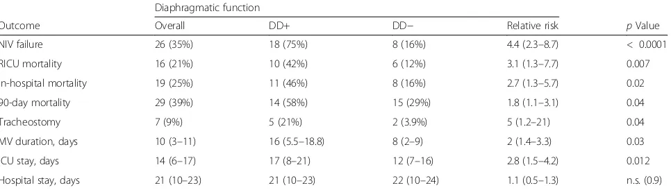
Table2shows the clinical outcomes in the study popula-tion. DD+ patients presented a higher risk for NIV failure than DD− patients (risk ratio 4.4; p< 0.001). Six DD+ patients who failed NIV then died, and twelve underwent endotracheal intubation. After intubation, two died while on MV, five underwent tracheostomy (two of whom died), and five were successfully weaned. Among DD−patients for whom NIV failed, four DD− patients for whom NIV failed then died, and four underwent endotracheal intub-ation (two died while on MV, and two underwent tracheos-tomy with one of them dying).
Among the secondary outcomes, DD+ correlated with higher short- and long-term mortality, longer stay in the RICU, prolonged MV, and higher tracheostomy rate (Table2).
In ROC analysis,ΔTdi < 20% showed higher accuracy in predicting NIV failure than baseline pH < 7.25, and both changes in arterial blood pH and PaCO2 did within 2 h
after NIV was started (Fig.2) (AUCs 0.84, 0.51, 0.56, and 0. 54, respectively; p< 0.0001). In addition, ΔTdi showed a very good correlation with Pdi sniff (Pearson’sr= 0.81;p= 0.004) (Fig. 3a); moreover,ΔTdi < 20% demonstrated the same accuracy as Pdi sniff in identifying DD+ (sensitivity 100%; 95% CI 0.6–1; specificity 100% 95% CI 0.51–1;p= 0. 0048) (Fig.3b).
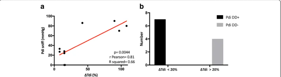
Figure4 reportsΔTdi and Pdi sniff values in the sub-group of ten patients tested with esophageal and gastric balloons (Fig.4a) and distribution of patients with ΔTdi < 20% or > 20% according to Pdi sniff (Fig. 4b). Among patients withΔTdi < 20%, two patients had a Pdi sniff of 0, meaning complete diaphragm paralysis, whereas four presented Pdi sniff values between 24 and 33 cmH2O.
Regarding diaphragm paralysis,ΔTdi demonstrated high sensitivity but low specificity in detecting this condition (sensitivity 100%, 95% CI 0.18–1; specificity 50%, 95% CI 0.22–0.78). Kaplan-Meier curves showed a significant
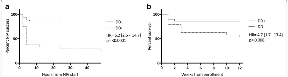
increase in NIV failure and 90-day mortality (Fig.5a and b, respectively) among DD+ versus DD−patients.
Discussion
In this single-center prospective observational study, we show that patients DD+ on their admission day in the RICU had a sixfold increased risk of NIV failure within the first 48 hours and an almost fivefold higher risk of dying during the follow-up. In addition, DD+ was associ-ated with increased RICU and hospital lengths of stay, prolonged use of MV, and higher tracheostomy rate. Finally, the presence/absence of DD as described by the
ΔTdi level (namely thethickening fraction) at US testing was strongly correlated with the diaphragmatic pressure-generating capacity as assessed by Pdi sniff. Thus, the early and noninvasive assessment of DD during severe AECOPD requiring NIV seems accurate and helpful in avoiding the risk of delayed intubation in patients with this critical condition.
Our study demonstrates a significant prevalence of DD in patients with severe AECOPD (32%). In a previous exploratory study on the same subject [12], we found that DD similarly assessed by US was reported in 22% of total patients; however, that sample had fewer patients and less severity than the present study (APACHE II score 20 vs 22,p= 0.041; and SAPS II score 41 vs 43,p= 0.036).
Demoule et al. reported that the prevalence of DD, as assessed by twitch tracheal pressure in response to bilateral anterior magnetic phrenic nerve stimulation, was 64% in a heterogeneous group of patients admitted to a general medical ICU [5]. However, the two populations seem difficult to compare because the population in our present study is limited to a specific subset of critical patients. Moreover, we used a more restricted definition of DD, the
[image:5.595.58.541.577.714.2]ΔTdi in the zone of apposition less than 20%, thus repre-senting a likely condition of very severe impairment, even confirmed by the transdiaphragmatic Pdi assessment as obtained in ten subjects.
Table 2Clinical outcomes (primary and secondary) of the study population
Diaphragmatic function
Outcome Overall DD+ DD− Relative risk pValue NIV failure 26 (35%) 18 (75%) 8 (16%) 4.4 (2.3–8.7) < 0.0001 RICU mortality 16 (21%) 10 (42%) 6 (12%) 3.1 (1.3–7.7) 0.007 In-hospital mortality 19 (25%) 11 (46%) 8 (16%) 2.7 (1.3–5.7) 0.02 90-day mortality 29 (39%) 14 (58%) 15 (29%) 1.8 (1.1–3.1) 0.04 Tracheostomy 7 (9%) 5 (21%) 2 (3.9%) 5 (1.2–21) 0.04 MV duration, days 10 (3–11) 16 (5.5–18.8) 8 (2–9) 2 (1.4–3.3) 0.03 ICU stay, days 14 (6–17) 17 (8–21) 12 (7–16) 2.8 (1.5–4.2) 0.012 Hospital stay, days 21 (10–23) 21 (10–23) 22 (10–24) 1.1 (0.5–1.3) n.s. (0.9)
The physiopathological mechanism(s) underlying the onset of DD during AECOPD is multifactorial. However, the synergic effect of mechanical disadvantage, functional (exhaustion) and biological (inflammation) impairment, and pharmacological damage (steroid-induced) could only be postulated.
In patients with COPD with frequent exacerbations the maximum pressure produced by the diaphragm’s contrac-tion is significantly than in individuals without COPD, in the same regard as the volitional (Pdi at total lung capacity and/or Pdi sniff) or nonvolitional (phrenic nerve stimula-tion) nature of the test applied [17–19]. This difference has been explained by the diaphragmatic shortening and the mechanical derangement following onset of progressive lung hyperinflation [17]. Indeed, patients with severe COPD produce higher transdiaphragmatic pressures at equivalent lung volumes than healthy control individuals [20]. The reason for this imbalance has been found in increased air-way resistance, reduced dynamic lung elastance, and raised PEEPi, which are related to the progressive air trapping [17, 20]. Thus, in a patient with AECOPD, the expiratory flow limitation and the increased respiratory rate with reduced expiratory time favor the onset of hyperinflation with sub-sequent respiratory muscle shortening and limited excur-sion. The reduction in muscle strength seems even more
inadequate to cover the excessive mechanical load imposed by the increased respiratory rate. In these conditions, the diaphragm soon exhausts its functional reserve, and ultim-ately mechanical impairment occurs.
In the present study, exposure to steroids in the previ-ous year was the only feature differentiating DD+ from DD−patients. To date, no studies have been conducted in humans to investigate the specific effect of steroid course on the respiratory muscles during AECOPD. Notwith-standing this, studies performed on the skeletal muscles clearly show an increase in proteolysis and a reduction in the expression of insulin-like growth factor (IGF)-1 following the use of steroids [21]. In animal models, there are studies showing damage to the diaphragmatic struc-ture after administration of systemic steroids [22,23], with proteolysis and downregulation of IGF-1 being the plaus-ible mechanisms responsplaus-ible for steroid-induced myop-athy [24]. Therefore, it is possible that previous or current (ab)use of systemic steroids could have contributed to the development of respiratory muscle weakness, thus leading to DD once AECOPD occurred.
Even despite the lack of a preadmission assessment of diaphragmatic function, some aspects seem to indicate that dysfunction might be related to the current acute condition rather than to a chronic exposure in our study population. First, the use of a very low cutoff limit ofΔTdi (< 20%) to identify DD+ let us individuate patients with a condition close to muscle paralysis, whose presence would already have been evident outside the acute phase [25]. Second, studies conducted with patients with stable severe COPD did not identify a significant difference in diaphragmatic function by US evaluation at Vtcompared with healthy
in-dividuals [26]. Third, in a study by Orozco-Levi et al., trans-diaphragmatic pressure values were similar in patients with stable COPD and normal control subjects, thus indicating a substantial functional preservation, although regional stresses and strains (muscle geometry, increased workload during exertion, mechanical stress, and metabolic factors) might induce or accentuate diaphragmatic injury [27]. Fig. 2ROC analysis comparing predictors for noninvasive ventilation
(NIV) failure at baseline and within 2 hours after NIV was started.
ΔTdiChange in diaphragm thickness
[image:6.595.57.291.88.199.2] [image:6.595.58.539.570.702.2]It is likely that these potential mechanisms of damage are amplified during AECOPD, when the diaphragm is called on to sustain stresses much greater than during clinical stability. Our study demonstrates a positive and significant correlation betweenΔTdi at US assessment and Pdi meas-urement at maximal inspiration. To our knowledge, this is the first time that US evaluation of the diaphragmatic function has been compared with standard techniques in subjects with AECOPD.
Kim et al. showed that US is reliable in identifying DD during weaning from MV and in estimating the work of breathing during NIV [28]. Gottesman et al. indicated that
ΔTdi < 20% is the cutoff limit for identifying diaphragm paralysis [25]. In our study, only two patients had Pdi sniff equal to 0 among those withΔTdi < 20% (see Fig.4). The sensitivity and specificity ofΔTdi (< 20%) to detect DD+ dropped to 50% in cases of diaphragm palsy.
Overall, the present study demonstrates that US is able to identify dysfunction but not palsy of the diaphragm, for which invasive maneuvers are needed. Given that these
patients usually present with acute hypercapnic respiratory failure, the use of volitional or invasive techniques such as transdiaphragmatic pressure might become extremely chal-lenging, in contrast to US assessment.
The main finding of our study is that DD+ patients, once confirmed by US, present a greater than fourfold risk of NIV failure during severe AECOPD (see Fig. 5). Interestingly, the two slopes start separating significantly 4–6 hours after starting NIV, then both groups gradually display a plateauing distribution over time.
Several studies have consistently demonstrated that the severity of hypercapnia and acidosis are associated with early NIV failure during AECOPD [13, 29–31]; however, no studies have assessed the impact of DD on NIV outcome. Indeed, our previous exploratory study demon-strated only that DD+ is related to NIV failure in a small population underpowered to test this hypothesis [9].
In the present study, we were able to demonstrate that DD+ as assessed by US may predict NIV failure with higher accuracy than both baseline pH < 7.25 and early Fig. 4aChange in diaphragm thickness (ΔTdi) values at ultrasound testing and transdiaphragmatic pressure capacity measured at maximal inspiration using the sniff maneuver (Pdi sniff) values in the subgroup of ten patients tested with esophageal and gastric balloons.bDistribution of patients withΔTdi < 20% or > 20% according to Pdi sniff.NIVNoninvasive ventilation
[image:7.595.58.540.88.260.2] [image:7.595.55.540.573.704.2]change of pH and PaCO2 (see Fig. 2). Notwithstanding
this, the reasons for the exceeding NIV failure rate are not completely clear, whereas severe expiratory flow limitation and excessive hyperinflation might have played a critical role in uncoupling patients’ efforts and MV. Because the use of NIV is increasing in different hospital settings, the present data may be of help to select patients with AECOPD who might be successfully ventilated in a general ward (patients without DD), rather than those who would benefit by being treated with MV in the ICU (patients with DD). Therefore, this finding increases the clinical importance of early identifi-cation of patients at greater risk for NIV failure even in the ward [32].
In our patients with AECOPD and DD+ short-term (in the RICU) and 90-day mortality are up to fivefold greater than in DD− patients. This finding is in line with data reported by Demoule et al., who described a higher mortal-ity rate in patients admitted to the ICU with low tracheal pressure in response to bilateral anterior magnetic phrenic nerve stimulation [5]. In that hospital setting, a potential explanation for increasing death rate following DD has been identified in the early impairment of nerve conduction and mitochondrial alterations due to the onset of systemic inflammation [33].
Our present data suggest only that the duration of venti-lation and the prolonged stay in the critical area following NIV failure may have resulted in a worse prognosis. Interestingly, we did not find any significant difference in hospital stay between patients with impaired or preserved diaphragmatic function. However, it could be hypothesized that other independent factors might have influenced the length of admission (e.g., home setting, chronic comorbidi-ties, lack of domestic facilities).
Our study provides new information on the pathophysi-ology of AECOPD needing NIV treatment; however, it has some limitations that need to be addressed. First, it was conducted in a single center, which means that the results need to be verified in a multicenter study. Because we performed a prespecified sample size analysis for the pri-mary outcome, the findings at least warrant further inves-tigations. Second, DD by US assessment was performed during spontaneous breathing in a critical care setting without any evaluation of lung volumes. Because different volumes highly correlate with the ability of the diaphragm to contract, measurement of volumes might have helped us to better understand the overall mechanism leading to progressive hyperinflation and then to DD. Third, we did not reassess diaphragmatic function over time, so data regarding potential spontaneous recovery are lacking. Last, we did not investigate the inflammatory status (i.e., the circulation levels of cytokines) in our patients, though this has been supposed to be a pathway of damage in the diaphragm (see paragraph above).
Conclusions
In this single-center trial, we observed that patients with severe AECOPD admitted to the RICU for NIV treatment have a significantly higher risk of failure and mortality when DD, as noninvasively assessed by US, occurs. US measurement of DD definitely correlates with transdiaph-ragmatic pressure-generating capacity in these subjects. Therefore, early evaluation of the diaphragm by US in this setting may help clinicians to identify patients with AECOPD at major risk for a negative prognosis.
Abbreviations
AECOPD:Acute exacerbation of chronic obstructive pulmonary disease; APACHE II: Acute Physiology and Chronic Health Evaluation II; COPD: Chronic obstructive pulmonary disease; DD: Diaphragmatic dysfunction; FiO2: Fraction
of inspired oxygen; FRC: Functional residual capacity; IGF-1: Insulin-like growth factor 1; MV: Mechanical ventilation; NIV: Noninvasive ventilation; PaCO2: Partial pressure of arterial carbon dioxide; PaO2: Partial pressure of
arterial oxygen; Pdi sniff: Transdiaphragmatic pressure capacity measured at maximal inspiration using the sniff maneuver; Pdi: Transdiaphragmatic pressure; PEEPi: Intrinsic positive end-expiratory pressure; Pes: Esophageal pressure; Pga: Gastric pressure; RICU: Respiratory intensive care unit; SAPS II: Simplified Acute Physiology Score II; US: Ultrasound; Vt: Tidal volume; ΔTdi: Change in diaphragm thickness
Acknowledgements
Not applicable.
Availability of data and materials
Data are available from the Respiratory Diseases Unit of the University Hospital of Modena, Italy.
Authors’contributions
AM and IC contributed equally to the manuscript. AM, RT, and IC reviewed the literature, designed the review, wrote the manuscript, and produced figures. RT and IC wrote the manuscript and produced figures. RF, SC, AV, and FG reviewed the literature and wrote the manuscript. VR edited the manuscript and reviewed it for English language. EC reviewed and edited the manuscript. MF and LT perform statistical analysis and wrote the manuscript. All authors read and approved the final manuscript.
Ethics approval and consent to participate
Approval from the local ethics committee of Modena was obtained (registered protocol number 839/C.E.). Written informed consent to participate was obtained from all enrolled patients or from their relatives, when appropriate.
Consent for publication
Consent for publication was obtained from all patients enrolled or from their relatives, when appropriate.
Competing interests
The authors declare that they have no competing interests with regard to any organization or entity with a financial interest in competition with the subject matter or materials discussed in this publication.
Publisher’s Note
Springer Nature remains neutral with regard to jurisdictional claims in published maps and institutional affiliations.
Author details 1
Respiratory Diseases Unit and Centre for Rare Lung Diseases, Department of Medical and Surgical Sciences, University of Modena Reggio Emilia, University Hospital of Modena, Modena, Italy.2University Hospital of Modena,
Received: 2 March 2018 Accepted: 9 April 2018
References
1. Global Initiative for Chronic Obstructive Lung Disease (GOLD). GOLD 2017 Global Strategy for the Diagnosis, Management and Prevention of COPD. Available from:http://goldcopd.org/.
2. Lightowler JV, Wedzicha JA, Elliott MW, Ram FS. Non-invasive positive pressure ventilation to treat respiratory failure resulting from exacerbations of chronic obstructive pulmonary disease: Cochrane systematic review and meta-analysis. BMJ. 2003;326(7382):185.
3. Rodriguez-Roisin R. Toward a consensus definition for COPD exacerbations. Chest. 2000;117(5 Suppl 2):398S–401S.
4. Brochard L, et al. Noninvasive ventilation for acute exacerbations of chronic obstructive pulmonary disease. N Engl J Med. 1995;333(13):817–22. 5. Demoule A, et al. Benefits and risks of success or failure of noninvasive
ventilation. Intensive Care Med. 2006;32(11):1756–65.
6. Unal O, et al. Evaluation of diaphragmatic movement with MR fluoroscopy in chronic obstructive pulmonary disease. Clin Imaging. 2000;24(6):347–50. 7. Gayan-Ramirez G, Decramer M. Mechanisms of striated muscle dysfunction
during acute exacerbations of COPD. J Appl Physiol. 2013;114(9):1291–9. 8. De Troyer A, Wilson TA. Effect of acute inflation on the mechanics of the
inspiratory muscles. J Appl Physiol. 2009;107:315–23.
9. Antenora F, et al. Prevalence and outcomes of diaphragmatic dysfunction assessed by ultrasound technology during acute exacerbation of chronic obstructive pulmonary disease: a pilot study. Respirology. 2017;22(2):338–44. 10. Davidson AC, et al. BTS/ICS guideline for the ventilatory management of acute
hypercapnic respiratory failure in adults. Thorax. 2016;71(Suppl 2):ii1–35. 11. Dellinger RP, et al. Surviving Sepsis Campaign Guidelines Committee
including the Pediatric Subgroup. Surviving Sepsis Campaign: international guidelines for management of severe sepsis and septic shock: 2012. Crit Care Med. 2013;41(2):580–637.
12. Fantini R, Mandrioli J, Zona S, Antenora F, Iattoni A, Monelli M, Fini N, Tonelli R, Clini E, Marchioni A. Ultrasound assessment of diaphragmatic function in patients with amyotrophic lateral sclerosis. Respirology. 2016;21(5):932–8.
13. Ambrosino N, Foglio K, Rubini F, Clini E, Nava S, Vitacca M. Non-invasive mechanical ventilation in acute respiratory failure due to chronic obstructive pulmonary disease: correlates for success. Thorax. 1995;50(7):755–7. 14. Higgs BD, Behrakis PK, Bevan DR, Milic-Emili J. Measurement of pleural
pressure with esophageal balloon in anesthetized humans. Anesthesiology. 1983;59(4):340–3.
15. Baydur A, Behrakis PK, Zin WA, Jaeger M, Milic-Emili J. A simple method for assessing the validity of the esophageal balloon technique. Am Rev Respir Dis. 1982;126(5):788–91.
16. Vogelmeier CF, Criner GJ, Martinez FJ, et al. Global strategy for the diagnosis, management, and prevention of chronic obstructive lung disease 2017 report: GOLD executive summary. Am J Respir Crit Care Med. 2017;195:557–82.
17. Similowski T, et al. Contractile properties of the human diaphragm during chronic hyperinflation. N Engl J Med. 1991;325:917–23. 18. Bellamare F, et al. Effects of emphysema and lung volume reduction
surgery on transdiaphragmatic pressure and diaphragm length. Chest. 2002; 121:1898–910.
19. Polkey MI, et al. Diaphragm strength in chronic obstructive pulmonary disease. Am J Respir Crit Care Med. 1996;154:1310–7.
20. Jubran A, Tobin MJ. Pathophysiologic basis of acute respiratory distress in patients who fail a trial of weaning from mechanical ventilation. Am J Respir Crit Care Med. 1997;155:906–15.
21. Crul T, et al. Gene expression profiling in vastus lateralis muscle during an acute exacerbation of COPD. Cell Physiol Biochem. 2010;25:491–500. 22. Arthurton L, et al. Membrane glucocorticoid receptors are localised in the
extracellular matrix and signal through the MAPK pathway in mammalian skeletal muscle fibres. J Physiol. 2015;593(12):2679–92.
23. Maes K, et al. Effects of acute administration of corticosteroids during mechanical ventilation on rat diaphragm. Am J Respir Crit Care Med. 2008;178:1219–26.
24. Dirks-Naylor AJ, Griffiths CL. Glucocorticoid-induced apoptosis and cellular mechanisms of myopathy. J Steroid Biochem Mol Biol. 2009;117:1–7. 25. Gottesman E, McCool ED. Ultrasound evaluation of the paralyzed
diaphragm. Am J Respir Crit Care Med. 1997;55:1734–9.
26. Baria MR, et al. B-mode ultrasound assessment of diaphragm structure and function in patients with COPD. Chest. 2014;146(3):680–5.
27. Orozco-Levi M, et al. Injury of the human diaphragm associated with exertion and chronic obstructive pulmonary disease. Am J Respir Crit Care Med. 2001;164:1734–9.
28. Kim WY, Suh HJ, Hong SB, Koh Y, Lim CM. Diaphragm dysfunction assessed by ultrasonography: influence on weaning from mechanical ventilation. Crit Care Med. 2011;39(12):2627–30.
29. Seneff MG, et al. Hospital and 1- year survival of patients admitted to intensive care units with acute exacerbation of chronic obstructive pulmonary disease. JAMA. 1995;274(23):1852–7.
30. Confalonieri M, Garuti G, Cattaruzza MS, Osborn JF, Antonelli M, Conti G, Kodric M, Resta O, Marchese S, Gregoretti C, Rossi A. Italian noninvasive positive pressure ventilation (NPPV) study group. A chart of failure risk for noninvasive ventilation in patients with COPD exacerbation. Eur Respir J. 2005;25(2):348–55.
31. Plant PK, Owen JL, Elliott MW. Early use of non-invasive ventilation for acute exacerbations of chronic obstructive pulmonary disease on general respiratory wards: a multicentre randomised controlled trial. Lancet. 2000;355:1931–5.
32. Chandra D, Stamm JA, Taylor B, Ramos RM, Satterwhite L, Krishnan JA, Mannino D, Sciurba FC, Holguín F. Outcomes of non-invasive ventilation for acute exacerbations of chronic obstructive pulmonary disease in the United States, 1998–2008. Am J Respir Crit Care Med. 2012;185(2):152–9. 33. Khan J, et al. Early development of critical illness myopathy and neuropathy