R E S E A R C H
Open Access
A
Plasmodium falciparum
copper-binding
membrane protein with copper transport motifs
David L Choveaux
1, Jude M Przyborski
2and JP Dean Goldring
1*Abstract
Background:Copper is an essential catalytic co-factor for metabolically important cellular enzymes, such as cytochrome-c oxidase. Eukaryotic cells acquire copper through a copper transport protein and distribute
intracellular copper using molecular chaperones. The copper chelator, neocuproine, inhibitsPlasmodium falciparum ring-to-trophozoite transitionin vitro, indicating a copper requirement for malaria parasite development. How the malaria parasite acquires or secretes copper still remains to be fully elucidated.
Methods:PlasmoDB was searched for sequences corresponding to candidateP. falciparumcopper-requiring proteins. The amino terminal domain of a putativeP. falciparumcopper transport protein was cloned and expressed as a maltose binding fusion protein. The copper binding ability of this protein was examined. Copper transport protein-specific anti-peptide antibodies were generated in chickens and used to establish native protein localization inP. falciparumparasites by immunofluorescence microscopy.
Results:SixP. falciparumcopper-requiring protein orthologs and a candidateP. falciparumcopper transport protein (PF14_0369), containing characteristic copper transport protein features, were identified in PlasmoDB. The
recombinant amino terminal domain of the transport protein bound reduced copperin vitroand withinEscherichia colicells during recombinant expression. Immunolocalization studies tracked the copper binding protein
translocating from the erythrocyte plasma membrane in early ring stage to a parasite membrane as the parasites developed to schizonts. The protein appears to be a PEXEL-negative membrane protein.
Conclusion:Plasmodium falciparumparasites express a native protein with copper transporter characteristics that binds copperin vitro. Localization of the protein to the erythrocyte and parasite plasma membranes could provide a mechanism for the delivery of novel anti-malarial compounds.
Keywords:Malaria, Copper transporter, Permeome, PEXEL-negative
Background
Malaria is a serious, acute and chronic relapsing infec-tion that kills close to one million people annually. More than 90% of these deaths are recorded in the sub-Saharan regions of Africa, with the majority being chil-dren under the age of five years [1]. Human mortality, as a result of malaria infection, is predominantly caused by
Plasmodium falciparum. Efforts to prevent and control the disease have, however, been hindered by an increased parasite resistance to currently available anti-malarial drugs, highlighting the need for novel anti-malarial drug development [2]. A group of proteins gaining increased
interest as potential therapeutic targets are the integral membrane proteins predicted to possess transport func-tions [3,4]. Despite these proteins playing key roles in
Plasmodium parasite growth and replication [4], they re-main poorly understood and underexploited [3]. Previous studies employing the intracellular copper chelator neocu-proine established that copper is an essential micronu-trient forin vitroparasite growth [5]. Rasolosonet al.[5] described the membrane-boundPfCuP-ATPase copper ef-flux protein and suggested this protein acts to reduce cop-per toxicity in P. falciparum. Studying P. falciparum
copper metabolism may lead to the identification of novel anti-malarial drug targets.
Copper is an essential micronutrient that plays import-ant catalytic and structural roles in numerous enzymes. * Correspondence:goldringd@ukzn.ac.za
1
Biochemistry, University of KwaZulu-Natal, P.B. X01, Carbis Road, Scottsville 3209, South Africa
Full list of author information is available at the end of the article
Cuproenzymes harness the ability of copper to cycle between a stable oxidized Cu(II) and unstable reduced Cu(I) state for various redox reactions. However, this property also makes copper potentially toxic to cells since it can undergo free radical producing Fenton chemistry [6]. Consequently, cells have evolved homeo-static mechanisms for the uptake, distribution, sequestra-tion and secresequestra-tion of copper to meet essential cellular requirements while reducing its toxic potential. From yeast to humans, copper acquisition is mediated by the high affinity copper transport protein, Ctr1 [7,8]. Copper metallochaperones subsequently distribute intracellular copper to specific proteins or organelles. The copper chaperone for superoxide dismutase (CCS) distributes copper to cuprozinc superoxide dismutase (Cu/Zn SOD) in the cytosol and mitochondrion, antioxidant protein 1 (Atox1) transfers copper to the secretory pathway and nucleus and an ensemble of proteins deliver copper to cytochrome-c oxidase (CCO) in the mitochondrion [7,8]. It has been suggested that Cu-ATPase-mediated secre-tion of excess copper is the main factor regulating copper homeostasis [9]. Equally important to mammalian growth and development, however, is the copper trans-porter Ctr1. Cell-specific knock-out of Ctr1 in mouse intestinal epithelial cells caused marked growth retard-ation, coupled with cardiac hypertrophy and overall viability defects that caused postnatal lethality around three weeks of age [10]. Similarly, knock-out of the
Drosophila melanogaster Ctr1Agene resulted in develop-mental arrest at early larval stages [11]. The C terminus of yeast copper transport protein, Ctr1, appears to have a role in copper regulation and stopping the build-up of copper to toxic concentrations [12].
Although aP. falciparumP-type ATPase copper efflux protein has been described [5], an additional protein-mediated mechanism for copper acquisition has not. The current study describes a novel membrane-bound
P. falciparumprotein with copper transporter characistics. A recombinant form of the protein's amino ter-minal region bound reduced copper in vitro and within
E. colihost cells. The nativeP. falciparum copper trans-port protein was found to initially localize to the infected erythrocyte membrane, followed by relocation to a para-site membrane through the development of the parapara-site’s erythrocytic cycle.
Methods Bioinformatics
The annotated Plasmodium database [13] was screened for the presence of copper-requiring protein orthologs using a BlastP search. Sequences used to screen the database are listed in Table 1. A putative Plasmodium
[image:2.595.64.540.508.733.2]copper transport protein was identified using theTheileria parva (Muguga stock) polymorphic immunodominant molecule (GenBank:AAA99499) [14] to BlastP screen PlasmoDB. In an effort to support sequence identity, the putativePlasmodium copper transport protein sequences were aligned with characterized copper transporter sequences fromHomo sapiens(NCBI:NP_001850), Arabi-dopsis thaliana(Genbank:BAE98928) andSaccharomyces cerevisiae (Genbank:AAB68064) using the ClustalWTM server. Transmembrane domains were identified in the putative Plasmodium copper transporters using the HMMTOP [15] and TMHMM 2.0 [16] topology pre-diction servers. The presence of potential signal sequences in each protein was established using the
Table 1 Copper-dependent protein orthologs toHomo sapiensproteins identified in theP. falciparumproteome
Enzyme name Accession number P. falciparumortholog identified PlasmoDB protein identifier
CCS CAG46726 No N/A
Cu/Zn Superoxide dismutase AAB05661 No N/A
Dopamine-β-monooxygenase PO9172 No N/A
Peptidylglycine monooxygenase P19021 No N/A
Metallothionein AAP97267 No N/A
S-adenosyl-L-homocysteine hydrolase P23526 Yes PFE1050w
Cox11 CAG46636 Yes PF14_0721
Cox17 AAA98114 Yes PF10_0252
Cox19 AAY35062 Yes PFL0090c
Cg3 O75880 Yes PF07_0034
Cytochrome-c oxidase subunit I ACR77861 Yes mal_mito_2
Cytochrome-c oxidase subunit II ABU47824 Yes PF14_0288 (IIa) PF13_0327 (IIb)
Cytochrome-c oxidase subunit III ABJ99455 Yes mal_mito_1
Cytochrome-c oxidase subunit Vb AAA52060 Yes PFI1365w
TMHMM server and the SignalP 3.0 program (Center for Biological Sequence Analysis, Lyngby, Denmark). A cladogram was generated from a ClustalW™alignment of six putative Plasmodium copper transporters with seven characterized copper transporters. The charac-terized transporters were from Homo sapiens, Mus musculus (NCBI:NP_780299), Rattus norvegicus (NCBI:NP_598284), Danio rerio (NCBI:NP_991280), Arabidopsis thaliana, Saccharomyces cerevisiae and Theileria parva.
Antibody production and purification
The amino acid sequence for a putative P. falciparum
copper transport protein (PF14_0369) was retrieved from PlasmoDB and subjected to Predict7 [17] analysis to identify immunogenic regions and the peptide CNLQKEEDTVVQLQD was selected for synthesis (GL Biochem Ltd. Shanghai, China). The selected peptide was coupled to rabbit albumin via the native amino terminal cysteine residue using m-maleimidobenzoyl-N-hydroxysuccinimide ester. Hy-Line Brown hens were used for antibody generation and antibodies were isolated from egg yolk as previously described [18,19]. Animal ethics clearance was obtained from the University of KwaZulu-Natal animal ethics committee (003/10/ Animal).
PCR amplification and cloning
Plasmodium falciparum (D10) gDNA was isolated from infected red blood cells using the FermentasTM DNA purification kit. The full-length PF14_0369, open reading frame was amplified using the following specific primers:
PfCtr369-fwd: 5'-atGAATTCGACAAAAGCGACAATA GTATTTG-3' and PfCtr369FL-rev: 5'-ACATCCACAA CAAGCTGGATC. For functional copper binding stud-ies, the coding domain for the protein's amino terminal domain minus signal peptide was amplified using the
PfCtr369-fwd primer containing an engineered EcoRI cleavage site and PfCtr369Nt-rev: 5'-caCTGCAGTTAC GATTTGGTTTCCCATTTG-3' containing a PstI clea-vage site (cleaclea-vage sites underlined). PCR products were analyzed on a 1% (w/v) agarose gel. The PCR amplicon for the PF14_0369 amino terminal domain (designated Nt) minus signal peptide (PfCtr369Nt-S) was cloned in frame with the maltose binding protein (MBP) gene in the pMal-p2x bacterial expression vector (New England BioLabs, USA), as previously described [18].The integrity of the resultant pMal-p2x/PfCtr369Nt-S plasmid insert was confirmed by DNA sequencing. Using the strategy employed for pMal-p2x/PfCtr369Nt-S, a plasmid con-taining pMal-p2X/PfCtr211Nt-S from the PF14_0211 gene was prepared.
Expression and purification of recombinant MBP-PfCtr369Nt-S
A single E. coli JM103[pMal-p2x/PfCtr369Nt-S] colony was grown overnight at 30°C in sterile LB medium sup-plemented with 100 μg.ml-1 ampicillin. The overnight culture was diluted in fresh broth, plus antibiotic, and incubated at 30°C until the A600 reached ~0.6.
MBP-PfCtr369Nt-Sexpression was induced with 0.3 mM IPTG and the cultures grown for a further 16 h before harvest-ing of cells by centrifugation (4000g, 15 min, 4°C). The
E. coli periplasmic content was isolated following the method described by French et al. [20] and the recom-binant protein purified by amylose affinity chromatog-raphy. Recombinant MBP-PfCtr211Nt-S was expressed using the same procedure. Protein samples were analyzed on a 12.5% SDS-PAGE gel [21] followed by Western blot-ting and probed with chicken PF14_0369 and anti-MBP antibodies [22] (see below). Protein concentration was determined with the Bradford dye-binding protein assay, using ovalbumin as the standard [23].
Parasite culture
Plasmodium falciparum 3D7 parasites were cultured in human O+ erythrocytes according to standard protocols, except cultures were incubated in gassed flasks [24]. Synchronized asexual parasites were obtained by sorbitol treatment [25].
Western blot analyses
For Western blot analyses ofP. falciparum lysates, pro-tein samples were prepared from saponin-isolated, sorbitol-synchronized parasites. Equivalent amounts of 1 x 10⁷ parasites were analysed by 12.5% SDS-PAGE and transferred to a nitrocellulose membrane. Primary chicken anti-PF14_0369 and chicken anti-P. falciparum
lactate dehydrogenase (LDH) antibodies against spe-cific peptides on each protein were employed and detected using rabbit anti-chicken horseradish peroxidase-conjugated secondary antibodies (DAKO, Santa Cruz). Immunoblots were developed using enhanced chemilu-minescence. Recombinant MBP-PfCtr369Nt-S expression was analysed by Western blot using chicken anti-MBP and PF14_0369 peptide antibodies. These anti-bodies were detected using rabbit anti-chicken horsera-dish peroxidase-conjugated secondary antibodies (Jackson Immuno-Research Laboratories, USA) and visualized by incubating the membrane in 0.06% (w/v) 4-chloro-1-naphthol and 0.0015% (v/v) H2O2.
Copper binding
For in vitro copper binding studies, purified
overnight dialysis against sodium phosphate buffer [0.1 M NaH2PO4, 0.01% (w/v) NaN3, pH 7.5] at 4°C. Copper binding to MBP-PfCtr369Nt-S and the oxidation state of bound copper was assessed using the bicincho-ninic acid (BCA) assay [26], in which protein-copper complexes are disrupted by acid denaturation and cop-per detected upon release. BCA binds reduced copcop-per (Cu+), forming a purple complex detectable at 354 nm. If copper is present in solution as Cu2+, ascorbate must be added to the sample following acid denaturation to yield a Cu+-BCA complex. However, if the addition of ascorbate does not influence Cu+-BCA complex forma-tion, this indicates copper was released from the protein in the Cu+ state. Solutions of CuCl2 in sodium phos-phate buffer were used as standards and affinity purified MBP served as a negative control. The ability of
MBP-PfCtr369Nt-S to bind copper withinE. colicells was also examined. Cells containing the recombinant protein were grown in the presence or absence of 0.5 mM CuCl2, and the recombinant protein purified as described above. Prior to cell disruption and MBP-PfCtr369Nt-Sisolation, pelleted cells were washed with a Tris buffer (0.2 M Tris–HCl, pH 7.5) to remove extraneous CuCl2. As with
in vitro copper binding, copper bound to
MBP-PfCtr369Nt-Swithin E. coli cells was detected using the BCA assay.
Ascorbate oxidation assay
The ability of MBP-PfCtr369Nt-S to inhibit the copper-catalysed oxidation of ascorbic acid (H2Asc) was tested
in vitro. In a typical experiment a mixture was prepared containing 5 μM MBP-PfCtr369Nt-S,, or 5 μM MBP (control), 8 μM CuCl2 and 120 μM H2Asc solution, adjusted to pH 4.5 with NaOH. The absorbance of this solution was monitored at 255 nm with readings taken every 5 s for 300 s. Reactions were carried out at RT.
Immunofluorescence
Immunofluorescence assays were carried out following cell fixation with 4% paraformaldehyde/0.00075% (w/v) glutaraldehyde as previously described [27] except fixa-tion was carried out at 37°C for 30 minutes and quench-ing was performed with 125 mM glycine/PBS. Chicken anti-peptide antibodies directed against P. falciparum
lactate dehydrogenase and PF14_0369 and a rabbit anti-body against Exp-1 were used. Primary antibodies were diluted in 3% (w/v) BSA/PBS and detected with either goat anti-chicken-Cy3 or goat anti-rabbit-Cy2 antibodies (DAKO, Santa Cruz). Hoechst 33258 (Molecular probes) was used at a concentration of 50 ng.ml-1to stain parasite DNA. All images were acquired at room temperature on a Zeiss Cell Observer using appropriate filter sets.
Results
Identification of copper-dependent protein orthologs in theP. falciparumproteome
Cytochrome-c oxidase (CCO) is a key multi-subunit catabolic protein containing three functionally important copper ions in its binuclear CuAand mononuclear CuB sites [28]. Metallation of the CuAand CuBsites requires the copper metallochaperones Cox17, Sco1 and Cox11 [29]. Identification of P. falciparum orthologs of these metallochaperones and other related copper-requiring proteins supports a plasmodial requirement for copper, as previously suggested [5]. A BlastP search of Plas-moDB, using mammalian and yeast proteins, identified six copper binding protein orthologs (Table 1). These were Cox11, Cox17, Cox19, Sco1, five CCO subunits and S-adenosyl-L-homocysteine hydrolase. The identifi-cation of CCO subunit I and II orthologs appears most important since these house the binuclear CuA and mononuclear CuB copper binding sites [28]. Interest-ingly, the P. falciparum proteome appears to lack an identifiable class of metallothioneins and Cu/Zn SOD (Table 1). In yeast and mammalian cells, these proteins are essential for copper storage and free radical detoxifi-cation, respectively [7,8].
Identification of a putativePlasmodiumcopper transport protein
Although P. falciparum appears to require copper for survival [5], the mechanism by which the parasite acquires copper remains to be formally identified. One suggested mechanism is acquisition via the ingestion and digestion of host erythrocyte Cu/Zn SOD [5]. A po-tential alternate mechanism is acquisition via a copper transport protein. The Theileria parva polymorphic immunodominant molecule sequence [14], which pos-sesses copper transport protein features, was used to screen PlasmoDB for a candidate Plasmodium copper transport protein. Full length, putative copper trans-porter sequences were identified for six Plasmodium
species, namely Plasmodium falciparum (PF14_0369 and PF14_0211), Plasmodium vivax (PVX_118540),
Plasmodium yoelIi (PY00413), Plasmodium berghei
extracellular amino-terminal domain (Figure 1A) and the essential MxxxM (Mx3M) and GxxxG (Gx3G) motifs (Figure 1B) in the second and third transmembrane domains respectively. An essential methionine residue, located 20 amino acid residues from the first putative trans-membrane domain [30], is present in all the Plasmodium
sequences (Additional file 1).
A cladogram was generated for the complete Plasmo-dium copper transporters to compare their similarity to seven characterized copper transporters. The Plasmodium
copper transporters formed a cluster related to, but separate from, the characterized copper transporters (Figure 1C). Of the characterized transporters, the sequence with the great-est similarity to that of Plasmodium parasites was from
Theileria parva. This was to be expected sinceT. parva
belongs to the same phylum asPlasmodiumparasites, the Apicomplexans [31]. Closer analysis of the Plasmodium
sequences indicated that the three rodent-infecting malaria parasites cluster together, whilstP. vivax,P. knowlesiand
P. falciparumform a separate but related cluster. Similar relationships have been observed from a phylogenetic analysis of thePlasmodiumcytochrome-b and small sub-unit rRNA sequences [32]. The separate cluster of the mammalian, yeast and plant reference sequences suggests a more distant similarity to thePlasmodiumsequences.
Considering its clinical importance, P. falciparumwas selected for further studies of the putative copper trans-porter. The gene encoding the putative copper transport protein (PF14_0369) is located on the positive strand of chromosome 14 as a two-exon gene with a 336 bp first exon and 372 bp second exon, translating to a 235 amino acid protein with a predicted mass of 27.15 kDa. The full length and amino terminal coding domain co-ding sequences for the putative P. falciparum copper transport protein (PF14_0369) were PCR amplified from genomic DNA, producing amplicons of approximately 890 bp and 290 bp, respectively (Figure 1D). An 820 bp product was amplified for the LDH control (Figure 1D). The identity of the amplicon for the putative transpor-ter’s amino terminal domain (PfCtr369Nt-S) was con-firmed by sequencing. This amplicon was cloned and expressed as a MBP fusion partner (MBP-PfCtr369Nt-S).
Recombinant expression of MBP-PfCtr369Nt-S
The amino terminal domain of yeast and mammalian copper transport proteins is essential for copper acquisi-tion [33], hence the corresponding domain of the puta-tive P. falciparum copper transporter was selected for copper binding studies. A similar approach has been adopted for characterization of the PfCuP-ATPase
A
B
C
D
1.5 1.0 0.8 0.6 0.4 0.2
M. 1. 2. 3. kb
Extracellular
Plasma membrane
Cytosol
1 2 3
MxxxM GxxxG
[image:5.595.60.537.91.320.2]Ct Nt
Figure 1Features characteristic of the copper transport protein family are conserved in the putativePlasmodiumcopper transporters. A. Three transmembrane (1–3), an extracellular amino terminal (Nt) and intracellular carboxy terminal (Ct) domains, methionine and MxxxM and GxxxG motifs of copper transport proteins.B. An aligned region of thePlasmodiumand other copper transporters showing the MxxxM and GxxxG motifs. Sequence identifiers are: Hs =Homo sapiens; Ms =Mus musculus; Tp =Theileria parva; Cp =Cryptosporidium parvum; Pf =P. falciparumPF14_0369; Pv =P. vivax; Py =P. yoelii; Pb =P. berghei; Pc =P. chabaudi; Pg =P. gallinaceum; Pk =P. knowlesi; Pr =P. reichenowi. Underlined amino acids (Hs) indicate the positions of transmembrane domains two and three.C. Cladogram comparing six completePlasmodium
copper efflux pump [5]. To limit the possibility of re-combinant MBP-PfCtr369Nt-S associating with a bacte-rial membrane, the predicted signal peptide was omitted and denoted as“-S”[34]. Expression of a 55 kDa protein was induced from the MBP-PfCtr369Nt-S plasmid and affinity purified from the bacterial periplasmic contents using an amylose affinity matrix (Figure 2A). The size of the purified protein corresponded well with that pre-dicted from the cloned sequence (54 kDa). An additional 42 kDa product, in the purified MBP-PfCtr369Nt-S frac-tion, is likely to be MBP lacking the fused protein. A Western blot using anti-MBP antibodies recognizes both the 42 kDa and 55 kDa bands (Figure 2B), whilst anti-bodies specific for PfCtr369Nt-S only recognize the 55 kDa band (Figure 2C). Also evident in the anti-MBP (Figure 2B) and anti-PfCtr369Nt-S blots (Figure 2C) are additional truncated forms of MBP-PfCtr369Nt-S be-tween the 55 kDa and 42 kDa bands. Recombinant MBP-PfCtr211Nt-S was expressed as a 46 kDa protein (Additional file 2).
MBP-PfCtr369Nt-Sbinds copper
In yeast and mammalian cells, copper appears to be transported across the membrane in its reduced form (Cu+) [30] and hence a Cu+preference was examined for the putative P. falciparum copper transporter. Purified MBP-PfCtr369Nt-S was incubated with CuCl2 in the presence or absence of the reducing agent ascorbate. Unbound copper was removed by dialysis and the bound copper detected using the BCA assay [26] with or with-out a second treatment of ascorbate. If ascorbate was required in the BCA assay for the formation of a purple complex it suggests Cu2+ is preferentially bound. Alter-natively, if ascorbate supplementation was not required for colour formation then Cu+ is suggested to be the preferred substrate. Purified MBP-PfCtr369Nt-S showed a binding preference for Cu+in vitro(Figure 3A). Exclu-sion of ascorbate from the initial CuCl2 incubation
resulted in low levels of detectable copper (Figure 3A), whereas including ascorbate resulted in a ~70% increase (p < 0.01) in the amount of copper detected. The absence of ascorbate from the BCA assay did not affect BCA-Cu+ complex formation for the sample originally incubated with ascorbate, thereby supporting a preference for Cu+ (Figure 3A).
The ability of MBP-PfCtr369Nt-S to bind copper within a cellular environment was examined by expres-sing the protein in the presence of 0.5 mM CuCl2. This concentration of copper does not affect E. coli growth [35]. MBP-PfCtr369Nt-S isolated and affinity purified from E. coli cells grown in copper-enriched media, bound copper (Figure 3B). Extraneous copper was removed by washing the cells prior to protein isolation. Protein isolated from cells grown without copper had negligible amounts of copper present (p < 0.01) (Figure 3B). The oxidation state of copper bound within the cell was inferred by including or excluding ascorbate from the BCA assay (Figure 3B). Like the in vitro ana-lysis, Cu+ appeared to be the preferred oxidation state bound by the protein within the E. coli periplasm. An interesting consequence when copper was added to the
E. coligrowth medium was a four-fold increase in
MBP-PfCtr369Nt-S yield from 1.53 mg to 5.98 mg per litre of culture (p < 0.01) (Figure 3B, inset). In all experiments MBP served as a negative control. Recombinant MBP did not bind copper and CuCl2 had no perceptible in-fluence on MBP expression (Figure 3).
Analysis of copper binding to MBP-PfCtr369Nt-Susing the rate of ascorbate oxidation
By taking advantage of the copper-catalysed oxidation rate of ascorbate and the ability of chelators to affect this rate, Jiang et al. [36] demonstrated that copper transport protein methionine motif peptides bind Cu+ with methionine-only coordination. The effect
[image:6.595.63.540.557.675.2]MBP-PfCtr369Nt-Shad on copper-catalysed ascorbate oxidation
Figure 2Expression and purification of recombinant MBP-PfCtr369Nt-S.Expression of MBP-PfCtr369Nt-Swas targeted to theE. coliperiplasm. A. Steps in the isolation of recombinant MBP-PfCtr369Nt-Swere analysed on a 12.5% reducing SDS-PAGE. Western blot of A. probed with either anti-MBPB. or anti-PF14_0369 antibodiesC. In all panels: lane 1, totalE. colilysate; lane 2, periplasmic proteins; lanes 3–5 represent
was examined by monitoring the loss of absorbance at 255 nm of a 120μM ascorbate solution at pH 4.5. This pH was optimal for several reasons. First, increased con-centrations of the ascorbate monoanion (HAsc-), which has a pKa of 4.1, cause increased oxidation rates thus ele-vated pH values can amplify unwanted errors. Second, Cu2+hydrolysis is minimal at a lower pH [36]. Last, and perhaps more relevant to the amino terminal domain of the putativeP. falciparumcopper transporter, is that the related human Ctr1 shows increased copper uptake at a low extracellular pH and yeast cells expressing Ctr1 acidify
their media to pH 4–5 [37]. Freshly prepared ascorbate showed a stable absorbance at 255 nm (A255) over 300 se-conds (Figure 4). The addition of 8 μM CuCl2 caused a rapid decrease in the A255 signal, indicative of oxidation by copper. The copper-catalysed oxidation appeared to be inhibited by the addition of 5 μM purified
MBP-PfCtr369Nt-S(Figure 4). This suggested MBP-PfCtr369Nt-S chelates copper in solution, thus preventing its participa-tion in the redox cycle of ascorbate oxidaparticipa-tion. Although this assay is not definitive of the copper species preferen-tially bound by MBP-PfCtr369Nt-S, it is presumably Cu+, Sample CuCl
2
only
MBP-PfCtr369Nt-S MBP only
Ascorbate - + - +
Sample MBP-PfCtr369Nt-S MBP only
CuCl2 - + - +
A354
A
A354 MBP
MBP-PfCtr369Nt-S
B
*
[image:7.595.57.540.90.255.2]* *
Figure 3MBP-PfCtr369Nt-Sbinds copperin vitroand withinE. colihost cells. A. Affinity-purified MBP-PfCtr369Nt-Sor MBP was incubated with CuCl2in the presence (+) or absence (−) of ascorbic acidin vitro. The BCA release assay detected copper with (solid bars) or without (open bars) the addition of ascorbic acid. The concentration of the copper standard (CuCl2only) was equimolar to the amount of protein used.B. CuCl2 was added to theE. colicell growth medium after the induction of recombinant protein expression. Following affinity purification, protein-bound copper was detected using the BCA release assay with (solid bars) or without (open bars) the addition of ascorbic acid. The addition of copper to theE. coligrowth medium increased recombinant protein yield (inset). In all panels, MBP serves as a control and * represents statistical
significance (p < 0.05). Results are means ± S.D. of triplicate measurements done for duplicate dialysis sacs.
A255
Time (sec)
Figure 4MBP-PfCtr369Nt-Sinhibits thecopper-catalysed oxidative degradation of ascorbate.Ascorbate does not auto-oxidize when
[image:7.595.53.540.502.683.2]based on results from the other copper-binding studies conducted for MBP-PfCtr369Nt-S (Figure 3). Control experiments indicated MBP had minimal influence on the copper-catalysed oxidation rate of ascorbate.
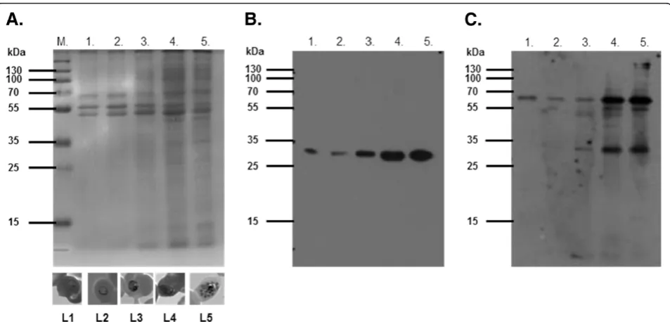
Native expression of a putativeP. falciparumcopper transport protein
An antibody probe specific for the putativeP. falciparum
copper transporter (PF14_0369) was designed to detect the native P. falciparum protein. Parasite lysates from sorbitol-synchronized P. falciparum parasite cultures were separated by SDS-PAGE (Figure 5A) and trans-ferred to a nitrocellulose membrane. Affinity purified anti-peptide antibodies targeting the N-terminal do-main of the putative P. falciparum copper transporter recognized protein bands of 34 kDa and 68 kDa in most stages of sorbitol-synchronized, asexual cultured para-sites (Figure 5C). These bands may represent mono-meric and dimono-meric species, as has been suggested for human Ctr1 [37]. Expression of the putative copper transporter appeared to increase in a similar manner to
Pf.LDH asP. falciparumprogressed through its asexual cycle (Figure 5B). The increase in protein expression is also seen as an increase in PF14_0369 transcrip-tion [38].
Localization of a putativeP. falciparumcopper transport protein
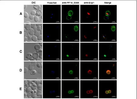
Characterized copper transport proteins are defined as integral membrane proteins that, depending on cell type, localize to either the plasma membrane or intracellular vesicles [8]. Analysis of the PF14_0369 protein sequence predicted an amino terminal signal peptide [39] and an apicoplast targeting signal [40]. Further analysis indi-cated that PF14_0369 lacked an identifiable PEXEL/HT motif. This suggested the putative copper transporter would be targeted to the parasite’s apicoplast and not to the parasite or erythrocyte plasma membranes. To de-fine the protein’s native sub-cellular location, the anti-peptide antibodies specific for the putative copper transporter were used in immunofluorescence assays. For this assay a modified form of the fixation protocol developed by Tonkin et al.[27] was employed. Advan-tages of this technique, over the traditional methanol or methanol/acetone fixation methods, include decreased auto-fluorescence and improved preservation of cell morphology [27].
[image:8.595.60.537.435.664.2]Sorbitol-synchronized parasites at various develop-mental stages were fixed as described above and probed with specific anti-peptide antibodies. In ring stage para-sites, the majority of the fluorescence signal could be detected in close proximity to the erythrocyte plasma
membrane (Figure 6A), with lower amounts of labelling found close to the body of the parasite. The signal asso-ciated with the parasite body overlaps considerably with that for Exp-1, a marker of the parasitophorous vacuole membrane [41]. As parasites progressed through the erythrocytic cycle, the erythrocyte plasma membrane staining became progressively weaker, with a concomi-tant increase in the signal co-localizing with Exp-1 (Figure 6B-E). The specificity of this signal migration becomes even more apparent in panels in which two dif-ferent parasites of slightly difdif-ferent developmental pro-gression show differing fluorescence phenotypes (Figure 6B and C). Towards the later stages of parasite development, the entire fluorescence signal can be seen in tight association with the parasite itself, with no signal detectable in the erythrocyte (Figure 6D and E). As the copper transporter is predicted to contain apicoplast tar-geting information, we carried out immuno-colocalization studies using antisera detecting the apicoplast resident
protein acyl-carrier protein. Even in parasite stages in which the copper transporter appears to localize with the body of the parasite, no significant signal co-localization was noted. All images were acquired using the same ex-posure time to allow for direct comparison of signal strength. Control experiments using pre-immune anti-body preparations or only secondary antibodies showed no antibody staining and additionally experiments target-ing theP. falciparumcytoplasmic proteins LDH, GAPDH and HSP86/90 showed a notably different localization from that of the putative copper transporter. No signal was seen to associate with non-infected erythrocytes (Figure 6A-E).
Analysis of PF14_0369 for novel export signals
Export of the putative copper transporter to the erythro-cyte surface was surprising considering the lack of an identifiable PEXEL/HT motif. Exported P. falciparum
[image:9.595.58.539.90.438.2]identified and are termed “PEXEL negative exported proteins” (PNEPs) [42]. Partial PEXEL motifs have been identified in the amino terminal domain of the PNEPs REX2 (LAE) [43] and SBP1 (LAD) [44] that are essential for export. Inspection of the PF14_0369 amino terminal domain identified a partial PEXEL motif identical to the important LAD motif identified in SBP1. Analysis of the essential transmembrane domains from SBP1 and REX2 highlighted a relative abundance of phenylalanine resi-dues [43] also found in the third transmembrane domain of PF14_0369 (Additional file 1).
Discussion
Maintenance of copper homeostasis is essential for cell survival. Consequently sophisticated mechanisms have evolved for copper acquisition, distribution and excre-tion. In mammalian cells, copper is acquired by the cop-per transport protein Ctr1 and distributed to target proteins via dedicated copper metallochaperones such as CCS, Atox1 and Cox17 [7,8]. To avoid the potentially toxic effects of copper, Cu-ATPase proteins mediate the excretion of excess copper [9] and a P. falciparum Cu-ATPase ortholog has been identified [5]. One P. falcip-arumcopper source appears to be from the ingestion of host erythrocyte Cu/Zn SOD [5]. A screen of the Plas-moDB database, performed in the present study, identi-fied sixP. falciparumcopper-requiring protein orthologs and a candidate copper transport protein. Native expres-sion of the putative copper transporter was confirmed by Western blot and the protein’s subcellular location identified by immunofluorescence microscopy. A recom-binant form of the transporter's amino terminal domain was shown to bind copper in vitro and within E. coli
host cells, supporting the possibility that the full-length protein functions to acquire copper.
Identification of a putativeP. falciparumcopper transport protein
The identification of six P. falciparum copper-requiring protein orthologs suggested an important role for copper in P. falciparum metabolism. Treating P. falciparum
parasites with the intracellular copper chelator, neocu-proine, inhibited ring-to-trophozoite transition in vitro
[5], highlighting the importance of copper for the para-site. During the course of its asexual cycle,P. falciparum
appears to digest host erythrocyte Cu/Zn SOD to release copper [5]. The presence of a membrane associated cop-per binding protein with copcop-per transport protein char-acteristics in plasmodia suggests a mechanism for the transport of copper similar to that played by the Ctr1 copper transport protein in yeast and mammalian cells [7,8]. Candidate copper transporter sequences were identified for eight species of the Plasmodium parasite and each sequence was shown to contain the essential
and largely definitive Mx3M and Gx3G motifs (Figure 1B) [30,45]. The predicted amino terminal do-main of each putative transporter was found to contain one or more methionine motifs (MxM or MxxM). Al-though not limited to copper transporters, these motifs are important for Ctr1 protein function [30].
The presence of three putative membrane-spanning regions is considered definitive for copper transport proteins [33] and topological analysis of the putative
P. falciparum copper transporter identified three such domains. Since three transmembrane domains cannot form a functional channel or pore for ion transport [10], a homotrimeric complex of copper transport protein mono-mers is formed for the characterized mammalian and yeast copper transport proteins [33]. The P. falciparum
copper transport protein may form a similar complex since the first transmembrane domain of the copper trans-port protein family serves as an adaptor allowing evolu-tionarily distant copper transporters to adopt a similar overall structure [46]. An immunoblot of yeast and mammalian copper transport proteins identified mono-meric and dimono-meric species of the trimono-meric complex [37]. Similar monomeric and dimeric species were detected in an immunoblot of the putative P. falciparum copper transport protein in this study.
Recombinant expression and copper binding to MBP-PfCtr369Nt-S
Signal peptides, transmembrane domains, rare codons, introns and genome AT-richness affect recombinant expression of P. falciparum proteins [34]. The amino terminal signal peptide of the putative P. falciparum
copper transporter was, therefore, excluded from the expression construct, resulting in periplasmic expression. The purified MBP-PfCtr369Nt-S recombinant protein bound reduced copper in vitro and within a cellular environment. A second copper transport protein,
MBP-PfCtr211Nt-S, from the PF14_0211 gene was recombi-nantly expressed and isolated. Expression of the native
P. falciparum protein encoded by this gene has not been ascertained at this stage.
In mammalian and yeast cells, the reduced cuprous ion (Cu+) is favoured for transfer and transport [30], since Cu+ is more exchange labile than Cu2+[36]. Cu+is highly reactive in an oxidizing environment and methio-nine motifs in the extracellular amino terminal domain of the copper transporter family are thought to sequester copper prior to its transport across the lipid bilayer [30]. This was supported by a study demonstrating that methionine motif peptides inhibited copper-catalysed ascorbate oxidation through Cu+ chelation [36].
it appeared that the amino terminal domain of the putative P. falciparum copper transporter preferably coordinates the Cu+ ion. Copper coordination was presumably a result of the methionine motif present in MBP-PfCtr369Nt-S.
The methionine motifs contained in the amino terminal domains of yeast and human copper transport proteins are essential for copper binding under copper limiting conditions [30]. The yeast copper transport protein con-tains eight methionine motifs, but only the last methio-nine of the eighth motif was shown by Puiget al.[30] to be essential for copper binding capability. The position of this methionine is conserved between copper transport proteins and is 20 amino acid residues N-terminal to the transporter’s first transmembrane domain [30]. Interest-ingly, the last methionine of the methionine motif in the amino terminal domain of the putative P. falciparum
copper transport protein is located in a similar position. However, the involvement of other amino acid residues, like cysteine and histidine residues that bind metal ions [47,48], and are present in this protein cannot be excluded. Identification of the specific residues coordi-nating copper will be explored in future experiments.
MBP-PfCtr369Nt-Srecombinant protein likely interacts withE. colicopper binding proteins
An interesting consequence of expressing
MBP-PfCtr369Nt-S in the presence of copper was a significant increase in recombinant protein yield. In the presence of excess copper it is a possibility that the copper bound to and stabilized the recombinant protein [48]. The cyto-plasm and pericyto-plasm of E. coli cells do, however, have different copper requirements since almost all bacterial copper proteins, such as multi-copper oxidases, amine oxidases and lysine oxidases are found in the periplasm or excreted extracellularly [49]. Copper availability inE. coli is thought to be regulated by the actions of the DNA-binding metal sensor CueR, which controls the ex-pression of genes encoding proteins involved in metal homeostasis [50]. A copper-requiring protein is thought to gain access to copper only if the protein’s affinity for the metal ion is greater than the buffered cellular con-centration of copper [51]. Interaction of a recombinant
P. falciparum protein with native E. coli proteins has been demonstrated for PfHsp70 [52], supporting the possibility that MBP-PfCtr369Nt-S interacts with native
E. colicopper proteins for copper loading. These interac-tions could, in turn, influence the availability of copper for native E. coli copper-requiring proteins resulting in tightly regulated MBP-PfCtr369Nt-S expression under standard growth conditions. The addition of excess extracellular copper appears to have alleviated this growth stress producing the increase in the expression of MBP-PfCtr369Nt-Sobserved here.
The putativeP. falciparumcopper transport protein shows stage-specific localization
During early stages of asexual development, the putative
P. falciparum copper transporter appears to be targeted to the plasma membrane of the infected erythrocyte. As the parasite matures through its asexual cycle the pro-tein was detected on a parasite membrane. This may represent a turnover of host cell membrane associated malarial proteins, followed by trafficking of the copper transporter to a parasite membrane. The precise localization of the copper transporter, when associated with the parasite, was not elucidated, due to limits to the fluorescence detection methods. The parasitopho-rous vacuolar membrane contains a “promiscuous”pore that permits the passage of solutes, nutrients and macro-molecules [53,54] and thus it is unlikely for there to be the need for a separate copper transporter. The parasite plasma membrane contains a variety of selective trans-porters (Reviewed by Martin et al. [4]). The copper transporter is thus suggested to be associated with the parasite plasma membrane.PfCuP-ATPase was similarly localized to a parasite plasma membrane and the surface of the infected erythrocyte. However, unlike the putative copper transport protein, PfCuP-ATPase was associated with both the parasite plasma membrane and the erythrocyte membrane at the same time in trophozoites and schizonts, whilst no expression was detected in rings [5]. These authors suggested that dual localization of
PfCuP-ATPase represents a novel mechanism by which the parasite reduces copper toxicity through copper ef-flux [5]. Reasons for the different localities of the copper transport protein are less apparent. There is the possibi-lity that during ring stages, extracellular copper is required until the parasite digests endocytosed host cell Cu/Zn SOD and translocates the copper transporter protein to a parasite membrane.
trophozoites to schizonts, the concentration of ingested Cu/Zn SOD decreases [5] and this coincides with data presented here showing the appearance of the putative copper transporter on parasite membranes, the highest level of expression of the protein, and mRNA levels described by Le Roch et al.[38]. Thus as the source of copper in the form of Cu/Zn SOD decreases in the para-site [5], the parapara-site compensates with an increase in ex-pression of the putative copper transport protein. Given the potential toxicity of copper and the need for the regulation of copper homeostasis, it is likely that the parasite employs a copper transport protein alongside the PfCuP-ATPase copper export protein described by Rasoloson et al.[5]. It is suggested that, given the pres-ence and membrane location of a copper binding protein with copper transporting motifs, and the increase in copper concentration in malaria infected red cells [57], that the protein described here has a role in copper transport. Data to support a transport role for the cop-per binding protein is being pursued.
Treatment ofP. falciparumparasites with neocuproine inhibited ring-to-trophozoite transition [5], implicating copper metabolism as a potential target for novel anti-malarial drug development. Copper, when the concen-tration is above a critical level, has been shown to be toxic to the parasite [58]. However, from in silico find-ings it seems unlikely thatP. falciparumcontains unique copper-dependent metabolic pathways. Copper-binding protein motifs also appear to be conserved from prokar-yotes to eukarprokar-yotes [59], suggesting that designing a compound specific for P. falciparum copper-dependent proteins could prove difficult. An alternative approach would be to deliver anti-malarial compounds to the parasite by exploiting transport mechanisms, as sug-gested for the P. falciparum new permeation pathway and choline carrier [60]. The human copper transport protein has been shown to transport the platinum con-taining anti-cancer drugs cisplatin and oxaliplatin [61]. Cisplatin also exhibits anti-malarial activity through DNA damage [62,63]. Given the structural similarities between the putative P. falciparum copper transport protein and the human copper transporter, it is possible that cisplatin is delivered to the parasite by the putative copper transport protein. This transport mechanism could perhaps be exploited for the delivery of novel platinum-based anti-malarial compounds.
Plasmodium falciparumactively remodels the erythro-cyte during infection, leading to an increase in the per-meability of the host cell membrane to low molecular weight solutes [64]. This increase is mediated by ‘new permeability pathways’ that have also been shown to greatly facilitate the uptake of the antibiotics fosmido-mycin and its derivative FR900098 [62]. Parasitized ery-throcytes also show a significant increase in the uptake
of a copper-neocuproine complex when compared to uninfected erythrocytes [65]. Association of the putative
P. falciparumcopper transport protein with the erythro-cyte membrane, during early asexual development, therefore makes it tempting to speculate that the copper transport protein’s mechanism accounts for the increased rate of copper-neocuproine complex uptake by parasitized erythrocytes. Complexation of the anti-malarial buparva-quone to copper(II) significantly enhanced its anti-malarial activity [66]. This was proposed to be a conse-quence of improved compound internalisation, which may be related to the transport mechanism of the putative cop-per transport protein. Localization of the P. falciparum
copper transport protein to the erythrocyte and parasite membranes in late ring and early trophozoite stages may explain the increased susceptibility of these stages to cis-platin [67].
The putative copper transport protein sequence lacks an identifiable export motif
Plasmodium falciparumprotein export beyond the para-sitophorous vacuole membrane was suggested to be mediated by the PEXEL/HT motif, but this motif was later shown not to be the sole determinant of protein ex-port. Over 300 PEXEL/HT proteins have been predicted, whereas only a few PEXEL negative export proteins (PNEPs) have been identified and a common PNEP motif is yet to be identified [68]. One important feature of PNEP proteins is the presence of a transmembrane domain, since removal of this domain from the PNEPs MAHRP1, SBP1 and REX2 inhibited protein export [43,44]. Similarly, the transmembrane domain of SUR-FIN4.2was essential for protein trafficking to the infected erythrocyte and Maurer’s clefts [69].
sequence identified a similar sequence upstream of the first predicted transmembrane domain (NKWETKS), thereby implicating this sequence as a potential con-tributor to successful protein export. The presence of motifs in PF14_0369 similar to those important for PNEP export suggests that these motifs play a role in the export of the putative copper transporter.
The amino terminal domain of the P. falciparum cop-per binding protein described here binds copcop-per in vitro
and in anin vivoexpression system. The protein has cop-per transport motifs and has been shown to be expressed by malaria parasites and locate to two different mem-branes (the erythrocyte and the parasite membrane) as the parasite develops within the infected red cell. This evidence implicates a copper transport role for the pro-tein in malaria infected erythrocytes and this implication is being explored. Whether the protein imports copper or has a copper export role alongside the Cu-ATPase protein described by others [5], remains to be elucidated.
Conclusions
A putative P. falciparum copper transport protein was identified and showed that the recombinant amino ter-minal domain binds copper in vitro and within E. coli
host cells. This ability, combined with conserved copper transport protein sequence features, suggest an additional malaria copper transport mechanism to that previously described [5]. Interestingly, the putative P. falciparum
copper transport protein initially localized to the erythro-cyte membrane and then relocated to a parasite mem-brane through the course of asexual development. The presence of a putative malaria copper transport protein has interesting implications for malaria copper metabo-lism and the protein may have a potential role as a novel anti-malarial drug delivery system.
Additional files
Additional file 1:Important features of the PF14_0369 amino acid sequence.Important features of the PF14_0369 amino acid sequence include a predicted N-terminal signal peptide (underlined), three transmembrane domains (black boxes and numbered 1,2,3), an essential methionine residueM, 20 amino acids N-terminal of the first
transmembrane domain, and the MX3M and GX3G motifs. Features thought to contribute to protein trafficking include a partial PEXEL motif (LAD) in the signal peptide and an enrichment of phenylalanine residues (F)in the third transmembrane domain.
Additional file 2:Expression and purification of recombinant MBP-PfCtr211Nt-S.Expression of MBP-PfCtr211Nt-Swas targeted to theE. coli
periplasm. Steps in the isolation of recombinant MBP-PfCtr211Nt-Swere analysed on a 10% reducing SDS-PAGE. Lane 1 and 2, totalE. colilysate; lane 3, periplasmic proteins; lane 4 represents proteins that did not bind and lanes 5–10 show protein eluted off the amylose resin. Fermentas unstained protein marker standards are shown to the left of each image.
Abbreviations
Atox1: Antioxidant protein 1; BCA: Bicinchoninic acid; CCO: Cytochrome-c oxidase; CCS: Copper chaperone for superoxide dismutase; Ctr1: Copper
transport protein 1; Cu/Zn SOD: Cuprozinc superoxide dismutase;
Exp1: Export protein 1; GAPDH: Glyceraldehyde 3-phosphate dehydrogenase; H2Asc: Ascorbic acid; HSP 86/90: Heat shock protein 86/90; LDH: Lactate dehydrogenase; MAHRP1: Membrane-associated histidine-rich protein 1; MBP: Maltose binding protein; PEXEL/HT:Plasmodiumexport element/host targeting signal;PfCtr369Nt-S:Plasmodium falciparumcopper transport protein PF14_0369 amino terminal domain minus signal peptide; PNEP: PEXEL negative exported protein; REX2: Ring exported protein 2; SBP1: Skeleton binding protein 1.
Competing interests
The authors declare that they have no competing interests.
Authors’contributions
All authors contributed to planning the experimental strategy, analysing the data and writing the paper. DLC conducted the experiments with the guidance of JPDG. DLC conducted the immunofluorescence experiments with help from JMP. All authors read and approved the final manuscript.
Acknowledgements
Our thanks to Peter Smith (University of Cape Town) for providing us with theP. falciparumD10 cultures and to Rob Skilton (International Livestock Research Institute) for supplying theT. parvagene sequence. We thank Prof Theresa Coetzer for critical reading of the manuscript.
Funding
We would like to thank the South African Department of Science and Technology S.A. Malaria Initiative, S.A .National Research Foundation, the S.A . Medical Research Council and the UKZN Research Incentive Fund for financial support. JMP is supported by the DFG German-Africa projects in Infectology programme.
Author details
1Biochemistry, University of KwaZulu-Natal, P.B. X01, Carbis Road, Scottsville
3209, South Africa.2Parasitology, Faculty of Biology, Philipps University Marburg, Karl von Frisch Str. 8, Marburg D-35043, Germany.
Received: 3 October 2012 Accepted: 20 November 2012 Published: 29 November 2012
References
1. WHO:World Malaria Report. Geneva: World Health Organization; 2011. http://who.int/malaria/world_malaria_report_2011/en/.
2. Wells TN, Poll EM:When is enough enough? The need for a robust pipeline of high-quality antimalarials.Discov Med2010,9:389–398. 3. Staines HM, Derbyshire ET, Slavic K, Tattersall A, Vial H, Krishna S:Exploiting
the therapeutic potential ofPlasmodium falciparumsolute transporters.
Trends Parasitol2010,26:284–296.
4. Martin RE, Ginsburg H, Kirk K:Membrane transport proteins of the malaria parasite.Mol Microbiol2009,74:519–528.
5. Rasoloson D, Shi L, Chong CR, Kafsack BF, Sullivan DJ:Copper pathways in Plasmodium falciparuminfected erythrocytes indicate an efflux role for the copper P-ATPase.Biochem J2004,381(Pt 3):803–811.
6. Halliwell B, Gutteridge JM:Oxygen toxicity, oxygen radicals, transition metals and disease.Biochem J1984,219:1–14.
7. Kim BE, Nevitt T, Thiele DJ:Mechanisms for copper acquisition, distribution and regulation.Nat Chem Biol2008,4:176–185.
8. Lutsenko S:Human copper homeostasis: a network of interconnected pathways.Curr Opin Chem Biol2010,14:211–217.
9. Tapiero H, Townsend DM, Tew KD:Trace elements in human physiology and pathology.Copper. Biomed Pharmacother2003,57:386–398. 10. Nose Y, Rees EM, Thiele DJ:Structure of the Ctr1 copper trans'PORE'ter
reveals novel architecture.Trends Biochem Sci2006,31:604–607. 11. Turski ML, Thiele DJ:DrosophilaCtr1A functions as a copper transporter
essential for development.J Biol Chem2007,282:24017–24026. 12. Wu X, Sinani D, Kim H, Lee J:Copper transport activity of yeast Ctr1 is
down-regulated via its C terminus in response to excess copper.J Biol Chem2009,284:4112–4122.
14. Toye P, Nyanjui J, Goddeeris B, Musoke AJ:Identification of neutralization and diagnostic epitopes on PIM, the polymorphic immunodominant molecule ofTheileria parva.Infect Immun1996,64:1832–1838.
15. Tusnady GE, Simon I:The HMMTOP transmembrane topology prediction server.Bioinformatics2001,17:849–850.
16. Krogh A, Larsson B, von Heijne G, Sonnhammer EL:Predicting transmembrane protein topology with a hidden Markov model: application to complete genomes.J Mol Biol2001,305:567–580. 17. Carmenes RS, Freije JP, Molina MM, Martin JM:Predict7, a program for
protein structure prediction.Biochem Biophys Res Commun1989,
159:687–693.
18. Hurdayal R, Achilonu I, Choveaux D, Coetzer THT, Goldring JPD:
Anti-peptide antibodies differentiate between plasmodial lactate dehydrogenases.Peptides2010,31:525–532.
19. Reininger L, Billker O, Tewari R, Mukhopadhyay A, Fennell C, Dorin-Semblat D, Doerig C, Goldring D, Harmse L, Ranford-Cartwright L, Packer J, Doerig, C:A NIMA-related protein kinase is essential for completion of the sexual cycle of malaria parasites.J Biol Chem2005,280:31957–31964.
20. French C, Keshavarz-Moore E, Ward JM:Development of a simple method for the recovery of recombinant proteins fromEscherichia coliperiplasm.
Enzyme Microb Technol1996,19:332–338.
21. Laemmli UK:Cleavage of structural proteins during the assembly of the head of bacteriophage T4.Nature1970,227:680–685.
22. Towbin H, Staehelin T, Gordon J:Electrophoretic transfer of proteins from polyacrylamide gels to nitrocellulose sheets: procedure and some applications.Proc Natl Acad Sci USA1979,76:4350–4354.
23. Goldring JP:Protein quantification methods to determine protein concentration prior to electrophoresis.Methods Mol Biol2012,869:29–35. 24. Spork S, Hiss JA, Mandel K, Sommer M, Kooij TW, Chu T, Schneider G, Maier UG,
Przyborski JM:An unusual ERAD-like complex is targeted to the apicoplast of Plasmodium falciparum.Eukaryot Cell2009,8:1134–1145.
25. Lambros C, Vanderberg JP:Synchronization ofPlasmodium falciparum erythrocytic stages in culture.J Parasitol1979,65:418–420.
26. Brenner AJ, Harris ED:A quantitative test for copper using bicinchoninic acid.Anal Biochem1995,226:80–84.
27. Tonkin CJ, van Dooren GG, Spurck TP, Struck NS, Good RT, Handman E, Cowman AF, McFadden GI:Localization of organellar proteins in Plasmodium falciparumusing a novel set of transfection vectors and a new immunofluorescence fixation method.Mol Biochem Parasitol2004,
137:13–21.
28. Tsukihara T, Aoyama H, Yamashita E, Tomizaki T, Yamaguchi H,
Shinzawa-Itoh K, Nakashima R, Yaono R, Yoshikawa S:The whole structure of the 13-subunit oxidized cytochrome c oxidase at 2.8 A.Science1996,
272:1136–1144.
29. Horng Y-C, Cobine PA, Maxfield AB, Carr HS, Winge DR:Specific copper transfer from the Cox17 metallochaperone to both Sco1 and Cox11 in the assembly of yeast cytochrome C oxidase.J Biol Chem2004,
279:35334–35340.
30. Puig S, Lee J, Lau M, Thiele DJ:Biochemical and genetic analyses of yeast and human high affinity copper transporters suggest a conserved mechanism for copper uptake.J Biol Chem2002,277:26021–26030. 31. Escalante AA, Ayala FJ:Evolutionary origin ofPlasmodiumand other
Apicomplexa based on rRNA genes.Proc Natl Acad Sci USA1995,
92:5793–5797.
32. Rathore D, Wahl AM, Sullivan M, McCutchan TF:A phylogenetic comparison of gene trees constructed from plastid, mitochondrial and genomic DNA of Plasmodium species.Mol Biochem Parasitol2001,
114:89–94.
33. De Feo CJ, Aller SG, Siluvai GS, Blackburn NJ, Unger VM:Three-dimensional structure of the human copper transporter hCTR1.Proc Natl Acad Sci USA
2009,106:4237–4242.
34. Vedadi M, Lew J, Artz J, Amani M, Zhao Y, Dong A, Wasney GA, Gao M, Hills T, Brokx S, Qiu W, Sharma S, Diassiti A, Alam Z, Melone M, Mulichak A, Wernimont A, Bray J, Loppnau P, Plotnikova O, Newberry K, Sundararajan E, Houston S, Walker J, Tempel W, Bochkarev A, Kozieradzki I, Edwards A, Arrowsmith C, Roos D, Kain K, Hui R:Genome-scale protein expression and structural biology ofPlasmodium falciparumand related Apicomplexan organisms.Mol Biochem Parasitol2007,151:100–110.
35. Lutsenko S, Petrukhin K, Cooper MJ, Gilliam CT, Kaplan JH:N-terminal domains of human copper-transporting adenosine triphosphatases (the Wilson's and Menkes disease proteins) bind copper selectively in vivo
and in vitro with stoichiometry of one copper per metal-binding repeat.
J Biol Chem1997,272:18939–18944.
36. Jiang J, Nadas IA, Kim MA, Franz KJ:A Mets motif peptide found in copper transport proteins selectively binds Cu(I) with methionine-only coordination.Inorg Chem2005,44:9787–9794.
37. Lee J, Pena MM, Nose Y, Thiele DJ:Biochemical characterization of the human copper transporter Ctr1.J Biol Chem2002,277:4380–4387. 38. Le Roch KG, Zhou Y, Blair PL, Grainger M, Moch JK, Haynes JD, De La Vega P,
Holder AA, Batalov S, Carucci DJ, Wenzler EA:Discovery of gene function by expression profiling of the malaria parasite life cycle.Science2003,
301:1503–1508.
39. Emanuelsson O, Brunak S, von Heijne G, Nielsen H:Locating proteins in the cell using TargetP, SignalP and related tools.Nat Protoc2007,2:953–971. 40. Foth BJ, Ralph SA, Tonkin CJ, Struck NS, Fraunholz M, Roos DS, Cowman AF,
McFadden GI:Dissecting apicoplast targeting in the malaria parasite Plasmodium falciparum.Science2003,299:705–708.
41. Gunther K, Tummler M, Arnold HH, Ridley R, Goman M, Scaife JG, Lingelbach K:An exported protein ofPlasmodium falciparumis synthesized as an integral membrane protein.Mol Biochem Parasitol1991,
46:149–157.
42. Spielmann T, Hawthorne PL, Dixon MW, Hannemann M, Klotz K, Kemp DJ, Klonis N, Tilley L, Trenholme KR, Gardiner DL:A cluster of ring stage-specific genes linked to a locus implicated in cytoadherence in Plasmodium falciparumcodes for PEXEL-negative and PEXEL-positive proteins exported into the host cell.Mol Biol Cell2006,17:3613–3624. 43. Haase S, Herrmann S, Gruring C, Heiber A, Jansen PW, Langer C, Treeck M,
Cabrera A, Bruns C, Struck NS, Kono M, Engelberg K, Ruch U, Stunnenberg HG, Gilberger TW, Spielmann T:Sequence requirements for the export of thePlasmodium falciparumMaurer's clefts protein REX2.Mol Microbiol
2009,71:1003–1017.
44. Saridaki T, Frohlich KS, Braun-Breton C, Lanzer M:Export of PfSBP1 to the Plasmodium falciparumMaurer's clefts.Traffic2009,10:137–152. 45. Aller SG, Eng ET, De Feo CJ, Unger VM:Eukaryotic CTR copper uptake
transporters require two faces of the third transmembrane domain for helix packing, oligomerization, and function.J Biol Chem2004,
279:53435–53441.
46. De Feo CJ, Mootien S, Unger VM:Tryptophan scanning analysis of the membrane domain of CTR-copper transporters.J Membr Biol2010,
234:113–123.
47. Davis AV, O'Halloran TV:A place for thioether chemistry in cellular copper ion recognition and trafficking.Nat Chem Biol2008,4:148–151. 48. Hodak M, Chisnell R, Lu W, Bernholc J:Functional implications of
multistage copper binding to the prion protein.Proc Natl Acad Sci USA
2009,106:11576–11581.
49. Rensing C, Grass G:Escherichia colimechanisms of copper homeostasis in a changing environment.FEMS Microbiol Rev2003,27:197–213.
50. Waldron KJ, Rutherford JC, Ford D, Robinson NJ:Metalloproteins and metal sensing.Nature2009,460:823–830.
51. Foster AW, Robinson NJ:Promiscuity and preferences of metallothioneins: the cell rules.BMC Biol2011,9:25.
52. Shonhai A, Boshoff A, Blatch GL:Plasmodium falciparumheat shock protein 70 is able to suppress the thermosensitivity of anEscherichia coli DnaK mutant strain.Mol Genet Genomics2005,274:70–78.
53. Desai SA, Krogstad DJ, McCleskey EW:A nutrient-permeable channel on the intraerythrocytic malaria parasite.Nature1993,362:643–646. 54. Spielmann T, Montagna GN, Hecht L, Matuschewski K:Molecular make-up
of thePlasmodiumparasitophorous vacuolar membrane.Int J Med Microbiol2012,302:179–186.
55. Alda JO, Garay R:Chloride (or bicarbonate)-dependent copper uptake through the anion exchanger in human red blood cells.Am J Physiol
1990,259(4 Pt 1):C570–C576.
56. Speisky H, Navarro P, Cherian MG, Jimenez I:Copper-binding proteins in human erythrocytes: searching for potential biomarkers of copper over-exposure.BioMetals2003,16:113–123.
57. Marvin RG, Wolford JL, Kidd MJ, Murphy S, Ward J, Que EL, Mayer ML, Penner-Hahn JE, Haldar K, O'Halloran TV:Fluxes in "free" and total zinc are essential for progression of intraerythrocytic stages ofPlasmodium falciparum.Chem Biol2012,19:731–741.
59. Andreini C, Banci L, Bertini I, Rosato A:Occurrence of copper proteins through the three domains of life: a bioinformatic approach.J Proteome Res2008,7:209–216.
60. Biagini GA, Ward SA, Bray PG:Malaria parasite transporters as a drug-delivery strategy.Trends Parasitol2005,21:299–301.
61. Howell SB, Safaei R, Larson CA, Sailor MJ:Copper transporters and the cellular pharmacology of the platinum-containing cancer drugs.Mol Pharmacol2010,77:887–894.
62. Baumeister S, Wiesner J, Reichenberg A, Hintz M, Bietz S, Harb OS, Roos DS, Kordes M, Friesen J, Matuschewski K, Lingelbach K, Jomaa H, Seeber F:
Fosmidomycin uptake intoPlasmodiumandBabesia-infected erythrocytes is facilitated by parasite-induced new permeability pathways.PLoS One2011,6:e19334.
63. Murray V, Campbell HM, Gero AM:Plasmodium falciparum: DNA sequence specificity of cisplatin and cisplatin analogues.Exp Parasitol2011,
128:396–400.
64. Tilley L, Dixon MW, Kirk K:ThePlasmodium falciparum-infected red blood cell.Int J Biochem Cell Biol2011,43:839–842.
65. Scheibel LW, Rodriguez S:Antimalarial activity of selected aromatic chelators V. Localization of 59Fe in Plasmodium falciparum in the presence of oxines.Prog Clin Biol Res1989,313:119–149.
66. Gokhale NH, Padhye SB, Croft SL, Kendrick HD, Davies W, Anson CE, Powell AK:
Transition metal complexes of buparvaquone as potent new antimalarial agents. 1. Synthesis, X-ray crystal-structures, electrochemistry and antimalarial activity against Plasmodium falciparum.J Inorg Biochem2003,
95:249–258.
67. Nair L, Bhasin VK:Cure with cisplatin (II) or murine malaria infection and in vitro inhibition of a chloroquine-resistantPlasmodium falciparum isolate.Jpn J Med Sci Biol1994,47:241–252.
68. Spielmann T, Gilberger TW:Protein export in malaria parasites: do multiple export motifs add up to multiple export pathways?Trends Parasitol2009,26:6–10.
69. Alexandre JS, Yahata K, Kawai S, Torii M, Kaneko O:PEXEL-independent trafficking of Plasmodium falciparum SURFIN4.2 to the parasite-infected red blood cell and Maurer's clefts.Parasitol Int2011,60:313–320. 70. Boddey JA, Moritz RL, Simpson RJ, Cowman AF:Role of the Plasmodium
export element in trafficking parasite proteins to the infected erythrocyte.Traffic2009,10:285–299.
doi:10.1186/1475-2875-11-397
Cite this article as:Choveauxet al.:APlasmodium falciparum
copper-binding membrane protein with copper transport motifs.Malaria Journal
201211:397.
Submit your next manuscript to BioMed Central and take full advantage of:
• Convenient online submission
• Thorough peer review
• No space constraints or color figure charges
• Immediate publication on acceptance
• Inclusion in PubMed, CAS, Scopus and Google Scholar
• Research which is freely available for redistribution