C A S E R E P O R T
Open Access
Infection due to
Mycoplasma hominis
after
left hip replacement: case report and
literature review
Lili Xiang
1,2and Binghuai Lu
3,4,5*Abstract
Background:Hip replacement is generally conducted in those with prolonged arthritis pain or hip fractures, and postoperative infection is a serious complication.Mycoplasma hominis, belonging to mycoplasma species, exists mainly in the genitourinary tract.M. hominisinfection after total hip replacement was rarely documented in literature.
Case presentation:A 59-year-old male was febrile after left total hip replacement. Empiric therapy with cefepime for suspected infection was ineffective. Specimens at the infection site were collected for culture, and pinpoint colonies grew after incubation at 35 °C for 48 h on blood agar plate. They grew to approximately 0.5 mm colonies in diameter after 7-day incubation, and were identified asM. hominis. Sequentially, combination therapy with clindamycin hydrochloride and moxifloxacin was initiated, and the patient defervesced within 3 days and was discharged home.
Conclusions:The study highlighted the potential pathogenicity ofM. hominisin postoperative infection. The possibility of this microorganism involvement should be valued if the patients who experienced the hip or joint replacement had inexplicable fever.
Keywords:Mycoplasma hominis, Postoperative infections, Hip replacement
Background
Mycoplasma hominis is a commensal bacterium of the urogenital tract and generally responsible for pelvic inflammatory illnesses and postpartum and neonatal in-fections [1–3].M. hominisinfections outside the genito-urinary tract occurred rarely. However, to date, wound infection [4], meningitis [5], postoperative infections [6–9] and other disseminated infections in immunocomprom-ised patients [10–12] due to the organism have been in-creasingly documented. Furthermore, hip replacement is commonly-conducted surgery to relieve obstinate arthritis pain or fractures in China. Herein, a case of extragenital infection caused byM. hominisafter hip replacement was
reported. Furthermore, we reviewed relevant literature to highlight the potential pathogenicity of M. hominis in postoperative infection.
Case presentation
Medical history
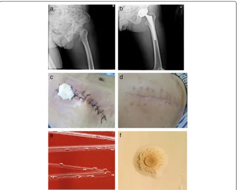
A 59-year-old male was admitted to Chongqing Sha-pingba District Chenjiaqiao hospital, Chongqing, China. He suffered the fractures of left femoral neck after falling to the ground (Fig. 1a). On July 16, 2017, the left total hip replacement was conducted and prosthetic hip in position was shown under X-ray (Fig. 1b). Cefazolin so-dium (1 g IV q8h) was started for prophylactic adminis-tration. His indwelling urinary catheter was removed after 24 h. On the 8th day after surgery, however, the pa-tient presented with left hip pain and clinical signs of in-fection, including fever (38.5 °C), redness and swelling around the surgical site (Fig. 1c), and he also reported local tenderness. His blood examination demonstrated the white blood cell (WBC), C-reactive protein (CRP), * Correspondence:zs25041@126.com
3Laboratory of Clinical Microbiology and Infectious Diseases, Department of
Pulmonary and Critical Care Medicine, China-Japan Friendship Hospital, Beijing, China
4Center for Respiratory Diseases, China-Japan Friendship Hospital, Beijing,
China
Full list of author information is available at the end of the article
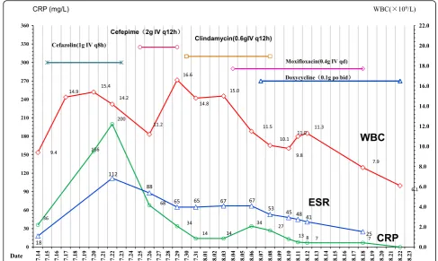
and erythrocyte sedimentation rate (ESR) significantly increased during postoperative period, as shown in Fig.2. Furthermore, the screening tests for human immuno-deficiency virus, hepatitis B virus and hepatitis C virus infections were non-reactive, and no abnormality in liver or renal function tests was observed. His T-lymphocyte subsets and gamma-globulin analysis were within nor-mal range.
On July 25, 2017, approximately 400-ml light-yellow, odorless subcutaneous fluid was punctured at surgical site, and forwarded to the microbiological laboratory for bacter-ial smear and culture. A large amount of
polymorpho-nuclear leucocytes (PMNs) were detected but no
microorganism on gram-staining smear. Moreover, there was a negative growth on the blood and chocolate agar plates. The repeated blood cultures using the BacT/ALERT
3D blood culture microbial detection system (bioMérieux SA, Marcy l’Étoile, France) were negative. The post-surgical infection was still under suspicion, and the wound was cleaned with iodophor and drainage gauze was placed. Ce-fepime (2 g IV q12h) was administrated. However, empiric therapy was still ineffective, the prosthetic hip infection de-teriorated, fever persisted, and on July 26, the debridement of left prosthetic hip was performed and the seroma, super-ficial fascia, deep fascia, deep tissue, as well as subcutaneous fluid collected during the surgery were sent for culture on blood and chocolate agar plates. Unexpectedly, tiny col-onies grew after 48 h, and they grew to approximately 0.5 mm colonies in diameter after 7-day incubation (Fig. 1e), and were identified as M. hominisby the bioMérieux® SA Mycoplasma IST2 kit (bioMérieux, France). The identifica-tion was also confirmed with 16S rRNA sequencing.
Fig. 1aFractures of left femoral neck under X-ray at admission.bProsthetic hip in right position (2 h after hip replacement under X-ray).c
Redness and swelling around surgical site, wound fluctuation in palpation (8 days after surgery).dRecovered after anti-M. hoministreatment with the combination of moxifloxacin, and doxycycline (18 days after surgery).eAfter 7 day of incubation of subcutaneous fluid collected during debridement on blood agar at 37 °C in a 5% CO2atmosphere, tiny, nonhemolytic, transparent colonies grew on Columbia blood agar plate.f
[image:2.595.58.540.86.472.2]In vitro antimicrobial susceptibility testing (AST) re-vealed that the organism was susceptible to doxycycline, clindamycin and levofloxacin, but resistant to azithromy-cin by the bioMérieux® SA Mycoplasma IST2 kit (Biomer-ieux, France). Combination therapy with clindamycin hydrochloride (0.6 g IV q12h) and moxifloxacin (400 mg IV QD) was initiated, as shown in Fig. 2. The patient defervesced within 3 days. His infection site recovered gradually (Fig.1d). Repeated X-ray scans before discharge showed marked improvement of his prosthetic hip. The patient was discharged on August 23, without further complications. No recurrence of symptoms and signs was reported during three-month outpatient follow-up.
Microbiological test
After 48 h of incubation on blood agar at 37 °C in a 5%
CO2 atmosphere, pinpoint-sized, non-hemolytic, and
transparent colonies were found on Columbia blood agar plate. The colonies were difficult to emulsify in saline water during the preparation of suspension solution for identifi-cation and AST. Both Gram stain and Wright-Giemsa mixed stain of the isolates demonstrated no bacterial morphology under × 1000 magnification, and only granular aggregates were detected. Fried-egg-type colonies of M. hominis growed on solid media (Zhongqisheng Hebei Bio-tech Co., Ltd.) 5 days after subculture (Fig.1f).
Discussion and conclusions
M. hominisis part of the normal inhabitant of the geni-tourinary tract [6]. However, in line with publicly-avail-able documents, it might disseminate to other body sites secondary to a disruption of the mucosa or in patients with autoimmune disorders, hypogammaglobulinemia, and other underlying immunosuppressions [3, 12–15]. Herein, we described the clinical circumstances, treat-ment, and outcomes of a postoperative septic complica-tion due to the microorganism after hip replacement. To the best of our knowledge, this is the first report of M. hominis as the causative, fastidious agent of prosthetic hip infection in China.
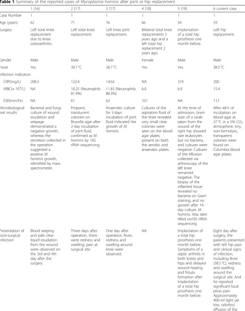
To better understand the characteristics of postoperative infection after hip or knee replacement, PubMed was searched for literature review and 5 cases byM. hominisin 4 reports were included for comparison (Table1) [16–19]. The literature review demonstrated that, including our case, the gender ratio of male/female sufferedM. hominis infec-tion after joint or tip replacement was 5:1. The median age was 64 years old. Furthermore, as documented, CRP con-centrations were available in 4 out of 5 cases, and all their CRP levels were higher than 100 mg/L. Our case also had an increased CRP (200 mg/L), hinting elevated CRP level would help in suspecting postoperative M. hominis infec-tion. The review also described that the most common and
[image:3.595.53.541.88.379.2]Table 1Summary of the reported cases ofMycoplasma hominisafter joint or hip replacement
1 [16] 2 [17] 3 [17] 4 [18] 5 [19] 6 current case
Case Number 1 1 1 1 1 1
Age (years) 62 71 76 66 54 59
Surgery Left total knee
replacement due to knee osteoarthritis.
Left total knee replacement.
Left knee joint replacement.
Bilateral total knee replacements 5 years ago and a left total hip replacement 2 years ago.
Implantation of a total hip prosthesis one month before.
Left hip replacement.
Gender Male Male Male Female Male Male
Fever Yes 39.7 °C 38.7 °C Yes Yes 38.5 °C
Infection indicators
CRP(mg/L) 208.3 122.4 143.6 NA 374 200
WBC(× 109/L) NA 10.25 (Neutrophils
81.4%)
11.83 (Neutrophils 88.3%)
6.0 6.9 15.4
ESR(mm/hr) NA 61 63 101 NA 112
Microbiological test results
Bacterial and fungi culture of wound exudation and seepage demonstrated a negative growth, whereas the secretion collected in the operation suggested a positiveM. hominisgrowth, identified by mass spectrometer.
Pinpoint, translucent colonies on Brucella agar after 2-day incubation of joint fluid, confirmed asM. hominisby 16S rRNA sequencing.
Anaerobic culture for 3 days incubation of joint fluid indicated the growth ofM. hominis.
Cultures of the aspiration fluid of the knee revealed very small clear colonies were seen on the blood agar plates, present on both the aerobic and anaerobic plates.
At the time of admission, Gram stain of a swab taken from the wound of the right hip showed rare leukocytes but no bacteria, and cultures were negative. Cultures of the effusion collected via arthroscopy of the left knee remained negative. The biopsy of the inflamed tissue revealed no bacteria on Gram staining, and no growth after 14-day culture.M. hominis. Was iden tifiedvia16S rRNA sequencing.
After 48 h of incubation on blood agar at 37 °C in a 5% CO2
atmosphere, tiny, non-hemolytic, transparent colonies were found on Columbia blood agar plates. Presentation of post-surgical infection Blood seeping and pale clear liquid exudation from the wound were observed on the 3rd and 4th day after the surgery.
Three days after operation, there were redness and swelling, pain at surgical site.
One day after operation, fever, redness and swelling around knee were observed.
NA Implantation of
a total hip prosthesis one month before. Symptoms of a septic arthritis in both knees and hips and delayed wound healing and fistula formation after implantation of a total hip prosthesis one month before.
effective test for diagnosing joint or hipM. hominis infec-tion was the culture of wound exudainfec-tion, joint fluid, and as-piration fluid of the knee [16–19]. In our case,M. hominis was recovered from subcutaneous puncture fluid and suc-cessfully identified. This showed that, if the tiny colonies grew on the blood agar plate without obvious bacteria shapes under gram-staining smears,M. hominisshould be suspected of being underlying pathogen [16,17]. Presently, the molecular methods, such as 16S rRNA sequencing or real-time PCR, might be used for the identification of infec-tions caused by the bacterium [17,19].
Furthermore, it is difficult to clarify the possible portal of entry of M. hominisin cases of this postoperative in-fection [20]. In accordance with previous reports, the source for an M. hominis in postoperative hip or joint infections might be explained by seeding of surgical site through transient bacteraemia. This bloodstream infec-tion occurred after urinary catheterizainfec-tion if the genito-urinary tract had been colonized by the microorganism. Indeed, urinary tract catheterization has been associated with mycoplasma bacteraemia leading to the seeding of brain-damaged tissues in brain abscess cases [5,6,9,21]. Similarly, in our case, a possible pathway might be in-dwelling catheter used during surgery and a possible route for hematogenous spread to surgical site. However, it is rather difficult to definitively identify the source of infection.
It presented a challenge to identifyM. hominisas patho-gen due to its elusiveness and fastidious slow-growing
nature [5, 8, 13, 22, 23]. This might be explained by the following reasons. Firstly, M. hominishas a 3-layer sterol membrane but lacks cell wall. Consequently, the inability to detectMycoplasma spp.by routine gram-staining con-tributes to the failure of detection in the clinical
speci-mens [24]. In present case, the gram-staining and
Wright-Giemsa mixed staining smear of subcutaneous fluid and the colonies demonstrated no bacterial morph-ology. Secondly, the slow-growth properties ofM. hominis made the detection on plates challenging, because it gen-erally takes several days (often ≥2 days) to grow into tiny colonies on the media commonly used in laboratory and the requirement for extended incubation period makes a timely diagnosis less likely. And moreover, the routine bio-chemical methods might fail to identify it correctly [18]. Thirdly, it is rather difficult to detect the growth of M. hominis in standard blood culture bottle solu-tions that use polyanethol sulfonate as an anticoagulant but rather requiring special methods for growth through automatic detection systems in those with sus-pected bacteremia, and false-negative results are likely yielded [16–19, 24]. Taken together, the post-surgical M. hominisinfection cases are not readily detected via
standard microbiology methods [7, 19]. Considering
[image:5.595.57.538.97.359.2]the high urethral carriage rate ofM. hominis (~ 15% of healthy adults) and catheterization is a common pro-cedure during operation, the possibility of postopera-tive Mycoplasma infection might be under-diagnosed or reported [8].
Table 1Summary of the reported cases ofMycoplasma hominisafter joint or hip replacement(Continued)
1 [16] 2 [17] 3 [17] 4 [18] 5 [19] 6 current case
Antibiotic prevention
Cefazolin Ceftazidime,
vancomycin
Vancomycin NA NA Cefazolin
Antibiotic treatment.
Cefazolin was replaced by vancomycin. Later transferred to the combination into erythromycin, clindamycin and minocyline.
Vancomycin, metronidazole.
Vancomycin. Later switched to azithromycin, doxycycline, moxifloxacin.
Cefazolin 500 mg of tetracycline iv every 8 h. After the first week, switched to oral doxycycline (200 mg/day), and over the next 3 weeks.
Ciprofloxacin and clindamycin; subsequently changed to cefazolin and clindamycin, continued for 4 weeks; and later changed to moxifloxacin and rifampin for a presumed chronic S. epidermidis infection. Treatment with moxifloxacin was initiated, however the patient’s condition continued to deteriorate.
Cefepime, clindamycin, moxifloxacin, and doxycycline.
Blood culture Negative Negative Negative Negative Negative Negative
Outcome Recovery Recovery Recovery Recovery Dead Recovery
Our Pubmed review showed that 83.3% (5/6) of the patients survived after appropriate antimicrobial treat-ment. Furthermore, previous studies demonstrated that the mycoplasmas resulted in serious infections without timely detection [15,19,22]. For example, a patient with implantation of a total hip prosthesis died due to a post-operative hip prosthesis and disseminated infection by M. hominis and Ureaplasma parvum [19]. Accordingly, if a patient developed unexplained post-surgical fever in cases of otherwise culture-negative infections, particu-larly if treat with wide-spectrum antibiotics meets with a poor response, it is especially important to consider the potential ofMycoplasmasas pathogens.
Empiric therapy for postoperative infections generally includes agents such as beta-lactams and vancomycin that act on the bacterial cell wall. Such an initial therapy will show no efficacy against M. hominis infections due to its lack of cell wall [16, 17]. Furthermore, M. hominis is, in contrast to most mycoplasmas, intrinsically resist-ant to currently available macrolide resist-antibiotics due to the mutations in the 23S rRNA gene and is the only mycoplasma susceptible to clindamycin, which often used for eradicating M. hominis with favorable results [16]. Quinolones (ciprofloxacin or moxifloxacin) or
tet-racyclines (minocyline) are active against M. hominis
and moxifloxacin appears to have the greatest activity as the most effective therapeutic agent. If M. hominis was correctly identified as underlying pathogen, the antibi-otics would be therefore switched to the right agents with a marked improvement of clinical syndromes and a favorable result [16,19]. The patient in our case had the regimen of the combination of moxifloxacin and doxy-cycline and had positive response.
In summary, the postoperative infection after hip
re-placement secondary to M. hominis is rare. Currently
there are 4 published reports of septic arthritis caused by M. hominis after hip or knee replacement in adults. Our case added to this body of evidence. The clinicians should recognize the possibility of M. hominis involve-ment in postoperative infections without microbiological findings or response to standard therapy, and consider changing antibiotic regimen.
Abbreviations
CRP:C-reactive protein (CRP); ESR: Erythrocyte sedimentation rate; WBC: White blood cell
Acknowledgements
Not applicable.
Funding
This study was supported by Beijing Municipal Science &Technology Commission, PR China (No. Z171100001017118). The funding bodies had no role in the design of the study and collection, analysis, and interpretation of data and in writing the manuscript.
Availability of data and materials
All the data and material involved in the current study are available from the corresponding author on reasonable request.
Authors’contributions
BHL was involved in the conception and design of the study, and writing, review and editing of the manuscript. LLX was involved in the data acquisition and analysis, and collected clinical data. Both authors read and approved the final manuscript
Ethics approval and consent to participate
The institutional review boards at the Chongqing Shapingba District Chenjiaqiao hospital approved the study protocol.
Consent for publication
Written informed consent was obtained from the patient for publication of this case report and any accompanying images. A copy of the written consent is available for review by the editor of this journal.
Competing interests
The authors declare that they have no competing interests.
Publisher’s Note
Springer Nature remains neutral with regard to jurisdictional claims in published maps and institutional affiliations.
Author details 1
Department of Laboratory Medicine, Chongqing Shapingba District Chenjiaqiao hospital, Chongqing, China.2Affiliated Hospital of Chongqing
Medical and Pharmaceutical College, Chongqing, China.3Laboratory of Clinical Microbiology and Infectious Diseases, Department of Pulmonary and Critical Care Medicine, China-Japan Friendship Hospital, Beijing, China.
4Center for Respiratory Diseases, China-Japan Friendship Hospital, Beijing,
China.5National Clinical Research Center of Respiratory Diseases, Beijing, China.
Received: 2 March 2018 Accepted: 7 January 2019
References
1. Allen-Daniels MJ, Serrano MG, Pflugner LP, Fettweis JM, Prestosa MA, Koparde VN, Brooks JP, Strauss JF 3rd, Romero R, Chaiworapongsa T, et al. Identification of a gene in Mycoplasma hominis associated with preterm birth and microbial burden in intraamniotic infection. Am J Obstet Gynecol. 2015;212(6):779 e771–779 e713.
2. Flouzat-Lachaniette CH, Guidon J, Allain J, Poignard A. An uncommon case of mycoplasma hominis infection after total disc replacement. Eur Spine J. 2013;22(Suppl 3):S394–8.
3. Phuah CL, Javid B, Aliyu SH, Lever AM. A case of mycoplasma hominis septic arthritis postpartum. J Infect. 2007;55(5):e135–7.
4. Krijnen MR, Hekker T, Algra J, Wuisman PI, Van Royen BJ. Mycoplasma hominis deep wound infection after neuromuscular scoliosis surgery: the use of real-time polymerase chain reaction (PCR). Eur Spine J. 2006;15(Suppl 5):599–603.
5. Zhou M, Wang P, Chen S, Du B, Du J, Wang F, Xiao M, Kong F, Xu Y. Meningitis in a Chinese adult patient caused by mycoplasma hominis: a rare infection and literature review. BMC Infect Dis. 2016;16(1):557. 6. Whitson WJ, Ball PA, Lollis SS, Balkman JD, Bauer DF. Postoperative
mycoplasma hominis infections after neurosurgical intervention. J Neurosurg Pediatr. 2014;14(2):212–8.
7. Yokoyama H, Domen T, Hiragata S, Ogawa T, Matsumoto T, Ishizuka O. Postoperative mycoplasma hominis infection after robot-assisted laparoscopic radical prostatectomy: a case report. Asian J Endosc Surg. 2016; 9(2):146–8.
8. Bergin SM, Mendis SM, Young B, Binti Izharuddin E. Postoperative mycoplasma hominis brain abscess: keep it in mind!. BMJ Case Rep. 2017; 2017:1–3.
10. Meyer RD, Clough W. Extragenital mycoplasma hominis infections in adults: emphasis on immunosuppression. Clin Infect Dis. 1993;17(Suppl 1):S243–9. 11. Miranda C, Camacho E, Reina G, Turino J, Rodriguez-Granger J, Yeste R,
Bautista MF, Garcia M, Alados JC, De la Rosa M. Isolation of mycoplasma hominis from extragenital cultures. Eur J Clin Microbiol Infect Dis. 2005;24(5): 334–7.
12. Fernandez S, Nicolas D, Pericas JM, Castro Rebollo P, Vila J, Miro JM, Blanco JL, Nicolas JM. A case of Mycoplasma hominis disseminated infection in a human immunodeficiency virus-1-infected pregnant woman with hypogammaglobulinemia. J Microbiol Immunol Infect. 2017;50(1):118–9. 13. Romeu Prieto JM, Lizcano Lizcano AM, Lopez de Toro Martin Consuegra I,
Largo Pau J, Lopez Almodovar LF, Garcia Camacho E. Culture-negative endocarditis: mycoplasma hominis infection. Rev Esp Cardiol. 2015;68(11): 1037–8.
14. Lee EH, Winter HL, van Dijl JM, Metzemaekers JD, Arends JP. Diagnosis and antimicrobial therapy of mycoplasma hominis meningitis in adults. Int J Med Microbiol. 2012;302(7–8):289–92.
15. Wylam ME, Kennedy CC, Hernandez NM, Peters SG, Maleszewski JJ, Cassivi SD, Scott JP. Fatal hyperammonaemia caused by mycoplasma hominis. Lancet. 2013;382(9908):1956.
16. Qiu HJ, Lu WP, Li M, Wang ZM, Du QY, Wang AM, Xiong Y. The infection of Mycoplasma hominis after total knee replacement: Case report and literature review. Chin J Traumatol. 2017;20(4):243–5.
17. Lee JH, Lee JH, Lee NY, Ha CW, Chung DR, Peck KR. Two cases of septic arthritis by mycoplasma hominis after total knee replacement arthroplasty. Korean J Lab Med. 2009;29(2):135–9.
18. Sneller M, Wellborne F, Barile MF, Plotz P. Prosthetic joint infection with mycoplasma hominis. J Infect Dis. 1986;153(1):174–5.
19. MacKenzie CR, Nischik N, Kram R, Krauspe R, Jager M, Henrich B. Fatal outcome of a disseminated dual infection with drug-resistant mycoplasma hominis and Ureaplasma parvum originating from a septic arthritis in an immunocompromised patient. Int J Infect Dis. 2010;14(Suppl 3):e307–9. 20. Pailhories H, Rabier V, Eveillard M, Mahaza C, Joly-Guillou ML, Chennebault
JM, Kempf M, Lemarie C. A case report of mycoplasma hominis brain abscess identified by MALDI-TOF mass spectrometry. Int J Infect Dis. 2014; 29:166–8.
21. Cuchý E, Cherta I, Garau J. Mycoplasma hominis catheter-related infection in a patient with multiple trauma. Clin Microbiol Infect. 2000;6(2):115–6. 22. Reissier S, Masson R, Guerin F, Viquesnel G, Petitjean-Lecherbonnier J, Pereyre S, Cattoir V, Isnard C. Fatal nosocomial meningitis caused by mycoplasma hominis in an adult patient: case report and review of the literature. Int J Infect Dis. 2016;48:81–3.
23. Koshiba H, Koshiba A, Daimon Y, Noguchi T, Iwasaku K, Kitawaki J. Hematoma and abscess formation caused by mycoplasma hominis following cesarean section. Int J Women's Health. 2011;3:15–8.