R E S E A R C H
Open Access
Moderate hyperoxic versus
near-physiological oxygen targets during and
after coronary artery bypass surgery: a
randomised controlled trial
Bob Smit
1*, Yvo M. Smulders
2, Monique C. de Waard
1, Christa Boer
3, Alexander B. A. Vonk
4, Dennis Veerhoek
4,
Suzanne Kamminga
3, Harm-Jan S. de Grooth
1, Juan J. García-Vallejo
5, Rene J. P. Musters
6, Armand R. J. Girbes
1,
Heleen M. Oudemans - van Straaten
1and Angelique M. E. Spoelstra - de Man
1Abstract
Background:The safety of perioperative hyperoxia is currently unclear. Previous studies in patients undergoing coronary artery bypass surgery suggest reduced myocardial damage when avoiding extreme perioperative hyperoxia (>400 mmHg). In this study we investigated whether an oxygenation strategy from moderate hyperoxia to a near-physiological oxygen tension reduces myocardial damage and improves haemodynamics, organ dysfunction and oxidative stress.
Methods:This was a single-blind, single-centre, open-label, randomised controlled trial in patients undergoing elective coronary artery bypass surgery. Fifty patients were randomised to a partial pressure of oxygen in arterial blood (PaO2) target of 200–220 mmHg during cardiopulmonary bypass and 130–150 mmHg during intensive care
unit (ICU) admission (control group) versus lower targets of 130–150 mmHg during cardiopulmonary bypass and 80–100 mmHg at the ICU (conservative group). Primary outcome was myocardial injury (CK-MB and Troponin-T) at ICU admission and 2, 6 and 12 hours thereafter.
Results:Weighted PaO2during cardiopulmonary bypass was 220 mmHg (interquartile range (IQR) 211–233) vs. 157
(151–162) in the control and conservative group, respectively (P< 0.0001). During ICU admission, weighted PaO2
was 107 mmHg (86–141) vs. 90 (84–98) (P= 0.03), respectively. Area under the curve of CK-MB was median 23.5μg/L/h (IQR 18.4–28.1) vs. 21.5 (15.8–26.6) (P= 0.35) and 0.30μg/L/h (0.25–0.44) vs. 0.39 (0.24–0.43) (P= 0.81) for Troponin-T. Cardiac index, systemic vascular resistance index, creatinine, lactate and F2-isoprostane levels were not different between groups.
Conclusions:Compared to moderate hyperoxia, a near-physiological oxygen strategy does not reduce myocardial damage in patients undergoing coronary artery bypass surgery. Conservative oxygen administration was not associated with increased lactate levels or hypoxic events.
Trial registration:Netherlands Trial Registry NTR4375, registered on 30 January 2014
Keywords:Hyperoxia, Oxygen, Intensive care unit, Cardiac surgery, CABG
* Correspondence:[email protected]
1Department of Intensive Care, VU University Medical Center, Amsterdam,
The Netherlands
Full list of author information is available at the end of the article
Background
During cardiac surgery, and especially during cardiopul-monary bypass (CPB), patients often receive supplemental oxygen as a precaution to prevent hypoxia. As a result, the arterial oxygen tension (partial pressure of oxygen in ar-terial blood; PaO2) often rises to supraphysiological levels (hyperoxia), which is by many considered as a salutary oxygen reserve. PaO2 levels of more than 200 to 300 mmHg during CPB are not exceptional [1–6] and hyperoxia is also common in mechanically ventilated pa-tients in the intensive care unit (ICU) [7, 8]. There are currently no guidelines on oxygenation during cardiac sur-gery. For intensive care, current target PaO2 recommenda-tions are limited to patients with acute respiratory distress syndrome [9].
The safety of hyperoxia is questionable [10, 11]. Besides the direct toxic effect of high fractions of inspired oxygen on lung tissue, animal [12–14] and human studies [15–19] show that hyperoxia induces vasoconstriction and may re-duce cardiac output and organ perfusion. The mechanism of hyperoxic vasoconstriction is unclear, but augmented production of reactive oxygen species (ROS) seems to play a pivotal role [20, 21]. Excessive ROS may, amongst other things, enhance ischaemia/reperfusion injury, which may translate into worse clinical outcome.
In patients after cardiac arrest, hyperoxia is associated with worse functional outcome and increased mortality [22–26]. In patients with cerebral [27] or myocardial farction [28], hyperoxia has been shown to increase in-farct size and mortality. Data on the effect of oxygen on cardiac markers are scarce, since in animal studies direct measurements of infarct size are preferred and human studies are lacking. However, the recently published AVOID trial shows increased Troponin-I and creatine kinase (CK) when patients received supplemental oxygen after myocardial infarction [28].
Patients undergoing coronary artery bypass graft (CABG) surgery may be particularly vulnerable to the ef-fects of hyperoxia because of fluctuations in cardiac func-tion due to anaesthesia, blood loss, altered temperature, non-pulsatile perfusion and fluid shifts post-surgery. Also, hyperoxia may have detrimental effects on CPB-related myocardial ischaemia/reperfusion injury, pulmonary re-perfusion and systemic inflammation.
The few clinical trials addressing the effect of hyper-oxia during or after CABG surgery [1–6, 29] suggest that avoidance of extreme hyperoxia reduces myocardial damage (>400 mmHg) [1, 2]. The design of these studies is variable, with exposure of patients to single [4, 5] or multiple short episodes [6] of hyperoxia and application of different oxygen tensions during reperfusion [2] or continuously throughout CPB [1]. In recognition of dir-ect pulmonary toxic effdir-ects of high inspiratory oxygen fractions these extreme PaO2 values (~400 mmHg) are
no longer used in current practice. Nevertheless, cur-rently employed oxygen levels remain high, which raises the question whether further PaO2reductions are benefi-cial. Additionally, no studies continued to control the ar-terial oxygen tension after admission to the ICU.
The aim of this randomised controlled trial (RCT) was to determine whether near-physiological oxygen targets during and after CABG surgery reduces myocardial dam-age and improves haemodynamics, organ function and oxidative stress compared to standard care, which involves moderate hyperoxia.
Methods
Study design and ethical approval
This investigator-initiated, single-centre, RCT (Netherlands Trial Register number NTR4375) was performed at the operating room (OR) and ICU of the VU University Medical Centre (Amsterdam, the Netherlands). The study protocol was approved by the Dutch Central Committee on Research Involving Human Subjects (NL43882.029.13).
Patients
Patients over 18 years of age with a body surface index ≥1.9 m2 and a preoperative haemoglobin con-centration ≥121 g/L, scheduled for elective, isolated CABG surgery with CPB were approached for participa-tion and included in the study after written informed con-sent. Body surface index and haemoglobin criteria were chosen to ensure sufficient oxygen-carrying capacity of blood after haemodiluton by the CPB circuit. Exclusion criteria were presence of a perioperative intra-aortic bal-loon pump or a medical history positive for chronic ob-structive pulmonary disease.
Clinical management Anaesthesia
Anaesthesia was induced with a combination of sufenta-nil (3–7 μg/kg), midazolam (0.1 mg/kg) and pancuro-niumbromide (0.1 mg/kg) or rocuronium (0.6 mg/kg). General anaesthesia was maintained using propofol (200–400 mg/h). After induction, all patients received dexamethasone (1 mg/kg), cefazoline (1000 mg) and tranexamic acid (1000 mg). Another 2000 mg of tranex-amic acid was administered following termination of CPB. After surgery, patients were supported with a low dose of dopamine.
Cardiopulmonary bypass
was washed, concentrated and returned to the patient. For more details, see Additional file 1.
Intensive care unit
Propofol sedation was stopped when haemodynamics were stable, chest drain production was minimal and pa-tient temperature was normal. When sufficiently awake, patients were extubated. Patients received three gifts of cefazoline (1000 mg) at 8-h intervals.
Randomisation and intervention
After inclusion, patients were randomised by drawing of sealed envelopes into the “control” or “conservative” group (1:1 ratio). Patients were enrolled and assigned by BS, who was not involved in the clinical care of patients. While on CPB, the control group was oxygenated, aiming for a target PaO2 of 200–220 mmHg during placement of the aortic cross-clamp as per current practice. After admission to the ICU, a target PaO2 of 130–150 mmHg was applied. The conservative group was oxygenated to a target PaO2of 130–150 mmHg dur-ing aortic cross-clampdur-ing and 80–100 mmHg at the ICU. Surgeons were unaware of group allocation.
Patients in both study groups were pre-oxygenated with a fraction of inspired oxygen (FIO2) of 1.0 and subse-quently intubated. In the control group, FIO2was set to a minimum of 0.4 and a maximum of 0.6. In the conserva-tive group, FIO2was set lower than or equal to 0.40, while ensuring an oxygen saturation≥94 %. During CPB, PaO2 was continuously monitored with a Terumo CDI 500 (Terumo Europe, Leuven, Belgium). After ICU admission, PaO2 was monitored approximately every hour by blood gas sampling. While patients were intubated, FIO2 was adjusted (with an upper limit of 60 %, higher only if clinic-ally necessary) to reach target PaO2. After extubation, pa-tients breathed room air or received supplemental oxygen through a nasal cannula or an oxygen mask. Other means of non-mechanical ventilation were only issued on clinical indication and not for study purposes.
Primary and secondary endpoints
The primary endpoint was myocardial damage, as reflected by area under the curve (AUC) of CK-MB. Sec-ondary endpoints were differences in haemodynamics, tis-sue perfusion, organ function, and oxidative stress. As safety outcomes, we monitored for desaturations (oxygen saturation <88 %) and hypoxic events (PaO2< 55 mmHg).
Myocardial damage/tissue perfusion, organ function and injury
Myocardial damage was determined by measuring CK-MB, Troponin-T and CK at the following time points: prior to induction of anaesthesia and 0, 2, 6 and 12 h after ICU admission. If no peak value was measured
within this timeframe, additional measurements were performed at 6-h intervals. As an estimation of organ perfusion and oxygenation, creatinine and lactate levels were measured at the same time points. Lung injury score (LIS) was assessed at ICU admission and 2 days post-operatively [30]. PaO2/FIO2ratio was measured ap-proximately every hour, simultaneously with blood gas analyses for intervention control.
Haemodynamics
In patients who received a pulmonary artery catheter (under the anaesthesiologist’s discretion), cardiac index (CI) and systemic vascular resistance index (SVRI) were measured prior to thoracotomy, after chest closure and 0, 2, 6 and 12 h after ICU admission.
Oxidative stress
Lipid peroxidation by ROS was assessed by measuring F2-isoprostanes (8-iso prostaglandin F2α, iPF2α-III) in plasma by liquid chromatography tandem mass spec-trometry. EDTA samples were obtained prior to induction of anaesthesia and 6 h after ICU admission. Directly after sampling, blood was centrifuged at 2500 g for 10 minutes and plasma was stored at–80 °C until analysis.
For the measurement of ROS production by circulat-ing polymorphonuclear leukocytes (PMN), samples were taken before induction of anaesthesia and at 1 h of CPB. A portion of whole blood was incubated with a fluores-cent ROS probe for 1 h (CellROX Green; Invitrogen, Grand Island, NY, USA). Mean fluorescence intensity (MFI) of 5000 cells, which is proportionate to the ROS produced, was then measured on a Beckton Dickinson FACS Calibur flowcytometer.
For a more elaborate description of these methods, see Additional file 2.
Statistics/analysis Sample size calculation
A previous small trial, which compared extreme versus less extreme hyperoxia during CPB, showed a periopera-tive increase in CK-MB of 20 to 80 μg/L and 20 to 45 μg/L, respectively [2]. In the present trial, we ex-pected the effect to be smaller. With an anticipated standard deviation of 20μg/L and anαof 0.05, inclusion of 25 patients in each arm provides a power of 80 % to detect a CK-MB difference of 20 %.
Analysis
between these measurements; the sum of all products was then divided by total time. Weighted PaO2/FIO2 ratios were calculated following the same principle. Differences between groups were tested with the non-parametric Mann-Whitney U test or Fisher’s exact test where appropriate.
Results
Patients
In the period between November 2013 and May 2015, 57 patients gave written informed consent for study par-ticipation. Seven patients were excluded prior to the start of the study protocol due to changes in the surgery schedule. The remaining 50 patients, 25 in each arm, completed the study protocol and were included for ITT analysis. The participants were Caucasian males with a median age of 67 years (IQR 63–71). For more detailed baseline and surgical characteristics of patients, see Table 1. For a CONSORT flow diagram, see Additional file 3.
Intervention
In the control group, weighted PaO2 during CPB was 220 mmHg (213–233) versus 157 (152–161) (P< 0.0001) in the conservative group. The lowest haemoglobin concentration during CPB was 92 g/L (87–97) vs. 89 (84–95) (P= 0.170). During aortic cross-clamping, weighted PaO2 was 214 mmHg (207–224) vs. 146 (141–151) (P< 0.0001). During ICU admission, weighted PaO2 was 107 mmHg (88–140) vs. 90 (85–97) (P= 0.034), respectively. Haemoglobin concentrations were 119 g/L (111–126) vs. 119 (113–122) (P= 0.690) on ICU admission. Patients in the control group were on mechanical ventilation for 4.1 h (3.2–5.0) with an FIO2of 0.52 (0.43–0.60). Patients in the conservative group were on mechanical ventilation for 4.3 h (3.1–6.2) (P= 0.383) with an FIO2of 0.38 (0.32–0.44) (P< 0.0001).
Post-operative course
Three patients underwent a re-thoracotomy for post-operative bleeding (one in the control group vs. two in the conservative group). One patient in the conservative group then developed cardiogenic shock (lowest PaO2= 76 mmHg). Two patients (one in either group) were scheduled for percutaneous coronary intervention after full recovery due to incomplete revascularisation. One patient in the conservative group had a ST-segment elevation myocardial infarction (STEMI) during ICU admission that did not require additional treatment (CK-MBmax= 74.9 μg/L). None of these events led to pro-longed ICU admission. Patients in the control group were admitted to the ICU for 22 h (19–24) versus 22.5 h (19.5– 24.5) for patients in the conservative group (P= 1.000).
Myocardial damage
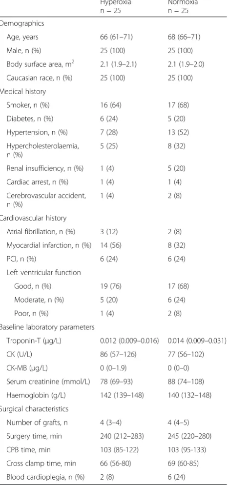
[image:4.595.306.540.104.602.2]CK-MB values generally peaked at 6 h after ICU admis-sion (Fig. 1a). AUC were 23.5 μg/L/h (18.4–28.1) in the control group vs. 21.5 (15.8–26.6) (P= 0.347) in the conservative group. Individual maximum values were 25.8μg/L (21.0–32.3) vs. 24.9 (18.4–31.5) (P= 0.528).
Table 1Baseline and surgical characteristics Hyperoxia n = 25
Normoxia n = 25
Demographics
Age, years 66 (61–71) 68 (66–71)
Male, n (%) 25 (100) 25 (100)
Body surface area, m2 2.1 (1.9–2.1) 2.1 (1.9–2.0)
Caucasian race, n (%) 25 (100) 25 (100)
Medical history
Smoker, n (%) 16 (64) 17 (68)
Diabetes, n (%) 6 (24) 5 (20)
Hypertension, n (%) 7 (28) 13 (52)
Hypercholesterolaemia, n (%)
5 (25) 8 (32)
Renal insufficiency, n (%) 1 (4) 5 (20)
Cardiac arrest, n (%) 1 (4) 1 (4)
Cerebrovascular accident, n (%)
1 (4) 2 (8)
Cardiovascular history
Atrial fibrillation, n (%) 3 (12) 2 (8)
Myocardial infarction, n (%) 14 (56) 8 (32)
PCI, n (%) 6 (24) 6 (24)
Left ventricular function
Good, n (%) 19 (76) 17 (68)
Moderate, n (%) 5 (20) 6 (24)
Poor, n (%) 1 (4) 2 (8)
Baseline laboratory parameters
Troponin-T (μg/L) 0.012 (0.009–0.016) 0.014 (0.009–0.031)
CK (U/L) 86 (57–126) 77 (56–102)
CK-MB (μg/L) 0 (0–1.9) 0 (0–0)
Serum creatinine (mmol/L) 78 (69–93) 88 (74–108)
Haemoglobin (g/L) 142 (139–148) 140 (132–148)
Surgical characteristics
Number of grafts, n 4 (3–4) 4 (4–5)
Surgery time, min 240 (212–283) 245 (220–280)
CPB time, min 103 (85-122) 103 (95-133)
Cross clamp time, min 66 (56-80) 69 (60-85)
Blood cardioplegia, n (%) 2 (8) 6 (24)
Troponin-T levels rose substantially after surgery and usually peaked at 6 h after ICU admission (Fig. 1b). AUC were not different between groups (0.30 μg/L/h (0.25–0.44) vs. 0.39 (0.24–0.43); P= 0.695)). Individual peak values were 0.35μg/L (0.30–0.46) vs. 0.42 (0.26–0.49) (P= 0.923).
CK levels rose gradually during ICU stay and did not de-cline in the following 12-h study period. AUC were similar at 550 U/L/h (347–706) vs. 376 (229–425) (P= 0.144). Individual maximum values measured were 663 U/L (403–977) vs. 511 (329–637) (P= 0.165).
To correct for an imbalance between groups in the number of patients who received warm blood cardiople-gia (Table 1; P= 0.247), we performed a multiple linear regression with CK-MB AUC as the dependent variable, and study group, minutes on CPB and type of cardio-plegia as independent variables. Neither study group (β= 0.024, P= 0.874), nor minutes on CPB (β= 0.200, P= 0.178) nor type of cardioplegia (β=–0.003,P= 0.984) were associated with CK-MB AUC (R2= 0.041).
Tissue perfusion, organ function and injury
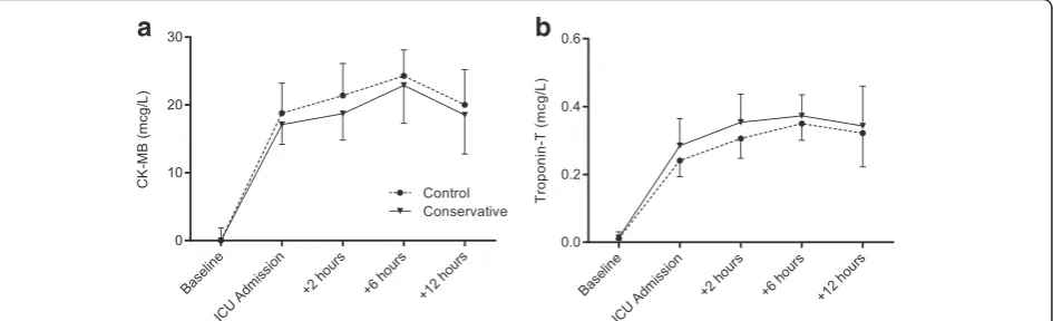
Creatinine remained stable throughout the study period (Fig. 2a). Lactate levels increased mildly in both groups and were not different between groups (Fig. 2b). On ICU ad-mission, lung injury scores were 1.3 (0.8–1.5) in the control group vs. 1.0 (0.7–1.5) (P= 0.290) in the conservative group. Two days later, LIS was 1.0 (0.8–1.5) vs. 1.0 (0–1.5) (P= 0.216). During ICU admission, weighted PaO2/FIO2 ra-tios were 236 (173–285) vs. 261 (199–311) (P= 0.244).
Haemodynamics
In 15 patients in the control group a pulmonary artery catheter was inserted. Preoperatively, 11 had good Left Ventricular Function (LVF), 3 moderate and 1 poor. In the conservative group, 18 patients received a pulmon-ary artery catheter, with 10 having good LVF, 6 moderate and 2 poor.
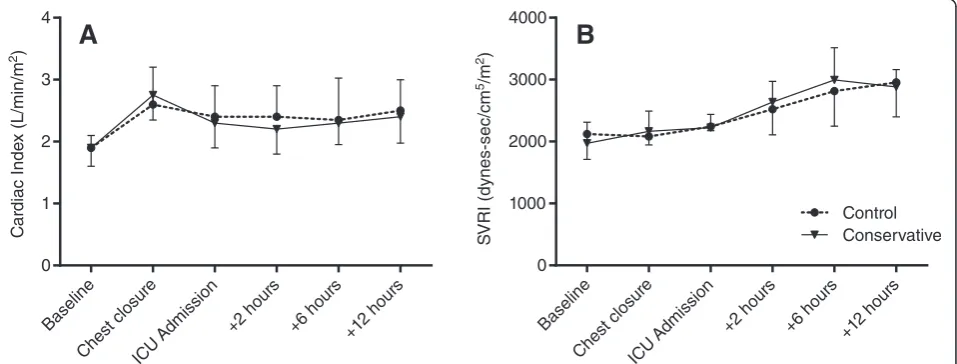
Before sternotomy, CI was similar between groups (1.9 L/min/m2 (1.6–2.1) vs. 1.9 (1.6–2.2); P= 1.000). After chest closure, CI was increased compared to base-line in both groups (2.6 (2.2–3.2) vs. 2.8 (2.4–3.0)). After admission to the ICU, CI decreased slightly and then remained stable throughout the next 12 hours (Fig. 3a). There was no difference between the two groups at any point. SVRI rose gradually in both groups (Fig. 3b), starting at 2120 dynes · s/cm5/m2 (1711–2254) in the control group and 1970 (1787–2311) in the conserva-tive group and reaching a maximum at 12 h of ICU admission (2955 (2398–3351) vs. 2883 (2564–3161); P= 0.861).
Nearly all patients in either group were supported with a low dose of dopamine in the first hours after ICU ad-mission (22/25 in the control group vs. 20/25 in the conservative group). Overall administration of vasoactive medicine was not different between groups (data not shown).
Oxidative stress
At baseline, there was no difference in F2-isoprostane levels between groups (32.1 ng/L (26.7–36.0) in the control group vs. 34.3 (24.8–43.2) in the conservative group; P= 0.467). Six hours after ICU admission, F2-isoprostane levels were lower in both groups compared to baseline (P< 0.001), but there was no difference be-tween groups (25.9 ng/L (23.7–28.5) vs. 27.3 (22.6–31.6); P= 0.399).
ROS production by PMN increased in the control group from a mean 21.3 MFI (95 % confidence interval 14.6–28) at baseline to 45.9 (21.8–70.0) (P= 0.001) at 1 h of CPB. In the conservative group, ROS production increased similarly from 15.6 MFI (10.1–20.7) at baseline to 45.2 (14.5–76.0) (P= 0.002) during CPB. There was no difference between groups either at baseline (P= 0.228) or at 1 h of CPB (P= 0.790).
a
b
[image:5.595.66.539.89.233.2]Safety outcome
All patients that participated in the study survived until hospital discharge. There were no hypoxic events (PaO2< 55 mmHg) in either study group.
Discussion
In the present RCT in patients undergoing scheduled isolated CABG surgery, we found no difference in markers of myocardial damage between a conventional moderate hyperoxic and a near-physiological oxygen strategy during and after surgery. The conservative oxy-genation strategy did not improve haemodynamics in comparison to moderate hyperoxia. However, lower oxy-gen targets during CPB and ICU admission could be ap-plied safely without risk of hypoxia.
With respect to myocardial damage, these findings are in apparent contrast to results from earlier clinical trials where the reduction of oxygen tension during CPB or reperfusion resulted in lower CK (672 ± 130 vs. 293 ± 21 U/l; P= 0.002) [1] or lower Troponin-T levels (~2.1 vs. ~0.8μg/L;P< 0.05) [2]. However, the high PaO2 investigated in the latter two studies are no longer applied today. The first study compared a PaO2 of ~400 mmHg during CPB in the control group to ~140 mmHg in the conservative group [2]. The second study compared reperfusion oxygen tensions of 450–550 mmHg to 200–250 mmHg [1]. This means that, in the conservative group, oxygen tension was reduced by 260–270 mmHg compared to the control group. In our study, the differ-ence was on average 70 mmHg during CPB and 20 mmHg during ICU admission. It is reasonable to assume that a
Fig. 2Perioperativeacreatinine andblactate levels. There were no differences between groups.ICUintensive care unit
Fig. 3Perioperative haemodynamic parameters.aCardiac index andbSVRI. There were no differences between groups.ICUintensive care unit,
[image:6.595.62.540.88.272.2] [image:6.595.60.539.521.703.2]smaller reduction in oxygen tension has a smaller effect on myocardial damage.
Our results are in contrast to those of the AVOID trial, which compared the effect of 8 L/min oxygen vs. no-oxygen supplementation in non-hypoxic patients with STEMI. The study showed increased CK, a trend for increased Troponin-I and increased infarction size when patients received supplemental oxygen [28]. Un-fortunately, the study was oxygen saturation-guided and there are no PaO2values available, so we cannot deduce whether oxygen tensions were in the same range. Of course, an obvious difference between the patient popu-lations is that, in STEMI, the insult is acute, while in CABG surgery the patient is prepared with moderate hypothermia and cardioplegia. This fundamental physio-logical difference may explain the contrasting results.
In addition, an important difference with previous CABG studies is that our patients received 1 mg/kg dexamethasone after anaesthesia induction [31]. Dexa-methasone is a potent glucocorticoid that reduces the formation of ROS by leukocytes in vivo [32] and, in CABG surgery, reduces the systemic inflammatory re-sponse syndrome [33, 34]. Since it is hypothesised that hyperoxia exerts its effects by augmenting production of ROS, it is possible that dexamethasone mitigates these effects. In anaesthetised [26, 33] and septic patients [34] supplemental oxygen can decrease CI and increase SVRI [14]. In these studies the contrast between the normoxic and hyperoxic state was high (i.e. switching from an FIO2of ~0.3 to 1.0) and short term (10–30 min). In our study, moderate hyperoxia appears to have no effect on CI or SVRI. Although the relatively lower PaO2 differ-ence between groups post-operatively may have led to a minor and immeasurable difference in haemodynamic parameters, it is also possible that the haemodynamic ef-fects are only temporary. Also, dexamethasone influ-ences vascular endothelial and smooth muscle Ca2+ mobilisation [35], which in turn modulates the balance between vasoconstriction and vasodilation which may have affected the vascular response to oxygen.
Over the years, CABG surgery has improved and the influence of oxygen tension may be less in current high-quality surgery and post-operative care. This view is sup-ported by observation that cardiac markers in the present study appear lower than in earlier studies. For instance, the lowest maximal Troponin-T reported in one study was ~0.8 ng/mL and the lowest maximal CK-MB ~50 ng/mL [2]. In our study, we found levels that were approximately 50 % lower (0.42 ng/mL for Troponin-T and 24.9 ng/mL for CK-MB). It should be noted, however, that the actual difference may be slightly different, since the assays to detect these markers have changed as well. However, in the case of Troponin-T, all commercial assays have been using the same set of
antibodies for years and comparability between assay generations is good in the upper range [36, 37].
The present study shows that employing a strategy with lower oxygenation targets can be performed safely in low-risk CABG surgery. There were no hypoxic events and there was no increase in lactate or creatinine levels, indicating adequate aerobic metabolism. This finding is in agreement with another study that focussed solely on strategies to avoid hyperoxia during CPB [3]. Only one other (retrospective) study reported post-operative creatinine levels, and, like our results, they did not find a difference between the control and conserva-tive group [38].
Study limitations
Although we anticipated that the current intervention would possibly find a smaller effect on cardiac markers compared to earlier trials [1, 2], we did not foresee the overall lower levels of Troponin-T and CK-MB. A re-vised power calculation with the present numbers indi-cates that 51 patients in either arm would be needed to detect a CK-MB difference of 20 % with 80 % power. However, amending the protocol to increase the sample size was deemed futile. If the sample size was to be dou-bled, the conditional power to detect the hypothesised effect (20 % difference in CK-MB) was 46 %. Assuming that the current trend continued, the conditional power would be only 11 %.
The PaO2of some patients in the control group were lower than the desired targets. Due to the toxicity of oxygen for lung tissue, we found it unethical and not compatible with daily practice to increase FIO2 above 0.60 for study purposes only. In these cases, we accepted adequate, but lower than target, PaO2. Although we think that it may have reduced differences between groups, it is unlikely that it entirely explains our results. For one, in previous studies PaO2was controlled during surgery only, suggesting that in CABG surgery intraop-erative oxygenation has the more profound impact on myocardial damage [1, 2]. Secondly, weighted PaO2was different between groups during ICU admission and therefore differential oxygen exposure was longer in our trial than in previous ones.
cardioplegia and CK-MB AUC using multiple linear regression.
All patients endured at least three short episodes of hyperoxia: 1) during pre-oxygenation; 2) during simul-taneous oxygenation through the endotracheal tube and the extracorporeal oxygenator; and 3) during transport from the OR to the ICU. That these short periods of hyperoxia explain our results seems unlikely, but cannot be ruled out either.
The current study was performed in a low-risk CABG population. The same intervention in high-risk patients or during combined surgery (CABG + valvular surgery) may give different results.
To reduce the impact of CPB haemodilution on oxy-genation, we included patients with a minimum body size and pre-operative haemoglobin level. Patients that are more affected by the volume of the CPB circuit (i.e. smaller or anaemic patients) might respond differently to either of the two oxygen strategies. Additionally, due to these criteria (especially body surface index) nearly all eligible participants were male, which makes the results of this trial gender specific.
Clinical implications and future directions
The most important note from this trial for clinical per-fusionists and physicians is that lower arterial oxygen targets can be applied safely during CPB and post-operative intensive care. These results are in accordance with several recently published implementation studies for conservative oxygen therapy in the ICU [40–42].
With regard to the effects of hyperoxia on the cardio-vascular system, it seems that perfusionists and physi-cians do not have to be particularly concerned about possible oxygen-induced haemodynamic disturbances in uncomplicated post-CABG patients, at least in the range of 80–150 mmHg.
Now that it is becoming clearer that aiming for lower arterial oxygen tensions is not harmful, we feel it is time for much larger RCTs in order to find optimal oxygen-ation targets in high-risk and combined cardiac surgery and intensive care. With this thought, we have initiated a multicentre RCT (O2-ICU, NCT02321072) to investi-gate optimal oxygenation targets in the ICU.
Conclusion
In the present RCT in elective isolated CABG surgery and post-operative intensive care, an oxygenation strat-egy towards near-physiological arterial oxygen tensions did not result in less myocardial damage or improved haemodynamics in comparison to moderate hyperoxia. However, the conservative oxygen strategy was safe and can be employed without concern for inadequate aerobic metabolism. Future, much larger RCTs are required to
determine optimal oxygenation targets in high-risk and combined cardiac surgery and intensive care.
Key messages
A near-physiological PaO2oxygenation strategy
during and after CABG surgery did not result in less myocardial damage, compared to moderate
hyperoxia
In elective isolated CABG surgery, a conservative
oxygen strategy does not improve haemodynamics, compared to moderate hyperoxia
A conservative oxygen strategy can be applied safely
both during CPB and ICU admission without hypoxic events
Additional files
Additional file 1:Cardiopulmonary bypass details. A more detailed description of the cardiopulmonary bypass used during the study. (DOCX 12 kb)
Additional file 2:Oxidative stress method details. A more detailed description of the methods used to measure F2-isoprostanes and reactive oxygen species production by polymorphonuclear cells during the study. (DOCX 14 kb)
Additional file 3:CONSORT 2010 flow diagram. Diagram of the inclusion and exclusion of study subjects according to the CONSORT 2010 statement. (DOC 47 kb)
Abbreviations
AUC:Area under the curve; CABG: Coronary artery bypass graft; CI: Cardiac index; CK: Creatinine kinase; CPB: Cardiopulmonary bypass; FIO2: Fraction of inspired oxygen; ICU: Intensive care unit; IQR: Interquartile range; ITT: Intention-to-treat; LIS: Lung injury score; LVF: Left ventricular function; MB: Creatinine kinase - muscle/brain; MFI: Mean fluorescence intensity; OR: Operating room; PaO2: Partial pressure of oxygen in arterial blood; PMN: Polymorphonuclear leukocytes; RCT: Randomised controlled trial; ROS: Reactive oxygen species; STEMI: ST-segment elevation myocardial infarction; SVRI: Systemic vascular resistance index.
Competing interests
The authors declare that they have no competing interests.
Authors’contributions
co-ordination and helped to draft the manuscript. All authors read and approved the final manuscript.
Acknowledgements
This investigation was supported by grants from ESICM NEXT Start-Up 2014 and The Netherlands Organisation for Health Research and Development project 836031007.
Author details
1Department of Intensive Care, VU University Medical Center, Amsterdam,
The Netherlands.2Department of Internal Medicine, VU University Medical Center, Amsterdam, The Netherlands.3Department of Anaesthesiology, VU University Medical Center, Amsterdam, The Netherlands.4Department of Cardiothoracic Surgery, VU University Medical Center, Amsterdam, The Netherlands.5Department of Molecular Cell Biology & Immunology, VU University Medical Center, Amsterdam, The Netherlands.6Department of Physiology, VU University Medical Center, Amsterdam, The Netherlands.
Received: 2 November 2015 Accepted: 10 February 2016
References
1. Ihnken K, Winkler A, Schlensak C, Sarai K, Neidhart G, Unkelbach U, et al. Normoxic cardiopulmonary bypass reduces oxidative myocardial damage and nitric oxide during cardiac operations in the adult. J Thorac Cardiovasc Surg. 1998;116:327–34.
2. Inoue T, Ku K, Kaneda T, Zang Z, Otaki M, Oku H. Cardioprotective effects of lowering oxygen tension after aortic unclamping on cardiopulmonary bypass during coronary artery bypass grafting. Circ J. 2002;66:718–22. 3. Toraman F, Evrenkaya S, Senay S, Karabulut H, Alhan C. Adjusting oxygen
fraction to avoid hyperoxemia during cardiopulmonary bypass. Asian Cardiovasc Thorac Ann. 2007;15:303–6.
4. Karu I, Tähepõld P, Ruusalepp A, Zilmer K, Zilmer M, Starkopf J. Effects of 60 minutes of hyperoxia followed by normoxia before coronary artery bypass grafting on the inflammatory response profile and myocardial injury. J Negat Results Biomed. 2012;11:14.
5. Karu I, Loit R, Zilmer K, Kairane C, Paapstel A, Zilmer M, et al. Pre-treatment with hyperoxia before coronary artery bypass grafting—effects on myocardial injury and inflammatory response. Acta Anaesthesiol Scand. 2007;51:1305–13.
6. Joachimsson PO, Sjöberg F, Forsman M, Johansson M, Ahn HC, Rutberg H. Adverse effects of hyperoxemia during cardiopulmonary bypass. J Thorac Cardiovasc Surg. 1996;112:812–9.
7. de Jonge E, Peelen L, Keijzers PJ, Joore H, de Lange D, van der Voort PHJ, et al. Association between administered oxygen, arterial partial oxygen pressure and mortality in mechanically ventilated intensive care unit patients. Crit Care. 2008;12:R156.
8. Suzuki S, Eastwood GM, Peck L, Glassford NJ, Bellomo R. Current oxygen management in mechanically ventilated patients: a prospective observational cohort study. J Crit Care. 2013;28:647–54.
9. Brower RG, Lanken PN, MacIntyre N, Matthay MA, Morris A, Ancukiewicz M, et al. Higher versus lower positive end-expiratory pressures in patients with the acute respiratory distress syndrome. N Engl J Med. 2004;351:327–36. 10. Sjöberg F, Singer M. The medical use of oxygen: a time for critical
reappraisal. J Intern Med. 2013;274:505–28.
11. Cornet AD, Kooter AJ, Peters MJ, Smulders YM. The potential harm of oxygen therapy in medical emergencies. Crit Care. 2013;17:313.
12. Pries AR, Heide J, Ley K, Klotz KF, Gaehtgens P. Effect of oxygen tension on regulation of arteriolar diameter in skeletal muscle in situ. Microvasc Res. 1995;49:289–99.
13. Cabrales P, Tsai AG, Intaglietta M. Nitric oxide regulation of microvascular oxygen exchange during hypoxia and hyperoxia. J Appl Physiol. 2006;100:1181–7.
14. Frisbee JC, Lombard JH. Elevated oxygen tension inhibits flow-induced dilation of skeletal muscle arterioles. Microvasc Res. 1999;58:99–107. 15. Gao Z, Spilk S, Momen A, Muller MD, Leuenberger UA, Sinoway LI. Vitamin
C prevents hyperoxia-mediated coronary vasoconstriction and impairment of myocardial function in healthy subjects. Eur J Appl Physiol. 2012; 112(2 PG - 483-492):483–92.
16. Orbegozo Cortés D, Puflea F, Donadello K, Taccone FS, Gottin L, Creteur J, et al. Normobaric hyperoxia alters the microcirculation in healthy volunteers. Microvasc Res. 2015;98(PG - 23-28):23–8.
17. Bak Z, Sjoberg F, Rousseau A, Steinvall I, Janerot-Sjoberg B, Sjöberg F, et al. Human cardiovascular dose-response to supplemental oxygen. Acta Physiol. 2007;191:15–24.
18. Mak S, Egri Z, Tanna G, Colman R, Newton GE. Vitamin C prevents hyperoxia-mediated vasoconstriction and impairment of endothelium-dependent vasodilation. Am J Physiol Heart Circ Physiol. 2002;282:H2414–21. 19. McNulty PH, Robertson BJ, Tulli MA, Hess J, Harach LA, Scott S, et al. Effect
of hyperoxia and vitamin C on coronary blood flow in patients with ischemic heart disease. J Appl Physiol. 2007;102(5 PG - 2040-5):2040–5. 20. Rubanyi GM, Vanhoutte PM. Superoxide anions and hyperoxia inactivate
endothelium-derived relaxing factor. Am J Physiol. 1986;250(5 Pt 2):H822–7. 21. Mouren S, Souktani R, Beaussier M, Abdenour L, Arthaud M, Duvelleroy M,
et al. Mechanisms of coronary vasoconstriction induced by high arterial oxygen tension. Am J Physiol. 1997;272(1 Pt 2):H67–75.
22. Wang C-H, Chang W-T, Huang C-H, Tsai M-S, Yu P-H, Wang A-Y, et al. The effect of hyperoxia on survival following adult cardiac arrest: a systematic review and meta-analysis of observational studies. Resuscitation. 2014;85:1142–8.
23. Pilcher J, Weatherall M, Shirtcliffe P, Bellomo R, Young P, Beasley R. The effect of hyperoxia following cardiac arrest—a systematic review and meta-analysis of animal trials. Resuscitation. 2012;83:417–22.
24. Janz DR, Hollenbeck RD, Pollock JS, McPherson JA, Rice TW. Hyperoxia is associated with increased mortality in patients treated with mild therapeutic hypothermia after sudden cardiac arrest. Crit Care Med. 2012;40:3135–9. 25. Couper K, Yeung J. Hyperoxia following cardiac arrest: how much is too
much? Resuscitation. 2014;85:1123–4.
26. Bellomo R, Bailey M, Eastwood GM, Nichol A, Pilcher D, Hart GK, et al. Arterial hyperoxia and in-hospital mortality after resuscitation from cardiac arrest. Crit Care. 2011;15:R90.
27. Rønning OM, Guldvog B. Should stroke victims routinely receive supplemental oxygen? A quasi-randomized controlled trial. Stroke. 1999;30:2033–7. 28. Stub D, Smith K, Bernard S, Nehme Z, Stephenson M, Bray JE, et al. Air
versus oxygen in ST-segment elevation myocardial infarction. Circulation. 2015;131:2143–50.
29. Harten JM, Anderson KJ, Kinsella J, Higgins MJ. Normobaric hyperoxia reduces cardiac index in patients after coronary artery bypass surgery. J Cardiothorac Vasc Anesth. 2005;19:173–5.
30. Murray JF, Matthay MA, Luce JM, Flick MR. An expanded definition of the adult respiratory distress syndrome. Am Rev Respir Dis. 1988;138:720–3. 31. Dieleman JM, Nierich AP, Rosseel PM, van der Maaten JM, Hofland J,
Diephuis JC, et al. Intraoperative high-dose dexamethasone for cardiac surgery: a randomized controlled trial. JAMA. 2012;308:1761–7.
32. Dandona P, Mohanty P, Hamouda W, Aljada A, Kumbkarni Y, Garg R. Effect of dexamethasone on reactive oxygen species generation by leukocytes and plasma interleukin-10 concentrations: a pharmacodynamic study. Clin Pharmacol Ther. 1999;66:58–65.
33. Cappabianca G, Rotunno C, de Luca Tupputi Schinosa L, Ranieri VM, Paparella D. Protective effects of steroids in cardiac surgery: a meta-analysis of randomized double-blind trials. J Cardiothorac Vasc Anesth. 2011;25:156–65.
34. El Azab SR, Rosseel PMJ, de Lange JJ, Groeneveld ABJ, van Strik R, van Wijk EM, et al. Dexamethasone decreases the pro- to anti-inflammatory cytokine ratio during cardiac surgery. Br J Anaesth. 2002;88:496–501.
35. Rogers KM, Bonar CA, Estrella JL, Yang S. Inhibitory effect of glucocorticoid on coronary artery endothelial function. Am J Physiol Heart Circ Physiol. 2002;283:H1922–8.
36. Hermsen D, Apple F, Garcia-Beltràn L, Jaffe A, Karon B, Lewandrowski E, et al. Results from a multicenter evaluation of the 4th generation Elecsys Troponin T assay. Clin Lab. 2007;53:1–9.
37. Saenger AK, Beyrau R, Braun S, Cooray R, Dolci A, Freidank H, et al. Multicenter analytical evaluation of a high-sensitivity troponin T assay. Clin Chim Acta. 2011;412:748–54.
38. Brown DM, Holt DW, Edwards JT, Burnett RJ. Normoxia vs. hyperoxia: impact of oxygen tension strategies on outcomes for patients receiving
cardiopulmonary bypass for routine cardiac surgical repair. J Extra Corpor Technol. 2006;38:241–8.
40. Helmerhorst HJF, Schultz MJ, van der Voort PHJ, Bosman RJ, Juffermans NP, de Wilde RBP, et al. Effectiveness and Clinical Outcomes of a Two-Step Implementation of Conservative Oxygenation Targets in Critically Ill Patients: A Before and After Trial. Crit Care Med. 2016;44:554–63.
41. Panwar R, Hardie M, Bellomo R, Barrot L, Eastwood GM, Young PJ, et al. Conservative versus liberal oxygenation targets for mechanically ventilated patients. a pilot multicenter randomized controlled trial. Am J Respir Crit Care Med. 2016;193:43–51.
42. Suzuki S, Eastwood GM, Goodwin MD, Noë GD, Smith PE, Glassford N, et al. Atelectasis and mechanical ventilation mode during conservative oxygen therapy: a before-and-after study. J Crit Care. 2015;30:1232–7.
• We accept pre-submission inquiries
• Our selector tool helps you to find the most relevant journal
• We provide round the clock customer support
• Convenient online submission
• Thorough peer review
• Inclusion in PubMed and all major indexing services • Maximum visibility for your research
Submit your manuscript at www.biomedcentral.com/submit