HIV-1 Nef function
Meuwissen
et al.
R E S E A R C H
Open Access
Identification of a highly conserved
valine-glycine-phenylalanine amino acid triplet required
for HIV-1 Nef function
Pieter J Meuwissen
1, Bettina Stolp
2, Veronica Iannucci
1, Jolien Vermeire
1, Evelien Naessens
1, Kalle Saksela
3,
Matthias Geyer
4, Guido Vanham
5, Kevin K Arien
1,6, Oliver T Fackler
2and Bruno Verhasselt
1*Abstract
Background:The Nef protein of HIV facilitates virus replication and disease progression in infected patients. This role as pathogenesis factor depends on several genetically separable Nef functions that are mediated by
interactions of highly conserved protein-protein interaction motifs with different host cell proteins. By studying the functionality of a series ofnefalleles from clinical isolates, we identified a dysfunctional HIV group O Nef in which a highly conserved valine-glycine-phenylalanine (VGF) region, which links a preceding acidic cluster with the
following proline-rich motif into an amphipathic surface was deleted. In this study, we aimed to study the functional importance of this VGF region.
Results:The dysfunctional HIV group O8nefallele was restored to the consensus sequence, and mutants of canonical (NL4.3, NA-7, SF2) and non-canonical (B2 and C1422) HIV-1 group Mnefalleles were generated in which the amino acids of the VGF region were changed into alanines (VGF!AAA) and tested for their capacity to interfere with surface receptor trafficking, signal transduction and enhancement of viral replication and infectivity. We found the VGF motif, and each individual amino acid of this motif, to be critical for downregulation of MHC-I and CXCR4. Moreover, Nef’s association with the cellular p21-activated kinase 2 (PAK2), the resulting deregulation of cofilin and inhibition of host cell actin remodeling, and targeting of Lck kinase to the trans-golgi-network (TGN) were affected as well. Of particular interest, VGF integrity was essential for Nef-mediated enhancement of HIV virion infectivity and HIV replication in peripheral blood lymphocytes. For targeting of Lck kinase to the TGN and viral infectivity, especially the phenylalanine of the triplet was essential. At the molecular level, the VGF motif was required for the physical interaction of the adjacent proline-rich motif with Hck.
Conclusion:Based on these findings, we propose that this highly conserved three amino acid VGF motif together with the acidic cluster and the proline-rich motif form a previously unrecognized amphipathic surface on Nef. This surface appears to be essential for the majority of Nef functions and thus represents a prime target for the pharmacological inhibition of Nef.
Keywords:HIV, Nef, Sequence motifs, SH3 domain binding, Cytoskeleton, Lck, Receptor downregulation, Infectivity, Replication
* Correspondence:[email protected]
1Department of Clinical Chemistry, Microbiology, and Immunology, Ghent
University, Ghent, (B-9000), Belgium
Full list of author information is available at the end of the article
Background
Primate immunodeficiency virus Nef is a 25–35 kDa pro-tein expressed by a conserved open reading frame, which partially overlaps with the 3 long terminal repeat (LTR) in the genome of HIV-1, HIV-2 or simian immunodeficiency virus (SIV). Nef is expressed early in the viral life cycle and is required for efficient viral replication and disease progression in the infected host. Therefore, the absence of Nef slows down or completely abolishes the progression towards acquired immunodeficiency syndrome (AIDS) [1,2]. Interestingly, a strong selective pressure has been demonstrated for a functional nef gene, since macaques infected with non-pathogenic, SIV nef-mutant show in vivo repair of the reading frame and subsequent pro-gression to AIDS-like disease [3]. Moreover, isolated ex-pression of Nef in transgenic mice induces a strong depletion of CD4+cells, resembling an AIDS-like pheno-type [4,5]. Additionally, cohorts of patients infected with HIV variants harboring Nef mutations and/or deletions in nef show a delayed onset of AIDS. While these studies clearly established Nef as a critical factor for AIDS patho-genesis, the underlying molecular mechanism remains to be fully elucidated.
Nef associates with host cell membranes through the N-terminal myristoyl group and functions as an adaptor protein, promoting viral pathogenicity probably by inter-acting with several classes of host cell proteins, mainly protein kinases and components of the endocytic traf-ficking machinery. For instance, Nef reduces surface ex-pression of the HIV entry receptor CD4 and co-receptors CCR5 and CXCR4 to prevent superinfection of already productively infected cells and possibly aiding virion release from these cells [5-9]. Nef also leads to reduced cell surface expression of MHC class I and MHC class II molecules to facilitate immune evasion of infected cells [6-8]. Finally, Nef interferes with the T cell receptor signal transduction machinery and enhances virion infectivity and viral replication [9,10].
Mutational analysis revealed that individual actions of Nef have distinct structural correlates. The protein has a flexible, myristoylated N-terminal anchor domain of vari-able length followed by a loop section containing a pro-line-rich type II helix, a core domain and a C-terminal flexible loop containing an endocytic di-leucine based sorting motif that is required for most trafficking func-tions of Nef [11]. Specifically, downregulation of CD4 requires conserved amino acid residues located at the N-terminal arm and the disordered C-N-terminal loop of Nef, whereas downregulation of MHC-I and CXCR4 depend on a cluster of acidic amino acid residues (EEEE, stretch of glutamic or aspartic acids) as well as the neighboring proline-rich motif that is part of a Src homology domain 3 (SH3) binding surface of Nef (PxxPxVPxRP, first four amino acids represented by PxxP, x is an unspecified
amino acid). A large number of cellular partners have been identified and for some of them the binding sites on Nef have been mapped [11,12].
In this study, we analyzed a panel of HIV-1 and HIV-2 nef alleles derived from clinical HIV isolates and identi-fied anefallele that was naturally mutated in an amphi-pathic stretch of amino acids in the PxxP loop region, compromising both the acidic cluster and the proline-rich motif. Analysis of this and other Nef mutants revealed a crucial role of the three amino acid valine-gly-cine-phenylalanine (VGF) motif which links the acidic cluster to the proline-rich motif. Without affecting the stability of the protein, specific mutations of the VGF motif interfered with a wide variety of Nef functions which are known to depend on the integrity of the pro-line-rich motif, including MHC-I and CXCR4 downre-gulation, association of PAK2 and Hck kinases and interference with T cell receptor signaling as well as en-hancement of viral infectivity and replication. For sur-face receptor trafficking and cytoskeletal functions each individual amino acid of the triplet is important; while for targeting of Lck kinase to the trans-golgi-network and viral infectivity, especially the phenylalanine of the triplet was essential. We propose that the highly con-served three amino acid VGF motif together with the adjacent acidic cluster and proline-rich motif form a previously unrecognized amphipathic surface on Nef that is essential for the majority of Nef functions.
Results
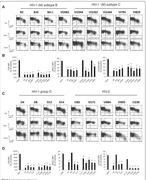
Nefalleles from different clades of HIV isolates modulate surface expression levels of human receptors
expression was more variable (Figure 1). The majority of the isolated HIV-1 group M, HIV-1 group O and HIV-2 Nef proteins downmodulated the expression of MHC-I and CXCR4 resulting in a 2- to 3-fold reduction in surface expression. Interestingly, two HIV-1 subtype C alleles (VI1044 and VI794; Figure 1 and 1B) and one HIV-1 group O allele (O8) did not show any effect on MHC-I downregulation (Figure 1C and 1D). All Nef proteins, ex-cept Nef O8 and HIV-2 VI171, downregulated the surface expression of CXCR4 (Figure 1C and 1D). In line with previous reports [15], nef alleles isolated from HIV-2 strains were much more efficient in downregulating CXCR4 than HIV-1 nefalleles (near 10-fold reduction of CXCR4 surface expression for HIV-2 compared to 2-3-fold for HIV-1). Clearly, there was no correlation between the ability of various Nef proteins to downregulate CXCR4, MHC-I and CD4, confirming that the mechan-isms of CD4 and MHC-I downregulations are at least in part genetically separable [15,18,19] and also suggesting that downregulation of CXCR4 and MHC-I depend on the interaction of Nef with distinct cellular ligands and machineries.
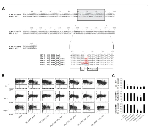
Mutational analysis of the natural Nef mutant HIV-1 O8 In our panel of primary nef alleles, we identified 4 nef alleles that were deficient for MHC-I and/or CXCR4 downregulation. A subtype C nef allele (VI1044) con-tained an insertion of a few amino-acids, which inter-rupted the structure of the N-proximal α-helical region, explaining the lack of MHC-I downregulation capacity of this allele (Additional file 1: Figure S1C) [20]. Nef alleles VI794 (subtype C) and VI171 (HIV-2) did not show any mutations in regions known to be important for Nef functions (Additional file 1: Figure S1C and Figure S1D). By contrast, Nef O8 showed mutations in an amphipathic stretch of amino acids in the Nef core domain encompassing the acidic cluster and the proline-rich motif (Figure 2A). More specific-ally, a point mutation changed the first proline of the polyproline stretch into an alanine. In addition to this point mutation in the PxxP motif, Nef O8 also showed a deletion of a three peptidic motif, i.e. valine-glycine-phenylalanine (VGF), located right in between the acidic cluster and the polyproline stretch. Based on the sequences found in the Los Alamos HIV sequence database consulted in December 2011, the VGF was highly conserved in HIV-1 and SIVcpz (V: 99.20 %; G: 98.85 %; F: 99.52 %). [21,22]. HIV-2 nef alleles instead contain a highly conserved VGV motif (V: 93.06 %; G: 97.22 %; V: 79.17 %, [22]) indicating that these residues are important for optimal Nef function. Despite inten-sive research on the functions of conserved regions in Nef, the importance of the VGF/V region has not yet been appreciated or investigated thoroughly.
To evaluate the individual contribution of the observed mutations for the defective phenotype of the O8 nef al-lele, we generated a panel of mutants in which the differ-ent mutations were sequdiffer-entially reverted to their consensus variant (Figure 2A) and re-evaluated their abil-ity to downregulate surface molecules. Neither restor-ation of the AxxP sequence into PxxP (i.e. EDED_PxxP) nor introduction of the deleted amino acids (i.e. EDED_VGF_AxxP) restored the capacity to interfere with MHC-I and CXCR4 surface expression (Figure 2B and 2C). Only simultaneous restoration of the PxxP and the VGF motif into the consensus variant (i.e. EDED_VGF_PxxP or EEEE_VGF_PxxP) rescued CXCR4 and MHC-I downregulation by O8 Nef. Interestingly, when all three amino acids of the VGF motif in Nef O8 were converted into alanine (i.e.EDED_AAA_PxxP), in the presence of an intact polyproline stretch, the cap-acity to affect MHC-I and CXCR4 surface expression was again lost (Figure 2B and 2C), indicating that spe-cific amino acids in both motifs are important.
An amphipathic surface in Nef is important for downregulation of CXCR4 and MHC
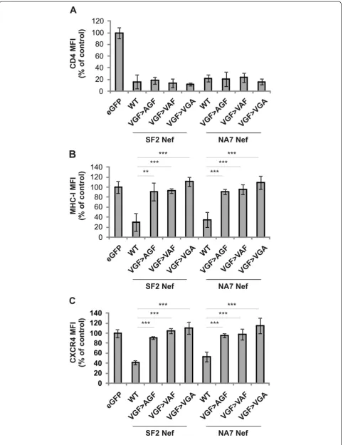
Depiction of the VGF motif within the structure of Nef showed that this motif is located within a loop region of Nef directly preceding the PxxP motif (Figure 3A). The motif is at the surface of Nef and highly accessible for protein-protein interactions [23]. To prove the func-tional conservation of the VGF motif, we initially evalu-ated the importance of this domain for MHC-I and CXCR4 downregulation innef alleles derived from HIV-1 group M, two canonical group (M) subtype B strains (i.e. NL4.3 and NA-7) and two non-canonical isolates: subtype B (i.e. B2) and subtype C (i.e. VI1422). Muta-tions were designed to generate triple mutaMuta-tions of VGF into alanines (VGF!AAA).
Figure 2Mutational analysis of Nef O8 identifies functional importance of the VGF region which links the acidic cluster and the proline-rich motif.(A) Alignment of the sequence of Nef O8 relative to a subtype O reference allele (O.BE.87.ANT70) obtained from the Los Alamos HIV sequence database reveals that the VGF motif is deleted in Nef O8. In addition, the first proline of the PxxP motif changed into alanine. A panel of mutants was generated in which the different mutations were sequentially reverted to their consensus variant (marked in red) or to a variant in which VGF was changed into triple alanines (AAA; blue). As reference, boxes below reverted sequences mark the acidic cluster (Ac) and proline-rich motif (Proline-motif) (B) Jurkat CD4-CCR5 cells were transduced with retroviral vectors expressing either eGFP alone or together (Nef-IRES-eGFP) with the indicatednefmutant and were analyzed by flow cytometry. Bivariate dot plots show expression of CD4, MHC-I, CXCR4 as a function of eGFP (Nef) expression. Dot plots shown are representative for results obtained in 3 independent experiments. (C) To determine relative surface levels, the MFI of transduced cells (eGFP+) was divided by the MFI of untransduced cells (eGFP-) in the same culture. Data represent the mean relative expression and standard deviations from 3 independent experiments. * over a bar indicates a statistically significant difference between the experimental construct compared with the eGFP control (P<0.01).
[image:6.595.58.538.198.609.2](See figure on previous page.)
these surface molecules in Nef O8, but also in HIV-1 group M Nef proteins.
Each amino acid residue of the VGF motif is highly conserved, suggesting a sequence specific requirement. However, the triple mutation of the VGF stretch might be structurally drastic and alter the functionality of adja-cent motifs. To exclude this and to confirm the se-quence specific requirement of the VGF motif, a panel of single mutants of the VGF motif, in which the differ-ent amino acids are changed into alanine (AGF, VAF and VGA), was generated and evaluated for their effect on Nef function. Similarly to the VGF!AAA mutant, single mutants failed to down regulate MHC-I and CXCR4,
while maintaining the capacity to downregulate CD4 (Figure 4). These results indicate that the VGF stretch as such is required for downregulation of MHC-I and CXCR4.
Integrity of the VGF motif is essential for effects of Nef on actin dynamics and Lck localization
[image:7.595.61.537.90.429.2]association of Nef with the cellular p21-associated kinase 2 (PAK2) [24]. Nef-PAK2 association occurs in a highly labile multiprotein complex of about 1 MDa and causes the phosphorylation and thereby inactivation of the actin severing factor cofilin [25,26]. While the instability of the Nef-PAK2 complex limits the reliability of biochemical quantification of this association between different nef alleles [24,26,27], the functional consequences including PAK2-dependent deregulation of cofilin and inhibition of actin dynamics can readily be quantified and are con-served among most Nef isolates [26,28]. We therefore analyzed the ability of Nef VGF!AAA mutants from SF2 and NA7 Nef to induce hyper-phosphorylation of cofilin downstream of PAK2, using a previously established sin-gle cell-based immunofluorescence quantification assay [20,21]. As expected, cofilin was hyper-phosphorylated in Jurkat T cells expressing WT SF2 and NA-7 Nef, but not in the presence of their AxxA mutants. Similarly, VGF!AAA mutated Nef failed to induce cofilin phos-phorylation (Figure 5A and 5B), suggesting that the VGF motif is essential for Nef-PAK2 association. To address this point directly, we performed anin vitrokinase assay to analyze the association of Nef with PAK2 activity. GFP-tagged SF2 Nef was used for this analysis due to more robust kinase signal detected with this Nef allele. Nef.GFP was immunoprecipitated from transiently trans-fected Jurkat T cells and subsequently subjected to an in vitro kinase assay, in which Nef-PAK2 association is detected by the presence of autophosphorylated PAK2 (62 kDa; p-PAK2) as well as of a yet unindentified 72 kDa substrate (p72). Expectedly [26,29], WT SF2 Nef but not its AxxA mutant efficiently associated with autopho-sphorylating PAK2; however, SF2 AxxA Nef was also expressed to lower levels than the WT in the experiment shown. In line with the results obtained from the cofilin-phosphorylation analyses, the VGF Nef mutant failed to associate with PAK2 activity despite robust expression and efficient immune isolation (Figure 5C). Since the de-regulation of cofilin by Nef results in impaired host cell actin remodeling following external stimulation, we next analyzed the role of the VGF motif for the Nef-dependent inhibition of actin remodeling following TCR-signaling [28,30,31]. T cells transiently expressing GFP-tagged ver-sions of WT, AxxA or VGF!AAA mutants from SF2 or NA7 Nef were plated onto anti-CD3 coated cover glasses and analyzed for the formation of circumferential F-actin rich rings by immuno fluorescence analysis. Expectedly,
WT Nef expressing cells interfered with actin remodeling and prevented the formation of F-actin rich circumferen-tial rings: as shown in Figure 4D, no actin ring is visible around green cells expressing WT Nef. In contrast, AxxA and VGF!AAA Nef variants were not able to interfere with actin polymerization (Figure 5D and 5E), so a clear circumferential staining is visible, despite Nef mutant pro-tein expression.
Similar to the VGF!AAA mutants, single mutants of the VGF motif (AGF, VAF and VGA) were significantly reduced in their effect on cofilin phosphorylation (Fig-ure 5A and 5B) and actin polymerization (Fig(Fig-ure 5D and 5E). It should, however, be noted that these single mutants had a slight but significant residual effect as compared to the triple mutant, indicating that some re-sidual activity remains for the alleles, especially for the VGF!AGF and VGF!VAF mutants. Nevertheless, these results demonstrate that the VGF motif in different nef alleles is critical for Nef-PAK2 association and thus consequential for Nef-mediated cofilin deregulation and disruption of host cell actin remodelling.
The above results demonstrated for differentnefalleles a requirement for both the PxxP and the VGF motif in Nef-PAK2 association and cell surface receptor down modulation. All of these activities, however, also depend on additional protein interaction motifs in Nef such as an intact protein interaction surface around aa191/195 or the acidic cluster [32] and thus did not allow us to conclude unambiguously whether the VGF and PxxP motifs exert independent functions or synergize in a common molecular interaction. We, therefore, assessed next the role of the VGF motif in Nef-mediated targeting of the TCR proximal kinase Lck to the trans-golgi network (TGN), a function of Nef for which so far no protein interaction surface in Nef other than the the PxxP motif has been implicated [30,33,34]. Jurkat T cells transiently expressing GFP-tagged Nef proteins were analyzed for TGN accumulation of Lck by im-munofluorescence analysis. While Lck appeared mostly at the plasma membrane with a minor intra-cellular fraction in GFP- and Nef AxxA-expressing cells, WT Nef expression resulted in a pronounced intracellular accumulation indicative of TGN targeting of the kinase (Figure 6A) as described previously [33]. VGF!AAA Nef mutants displayed intermediate activ-ity in this assay and failed to affect the subcellular localization of Lck in most cells; however, some cells (See figure on previous page.)
still showed slight TGN recruitment of Lck (Fig-ures 6A and 6B). In contrast, single substitutions of the valine (VGF!AGF) and the glycine (VGF!VAF) respectively did not or did marginally affect Lck accu-mulation. However, mutation of the phenylalanine,
[image:10.595.62.539.87.515.2](VGF!VGA) was sufficient to abrogate this Nef func-tion similar to the VGF!AAA mutant, indicating that Lck accumulation by Nef depends at least in part on the VGF motif and that the phenylalanine which dir-ectly precedes the proline-rich stretch is crucial for this. Figure 5The VGF region is important for Nef-PAK2 association, cofilin hyper- phopsorylation and inhibition of F-actin rich
circumferential ring formation by Nef.(A) Jurkat TAg cells were transfected arrows with WT, AxxA, VGF!AAA, VGF!AGF, VGF!VAF or VGF!VGA mutant Nef.GFP fusion proteins, plated onto poly-L-lysine (PLL)–coated cover glasses, fixed and stained for phospho-cofilin (p-cofilin). Images show intense staining for p-cofilin (red-orange) only in cells expressing Nef (green) of wild-type, but not of mutated sequence. Scale bar = 10μm (B) Frequency of the cells from cultures as shown in panel A that were scored high in p-cofilin. Values are the means of 3 independent experiments, and error bars represent SD from the mean;≥100 cells were analyzed per transfection; * indicates P<0.01, ** indicates P<0.001 and *** indicates P<0.0001. (C) Lysates of Jurkat TAg cells expressing eGFP or WT, VGF!AAA or AxxA SF2 Nef.GFP fusion proteins. Lower panels (input) show Western blot for Nef and loading control (transferrin receptor). These lysates were immunoprecipitated for Nef.GFP (rαGFP-IP), and subsequently used forin vitrokinase assay (IVKA). Nef-PAK2 association is detected in the IVKA only with WT Nef, by the presence of autophosphorylated PAK2 (62 kDa; p-PAK2) as well as of a yet unindentified 72 kDa substrate (p72). (D) Jurkat TAg cells were transfected with WT, AxxA, VGF!AAA, VGF!AGF, VGF!VAF or VGF!VGA mutant Nef.GFP fusion proteins, plated onto anti-CD3–coated cover glasses, fixed and stained with phalloidin to reveal F-actin (red). Scale bar = 10μm (E) Frequency of the cells from cultures as shown in panel A that to form F-actin–rich circumferential rings. Values are the means of 3 independent experiments, and error bars represent SD from the mean;
To address whether the VGF motif is directly involved in protein interactions of the adjacent PxxP motif, we probed its impact on the direct interaction of Nef with its best characterized and highest affinity PxxP ligand, the Src-family kinase Hck [35]. Nef and Hck were co-expressed in 293 T cells and Nef.GFP was immunoprecipi-tated. Subsequently, Co-precipitating Hck was detected by Western Blot. While different isoforms of Hck co-precipi-tated efficiently with WT (NA-7 or SF2) Nef, the AxxA and VGF!AAA Nef mutants were completely defective in Hck binding. Single amino acid substitution mutants did not bind the low molecular weight isoform as WT preferentially did, although some residual binding was sometimes observed with the high molecular weight
isoform (Figure 6C). Together, we concluded that the in-tegrity of the VGF motif is essential for most, if not all, functions attributed to the PxxP motif and that both motifs may be part of a common interaction surface.
The VGF motif is essential for the effect of Nef on viral infectivity and replication
[image:11.595.57.543.89.445.2]mutant HIV. We constructed a panel of replication com-petent HIVNL4.3-based reporter viruses expressing NL4.3
WT, AxxA and VGF!AAA Nef. These viruses express Nef together with mouse-derived heat stable antigen (HSA) from a single bicistronic mRNA (HSA-IRES-Nef) [40]. It was shown before that key activities of Nef such as enhancement of virion infectivity and increase of viral rep-lication remain conserved in such chimeric viruses and that marker gene expression correlates with Nef expres-sion in productively infected cells [41,42]. As control, we
also constructed a NefstopHIV reporter virus, which does
not express Nef due to the insertion of multiple premature stop codons [43].
[image:12.595.58.542.216.631.2]We evaluated whether the VGF!AAA mutation affected the infectivity of HIV particles in HeLa P4R5β -galactosidase indicator cells. Cells were infected with virus preparations corresponding to equal amounts of p24 and HIV-infection was quantified on the basis ofβ -galactosidase activity, 48 h post infection (Figure 7A). As expected, the Nefstop HIV, deficient in Nef expression
was almost 8-fold less infectious than HIV-1 WT virions. Both the VGF!AAA and AxxA Nef mutated HIV were severely less infective. Results shown were obtained with the NL4-3 nef allele and similar reduction in infectivity was seen using SF2 and NA-7 nef alleles (data not shown). Similary to the VGF!AAA mutant of NL4.3 Nef, single mutants were significantly less infective rela-tive to the wt virus. Of note, single substitution mutants of the valine (VGF!AGF) and the glycine (VGF!VAF), but not of the phenylalanine (VGF!VGA), were slightly more infective than the triple mutation, resulting in an intermediate phenotype.
As a final functional correlate, we evaluated if the in-tegrity of the VGF motif is essential for Nef enhanced HIV replication in peripheral blood lymphocytes (PBL). CD4+ T cells were isolated and stimulated with IL-2/ PHA, prior to infection with isogenic viruses. Cells were infected with 293 T-derived viral supernatants corre-sponding to the same amount of p24. After 1, 3, 5, 7, 9 and 11 days of culture, HIV replication was monitored by determining the amount of p24 antigen in the super-natants (Figure 7B). WT NL4.3 HIV-1 replicated much more efficiently in IL-2 stimulated PBL’s relative to the nef stop variant. As expected the mutants of the PxxP motif were less efficient in enhancing viral replication [44,45]. Similarly, the VGF mutated Nef HIV replicated less efficiently compared to WT HIV, although the effect was attenuated in some donors when compared to the PxxP Nef mutated HIV. All donors tested showed a sig-nificantly reduced replication rate of HIV VGF!AAA Nef mutated virus compared to WT Nef HIV. After day 9 of culture, viability in infected cultures decreased sig-nificantly, explaining the general drop in p24 levels at day 11. As a control, we also evaluated the amount of HSA expressing cells at several time points by flow cyto-metry as an alternative measure for HIV expression, with similar results (data not shown).
Discussion
Although not essential for viral replication, HIV-1 Nef is important for disease progression and, therefore, is con-sidered a pathogenic factor in primate lentiviridae [10]. For its function, Nef depends on specific surfaces to interact with host cell proteins. Therefore, a strong posi-tive selection pressure exists to keep the property of these surfaces conserved [46]. It is key to identify con-served sites in Nef with functional importance in pro-tein-protein interactions, since they might serve as potential target sites for pharmacological intervention.
In the present study, we identified, by functional ana-lysis of a panel of clinical HIV-1 and HIV-2nefalleles, a previously unrecognized conserved region. A HIV-1 group O nef allele, was mutated in an amphipathic stretch of amino acids located in the core domain
encompassing the acidic cluster, a VGF region and the PxxP motif, resulting in the loss of downregulation of MHC-I and CXCR4, but not of CD4. By selective muta-tion of this O8nefallele back to the consensus sequence, we show that both an intact PxxP motif in conjunction with an intact VGF region are needed to restore the defect in receptor trafficking. We could extend this observation by showing that the VGF region is essential for the associ-ation of Nef with active PAK2 and consequently hyper-phosphorylation of cofilin resulting in the inhibition of actin remodeling following TCR triggering. In addition, Nef-induced Lck accumulation in the TGN also requires integrity of the VGF region. The importance of the pro-line-rich region for this Nef function has been studied ex-tensively [10,47]. However, despite its high conservation in HIV-1 and SIVcpznefalleles, the function of the VGF re-gion remained poorly investigated.
Modulation of cell surface molecules such as MHC-I and chemokine receptors or the TNF receptor-associated factor TRAF2 were shown to depend on the conserva-tion of the PxxP motif as well as the acidic cluster (EEEE) [19,48,49]. How the acidic cluster contributes to downregulation of MHC-I is controversial. Several stud-ies have observed that the four contiguous glutamate residues interact with PACS-1 and PACS-2 to initiate as-sembly of a multiprotein complex which targets MHC-I to the trans-Golgi network for subsequent degradation [50,51]. However, in an alternative model for MHC-I downregulation, in which Nef interacts with both MHC-I and the μ subunit of the AP-1 endosomal coat com-plex, the EEEE motif plays only a stabilizing role [52,53]. The PxxP motif on the other hand is essential for the interaction of Nef with Src kinases (like Hck); misrout-ing of Lck as well as association of Nef with the cellular kinase PAK2, results in elevated cellular levels of inacti-vated phosphorylated cofilin, a deregulation that is in-strumental for the inhibition of TCR- or chemokine-induced F-actin remodeling [26,28-31,34].
Triple mutants of the VGF motif (VGF!AAA) lost both
‘trafficking’(MHC-I, CXCR4) as well as‘signaling' (PAK2
association, cofilin hyper-phosphorylation and inhibition of actin ring formation) functions, suggesting that the VGF region is functionally linked with the PxxP motif. Moreover, Baugh et al. showed that similar to the PxxP motif, MHC-I downregulation, interaction with PAK2, and to a lesser degree enhancement of virion infectivity are dependent on the acidic cluster [32]. Our observation that mutation of VGF into triple alanines similarly abro-gates these multiple effects of Nef is a hint that the acidic cluster-VGF-PxxP triplet forms a functional unit import-ant for protein-protein interactions with host cell factors.
therefore be indirect, meaning that only the correct spa-cing between the acidic cluster EEEE motif and the PxxP motif is required for Nef functionality, i.e. the correct positioning of the EEEE motif and potentially the pre-ceding CAWL protease recognition motif with respect to the Nef core domain. Alternatively, the effect of Nef dysfunction could be direct through binding of these amino acids to the primary receptors itself or to any stimulatory co-factor required for receptor downregula-tion. As mutation of the VGF motif to AAA in O8 Nef did not restore the Nef trafficking functions, the effect of mutation is most likely direct. Our finding that indi-vidual mutation of the three amino acids (AGF, VAF, VGA) is sufficient to interfere with the effect of Nef on receptor trafficking and TCR signaling further supports this hypothesis and shows that the function of V-G-F is sequence specific. The integrity of the phenylalanine appears to be specifically essential for functions related to the proline-rich motif. In contrast to single mutants of the valine and the glycine (VGF!AGF and VGF!VAF), sin-gle mutants of this amino-acid (VGF!VGA) did not ac-cumulate Lck. Of note, several SH3 binding regions of mammalian proteins contain a N-terminal phenylalanine, which is required for efficient ligand binding [54]. These observations suggest that the effect of Nef on TCR induced actin remodeling and on Lck targeting to the TGN are mechanistically different, as was shown before [30].
Sheltonet al. showed recently that the VGFPV region, in their study identified as the secretion modification re-gion, forms a specific binding surface for mortalin which subsequently promotes cellular secretion of extracellular Nef [55]. The phenotypes which we analysed in this study are all cell intrinsic and are unlikely to be explained by differences in levels of extracellular Nef. The results of Sheltonet al. and ours together show that the VGF region is not just a mere spacer between the acidic cluster and the proline rich region. Instead, it acts as an interaction surface, which together with its neigh-boring motifs forms a functional unit.
Replication of HIV-1 in primary T lymphocytes is tightly coupled to their activation state. While HIV-1 undergoes early replication events in quiescent CD4+ T cells, subsequent steps in the viral life cycle require T cell activation. One of the prime roles of Nef in vivo is to fine tune activation states in infected T cells [56]. Nef interferes with TCR proximal signaling to prevent T cell activation and activation–induced cell death upon anti-genic stimulation [28,30,31,55], while at the same time enhancing distal TCR signaling effects to promote pro-viral expression [33,57,58]. Both functions of Nef depend on the interaction of several specific SH3-domain con-taining host cell proteins with the conserved PxxP motif. For example, the PxxP motif interacts with the
SH3-domain of Lck, misrouting the TCR proximal kinase Lck from the plasma membrane to the trans Golgi-network (TGN), altering TCR proximal signaling events [28,30,33,59]. In our HIV replication assay, WT HIV viruses replicated much more efficiently then HIV Nefstop viruses. The requirement of PxxP residues for efficient HIV-1 replication in T-cells is controversial [35,39,44,59]. Lundquist and coworkers did not observe significant differ-ences between the replication-efficiency of WT, AxxA mu-tant and polyproline deleted viruses [60], while Saksela and co-workers reported that AxxA mutant replicate as delta-Nef viruses [35]. In our hands, the replication of the HIV Nef AxxA viruses was significantly attenuated compared to WT viruses. The fact that we use a different proviral back-bone and other culture methods for CD4+ lymphocyte stimulation may account for the discrepancy between our results and observations from others. Similar to Nef AxxA viruses, VGF!AAA mutants replicated much less effi-ciently than WT HIV viruses. We recently showed that the accumulation of Lck is required for TGN-associated Ras-Erk signaling, which in turn was shown to promote IL-2 production and enhance virus spread [33]. As HIV viruses harboring AxxA or VGF!AAA mutations in Nef fail to efficiently accumulate Lck [30], this could ex-plain the altered replication kinetics of these mutants. Interestingly, our previous observation that the HIV O8 isolate showed reduced fitness compared to other iso-lates of the same group might be due to the deletion of the VGF domain [16].
Despite the opposing findings concerning the role of the PxxP motif for replication of HIV, there is general consen-sus that this domain is required for Nef to enhance the in-fectivity of HIV virions [61-63]. The mechanism by which Nef enhances viral infectivity remains unclear, but is thought to involve alterations of specific signaling and trafficking events in the producer cell. [9]. We observed that, in line with our other results, VGF!AAA mutants were similarly less infective than wild type viruses.
In our study, we only analyzed the importance of the VGF motif in Nef for HIV-1 function. Despite VGF con-servation in HIV-1 and SIVcpz; HIV-2 and SIVsmm instead
Conclusions
Our results extend the previous research performed on the function of the PxxP motif. Based on our and earlier observations from other groups, we propose a new model in which the PxxP motif operates in the context of a larger amphipathic protein surface, encompassing both the acidic cluster and the VGF linker region. Moreover, integ-rity of the VGF region is required for SH3 binding and likely for subsequent formation of multi-protein com-plexes. The fact that a multitude of Nef functions required for enhancement of viral replication and disease progres-sion are dependent on the formation of these protein complexes suggests that the amphipathic region could serve as a prime target for pharmaceutical intervention.
Availability of supporting data
Nucleotide sequences of isolated HIV-1 and HIV-2 Nef alleles used in this study were submitted to Public sequence repository (Genbank accession numbers JQ 990942 - JQ990960).
Methods
Viruses and Nef alleles
HIV-1 group M and group O isolates were obtained from the AIDS Research and Reference Reagent Program, while the HIV-2 strains were previously isolated from patients attending the AIDS clinic at the Institute of Tropical Medi-cine in Antwerp, Belgium, with the approval of the ethical committee. Virus stocks were propagated and expanded in short-term cultures of PBMCs treated with PHA (1μg/mL) and IL-2 (10 ng/mL). Genomic DNA was extracted using the Qiagen Blood kit (Qiagen, Venlo, The Netherlands) according to the manufacturer’s instructions. Nef alleles from different HIV subtypes were amplified by semi-nested polymerase chain reaction (PCR) with Platinum Pfx DNA polymerase (Invitrogen, Merelbeke, Belgium). Primers rec-ognizing Env and the 3’LTR regions of the HIV genome used for the first round of PCR were forward:
5’-GCACT-CAAGGCAAGCTTTATTGAGGC-3’and reverse
5’-CCA-CATACCTAGAAGAATAAGACAGG-3’. A nested PCR reaction was performed after sequencing and using allele-specific forward and reverse primers covering the complete nef coding sequence and containing restriction sites for BamHI and EcoRI, respectively (see Additional file 1: Table S1A). Site specific mutants of Nef O8, VI1422, B2 and NA-7 in LZRS-IRES-eGFP were generated with PCR using allele specific primers described in Additional file 1: Table S1B and Table S1C.
Plasmid construction
Nef amplicons were cloned into the LZRS-IRES-eGFP retroviral vector, using BamHI and EcoRI restrictions sites [64]. Plasmids were purified with the Qiaprep Mini-prep kit (Qiagen). The integrity of the constructs and
the nefgenes was confirmed by direct sequencing (ABI, Foster City, California). Alignments of thenefsequences together with selected reference sequences were per-formed using BioEdit version 5.0.9. Type, group and subtype designation was confirmed by constructing phylogenetic trees by the neighbor joining method using MEGA 4 [65] (see Additional file 1: Figure S1).
Expression constructs for GFP fusion proteins of HIV-1 SF2 Nef, HIV-1 NA-7 Nef and SF2 AxxA were already described and were constructed by cloning PCR fragments of the respective nef genes into pEGFP-N1 (Clontech) under the control of the cytomegalovirus promoter [26,31]. The expression constructs for NA7 Nef VGF!AAA and SF2 Nef VGF!AAA and NA-7 AxxA were generated by site directed mutagenesis from the wild-type (WT) plasmid (primers Additional file 1: Table S1D).
The proviral constructs used for HIV infectivity and repli-cation assays are derived from the NLENG1-IRES vector [66], which expresses HSA-IRES-Nef cloned in HIV-1 NL4-3 provirus, as described before [40]. These constructs express from the nef reading frame (together with Nef as two indi-vidual proteins) mouse heat-stable antigen as a protein re-porter. A BamH1/NgoMIV fragment corresponding to the second exon of tatto the end of nefwas cut out from the vector and was ligated into pSUPER [67]. Next, SF2 and NA-7nefalleles and their specific AxxA and VGF mutants were amplified by PCR using primers containing restriction sites for BstXI and BspEI from the pEGFP-N1 vectors described above. A nefallele, with deleted initiation codon, two in frame stop codons at amino acid positions 4 and 5 and premature stop codons at positions 73 and 74 in thenef ORF, disrupting thenefgene was generated by standard PCR and cloning techniques, similarly to what was described be-fore (Nefstop) [43]. The amplified nef alleles were
subse-quently cut with BstXI and BspE and ligated into the pSUPER plasmid, swapping it with the WT nef present. NL4.3 AxxA and VGF!AAA mutants were generated based on the pSUPER vectors by site directed mutagenesis. Next, BamH1/NgoMIV fragments of newly constructed pSUPER plasmids were ligated into the NL4-3 HSA-IRES-Nef proviral construct. All these final proviral constructs were verified for correctnefallele sequence by direct sequencing.
The vector for the 764 amino acid p59Hck-mCherry fu-sion protein was constructed by PCR amplifying human Hck cDNA insert from the image clone 4855747 using primers containing BglII (sense) and KpnI (antisense), and inserting it between the BamHI and KpnI sites of the pEBB vector [68], followed by cloning of red fluorescent protein mCherry (Clontech) between the KpnI and EagI sites after the p59Hck insert.
Jurkat E6.1-derived cell lines. The easy to transfect Jurkat TAg cells express the large T antigen of simian virus 40 and were grown in RPMI 1640 medium supplemented with 10% fetal calf serum (FCS, Hyclone), 100 U/mL penicillin, and 100 g/mL streptomycin (both from Gibco). Jurkat CD4-CCR5 cells were grown in IMDM medium supple-mented with 2 mM L-glutamin, 10% heat-inactivated fetal calf serum (FCS), 293 T cells (American Type Culture Col-lection (ATCC) Manassas, VA) were maintained in IMDM medium, supplemented with 10% FCS, 2 mM L-glutamin, 100 U/mL penicillin, and 100 U/mL streptomycin.
Mouse antihuman monoclonal antibodies used were: CD4 (clone SK3; [PE] and [APC]), CXCR4, anti-MHC-I [PE] (anti-human histocompatibility leukocyte antigens [HLA]-A,-B,-C), all purchased from Becton Dickinson Immunocytometry systems (Beckton Dickinson, Erembodegem, Belgium) and anti-mouse-CD24-APC (HSA, heat stable antigen; BioLegend, San Diego, CA). The cells were analyzed on a FACSCalibur flow cytometer (Becton Dickinson). Forward light scattering, orthogonal scattering, and fluorescence signals were stored and ana-lyzed using the CellQuest software (Becton Dickinson).
Cells were transfected using either calcium-phosphate precipitation (Invitrogen, Merelbeke, Belgium) or JetPei (Polyplus, Sélestat, France) according to the description of the manufacturer. For transduction of suspension cell lines, cells were mixed with retroviral supernatant, pre-incubated with DOTAP (Roche diagnostics). To increase transduction efficiency, cells were centrifuged (90 min-utes, 950 g, 32°C) [64].
Production of retroviral vectors and replication competent HIV
To produce retroviral vectors, the Phoenix-Amphotropic packaging cell line was transfected with LZRS-IRES-eGFP (control) and LZRS-Nef-IRES-eGFP plasmids using calcium-phosphate precipitation as previously described [17,64,69].
Replication competent HIV was produced by trans-fecting 293 T cells with the HIV-1 NL4-3 –HSA-IRES-Nef proviral constructs described above using the JetPEI method. The medium was changed after overnight incu-bation, and virus was harvested 24 h later and stored at −80°C. The content of viral p24 antigen was quantified using a HIV p24 enzyme-linked immunosorbent assay kit (Innogenetics, Ghent, Belgium).
Immunofluorescence
Immunofluorescence stainings were performed as previ-ously described [26,28]. Briefly, for staining of F-actin (0.5 ng/ml phalloidin-TRITC), cells were fixed for 15 min with 3,7% paraformaldehyde (PFA)/phosphate-buffered saline (PBS), permeabilized with 0.1% Triton X-100/PBS for 2 min and subsequently blocked for
unspecific binding with 1% bovine serum albumin (BSA)/PBS for 15 min. Hoechst 33258 was used in a concentration of 1 ng/ml. For staining of phospho-cofilin (p-cofilin) (cell signaling) (1:50), and Lck (Santa Cruz) (1:50) all solutions, dilutions, and wash steps were done with Tris-buffered saline (TBS; 50 mM Tris, 150 mM NaCl [pH 7.5]). Blocking was performed for 30 min, and incubation was performed with the first antibody over-night at 4°C. Confocal pictures of p-cofilin in Jurkat T cells were acquired using a Zeiss LSM 510 Axiovert micro-scope and LSM Meta software. Images were processed using Adobe Photoshop CS3.
In vitro kinase assay
In vitrokinase assays (IVKA) were essentially performed as described previously [26]. Briefly, Jurkat T lympho-cytes were transfected with expression plasmids for Nef. eGFP. After 24 h, cells were lysed in KEB (137 mM NaCl, 50 mM Tris/HCl [pH 8], 2 mM EDTA, 0.5% Non-indet P-40, and protease inhibitors) supplemented with Na3VO4, and cleared lysates were sampled for Western blot and immunoprecipitated with a rabbit anti-GFP antibody. After intensive washing in KEB, the immuno-precipitates were resuspended in KAB (50 mM HEPES [pH 8], 150 mM NaCl, 5 mM EDTA, 0.02% Triton X-100, 10 mM MgCl2) containing 10 μCi of [γ-32P]ATP per reaction. After incubation for 10 min, samples were washed, and bound proteins were separated by SDS-PAGE and subjected to autoradiography. The kinase sig-nal was quantified relative to the SF2 nef allele using Quantity One (Bio-Rad).
Co-immunoprecipitation
For the Nef–Hck co-immunoprecipitation experiments, 293 T cells were transfected with expression plasmids for Nef.eGFP and p59Hck (Cherry fusion protein). After 24 h, cells were lysed in KEB (137 mM NaCl, 50 mM Tris/HCl [pH 8], 2 mM EDTA, 0.5% Nonidet P-40, and protease inhibitors) supplemented with Na3VO4, and cleared lysates were immunoprecipitated with a GFP-Trap (Chromotek, Planegg-Martinsried, Germany). After intensive washing in KEB, the precipitate was incubated in anti-Hck Santa Cruz Biotechnology inc. Santa Cruz, US), diluted 1:1000 in 50% blocking buffer.
Western blotting
was done using enhanced chemiluminescence (GE Healthcare, Chalfont St. Gilles, UK). To control that an equal amount of protein was loaded for each sample, blots were also stained for β-actin (primary anti-β-actin antibody: clone C4, ICN, Aurora, OH, USA).
Infectivity assays
Virus infectivity was determined using P4R5 HeLa reporter cells (NIH AIDS Reference and Reagent Program) [70]. Briefly, the cells were sown out in 96-well dishes in a vol-ume of 200 μl IMDM supplemented with 2 mM L-gluta-min and 10% FCS and infected after overnight incubation with virus stocks containing 40 ng of p24 antigen (Innoge-netics, Zwijnaarde, Belgium) produced by transiently trans-fected 293 T cells and centrifuged (90 minutes, 950 g, 32° C). 48 hours postinfection viral infectivity was detected using a beta-galactosidase screen kit from Thermo scien-tific (Pierce Biotechnology, Rockford, USA) as recom-mended by the manufacturer. β-Galactosidase activities were quantified as relative light units per second using an Orion Microplate Luminometer. To calculate percent values, relative light units per second obtained for wild-type HIV-1 NL4-3 HSA-IRES Nef infection were set to 100%.
Viral replication in CD4+ PBL cultures
To determine the efficiency of HIV-1 replication in per-ipheral blood CD4+lymphocytes, we isolated CD4+cells from buffy coat peripheral blood mononuclear cells (normal blood donors, Red Cross, Ghent, Belgium) by negative selection using paramagnetic beads (MACS; Miltenyi Biotec, Bergish Gladbach, Germany). After iso-lation, the cells were cultured in RPMI medium supple-mented with 2 mM L-glutamin, 10% heat-inactivated fetal calf serum, phytohemagglutinin (1μg/mL; Thermo Fisher Scientific, Waltham, USA), 20 ng/mL IL-2 (Peprotech, Rocky Hill, USA), 100 U/mL penicillin, and 100 g/mL streptomycin. Thereafter, 1 ng of p24 antigen was added to 2,5*105PBLs, and the culture was spinocu-lated at 2,300 rpm for 90 minutes at 32°C. After centri-fugation, the supernatant was removed and the cells were further cultured in RPMI supplemented with 20 ng/mL IL-2. HIV replication was monitored at 1, 3, 5, 7, 9 and 11 days post-infection by measuring the amount of p24 antigen present in the supernatants, using a HIV p24 enzyme-linked immunosorbent assay kit (Innoge-netics, Ghent, Belgium). Alternatively, we also deter-mined the expression of the HSA reporter gene at each time point using flow cytometry, as was described before [40].
Statistical analysis
Data were analysed with non-parametric Mann– Whit-ney U test, one-tailed (SPSS, version 17; SPSS, Chicago, USA).
Additional File
Additional file 1:Figure S1. Alignment of the protein sequences and evolutionary relationship of the isolated HIV-1 and HIV-2 Nef proteins. (A)The evolutionary history was inferred using the neighbor-joining (NJ) method as conducted in MEGA 4.(B–E)Amino acid sequence alignments relative to references sequences, obtained from the Los Alamos database HIV sequence database. Dots connote identity with the reference sequence, whereas individual variations are shown by the single letter amino acid code. Dashes indicate gaps introduced into the sequence to optimize the alignment.(B)Alignment of HIV-1 group M subtype B Nef amino acid sequences.(C)Alignment of HIV-1 group M subtype C Nef amino acid sequences.(D)Alignment of HIV-1 group O Nef amino acid sequences.(E)Alignment of HIV-2 Nef amino acid sequences.Table S1. A)Allele-specific primers covering the complete Nef coding sequence.B)Primers to generate site-specific mutants in Nef O8.C)Primers to generate VGF!AAA mutants (indicated byΔVGF) in B2, VI1422, NA-7 and NL4.3 Nef.D)Primers for site specific mutagenesis of NA-7.GFP and SF2.GFP fusion proteins and NL4.3.
Abbreviations
HIV: Human immunodeficiency virus; SIV: Simian immunodeficiency virus; AIDS: Acquired immunodeficiency syndrome; TCR: T cell receptor; MHC-I/ II: Major histocompatibility complex class-I/ II; PAK2: P21-activated kinase 2; Lck: Lymphocyte-specific protein tyrosine kinase; TGN: Trans-golgi-network; PBL: Peripheral blood lymphocytes; IL-2: Interleukin-2; PHA: Phytohaemaglutinin A; eGFP: Enhanced green fluorescent protein; PACS-1/2: Phosphofurin acidic cluster- sorting protein-1/2; HSA: Heat stable antigen; AP-1: Adaptor protein 1; DNA: Deoxyribonucleic acid; IRES: Internal ribosomal entry site; FCS: Fetal calf serum; PE: Phycoerythrine; APC: Allophycocyanine; PFA: Paraformaldehyde; IVKA: In vitro kinase assay; PVDF: Polyvinyldifluoride.
Competing interests
The authors declare that they have no competing interests.
Author’s contributions
PM was responsible for the concept and design of the study, acquisition of most of the data and the writing of the the manuscript; BS was responsible forin vitrokinase and actin ring assays and helped with p-cofilin and Lck assays and critically revised the manuscript; EN gave substantial practical support during acquisition of the data; JV and VI optimized the HIV replication assay, helped with proviral construction and critically revised the manuscript; MG performedin silicomodeling, gave substantial scientific input and critically revised the manuscript; KS constructed the p59-HcK expression plasmids and critically revised the manuscript; GV provided the clinical HIV1- and HIV-2 isolates and critically revised the manuscript; KKA performedin vitroculture of primary isolates, is responsible for design of the study and critically revised the article. OF gave substantial scientific input and critically revised the manuscript; BV was responsible for the concept and design of the study, gave substantial scientific input and wrote the manuscript. All authors read and approved the final manuscript.
Acknowledgements
–Flanders (FWO) to BV. PJM and JV are PhD. fellows; KKA is a postdoctoral researcher; and BV is a Senior Clinical Investigator of the Fund for Scientific Research-Flanders. VI is a PhD fellow supported by the BOF program of the Ghent University. OTF and BS were supported by the Deutsche Forschungsgemeinschaft (SFB638, TRR83 and GRK1188) and by a fellowship to BS from the Leopoldina/Nationale Akademie der Wissenschaften.
Author details
1Department of Clinical Chemistry, Microbiology, and Immunology, Ghent
University, Ghent, (B-9000), Belgium.2Department of Infectious Diseases, Virology, University Hospital Heidelberg, INF 324, Heidelberg, (D-69120), Germany.
3
Department of Virology, Haartman Institute, University of Helsinki and Helsinki University Central Hospital, Helsinki, (FIN-00014), Finland.4Max Planck Institute for
Molecular Physiology, Dortmund, (D-44227), Germany.5Department of Biomedical Sciences, Virology Unit, Institute of Tropical Medicine, Antwerp, (B-2000), Belgium.
6
Present Address: Department of Biomedical Sciences , Virology Unit, Institute of Tropical Medicine, Antwerp, Belgium.
Received: 16 December 2011 Accepted: 27 April 2012 Published: 27 April 2012
References
1. Daniel MD, Kirchhoff F, Czajak SC, Sehgal PK, Desrosiers RC:Protective effects of a live attenuated SIV vaccine with a deletion in the nef gene.
Science1992,258:1938–1941.
2. Kestler HW, Ringler DJ, Mori K, Panicali DL, Sehgal PK, Daniel MD, Desrosiers RC:Importance of the Nef Gene for Maintenance of High Virus Loads and for Development of Aids.Cell1991,65:651–662.
3. Sawai ET, Hamza MS, Ye M, Shaw KES, Luciw PA:Pathogenic Conversion of Live Attenuated Simian Immunodeficiency Virus Vaccines Is Associated with Expression of Truncated Nef.J Virol2000,74:2038–2045.
4. Simard MC, Chrobak P, Kay DG, Hanna Z, Jothy S, Jolicoeur P:Expression of simian immunodeficiency virus nef in immune cells of transgenic mice leads to a severe AIDS-like disease.J Virol2002,76:3981–3995. 5. Hanna Z, Kay DG, Rebai N, Guimond A, Jothy S, Jolicoeur P:Nef harbors a
major determinant of pathogenicity for an AIDS-like disease induced by HIV-1 in transgenic mice.Cell1998,95:163–175.
6. Schwartz O, Marechal V, Le Gall S, Lemonnier F, Heard JM:Endocytosis of major histocompatibility complex class I molecules is induced by the HIV-1 Nef protein.Nat Med1996,2:338–342.
7. Landi A, Iannucci V, Van Nuffel A, Meuwissen PJ, Verhasselt B:One protein to rule them all: modulation of cell surface receptors and molecules by HIV Nef.Curr HIV Res2011,9:496–504.
8. Collins KL, Chen BK, Kalams SA, Walker BD, Baltimore D:HIV-1 Nef protein protects infected primary cells against killing by cytotoxic T lymphocytes.Nature1998,391:397–401.
9. Vermeire J, Vanbillemont G, Witkowski W, Verhasselt B:The Nef-infectivity enigma: mechanisms of enhanced lentiviral infection.Curr HIV Res2011, 9:474–489.
10. NJ Arhel F Kirchhoff 2009 Implications of Nef: Host Cell Interactions in Viral Persistence and Progression to AIDSP Spearman EO Freed HIV Interactions with Host Cell Proteins. Current Topics in Microbiology and Immunology Volume 339 Springer-Verlag Berlin Berlin 147175 Arhel NJ, Kirchhoff F: Implications of Nef: Host Cell Interactions in Viral Persistence and Progression to AIDS. InHIV Interactions with Host Cell Proteins. Current Topics in Microbiology and Immunology, Volume Volume 339. Edited by Spearman P, Freed EO. Berlin: Springer-Verlag Berlin; 2009:147–175. 11. Geyer M, Fackler OT, Peterlin BM:Structure-function relationships in HIV-1
Nef.EMBO2001,2:580–585.
12. Foster J, Denial S, Temple B, Garcia J:Mechanisms of HIV-1 Nef Function and Intracellular Signaling.J Neuroimm Pharmacol2011,6:230–246. 13. show[?twb.4w][?tlsb.09pt]?>Specht A, DeGottardi MQ, Schindler M, Hahn B,
Evans DT, Kirchhoff F:Selective downmodulation of HLA-A and -B by Nef alleles from different groups of primate lentiviruses.Virology2008, 373:229–237.
14. Schindler M, Würfl S, Benaroch P, Greenough TC, Daniels R, Easterbrook P, Brenner M, Münch J, Kirchhoff F:Down-Modulation of Mature Major Histocompatibility Complex Class II and Up-Regulation of Invariant Chain Cell Surface Expression Are Well-Conserved Functions of Human and Simian Immunodeficiency Virus nef Alleles.J Virol2003,
77:10548–10556.
15. Hrecka K, Swigut T, Schindler M, Kirchhoff F, Skowronski J:Nef Proteins from Diverse Groups of Primate Lentiviruses Downmodulate CXCR4 To Inhibit Migration to the Chemokine Stromal Derived Factor 1.J Virol
2005,79:10650–10659.
16. Arien KK, Abraha A, Quinones-Mateu ME, Kestens L, Vanham G, Arts EJ:The replicative fitness of primary human immunodeficiency virus type 1 (HIV-1) group M, HIV-1 group O, and HIV-2 isolates.J Virol2005, 79:8979–8990.
17. Verhasselt B, Naessens E, Verhofstede C, De Smedt M, Schollen S, Kerre T, Vanhecke D, Plum J:Human immunodeficiency virus nef gene expression affects generation and function of human T cells, but not dendritic cells.
Blood1999,94:2809–2818.
18. Michel N, Ganter K, Venzke S, Bitzegeio J, Fackler OT, Keppler OT:The nef protein of human immunodeficiency virus is a broad-spectrum modulator of chemokine receptor cell surface levels that acts independently of classical motifs for receptor endocytosis and G alpha(i) signaling.Mol Biol Cell2006,17:3578–3590.
19. Venzke S, Michel N, Allespach I, Fackler OT, Keppler OT:Expression of Nef downregulates CXCR4, the major coreceptor of human
immunodeficiency virus, from the surfaces of target cells and thereby enhances resistance to superinfection.J Virol2006,80:11141–11152. 20. Mangasarian A, Piguet V, Wang J-K, Chen Y-L, Trono D:Nef-Induced CD4
and Major Histocompatibility Complex Class I (MHC-I) Down-Regulation Are Governed by Distinct Determinants: N-Terminal Alpha Helix and Proline Repeat of Nef Selectively Regulate MHC-I Trafficking.J Virol1999, 73:1964–1973.
21. O'Neill E, Kuo LS, Krisko JF, Tomchick DR, Garcia JV, Foster JL:Dynamic Evolution of the Human Immunodeficiency Virus Type 1 Pathogenic Factor, Nef.J Virol2006,80:1311–1320.
22. HIV sequence database, [http:// HIV.lanl.gov], consulted December 15th2011. 23. Breuer S, Schievink SI, Schulte A, Blankenfeldt W, Fackler OT, Geyer M:
Molecular Design, Functional Characterization and Structural Basis of a Protein Inhibitor Against the HIV-1 Pathogenicity Factor Nef.PLoS One
2011,6:e20033.
24. Manninen A, Hiipakka M, Vihinen M, Lu W, Mayer BJ, Saksela K:SH3-Domain Binding Function of HIV-1 Nef Is Required for Association with a PAK-Related Kinase.Virology1998,250:273–282.
25. Fackler OT, Luo W, Geyer M, Alberts AS, Peterlin BM:Activation of Vav by Nef Induces Cytoskeletal Rearrangements and Downstream Effector Functions.Mol Cell1999,3:729–739.
26. Stolp B, Abraham L, Rudolph JM, Fackler OT:Lentiviral Nef Proteins Utilize PAK2-Mediated Deregulation of Cofilin as a General Strategy To Interfere with Actin Remodeling.J Virol2010,84:3935–3948.
27. Sawai ET, Khan IH, Montbriand PM, Peterlin BM, Cheng-Mayer C, Luciw PA: Activation of PAK by HIV and SIV Nef: importance for AIDS in rhesus macaques.Curr Biol1997,6:1519–1527.
28. Rudolph JM, Eickel N, Haller C, Schindler M, Fackler OT:Inhibition of T-Cell Receptor-Induced Actin Remodeling and Relocalization of Lck Are Evolutionarily Conserved Activities of Lentiviral Nef Proteins.J Virol2009, 83:11528–11539.
29. Stolp B, Reichman-Fried M, Abraham L, Pan X, Giese SI, Hannemann S, Goulimari P, Raz E, Grosse R, Fackler OT:HIV-1 Nef Interferes with Host Cell Motility by Deregulation of Cofilin.Cell Host & Microbe2009,6:174–186. 30. Haller C, Rauch S, Fackler OT:HIV-1 Nef Employs Two Distinct Mechanisms
to Modulate Lck Subcellular Localization and TCR Induced Actin Remodeling.PLoS One2007,2:e1212.
31. Haller C, Rauch S, Michel N, Hannemann S, Lehmann MJ, Keppler OT, Fackler OT:The HIV-1 Pathogenicity Factor Nef Interferes with Maturation of Stimulatory T-lymphocyte Contacts by Modulation of N-Wasp Activity.
J Biol Chem2006,281:19618–19630.
32. Baugh LL, Garcia JV, Foster JL:Functional Characterization of the Human Immunodeficiency Virus Type 1 Nef Acidic Domain.J Virol2008,82:9657–9667. 33. Pan X, Rudolph JM, Abraham L, Habermann A, Haller C, Krijnse-Locker J,
Fackler OT:HIV-1 Nef compensates for disorganization of the
immunological synapse by inducing TGN-associated Lck signaling.Blood
2011,119: 786–797.
34. Thoulouze MI, Sol-Foulon N, Blanchet F, Dautry-Varsat A, Schwartz O, Alcover A:Human Immunodeficiency Virus Type-1 Infection Impairs the Formation of the Immunological Synapse.Immunity2006,24:547–561. 35. Saksela K, Cheng G, Baltimore D:Proline-rich (PxxP) motifs in HIV-1 Nef
enhanced growth of Nef + viruses but not for down-regulation of CD4.
EMBO1995,14:484–491.
36. Hammes SR, Dixon EP, Malim MH, Cullen BR, Greene WC:Nef protein of human immunodeficiency virus type 1: evidence against its role as a transcriptional inhibitor.Proc Nat Acad Sci1989,86:9549–9553. 37. Kim S, Ikeuchi K, Byrn R, Groopman J, Baltimore D:Lack of a negative
influence on viral growth by the nef gene of human immunodeficiency virus type 1.Proc Nat Acad Sci1989,86:9544–9548.
38. Spina CA, Kwoh TJ, Chowers MY, Guatelli JC, Richman DD:The importance of nef in the induction of human immunodeficiency virus type 1 replication from primary quiescent CD4 lymphocytes.J Exp Med1994, 179:115–123.
39. Homann S, Tibroni N, Baumann I, Sertel S, Keppler O, Fackler O: Determinants in HIV-1 Nef for enhancement of virus replication and depletion of CD4+ T lymphocytes in human lymphoid tissue ex vivo.
Retrovirology2009,6:6.
40. Imbeault Ml, Lodge R, Ouellet M, Tremblay MJ:Efficient magnetic bead-based separation of HIV-1-infected cells using an improved reporter virus system reveals that p53 up-regulation occurs exclusively in the virus-expressing cell population.Virology2009,393:160–167.
41. Fackler OT, Moris A, Tibroni N, Giese SI, Glass Br, Schwartz O, Krausslich H-G: Functional characterization of HIV-1 Nef mutants in the context of viral infection.Virology2006,351:322–339.
42. Rücker E, Münch J, Wildum S, Brenner M, Eisemann J, Margolis L, Kirchhoff F:A Naturally Occurring Variation in the Proline-Rich Region Does Not Attenuate Human Immunodeficiency Virus Type 1 Nef Function.J Virol
2004,78:10197–10201.
43. Münch J, Rajan D, Schindler M, Specht A, Rücker E, Novembre FJ, Nerrienet E, Müller-Trutwin MC, Peeters M, Hahn BH, Kirchhoff F:Nef-Mediated Enhancement of Virion Infectivity and Stimulation of Viral Replication Are Fundamental Properties of Primate Lentiviruses.J Virol2007, 81:13852–13864.
44. Fackler OT, Wolf D, Weber HO, Laffert B, D'Aloja P, Schuler-Thurner B, Geffin R, Saksela K, Geyer M, Peterlin BM,et al:A natural variability in the proline-rich motif of Nef modulates HIV-1 replication in primary T cells.Curr Biol
2001,11:1294–1299.
45. Brown A, Moghaddam S, Kawano T, Cheng-Mayer C:Multiple human immunodeficiency virus type 1 Nef functions contribute to efficient replication in primary human macrophages.J Gen Virol2004,85:1463–1469. 46. Geyer M, Peterlin BM:Domain assembly, surface accessibility and
sequence conservation in full length HIV-1 Nef.FEBS2001,496:91–95. 47. Saksela K:Interactions of HIV/SIV pathogenicity factor nef with SH3
domain containing host cell proteins.Curr HIV Res2011,9: S31–S42. 48. Greenberg ME, Iafrate AJ, Skowronski J:The SH3 domain-binding surface
and an acidic motif in HIV-1 Nef regulate trafficking of class I MHC complexes.EMBO1998,17:2777–2789.
49. Mangino G, Percario ZA, Fiorucci G, Vaccari G, Acconcia F, Chiarabelli C, Leone S, Noto A, Horenkamp FA, Manrique S,et al:HIV-1 Nef Induces Proinflammatory State in Macrophages through Its Acidic Cluster Domain: Involvement of TNF Alpha Receptor Associated Factor 2.PLoS One2011,6:e22982.
50. Blagoveshchenskaya AD, Thomas L, Feliciangeli SF, Hung C-H, Thomas G: HIV-1 Nef Downregulates MHC-I by a PACS-1- and PI3K-Regulated ARF6 Endocytic Pathway.Cell2002,111:853–866.
51. Atkins KM, Thomas L, Youker RT, Harriff MJ, Pissani F, You H, Thomas G: HIV-1 Nef Binds PACS-2 to Assemble a Multikinase Cascade That Triggers Major Histocompatibility Complex Class I (MHC-I) Down-regulation.J Biol Chem2008,283:11772–11784.
52. Schaefer MR, Wonderlich ER, Roeth JF, Leonard JA, Collins KL:HIV-1 Nef Targets MHC-I and CD4 for Degradation Via a Final Common beta-COP-Dependent Pathway in T Cells.PLoS Pathog2008,4:e1000131.
53. Singh RK, Lau D, Noviello CM, Ghosh P, Guatelli JC:An MHC-I Cytoplasmic Domain/HIV-1 Nef Fusion Protein Binds Directly to the mu Subunit of the AP-1 Endosomal Coat Complex.PLoS One2009,4:e8364.
54. Ball LJ, Jarchau T, Oschkinat H, Walter U:EVH1 domains: structure, function and interactions.FEBS Lett2002,513(1):45–52.
55. Shelton MN, Huang MB, Ali SA, Powell MD, Bond VC:Secretion
modification region-derived peptide disrupts HIV-1 Nef's interaction with mortalin and blocks virus and nef exsosome release.J Virol2012,86 (1):406–19.
56. Stevenson M:HIV-1 pathogenesis.Nat Med2003,9:853–860.
57. Arhel N, Lehmann M, Clauss K, Nienhaus GU, Piguet V, Kirchhoff F:The inability to disrupt the immunological synapse between infected human T cells and APCs distinguishes HIV-1 from most other primate lentiviruses.J Clin Invest2009,119:2965–2975.
58. Simmons A, Aluvihare V, McMichael A:Nef Triggers a Transcriptional Program in T Cells Imitating Single-Signal T Cell Activation and Inducing HIV Virulence Mediators.Immunity2001,14:763–777.
59. Fenard D, Yonemoto W, de Noronha C, Cavrois M, Williams SA, Greene WC: Nef Is Physically Recruited into the Immunological Synapse and Potentiates T Cell Activation Early after TCR Engagement.J Immunol
2005,175:6050–6057.
60. Lundquist CA, Tobiume M, Zhou J, Unutmaz D, Aiken C:Nef-Mediated Downregulation of CD4 Enhances Human Immunodeficiency Virus Type 1 Replication in Primary T Lymphocytes.J Virol2002,76:4625–4633. 61. Madrid R, Janvier K, Hitchin D, Day J, Coleman S, Noviello C, Bouchet J,
Benmerah A, Guatelli J, Benichou S:Nef-induced Alteration of the Early/ Recycling Endosomal Compartment Correlates with Enhancement of HIV-1 Infectivity.J Biol Chem2005,280:5032–5044.
62. Goldsmith MA, Warmerdam MT, Atchison RE, Miller MD, Greene WC: Dissociation of the CD4 downregulation and viral infectivity enhancement functions of human immunodeficiency virus type 1 Nef.
J Virol1995,69:4112–4121.
63. Chutiwitoonchai N, Hiyoshi M, Mwimanzi P, Ueno T, Adachi A, Ode H, Sato H, Fackler OT, Okada S, Suzu S:The Identification of a Small Molecule Compound That Reduces HIV-1 Nef-Mediated Viral Infectivity Enhancement.PLoS One2011,6:e27696.
64. Stove V, Naessens E, Stove C, Swigut T, Plum J, Verhasselt B:Signaling but not trafficking function of HIV-1 protein Nef is essential for Nef-induced defects in human intrathymic T-cell development.Blood2003, 102:2925–2932.
65. Tamura K, Dudley J, Nei M, Kumar S:MEGA4: Molecular evolutionary genetics analysis (MEGA) software version 4.0.Mol Biol Evol2007, 24:1596–1599.
66. Levy DN, Aldrovandi GM, Kutsch O, Shaw GM:Dynamics of HIV-1 recombination in its natural target cells.Proc Nat Acad Sci2004, 101:4204–4209.
67. Brummelkamp TR, Bernards R, Agami R:A system for stable expression of short interfering RNAs in mammalian cells.Science2002,296:550–553. 68. Tanaka M, Gupta R, Mayer BJ:Differential inhibition of signaling pathways
by dominant-negative SH2/SH3 adapter proteins.Mol Cell Biol1995, 15:6829–6837.
69. Smits K, Iannucci V, Stove V, Van Hauwe P, Naessens EL, Meuwissen PJ, Arien KK, Bentahir M, Plum J, Verhasselt B:Rho GTPase Cdc42 is essential for human T-cell development.Haematologica2010,95:367–375. 70. Deng H, Liu R, Ellmeier W, Choe S, Unutmaz D, Burkhart M, Marzio PD,
Marmon S, Sutton RE, Hill CM,et al:Identification of a major co-receptor for primary isolates of HIV-1.Nature1996,381:661–666.
doi:10.1186/1742-4690-9-34
Cite this article as:Meuwissenet al.:Identification of a highly conserved valine-glycine-phenylalanine amino acid triplet required for HIV-1 Nef function.Retrovirology20129:34.
Submit your next manuscript to BioMed Central and take full advantage of:
• Convenient online submission • Thorough peer review
• No space constraints or color figure charges • Immediate publication on acceptance
• Inclusion in PubMed, CAS, Scopus and Google Scholar
• Research which is freely available for redistribution