R E S E A R C H
Open Access
pLDH level of clinically isolated
Plasmodium vivax
and detection limit of pLDH based malaria rapid
diagnostic test
Jin Woo Jang
1†, Chi Hyun Cho
1†, Eun Taek Han
2, Seong Soo A An
3and Chae Seung Lim
1*Abstract
Background:The malaria rapid diagnostic tests (RDTs) are now widely used in the world. Compared toPlasmodium falciparum, a poor sensitivity of RDTs was reported againstPlasmodium vivaxbased on the adopted antibody against pan-Plasmodiumantigen lactate dehydrogenase (pLDH) or aldolase. Levels of pLDH were measured from patient withP. vivax, and the correlations between the levels of pLDH and the sensitivities of RDTs were analysed among Republic of Korea (ROK) isolates.
Methods:Three RDTs, OptiMAL test, SD BIOLINE Malaria Ag P.f/Pan test, Humasis Malaria Pf/Pan antigen test, and the Genedia pLDH antigen ELISA were performed with blood samples from 152 febrile patients and 100 healthy controls. Results:Three malaria RDTs revealed sensitivities between 85.5 (131/152) and 86.8% (132/152) with highest sensitivity for the detection ofP.vivaxby pLDH antigen ELISA test (145/152, 95.4%) in comparison to traditional microscopy using Giemsa–stained slides. None of the healthy control tested positive by three RDTs or ELISA, indicating 100% specificity in their respective test. Levels of pLDH among KoreanP. vivaxisolates ranged between 0 ng/mL and 22,387.2 ng/mL (mean ± standard deviation 3,917.5 ± 6,120.9 ng/mL). The lower detection limits of three RDTs were between 25 and 50 ng/mL with artificially diluted samples. The moderate degree of correlation was observed between parasitaemia and concentrations of pLDH (r = 0.4, p < 0.05).
Conclusion:The pLDH levels ofP. vivaxare the main explanation for the variations in the performance of pLDH-based RDTs. Therefore, comparing sensitivities of RDT may need to include targeted biomarker value of patients.
Keywords:Plasmodium vivax, Standard deviation, OptiMAL, Sensitivity, Specificity, pLDH
Background
Malaria remains to be one of the most difficult infectious diseases to control. Even though many new efforts are be-ing made to eradicate the disease, the incidences and prevalence of malaria does not decrease and is worsening in some incidents [1]. Early diagnosis and treatment are es-sential for reducing its morbidity and mortality and for this reason, rapid and accurate diagnosis of malaria is import-ant. The Giemsa-stained thin and thick peripheral blood smears and PCR methods are acceptable as gold standard, but the relative long assay time and the requirement of
staff training or special instruments are limitations that are not easy to overcome [2,3]. Hence, effective and rapid immunochromatographic tests with simple procedures were developed for diagnosing malaria [4,5]. Even though the immunochromatography-based kit has proven to be an easy and rapid method for the clinical diagnosis of malaria by inexperienced personnel, few antigens were considered as targets for adoption in malaria rapid diagnostic tests (RDTs) until now.
The major target antigens of malaria RDTs were spe-cific histidine-rich protein 2 (PfHRP-2), andPlasmodium lactate dehydrogenase in Plasmodium falciparum and pan-specific Plasmodium lactate dehydrogenase and al-dolase were used for detecting other humanPlasmodium species. While sensitivities of currently available malaria assay kits in the market would fluctuate by field conditions, * Correspondence:[email protected]
†Equal contributors
1
Department of Laboratory Medicine, College of Medicine, Korea University Guro Hospital, Guro 2 Dong, Guro Gu, Seoul 152-703, Republic of Korea Full list of author information is available at the end of the article
relatively poor sensitivities of RDTs forPlasmodium vivax with reactive antibodies to pan-Plasmodiumantigen lactate dehydrogenase (pLDH) or aldolase was reported in com-parison to RDTs for P. falciparum[2,6-8]. The sensitivity of malaria RDTs depended usually on parasitaemia of pa-tients, however, a common discrepancy was observed in treatment monitoring cases with gametocytaemia [9]. Monitoring the treatment outcomes through pLDH-based tests could be limited [9].
Among malaria RDTs, pLDH-based malaria RDTs present better sensitivities, corresponding well to the respective levels of parasitaemia. The levels of pLDH reflect the presence of metabolically viableP. vivax par-asites, since pLDH usually disappear rapidly within three to five days in the body [10]. Hence, pLDH-based malaria RDTs could also be used to monitor patient re-sponses to anti-malarial therapy. Overall, pLDH is the preferred target in developing many malaria RDTs in the field. While the performances of malaria RDTs were evaluated according to parasitaemia levels of patients in many studies [10-14], it should be noted that the target of rapid kit is antigens, not parasite itself. Until now, distribu-tion of pLDH levels inP.vivaxinfected patients and sensi-tivities of currently available malaria RDTs were not reported. In addition, the association from RDT results be-tween pLDH levels and parasitaemia was not evaluated.
In the present study, levels of pLDH with pLDH spe-cific ELISA test were measured in diagnosed patients with P. vivax in comparison with three simultaneous RDT tests. The results of both RDT and pLDH ELISA assays for P. vivaxwere analysed and compared against parasitaemia levels from Giemsa-stain microscopy for calculating the detection limit of pLDH-based RDT kits.
Methods
Subjects
A total of 252 samples, including 100 healthy controls, were collected between April 2009 and November 2012 at Korea University Hospital, Republic of Korea (ROK). Patients (n = 152) had fever or a recent history of fever within a week of their return from malaria-endemic re-gions. All patients and control subjects gave their in-formed written consent to participate in the study. This study was approved by the Ethical Committee of the Korea University Guro Hospital.
Thick and thin blood films were prepared for the diag-nosis of malaria. The species and density of the plasmo-dial parasites were blindly determined by two experts in malaria diagnosis, using microscopic examination of Giemsa-stained thick and thin blood films. Parasitaemia was indirectly calculated by counting the parasite numbers per 200 white blood cells (WBC) in blood film, and the WBC counts from the automatic blood cell counter (Cell-Dyn 4000, Abbott Diagnostics, USA).
Polymerase chain reaction (PCR)
Genomic DNA was extracted from frozen pellets using a blood genomic DNA extraction kit (Bio-Solution, ROK) and stored at −80°C. Circumsporozoite protein (CSP) genes of P. vivax (Belem strain, M11926) were ampli-fied as previously described [8].
Rapid diagnostic tests for malaria
The three RDTs, OptiMAL test, SD BIOLINE Malaria Ag Pf/Pan test and Humasis Malaria Pf/Pan antigen test, were used in this study. Humasis Malaria Pf/Pan antigen test kits (AMAL-7025) were provided by the manufacturer for the evaluation, and the OptiMAL-IT test (710024) and SD BIOLINE Malaria Ag Pf/Pan test (05FK60) kits were purchased from BioRad Inc (Hercules, USA) and SD Inc (Seoul, ROK), respectively. OptiMAL test incorporated pLDH from P. falciparum and pan-Plasmodium-specific pLDH fromP. vivax, as major specific target antigens. The other two tests used HRP-II fromP. falciparumand pan-Plasmodium-specific pLDH from P. vivax for the specific target antigens. The assays were performed following each manufacturer’s instructions. All RDTs were read by differ-ent technicians, who were blinded against the results from the other diagnostic techniques and RDTs. Detection limit of pLDH and parasitaemia in three RDTs from five individ-ual kits was determined from artificially diluted P. vivax samples with known parasitaemia (8,700/μL) and pLDH level (5,000 ng/ml).
pLDH level measurement by pLDH antigen ELISA test
at 650 nm was subtracted from the OD values at 450 nm for each sample.
The obtained OD values of the standard references were plotted against their concentrations, either on semi-logarithmic graph paper or using an automated method. The concentrations of the samples were read directly from the standard reference curve. Samples with higher concentrations than the highest standard refer-ence were diluted, re-assayed, and calculated according to the dilution factor. For the data analysis of dichotom-ous groups, cut-off value (CO) for the positive ELISA signals was set at 0.1 plus mean OD of negative control (as recommended by manufacturer). S/CO value was counted as S divided by CO. If S/CO value was greater or less than 1.0 (≥1.0 or <1.0), the samples were interpreted as positive or negative, respectively.
Results
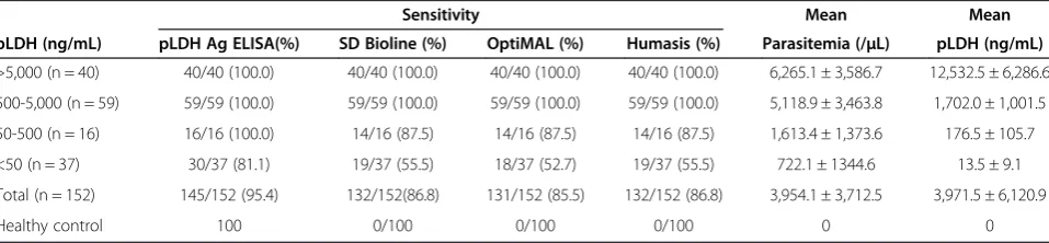
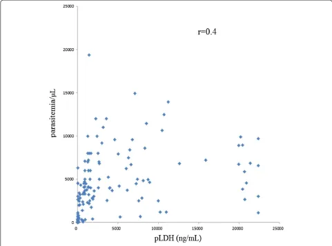
During the study period, 152 patients and 100 healthy con-trols were enrolled and diagnosed for malaria parasites by PCR and microscopic examination of Giemsa-stained blood smear. CSP genes were successfully amplified from parasite genomic DNA from 152 patients. Mean (±SD) parasitaemia level was 3954.1 ± 3712.5/μL with range be-tween 2.0 and 19,382.0/μL, and mean (±SD) pLDH level of P. vivaxinfected patients was 3,971.5 ± 6120.9 ng/mL with range between 0 and 22,387.2 ng/ml (Table 1). Major pLDH levels ofP. vivax(38.8%, 59/152) fell between 500– 5,000 ng/mL (mean ± SD, 1702.0 ± 1001.5 ng/mL), and the second frequent level was over 5,000 ng/mL (25.1%, 40/152). Thirty seven cases (20.1%) revealed extremely low pLDH levels, less than 50 ng/mL. In healthy controls, pLDH was not detected. The lowest detectable level of pLDH antigen ELISA was 6.3 ng/mL, and lowest parasit-aemia level of patients detected by pLDH antigen ELISA was five parasites/μL in clinical samples. Direct correlation was observed between pLDH level and parasitaemia (r = 0.4, p < 0.05) (Figure 1). However, some discrepancies were observed between parasitaemia and pLDH levels (Figure 1). Seven cases of P. vivax patients with parasitaemia over 1,000/μL presented very low pLDH levels (<100 ng/mL).
The diagnostic sensitivity of pLDH antigen ELISA was 95.4% (145/152) among microscopy-positive cases with 100% specificity (0/100), which was superior to three malaria RDTs. OptiMAL test, SD BIOLINE Malaria Ag P.f/Pan test, and Humasis Malaria Pf/Pan antigen test had sensitivity of 85.5% (131/152), 86.8% (132/152), and 86.8% (132/152) with each 100% specificity (0/100), re-spectively (Table 1).
From five repeated test by each kit, detection limits of pLDH in three RDTs were determined as 50 ng/mL for OptiMAL test and 25 ng/ml for SD BIOLINE Malaria Ag P.f ⁄ Pan test and Humasis Malaria P.f ⁄ Pan antigen test with artificially diluted samples (Table 2). The pLDH levels in negative cases by all three RDTs (n = 19) were approximately 19.7 ± 28.4 ng/mL, while values of parasit-aemia were observed at 348.8 ± 602.4/μL. In all positive cases, much higher levels of pLDH from RDTs were ob-served at 4,651.2 ± 6374.1 ng/mL with corresponding parasitaemia of 4,586.1 ± 6,666.8/μL (Table 3).
Discussion
Malaria diagnostic tests should be easy to perform and the interpretation of the results by examiners needs to be simple, however, in many RDTs sensitivities and spec-ificities fluctuate in various field settings. The evalua-tions of pLDH-based RDTs for P. vivax indicate that quality and performance are not satisfactory due to low sensitivity. Poor manufacturing conditions or inadequate precautions in transportation and storage of RDTs result in low sensitivity and reproducibility [11,12].
[image:3.595.59.538.622.733.2]Even though patient parasitaemia levels are the most important determining factor for sensitivity of RDTs, several exceptional cases could not be explained by para-sitaemia factors only. Traditionally, the performances of malaria RDTs are analysed by comparing the value with the parasitaemia levels from microscopic examinations. Microscopy-based evaluations reveal frequent limitations due to incorrect interpretation results dependent on ex-aminers’ training and experience. Hence, the measured value of pLDH by ELISA could be more objective than the parasitaemia value, and the value of pLDH could
Table 1 The sensitivities of rapid diagnostic tests compared against the levels of pLDH in detectingPlasmodium vivax infection
Sensitivity Mean Mean
pLDH (ng/mL) pLDH Ag ELISA(%) SD Bioline (%) OptiMAL (%) Humasis (%) Parasitemia (/μL) pLDH (ng/mL)
>5,000 (n = 40) 40/40 (100.0) 40/40 (100.0) 40/40 (100.0) 40/40 (100.0) 6,265.1 ± 3,586.7 12,532.5 ± 6,286.6
500-5,000 (n = 59) 59/59 (100.0) 59/59 (100.0) 59/59 (100.0) 59/59 (100.0) 5,118.9 ± 3,463.8 1,702.0 ± 1,001.5
50-500 (n = 16) 16/16 (100.0) 14/16 (87.5) 14/16 (87.5) 14/16 (87.5) 1,613.4 ± 1,373.6 176.5 ± 105.7
<50 (n = 37) 30/37 (81.1) 19/37 (55.5) 18/37 (52.7) 19/37 (55.5) 722.1 ± 1344.6 13.5 ± 9.1
Total (n = 152) 145/152 (95.4) 132/152(86.8) 131/152 (85.5) 132/152 (86.8) 3,954.1 ± 3,712.5 3,971.5 ± 6,120.9
provide more reliable analysis of selecting adequate mal-aria RDTs.
Without adequate baseline data of pLDH levels from clinically isolatedP. vivax, it is difficult to correct the inter-pretation of results to make better decisions for selecting
reliable malaria RDTs. Since none of the malaria RDTs de-tect the parasite itself, the secreted biomarkers became the diagnostic targets. To date, the level of target biomarkers using a standardized protocol for clinically isolated malaria samples are rarely reported [13].
[image:4.595.56.538.90.447.2] [image:4.595.58.290.598.732.2]Here, In this study, the distribution of pLDH in clinic-ally isolated P. vivax samples, and the relationships be-tween levels of pLDH and parasitaemia and bebe-tween levels of pLDH and sensitivities of pLDH-based RDTs are Figure 1Correlations between parasitaemia and the levels of pLDH among isolatedPlasmodium vivaxpatients with clinical symptoms.
Table 2 Limit of detection for pLDH in three rapid diagnostic tests by using artificially diluted samples of Plasmodium vivax
pLDH (ng/mL)
Parasitaemia (/μL)
SD Bioline (n = 5)
OptiMAL (n = 5)
Humasis (n = 5)
5,000 8,700.0 100% 100% 100%
500 870.0 100% 100% 100%
250 435.0 100% 100% 100%
50 87.0 100% 100% 100%
25 44.0 100% 0% 100%
5 8.7 0% 0% 0%
0.5 0.9 0% 0% 0%
0 0 0% 0% 0%
Table 3 The sensitivities of rapid diagnostic tests were compared to the levels of pLDH in detectingPlasmodium vivaxinfection
Mean Mean
RDT N Parasitaemia(/μL) pLDH (ng/mL)
All RDT Positive 130 4,586.1 ± 6,666.8 4,651.2 ± 6,374.1
2 RDT positive 3 777.7 ± 1076.5 5.0 ± 7.1
All RDT Negative 19 348.8 ± 602.4 19.7 ± 28.4
[image:4.595.304.540.651.733.2]assessed. In WHO/FIND malaria RDT testing round 4 report, Humasis Malaria P.F/Pan Antigen test (AMAL-7025) showed panel detection score (PDS) of 0% at both 200 and 2000 parasites/μL of P. vivax[14]. In addition, false positive rate of Humasis kit was 97.8%. While the performance parameter in this study was not PDS, but clinical sensitivity, Humasis kit in this study demon-strated the clinical sensitivity of 55.5 ~ 100.0% at similar parasitaemia (Table 1). Next, specificity of Humasis kit for P. vivaxwas 100% with no observed false positivity. As for OptiMAL-IT kit (710024), round 4 report presented PDS of 97.1 and 68.6% at 200 and 2,000 ~ 5,000 parasites/μL of P. vivax, respectively. On the other hand, OptiMAL-IT showed increased clinical sensitivity from 52.7 to 100%, as the P. vivax parasitaemia increased. As for the SD Bioline Malaria Ag P.f/Pan kit (05 F60), round 4 report showed PDS of 97.1 and 100.0% at 200 and 2,000 ~ 5,000 parasites/μL of P. vivax, respectively. However, SD Bioline demonstrated slightly decreased clinical sensitivity of 87.5 ~ 100.0% (Table 1). In this study, lower sensitivities in all three RDTs were observed from groups with lower pLDH antigen levels (Table 1). From the study for deter-mining the limit of detection, lower levels pLDH associated with lower sensitivities in all three RDTs (Table 2). These direct correlations were similar to the levels of parasitaemia in all three RDTs. Above results suggested the possibility of pLDH, as a performance parameter for malaria RDTs. This information on currently available RDTs is valuable in re-vealing detection limits against pLDH. Therefore, the current results could provide one more reasonable proto-col for comparing various malaria RDTs and their practical guidelines in developing sensitive malaria RDTs.
pLDH antigen ELISA system showed higher sensitivity (95.4%) in comparison with three RDTs (85.5-86.8%), which might be associated with the fact that ELISA uses 50μL of blood, while RDTs used only 5–10μL of blood. RDTs are fast and convenient diagnostic tools. Parasit-aemia levels of P. vivax in ROK ranged 2–25,000/μL with mean level of 3,954.1 ± 3712.5/μL, while pLDH levels of P. vivaxranged between 0–22,387 ng/mL with mean value of 3,971.5 ± 6120.9 ng/mL. pLDH levels showed moderate correlation with parasite density of P. vivax, as determined by microscopy (r = 0.4, p < 0.05).
From three tested RDTs, pLDH levels and parasit-aemia levels of three RDTs, positive cases were higher than two RDT positive cases or all RDT negative cases. One positive sample from two RDT tests (n = 3) showed disparity between parasitaemia and pLDH levels, where the pLDH and parasitaemia levels were 15 ng/mL and 2,300/μL, respectively. These levels of parasitaemia most likely produce clinical symptoms. RDTs are fast and con-venient diagnostic tools. Using only RDTs, malaria diagno-sis might be difficult and patients might be left untreated, leading to severe complications. Since the evaluation of
RDTs is currently based on patients’ parasitaemia levels, with little information on targeted biomarkers in clinical isolates, the current study provides additional information for selecting adequate malaria RDT in fields.
Competing interests
The authors have declared that they have no competing interests.
Authors’contributions
JWJ carried out the molecular work and RDT test. CHC carried out the immunoassays. ETH & SSA participated in the design of the study and performed the statistical analysis. CSL contributed to the design and coordination of the study, assisted with data entry, analysis and interpretation. All authors read and approved the final manuscript.
Acknowledgements
This work was supported by National Research Foundation of Korea Grant (2012R1A1A2006650) funded by the Korean Government.
Author details
1
Department of Laboratory Medicine, College of Medicine, Korea University Guro Hospital, Guro 2 Dong, Guro Gu, Seoul 152-703, Republic of Korea.
2Department of Parasitology, Kangwon National University College of
Medicine, Chuncheon, Republic of Korea.3College of Bionano Technology,
Gachon Bionano Research Institute, Kyungwon University, Seongnam-si, Gyeonggi Do, Republic of Korea.
Received: 29 March 2013 Accepted: 30 May 2013 Published: 3 June 2013
References
1. Nafo Traore F:Rolling back malaria: opportunities and challenges.Trans R Soc Trop Med Hyg2005,99:403–406.
2. Murray CK, Bennett JW:Rapid diagnosis of malaria.Interdiscip Perspect Infect Dis2009,2009:415953.
3. Maltha J, Gillet P, Jacobs J:Review: malaria rapid diagnostic tests in endemic settings.Clin Microbiol Infect2013,19:399–407.
4. Beadle C, Long GW, Weiss WR, McElroy PD, Maret SM, Oloo AJ, Hoffman SL:
Diagnosis of malaria by detection ofPlasmodium falciparumHRP-2 antigen with a rapid dipstick antigen-capture assay.Lancet1994,
343:564–568.
5. Makler MT, Hindrich DJ:Measurement of the lactate dehydrogenase activity ofPlasmodium falciparumas an assessment of parasitemia.Am J Trop Med Hyg1993,48:205–210.
6. Mai Huong NM, Davis TM, Hewitt S, Huong NV, Uyen TT, Nhan DH, le Cong D:
Comparison of three antigen detection methods for diagnosis and therapeutic monitoring of malaria: a field study from southern Vietnam.
Trop Med Int Health2002,7:304–308.
7. Singh N, Saxena A, Valecha N:Field evaluation of the ICT malaria P.f⁄P.v immunochromatographic test for diagnosis ofPlasmodium falciparum
andP. vivaxinfection in forest villages of Chhindwara, central India.Trop Med Int Health2000,5:765–770.
8. Cho CH, Nam MH, Kim JS, Han ET, Lee WJ, Oh JS, An SS, Lim CS:Genetic variability inP. vivaxaldolase gene in Korean isolates and the sensitivity of the Binax Now malaria test.Trop Med Int Health2011,16:223–226. 9. Mueller I, Betuela I, Ginny M, Reeder JC, Genton B:The sensitivity of the
OptiMAL rapid diagnostic test to the presence ofPlasmodium falciparum
gametocytes compromises its ability to monitor treatment outcomes in an area of Papua New Guinea in which malaria is endemic.J Clin Microbiol2007,45:627–630.
10. Piper R, Lebras J, Wentworth L, Hunt-Cooke A, Houze S, Chiodini P, Makler M:
Immunocapture diagnostic assays for malaria using Plasmodium lactate dehydrogenase (pLDH).Am J Trop Med Hyg1999,60:109–118. 11. Jorgensen P, Chanthap L, Rebueno A, Tsuyuoka R, Bell D:Malaria rapid
diagnostic tests in tropical climates: the need for a cool chain.Am J Trop Med Hyg2006,74:750–754.
12. Rennie W, Phetsouvanh R, Lupisan S, Vanisaveth V, Hongvanthong B, Phompida S, Alday P, Fulache M, Lumagui R, Jorgensen P, Bell D, Harvey S:
instructions and health worker performance.Trans R Soc Trop Med Hyg 2007,101:9–18.
13. Hendriksen IC, Mwanga-Amumpaire J, von Seidlein L, Mtove G, White LJ, Olaosebikan R, Lee SJ, Tshefu AK, Woodrow C, Amos B, Karema C, Saiwaew S, Maitland K, Gomes E, Pan-Ngum W, Gesase S, Silamut K, Reyburn H, Joseph S, Chotivanich K, Fanello CI, Day NP, White NJ, Dondorp AM:Diagnosing severe falciparum malaria in parasitaemic African children: a prospective evaluation of plasma PfHRP2 measurement.PLoS Med2012,9:e1001297.
14. WHO:Malaria rapid diagnostic test performance: results of WHO product testing of malaria RDTs: round 4.Geneva: World Health Organization; 2012.
doi:10.1186/1475-2875-12-181
Cite this article as:Janget al.:pLDH level of clinically isolated
Plasmodium vivaxand detection limit of pLDH based malaria rapid diagnostic test.Malaria Journal201312:181.
Submit your next manuscript to BioMed Central and take full advantage of:
• Convenient online submission
• Thorough peer review
• No space constraints or color figure charges
• Immediate publication on acceptance
• Inclusion in PubMed, CAS, Scopus and Google Scholar
• Research which is freely available for redistribution